HAL Id: hal-00735019
https://hal.archives-ouvertes.fr/hal-00735019
Submitted on 17 Feb 2016
HAL is a multi-disciplinary open access
archive for the deposit and dissemination of
sci-entific research documents, whether they are
pub-lished or not. The documents may come from
teaching and research institutions in France or
abroad, or from public or private research centers.
L’archive ouverte pluridisciplinaire HAL, est
destinée au dépôt et à la diffusion de documents
scientifiques de niveau recherche, publiés ou non,
émanant des établissements d’enseignement et de
recherche français ou étrangers, des laboratoires
publics ou privés.
AT_CHLORO: A Chloroplast Protein Database
Dedicated to Sub-Plastidial Localization.
Christophe Bruley, Véronique Dupierris, Daniel Salvi, Norbert Rolland,
Myriam Ferro
To cite this version:
Christophe Bruley,
Véronique Dupierris,
Daniel Salvi,
Norbert Rolland,
Myriam Ferro.
AT_CHLORO: A Chloroplast Protein Database Dedicated to Sub-Plastidial Localization.. Frontiers
in Plant Science, Frontiers, 2012, 3, pp.205. �10.3389/fpls.2012.00205�. �hal-00735019�
AT_CHLORO: a chloroplast protein database dedicated to
sub-plastidial localization
Christophe Bruley1,2,3, Véronique Dupierris1,2,3, Daniel Salvi3,4,5,6, Norbert Rolland3,4,5,6* and Myriam Ferro1,2,3 1CEA, DSV, IRTSV, Laboratoire Biologie à Grande Echelle, Institut de Recherches en Technologie et Sciences pour le Vivant, Grenoble, France
2INSERM, U1038, Grenoble, France 3
Université Joseph Fourier, Grenoble 1, Grenoble, France 4
CEA, DSV, IRTSV, Laboratoire de Physiologie Cellulaire et Végétale, Grenoble, France 5
CNRS, UMR5168, Grenoble, France 6INRA, USC1359, Grenoble, France
Edited by:
Joshua L. Heazlewood, Lawrence Berkeley National Laboratory, USA
Reviewed by:
Wolfgang P. Schröder, Umeå University, Sweden
Torsten Kleffmann, University of Otago, New Zealand
*Correspondence:
Norbert Rolland , Laboratoire de Physiologie Cellulaire et Végétale, Institut de Recherches en Technologies et Sciences pour le Vivant, CEA Grenoble, 17 rue des Martyrs, 38054 Grenoble Cedex 9, France.
e-mail: [email protected]
AT_CHLORO (www.grenoble.prabi.fr/at_chloro) is a database dedicated to sub-plastidial localization of A. thaliana chloroplast proteins. This information was infered from pro-teomics experiments obtained from a comprehensive study that allowed the identification of proteins from envelope, stroma, and thylakoid sub-compartmentsFerro et al., 2010. In addition to current knowledge regarding sub-plastidial localization, AT_CHLORO provides experimental data that allowed curated information regarding subcellular localizations of chloroplast proteins to be given. A specific focus was given to proteins that were identified in envelope fractions and for which expert functional annotation was provided. The present mini review shows the specificities of AT_CHLORO with respect to available information, data export options and recent improvements in data representation.
Keywords: chloroplast, proteomics, sub-plastidial localization, chloroplast envelope, database
INTRODUCTION
As chloroplasts are Earth’s main solar energy converters, much interest has emerged for a better knowledge of chloroplast biology. Indeed, most renewable carbon is fixed by photosynthetic organ-isms through a process made possible by their chloroplasts. Being the location of essential metabolic pathways (photosynthesis, syn-thesis of lipids, pigments, amino acids, vitamins, starch, precursors of plant hormones, etc.), the chloroplast is an established target for metabolic engineering of crop plants to improve productiv-ity. In order to carry out such an array of biochemical reactions, chloroplasts need to coordinate the functions of three main sub-compartments: the envelope, the stroma, and the thylakoids. The envelope comprises a pair of membranes surrounding the chloro-plast and controls the dialog between the chlorochloro-plast and the rest of the cell (Block et al., 2007). In addition, those membranes are also involved in many other essential metabolic reactions (e.g., lipid, pigment, or vitamin synthesis). The stroma, the soluble phase of the chloroplast, is the main place for the conversion of carbon dioxide into carbohydrates. Other catalytic reactions occur in the stroma that allow the synthesis of compounds such as amino acids. The thylakoids are a highly organized internal membrane network where solar energy is collected and converted into chemical energy (ATP and NADPH).
In order to investigate chloroplast metabolism and main func-tions, sub-plastidial localization is a crucial piece of information required to select proteins in the context of targeted functional characterization. In that context, recent advances in the proteomic field have allowed high throughput experiments to be conducted
on chloroplast samples and to provide additional information about functional compartmentalization (Agrawal et al., 2011;van Wijk and Baginsky, 2011). The spatial distribution of proteins within chloroplasts has been investigated from various indepen-dent studies which aimed to establish the proteome repertoire of sub-plastidial compartments: the thylakoids (e.g.,Friso et al., 2004;Peltier et al., 2004), the stroma (e.g.,Zybailov et al., 2008), the plastoglobules (e.g.,Vidi et al., 2006;Ytterberg et al., 2006; Lundquist et al., 2012), and the envelope (e.g.,Ferro et al., 2002, 2003; Froehlich et al., 2003). This targeted repertoire allowed the identification of minor components of each of these sub-compartments. Whereas these repertoires are highly informative, the actual sub-plastidial localization of some proteins might be questionable as they were identified in different chloroplast sub-fractions or in other subcellular compartments. Indeed, the actual localization of proteins within a fraction that has been used for proteomics analyses is related to cross-contamination issues. Thus, MS-based quantification strategies, must be applied to discrimi-nate between true and false protein localization assignments (e.g., Dunkley et al., 2006). In that context, as the accurate localization of many chloroplast proteins remained hypothetical, we set up a proteomics strategy which aimed to ascertain the sub-plastidial localization of chloroplast proteins (Ferro et al., 2010). Using a MS-based semi-quantitative strategy (spectral count), we chose to revisit the sub-plastidial localization of chloroplast proteins. In order to gage sub-plastidial cross-contamination, we started from purified sub-fractions retrieved from the same chloroplast sam-ples. MS-based sub-plastidial localization of identified proteins
Bruley et al. AT_CHLORO, a sub-plastidial localization database
was stored in the AT_CHLORO database1which compiles results from LC-MS/MS analyses of highly purified sub-fractions of the three major chloroplast sub-compartments. From the MS analyses, about 1,300 proteins were identified, of which more than 800 pro-teins could be assigned a sub-plastidial localization. In addition, the AT_CHLORO database condenses public and curated infor-mation related to protein function and localization, especially for envelope proteins.
PRESENTATION OF THE DATABASE
AT_CHLORO is one of the databases dedicated to the chloro-plast proteome of Arabidopsis thaliana and specifically gathers information for proteins that have been identified from the main chloroplasts sub-fractions: envelope, stroma, and thylakoids (Demartini et al., 2011). Sub-plastidial localization was assessed using MS-based data corresponding to the three purified chloro-plast compartments – envelope, thylakoids, and stroma – that had been, for the first time, analyzed in the same set of experiments (Ferro et al., 2010). Briefly, purification of those three chloro-plast sub-fractions was achieved using sucrose gradients (Salvi et al., 2011). Envelope, stroma, and thylakoid fractions were either digested in solution or analyzed by SDS-PAGE prior to trypsin digestion (Salvi et al., 2008). Then, generated samples were sub-mitted to LC-MS/MS analysis for identification purposes. About 500 LC-MS/MS analyses were performed, ending up with the identification of 1,323 proteins. As chloroplast sub-fractions were prepared with a low level of contamination, as determined by Western blot analyses, and starting from the same chloroplast sam-ples, semi-quantitative spectral count data allowed assessment of protein relative abundances in each of the three sub-fractions. Thus, the partitioning of each of the 1,323 proteins in envelope, stroma, and thylakoids was calculated based on normalized spec-tral count data, from which a percentage of occurrence in each
1http://www.grenoble.prabi.fr/at_chloro/
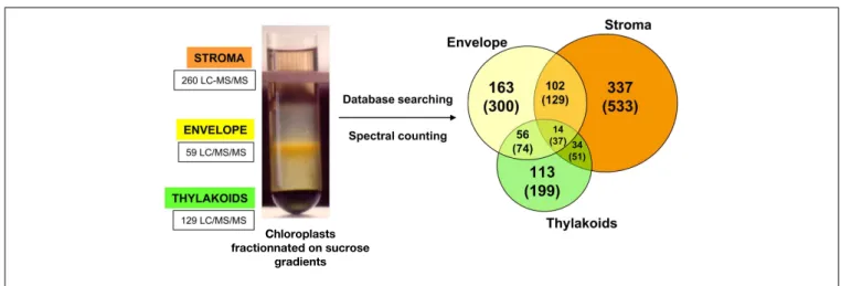
sub-compartment was deduced. Amongst the 1,323 proteins, sta-tistical analysis allowed the accurate localization of 819 proteins (Figure 1).
Four types of information can be found in the AT_CHLORO database: (i) the proteomics-based sub-plastidial localization as revealed by spectral counting; (ii) analytical coordinates (HPLC retention time, RT; peptide molecular weight, Mr) of all the peptides corresponding to the proteins stored in the database; (iii) curated localization and function of proteins, especially the ones that were identified in envelope fractions, and (iv) information from public databases such as TAIR2, or PPDB3 (Sun et al., 2009). Some data from AT_CHLORO can also be retrieved from the MASCGator Portal (Joshi et al., 2011). Informa-tion related to the sub-plastidial localizaInforma-tion was also submitted to TAIR.
DESCRIPTION OF THE DIFFERENT TYPES OF INFORMATION
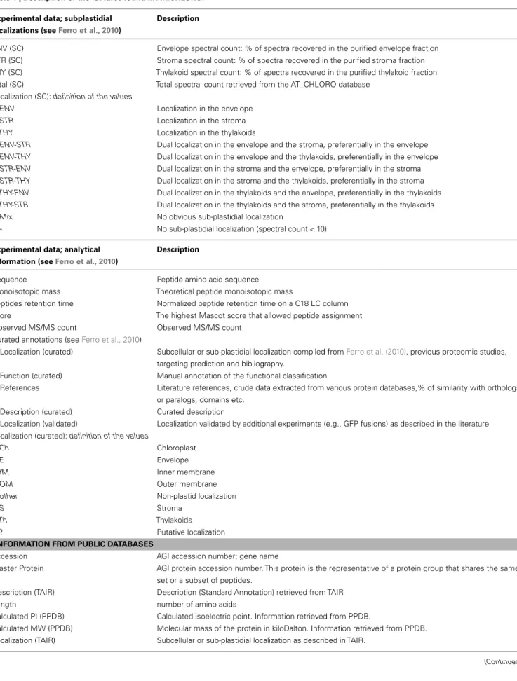
AT_CHLORO gathers three levels of information: MS-based experimental data, curated annotations, and public information. All this information gives a comprehensive overview of current knowledge about the localization and the function of identified chloroplast proteins. Definitions of the different types of infor-mation can be found in Table 1. Fields or columns in which the different types of information can be found appear in italics in the text below.
EXPERIMENTAL DATA: PROTEOMICS-BASED SUB-PLASTIDIAL LOCALIZATION AS DEDUCED FROM SPECTRAL COUNTING
Experimental data were extracted from (Ferro et al., 2010). Briefly, for each protein and each chloroplast sub-fraction (envelope, stroma, and thylakoids), the number of associated spectra was retrieved from LC-MS/MS and database searching data. Because spectral counting is a semi-quantitative approach, significant
2http://www.arabidopsis.org/ 3http://ppdb.tc.cornell.edu/
FIGURE 1 | Distribution of protein sub-plastidial localizations as determined using spectral count data for a subset of 819 selected proteins. Number into brackets correspond to the total 1323 proteins. Left
part: fractionation of chloroplasts on a sucrose gradient (0.3, 0.6, and 0.93 M sucrose layers from top to bottom) after osmotic shock performed
on Percoll-purified chloroplasts. After centrifugation, the envelope membrane and the thylakoids are present as a yellow band at the 0.93/0.6 M interface and as a dark green band at the bottom of the tube, respectively. The soluble fraction containing the stroma remains on top of this gradient (see,Salvi et al., 2011).
Table 1 | Description of the features found in AT_CHLORO.
Experimental data; subplastidial localizations (seeFerro et al., 2010)
Description
ENV (SC) Envelope spectral count: % of spectra recovered in the purified envelope fraction STR (SC) Stroma spectral count: % of spectra recovered in the purified stroma fraction THY (SC) Thylakoid spectral count: % of spectra recovered in the purified thylakoid fraction Total (SC) Total spectral count retrieved from the AT_CHLORO database
Localization (SC): definition of the values
ENV Localization in the envelope
STR Localization in the stroma
THY Localization in the thylakoids
ENV-STR Dual localization in the envelope and the stroma, preferentially in the envelope
ENV-THY Dual localization in the envelope and the thylakoids, preferentially in the envelope
STR-ENV Dual localization in the stroma and the envelope, preferentially in the stroma
STR-THY Dual localization in the stroma and the thylakoids, preferentially in the stroma
THY-ENV Dual localization in the thylakoids and the envelope, preferentially in the thylakoids
THY-STR Dual localization in the thylakoids and the stroma, preferentially in the thylakoids
Mix No obvious sub-plastidial localization
- No sub-plastidial localization (spectral count< 10) Experimental data; analytical
information (seeFerro et al., 2010)
Description
Sequence Peptide amino acid sequence Monoisotopic mass Theoretical peptide monoisotopic mass
peptides retention time Normalized peptide retention time on a C18 LC column score The highest Mascot score that allowed peptide assignment Observed MS/MS count Observed MS/MS count
Curated annotations (seeFerro et al., 2010)
Localization (curated) Subcellular or sub-plastidial localization compiled fromFerro et al. (2010), previous proteomic studies, targeting prediction and bibliography.
Function (curated) Manual annotation of the functional classification
References Literature references, crude data extracted from various protein databases,% of similarity with orthologs or paralogs, domains etc.
Description (curated) Curated description
Localization (validated) Localization validated by additional experiments (e.g., GFP fusions) as described in the literature Localization (curated): definition of the values
Ch Chloroplast
E Envelope
IM Inner membrane
OM Outer membrane
other Non-plastid localization
S Stroma
Th Thylakoids
? Putative localization
INFORMATION FROM PUBLIC DATABASES
Accession AGI accession number; gene name
Master Protein AGI protein accession number. This protein is the representative of a protein group that shares the same set or a subset of peptides.
Description (TAIR) Description (Standard Annotation) retrieved from TAIR
Length number of amino acids
Calculated PI (PPDB) Calculated isoelectric point. Information retrieved from PPDB.
Calculated MW (PPDB) Molecular mass of the protein in kiloDalton. Information retrieved from PPDB. Localization (TAIR) Subcellular or sub-plastidial localization as described in TAIR.
Bruley et al. AT_CHLORO, a sub-plastidial localization database
Table 1 | Continued
MapManBin (PPDB) MapMan functional classification. Information retrieved from PPDB.
ChloroP Prediction of plastid localization using ChloroP (http://www.cbs.dtu.dk/services/ChloroP/) Curated localization (PPDB) Curated subcellular or sub-plastidial localization as stated in PPDB.
TargetP Prediction of subcellular localization using TargetP (http://www.cbs.dtu.dk/services/TargetP/) Aramemnon Number of transmembrane helices as found in the Aramemnon database
(http://aramemnon.botanik.uni-koeln.de/)
Publications (PPDB) List of most subcellular targeted proteomics studies (MedLine numbers) in which the protein was identified. Information retrieved from PPDB.
ChloroP: definition of values
Y The protein has a predicted chloroplast transit peptide TargetP: definition of values
C Chloroplast
M Mitochondria
SP Secretory pathway
– No prediction
“?” indicates that the proposed subcellular or subplastidial localizations remain putative. In the database, the “?” is associated to localization classes (e.g. Ch?, Ch/E?, ...).
ratios, and thresholds are generally high. Consequently, only 819 proteins identified with at least 10 spectral counts were taken into account for being assigned an accurate sub-plastidial localization. Spectral counts were normalized with respect to the number of assigned MS/MS spectra in each fraction and a percentage of occurrences in each sub-fraction was calculated for all proteins (ENV SC; STR SC; THY SC). The localization given by normalized spectral counts was verified using a logistic regression model. From the calculated percentages, proteins were attributed a single, dual, or mixed sub-plastidial localization. A single localization was thus assigned to proteins for which the percentages of occurrence in the two other sub-fractions were below a threshold level fixed at 15%. This percentage was set to 15%, above the cross-contamination level as estimated by Western blotting. Dual localization was assigned to proteins with a major localization (occurrence ≥50%) and a secondary localization (occurrence ≥15%). The remaining proteins were considered to have a mixed localization between the three sub-plastidial compartments [Localization (SC)]. For all proteins, the total number of spectral counts can also be viewed and gives an assessment of the relative amount of a given protein in the chloroplast [Total (SC)].
EXPERIMENTAL DATA: ANALYTICAL COORDINATES FOR LABEL-FREE QUANTIFICATION
The AT_CHLORO database is not only a repository of chloro-plast proteins but also gathers information related to peptides that have allowed protein identification. Thus peptide sequences (sequence), theoretical molecular weight (monoisotopic mass), chromatographic retention times (Peptides retention time), the score that allowed peptide identification (score), and spectral count (observed MS/MS count ) can be found in the window dedicated to each protein. Theoretical molecular weight and chromatographic retention times can be particularly useful for label-free based quantification studies using the AMT strategy (Lipton et al., 2006). Indeed, the accurate mass and time tags (AMT) method, combines identification, and quantification issues in the context of high throughput quantitative experiments. In a first stage, standard
shotgun proteomics approaches are undertaken on extensively fractionated proteins to yield peptide identification. Those exper-iments yield a database containing the calculated masses based on putative peptide sequences and their corresponding measured chromatographic retention times. Thus, AT_CHLORO is also an AMT database dedicated to the chloroplast. Accurate mass and time tags can subsequently be used, in the course of “simple” LC-MS measurements, as biomarkers of the presence of a given protein without resorting systematically to MS/MS for identification. Con-sequently, it becomes possible to identify hundreds of proteins in a single MS spectrum in all subsequent LC-MS experiments, using high resolution mass spectrometers, such as the Orbitrap.
CURATED LOCALIZATION AND FUNCTION OF PROTEINS
From experimental data and information retrieved from public repositories, curated localizations and functions were given. As sub-plastidial localization is the main focus of AT_CHLORO spe-cial care was taken with regard to localization annotations. Thus experimental sub-plastidial localization, previous proteomic stud-ies, targeting prediction, and bibliography were compiled in order to assign a curated localization [Localization (curated)]. In this context, since the first release of AT_CHLORO, we improved the curated localization of some proteins by providing information about the lumenal localization of a given set of thylakoid pro-teins, as selected from two reference papers in the field (Peltier et al., 2002; Schubert et al., 2002). Also manual annotation of protein function was undertaken [Function (curated)]. A specific emphasis was given to about 700 proteins identified in envelope sub-fractions. Indeed, as most available chloroplast proteomics data provide information about proteomes from thylakoids and stroma compared to the envelope, we paid specific attention to analyzing the proteome of the two envelope membranes. Exter-nal sources that were used for curated annotations, such as lit-erature references, similarity values with orthologs or protein domains can be found in the references field. This recent update of the AT_CHLORO database also includes citation of more recent publications, in the references field, for some selected proteins.
INFORMATION FROM PUBLIC DATABASES
Public information was retrieved from TAIR4 and PPDB5 (Sun
et al., 2009) and are listed in Table 1.
HOW TO USE THE AT_CHLORO DATABASE
AT_CHLORO is organized around four main types of pages: the main page, the search page, the protein list page and the protein ID page.
THE MAIN PAGE
The main page of AT_CHLORO presents a short description of the data that were generated to build the database with associated references and contact. The top main menu contains six options. The user can choose to get the list of all the proteins identified by (Ferro et al., 2010; all) or to visualize the list of proteins specifically identified in one of the three chloroplast sub-fractions (Envelope,
Stroma, Thylakoids). Also, users can search for a given protein
or a list of proteins using different features (Search). Results can be viewed by clicking on the “Search Results” option. The list of features with associated description can be accessed from the main page.
THE SEARCH PAGE
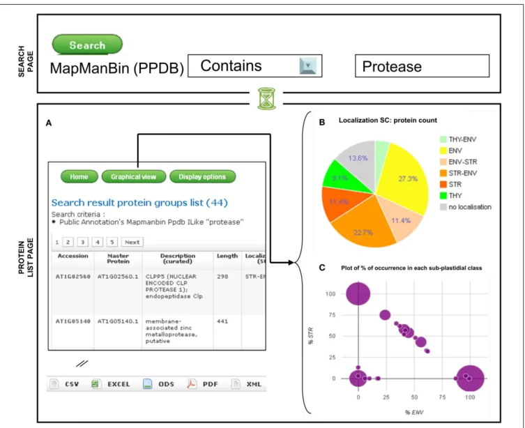
The search page can be accessed from the main page top menu using the “Search” option. In the first version of AT_CHLORO, proteins could be selected by loading the accession number, or the protein’s description, localization, or function. In the cur-rent version it is also now possible to retrieve proteins accord-ing to selected values related to one or several features. For instance, proteins can be selected according to a particular func-tion either from the Funcfunc-tion (curated) or the MapManBin (PPDB) features (Figure 2). As written in the search page, the different search criteria are combined using the AND Boolean Operator.
THE PROTEIN LIST PAGE
A table summarizing the features of a list of proteins can be obtained directly from the main page with the “All,” “Envelope,” “Stroma,”“Thylakoids,”“Search Results,” options (Figure 2A). Users can customize the display, using the “Display option,” button, so that only selected features, amongst the one listed above, are visible on the screen. Recently, graphical representation of the data were added (“graphical view” option). As the specificity of AT_CHLORO relies on localization information, proteins can be classified according to their sub-plastidial localization, as given by spectral count data. As shown in Figure 2B, pie-charts can be generated and allow a quick overview of the distribution of pro-teins within the chloroplast sub-compartments according to the “Localization (SC)” feature. A pie chart is available which takes into account the protein counts of “Localization (SC)” classes (Figure 2B). Together with the pie-charts, a bar-chart allows a potential enrichment in one of the sub-fraction with respect to the whole database to be assessed. In addition, a diagram which plots the% of occurrence in the envelope according to the% of
4http://www.arabidopsis.org/ 5http://ppdb.tc.cornell.edu/
occurrence in the stroma has been set up which gives a quick overview of the sub-plastidial partitioning of selected proteins (Figure 2C).
From the protein list page it is possible to export data in dif-ferent formats: csv (comma separated values), xls (Excel), ods (Open Document Spreadsheet), pdf (portable document format), and xml (extensible markup language). Thus the user can eas-ily retrieve protein lists with associated sub-plastidial information for data mining purposes. For instance in a recent report (Tanz et al., 2012) attributes on protein localization were retrieved from AT_CHLORO and were integrated in the widely used Cytoscape tool6.
THE PROTEIN ID PAGE
From a protein list page, users can select one particular protein, for which different types of information are displayed in an addi-tional window. All the features listed in Table 1 can be found in the protein ID page and additional information from TAIR, PPDB, Atproteome7, SUBA8, POGs9, and Aramemnon10can be accessed from appropriate links.
CONCLUSIONS AND PERSPECTIVES
The AT_CHLORO database represents a dedicated resource for getting sub-plastidial localization and functional annotation of Arabidopsis chloroplast proteins, especially for envelope proteins. As revealed by the increasing number of visits and recent pub-lications, information derived from the AT_CHLORO database proved to be of valuable interest for the plant community to ascertain protein sub-plastidial localization, to get insight over metabolic mechanisms, or for data mining purposes. Indeed infor-mation regarding the sub-plastidial localization of proteins has proved to be of major interest to confirm the subcellular localiza-tion of particular proteins or to investigate biological processes at a larger scale. For instance, in order to decipher mechanistic details of thylakoids biogenesis, analysis, and characterization of mutants can be particularly useful (Adam et al., 2011). The knowledge of the sub-plastidial localization of proteins whose correspond-ing mutant shows defects in thylakoid network formation, can give insight over the actual role of such proteins. Indeed, as the inner envelope membrane is likely to be the source of internal membrane structures, knowing whether a protein is located in the envelope, or in the thylakoids might help in underlining its role during thylakoid genesis, e.g., with regards to lipid traffick-ing. In order to determine the subcellular localization of a given protein, Western blotting has been the conventional method for many years. With recent advances in proteomics science, mass spectrometry detection has emerged as an alternative or a comple-mentary approach to Western blots (Mann, 2008). In that context, sub-plastidial information retrieved from AT_CHLORO was also used to ascertain protein localization. For instance, in a recent paper (Karamoko et al., 2011), AT_CHLORO spectral count-based
6http://www.cytoscape.org/ 7http://fgcz-atproteome.unizh.ch/ 8http://suba.plantenergy.uwa.edu.au/ 9http://plantrbp.uoregon.edu/ 10http://aramemnon.uni-koeln.de/
Bruley et al. AT_CHLORO, a sub-plastidial localization database
FIGURE 2 | Visualization of the sub-plastidial localization of a selected set of proteins: example of a search. Search results (A) Protein groups list; (B) Pie
chart with sub-plastidial localization;(C) Plot of% of occurrence in each sub-plastidial class.
data reinforced immunoblot analyses showing that two FtsZ2 iso-forms were associated with the thylakoid membranes. Another example concerns the CJD1 protein that influences fatty acid com-position of chloroplast lipids (Ajjawi et al., 2011). GFP fusion experiments suggested that the CJD1 protein was located in the inner envelope of the chloroplast. This information was strength-ened by proteomics studies which allowed identification of the CJD1 protein in chloroplast envelope fractions (Ajjawi et al., 2011). Sub-plastidial proteomics data might also be useful to provide a more precise view of sub-organellar compartmentation of biosyn-thetic pathways, such as isoprenoid (Joyard et al., 2009) or lipid (Joyard et al., 2010) metabolism. In the context of lipid metabo-lism, sub-plastidial data stored in AT_CHLORO proved to be of strong added value in acknowledging the envelope as a central location for lipid synthesis. For instance, the survey performed by (Joyard et al., 2010) indicated that once the fatty acids are esteri-fied to glycerol-3-phosphate the envelope becomes the key player
in glycerolipid biosynthesis, as indicated by the proteomics-based localization of the dedicated enzymes. Thus, sub-plastidial local-ization found in AT_CHLORO proved to be important and useful with respect to the study of metabolic pathways or of specific proteins.
AT_CHLORO, being targeted to chloroplast proteins, is com-plementary to more generic plant databases such as PPDB (Sun et al., 2009) or pep2pro (Baerenfaller et al., 2011).
Since the first release of AT_CHLORO we have improved the outputs of the results, especially by providing a graphical overview of sub-plastidial localization of a given set of pro-teins. The AT_CHLORO database aims at being updated with in-house experiments and curated information related to chloro-plast proteins, especially those identified in envelope fractions in order to get the most accurate picture of chloroplast sub-compartmentalization. Indeed we plan to integrate forthcom-ing experiments that will allow additional identification and
quantification data to be produced. Finally, we welcome colleagues from the plant community to provide updated and additional curated information as well as suggestions regarding the different data outputs.
ACKNOWLEDGMENTS
Yves Vandenbrouck is acknowledged for having made the AT_CHLORO database available on line. James Connorton is
acknowledged for critical reading and editing of the manuscript. Claire Adam is acknowledged for the drawing of the figures. Daphné Seigneurin-Berny and Gilles Curien are acknowledged for updating the AT_CHLORO database with recent references. Christophe Bruley, Véronique Dupierris, Daniel Salvi, Norbert Rolland, and Myriam Ferro received financial support from the French National Research Agency (ANR-2010-GENOM-BTV-002-01 Chloro-Types, ANR-2010-BLAN-1610-01 Chloro-Pro).
REFERENCES
Adam, Z., Charuvi, D., Tsabari, O., Knopf, R. R., and Reich, Z. (2011). Biogenesis of thylakoid net-works in angiosperms: knowns and unknowns. Plant Mol. Biol. 76, 221–234.
Agrawal, G. K., Bourguignon, J., Rol-land, N., Ephritikhine, G., Ferro, M., Jaquinod, M., Alexiou, K. G., Chardot, T., Chakraborty, N., Jolivet, P., Doonan, J. H., and Rakwal, R. (2011). Plant organelle pro-teomics: collaborating for optimal cell function. Mass Spectrom. Rev. 30, 772–852.
Ajjawi, I., Coku, A., Froehlich, J. E., Yang, Y., Osteryoung, K. W., Ben-ning, C., and Last, R. L. (2011). A J-like protein influences fatty acid composition of chloroplast lipids in
Arabidopsis. PLoS ONE 6, e25368.
doi:10.1371/journal.pone.0025368 Baerenfaller, K., Hirsch-Hoffmann, M.,
Svozil, J., Hull, R., Russenberger, D., Bischof, S., Lu, Q., Gruis-sem, W., and Baginsky, S. (2011). pep2pro: a new tool for compre-hensive proteome data analysis to reveal information about organ-specific proteomes in Arabidopsis
thaliana. Integr. Biol. (Camb.) 3,
225–237.
Block, M. A., Douce, R., Joyard, J., and Rolland, N. (2007). Chloroplast envelope membranes: a dynamic interface between plastids and the cytosol. Photosyn. Res. 92, 225–244.
Demartini, D. R., Carlini, C. R., and Thelen, J. J. (2011). Proteome data-bases and other online resources for chloroplast research in
Ara-bidopsis. Methods Mol. Biol. 775,
93–115.
Dunkley, T. P. J., Hester, S., Shad-forth, I. P., Runions, J., Weimar, T., Hanton, S. L., Griffin, J. L., Bessant, C., Brandizzi, F., Hawes, C., Watson, R. B., Dupree, P., and Lilley, K. S. (2006). Mapping the Arabidopsis organelle proteome.
Proc. Natl. Acad. Sci. U.S.A. 103,
6518–6523.
Ferro, M., Brugière, S., Salvi, D., Seigneurin-Berny, D., Court, M., Moyet, L., Ramus, C., Miras, S., Mellal, M., Le Gall, S., Kieffer-Jaquinod, S., Bruley, C., Garin, J., Joyard, J., Masselon, C., and Rolland, N. (2010). AT_CHLORO, a compre-hensive chloroplast proteome data-base with subplastidial localization and curated information on enve-lope proteins. Mol. Cell. Proteomics 9, 1063–1084.
Ferro, M., Salvi, D., Brugière, S., Miras, S., Kowalski, S., Louwagie, M., Garin, J., Joyard, J., and Rolland, N. (2003). Proteomics of the chloroplast enve-lope membranes from Arabidopsis
thaliana. Mol. Cell. Proteomics 2,
325–345.
Ferro, M., Salvi, D., Riviere-Rolland, H., Vermat, T., Seigneurin-Berny, D., Grunwald, D., Garin, J., Joyard, J., and Rolland, N. (2002). Integral membrane proteins of the chloroplast envelope: identification and subcellular localization of new transporters.
Proc. Natl. Acad. Sci. U.S.A. 99,
11487–11492.
Friso, G., Giacomelli, L., Ytterberg, A. J., Peltier, J., Rudella, A., Sun, Q., and Wijk, K. J. V. (2004). In-depth analysis of the thylakoid membrane proteome of
Arabidop-sis thaliana chloroplasts: new
pro-teins, new functions, and a plastid proteome database. Plant Cell 16, 478–499.
Froehlich, J. E., Wilkerson, C. G., Ray, W. K., McAndrew, R. S., Osteryoung, K. W., Gage, D. A., and Phinney, B. S. (2003). Proteomic study of the
Ara-bidopsis thaliana chloroplastic
enve-lope membrane utilizing alterna-tives to traditional two-dimensional electrophoresis. J. Proteome Res. 2, 413–425.
Joshi, H. J., Hirsch-Hoffmann, M., Baerenfaller, K., Gruissem, W., Baginsky, S., Schmidt, R., Schulze, W. X., Sun, Q., van Wijk, K. J., Egelhofer, V., Wienkoop, S., Weck-werth, W., Bruley, C., Rolland, N., Toyoda, T., Nakagami, H., Jones, A.
M., Briggs, S. P., Castleden, I., Tanz, S. K., Millar, A. H., and Heazle-wood, J. L. (2011). MASCP Gator: an aggregation portal for the visu-alization of Arabidopsis proteomics data. Plant Physiol. 155, 259–270. Joyard, J., Ferro, M., Masselon, C.,
Seigneurin-Berny, D., Salvi, D., Garin, J., and Rolland, N. (2009). Chloroplast proteomics and the compartmentation of plastidial iso-prenoid biosynthetic pathways. Mol.
Plant 2, 1154–1180.
Joyard, J., Ferro, M., Masselon, C., Seigneurin-Berny, D., Salvi, D., Garin, J., and Rolland, N. (2010). Chloroplast proteomics highlights the subcellular compartmentation of lipid metabolism. Prog. Lipid Res. 49, 128–158.
Karamoko, M., El-Kafafi, E., Man-daron, P., Lerbs-Mache, S., and Fal-conet, D. (2011). Multiple FtsZ2 iso-forms involved in chloroplast divi-sion and biogenesis are developmen-tally associated with thylakoid mem-branes in Arabidopsis. FEBS Lett. 585, 1203–1208.
Lipton, M. S., Romine, M. F., Mon-roe, M. E., Elias, D. A., Pasa-Tolic, L., Anderson, G. A., Anderson, D. J., Fredrickson, J., Hixson, K. K., Masselon, C., Mottaz, H., Tolic, N., and Smith, R. D. (2006). AMT tag approach to proteomic char-acterization of Deinococcus
radio-durans and Shewanella oneiden-sis. Methods Biochem. Anal. 49,
113–134.
Lundquist, P. K., Poliakov, A., Bhuiyan, N. H., Zybailov, B., Sun, Q., and van Wijk, K. J. (2012). The functional network of the Arabidopsis plas-toglobule proteome based on quan-titative proteomics and genome-wide coexpression analysis. Plant
Physiol. 158, 1172–1192.
Mann, M. (2008). Can proteomics retire the western blot? J. Proteome Res. 7, 3065.
Peltier, J., Ytterberg, A. J., Sun, Q., and van Wijk, K. J. (2004). New functions of the thylakoid mem-brane proteome of Arabidopsis
thaliana revealed by a simple,
fast, and versatile fractionation strategy. J. Biol. Chem. 279, 49367–49383.
Peltier, J. B., Emanuelsson, O., Kalume, D. E., Ytterberg, J., Friso, G., Rudella, A., Liberles, D. A., Söderberg, L., Roepstorff, P., von Heijne, G., and van Wijk, K. J. (2002). Central func-tions of the lumenal and peripheral thylakoid proteome of Arabidopsis determined by experimentation and genome-wide prediction. Plant Cell 14, 211–236.
Salvi, D., Moyet, L., Seigneurin-Berny, D., Ferro, M., Joyard, J., and Rol-land, N. (2011). Preparation of envelope membrane fractions from Arabidopsis chloroplasts for proteomic analysis and other studies. Methods Mol. Biol. 775, 189–206.
Salvi, D., Rolland, N., Joyard, J., and Ferro, M. (2008). Purification and proteomic analysis of chloroplasts and their sub-organellar compart-ments. Methods Mol. Biol. 432, 19–36.
Schubert, M., Petersson, U. A., Haas, B. J., Funk, C., Schröder, W. P., and Kieselbach, T. (2002). Proteome map of the chloroplast lumen of
Ara-bidopsis thaliana. J. Biol. Chem. 277,
8354–8365.
Sun, Q., Zybailov, B., Majeran, W., Friso, G., Olinares, P. D. B., and van Wijk, K. J. (2009). PPDB, the plant pro-teomics database at Cornell. Nucleic
Acids Res. 37, D969–D974.
Tanz, S. K., Kilian, J., Johnsson, C., Apel, K., Small, I., Harter, K., Wanke, D., Pogson, B., and Albrecht, V. (2012). The SCO2 protein disulphide isomerase is required for thylakoid biogenesis and interacts with LCHB1 chloro-phyll a/b binding proteins which affects chlorophyll biosynthesis in
Arabidopsis seedlings. Plant J. 69,
743–754.
van Wijk, K. J., and Baginsky, S. (2011). Plastid proteomics in higher plants: current state and future goals. Plant
Bruley et al. AT_CHLORO, a sub-plastidial localization database
Vidi, P. A., Kanwischer, M., Bagin-sky, S., Austin, J. R., Csucs, G., Dörmann, P., Kessler, F., and Bréhélin, C. (2006). Tocopherol cyclase (VTE1) localization and vitamin E accumulation in chloro-plast plastoglobule lipoprotein particles. J. Biol. Chem. 281, 11225–11234.
Ytterberg, A. J., Peltier, J., and van Wijk, K. J. (2006). Protein profiling of plastoglobules in chloroplasts and chromoplasts. A surprising site for differential
accumulation of metabolic enzymes. Plant Physiol. 140, 984–997.
Zybailov, B., Rutschow, H., Friso, G., Rudella, A., Emanuelsson, O., Sun, Q., and van Wijk, K. J. (2008). Sorting signals, N-terminal modifications and abundance of the chloroplast proteome. PLoS ONE 3, e1994. doi:10.1371/journal.pone.0001994
Conflict of Interest Statement: The authors declare that the research was
conducted in the absence of any commercial or financial relationships that could be construed as a potential conflict of interest.
Received: 30 April 2012; accepted: 14 August 2012; published online: 11 Sep-tember 2012.
Citation: Bruley C, Dupierris V, Salvi D, Rolland N and Ferro M (2012) AT_CHLORO: a chloroplast protein database dedicated to sub-plastidial localization. Front. Plant Sci. 3:205. doi: 10.3389/fpls.2012.00205
This article was submitted to Frontiers in Plant Proteomics, a specialty of Frontiers in Plant Science.
Copyright © 2012 Bruley, Dupier-ris, Salvi, Rolland and Ferro. This is an open-access article distributed under the terms of the Creative Com-mons Attribution License, which per-mits use, distribution and reproduc-tion in other forums, provided the orig-inal authors and source are credited and subject to any copyright notices concerning any third-party graphics etc.