HAL Id: hal-02464533
https://hal-amu.archives-ouvertes.fr/hal-02464533
Submitted on 3 Feb 2020
HAL is a multi-disciplinary open access
archive for the deposit and dissemination of sci-entific research documents, whether they are pub-lished or not. The documents may come from teaching and research institutions in France or abroad, or from public or private research centers.
L’archive ouverte pluridisciplinaire HAL, est destinée au dépôt et à la diffusion de documents scientifiques de niveau recherche, publiés ou non, émanant des établissements d’enseignement et de recherche français ou étrangers, des laboratoires publics ou privés.
family of giant viruses.
Eugene Christo-Foroux, Jean-Marie Alempic, Audrey Lartigue, Sébastien
Santini, Karine Labadie, Matthieu Legendre, Chantal Abergel, Jean-Michel
Claverie
To cite this version:
Eugene Christo-Foroux, Jean-Marie Alempic, Audrey Lartigue, Sébastien Santini, Karine Labadie, et al.. Characterization of Mollivirus kamchatka , the first modern representative of the proposed Molliviridae family of giant viruses.. Journal of Virology, American Society for Microbiology, 2020, �10.1101/844274�. �hal-02464533�
1 1
Characterization of Mollivirus kamchatka, the first modern representative 2
of the proposed Molliviridae family of giant viruses. 3
4
Running title: characterization of the first modern mollivirus 5
6 7
Eugene Christo-Foroux#a, Jean-Marie Alempica, Audrey Lartiguea, Sebastien Santinia, 8
Karine Labadieb, Matthieu Legendrea , Chantal Abergela , Jean-Michel Claverie#a 9
10 11
a Aix Marseille Univ., CNRS, IGS, Information Génomique & Structurale (UMR7256), 12
Institut de Microbiologie de la Méditerranée (FR 3489), Marseille, France 13
14
b Genoscope, Institut François Jacob, CEA, Université Paris-Saclay, Évry, France 15
16
# Correspondance to: Eugene.christo-foroux@igs.cnrs-mrs.fr, jean-michel.claverie@univ-amu.fr 17
18 19
Keywords: Paleovirology; Kamchatka; Comparative Genomics; Nucleo-cytoplasmic Large DNA 20
viruses; Genome Structure. 21
22 23
2 Abstract
24
Microbes trapped in permanently frozen paleosoils (permafrost) are the focus of increasing 25
researches in the context of global warming. Our previous investigations led to the discovery and 26
reactivation of two Acanthamoeba-infecting giant viruses, Mollivirus sibericum and Pithovirus 27
sibericum from a 30,000-year old permafrost layer. While several modern pithovirus strains have
28
since been isolated, no contemporary mollivirus relative was found. We now describe Mollivirus 29
kamchatka, a close relative to M. sibericum, isolated from surface soil sampled on the bank of the
30
Kronotsky river in Kamchatka. This discovery confirms that molliviruses have not gone extinct and 31
are at least present in a distant subarctic continental location. This modern isolate exhibits a 32
nucleo-cytoplasmic replication cycle identical to that of M. sibericum. Its spherical particle (0.6-μm 33
in diameter) encloses a 648-kb GC-rich double stranded DNA genome coding for 480 proteins of 34
which 61 % are unique to these two molliviruses. The 461 homologous proteins are highly 35
conserved (92 % identical residues in average) despite the presumed stasis of M. sibericum for the 36
last 30,000 years. Selection pressure analyses show that most of these proteins contribute to the 37
virus fitness. The comparison of these first two molliviruses clarify their evolutionary relationship 38
with the pandoraviruses, supporting their provisional classification in a distinct family, the 39
Molliviridae, pending the eventual discovery of intermediary missing links better demonstrating
40
their common ancestry. 41
3 43
Importance 44
Virology has long been viewed through the prism of human, cattle or plant diseases leading to a 45
largely incomplete picture of the viral world. The serendipitous discovery of the first giant virus 46
visible under light microscopy (i.e., >0.3μm in diameter), mimivirus, opened a new era of 47
environmental virology, now incorporating protozoan-infecting viruses. Planet-wide isolation 48
studies and metagenomes analyses have shown the presence of giant viruses in most terrestrial 49
and aquatic environments including upper Pleistocene frozen soils. Those systematic surveys have 50
led authors to propose several new distinct families, including the Mimiviridae, Marseilleviridae, 51
Faustoviridae, Pandoraviridae, and Pithoviridae. We now propose to introduce one additional
52
family, the Molliviridae, following the description of M. kamchatka, the first modern relative of M. 53
sibericum, previously isolated from 30,000-year old arctic permafrost.
54 55
4 56
Introduction 57
Studies started about 30 years ago, have provided multiple evidence that soils frozen since the late 58
Pleistocene and predominantly located in arctic and subarctic Siberia, do contain a wide diversity 59
of microbes that can be revived upon thawing (1-3) after tens of thousands of years. These studies 60
culminated by the regeneration of a plant from 30,000 year-old fruit tissue (4). Inspired by those 61
studies, we then isolated from a similar sample two different Acanthamoeba-infecting large DNA 62
viruses, named Pithovirus sibericum and Mollivirus sibericum, demonstrating the ability for these 63
viruses, and maybe many others, to remain infectious after similarly long periods of stasis in 64
permafrost (5-6). Several modern relatives to the prototype Pithovirus have since been isolated 65
and characterized, leading to the emergence of the new proposed Pithoviridae family (7-10). In 66
contrast, and despite the increasing sampling efforts deployed by several laboratories, no other 67
relative of M. sibericum was found. Without additional isolates, its classification as a prototype of a 68
new family, or as a distant relative of the pandoraviruses (with which it shared several 69
morphological features and 16% of its gene content) (6) remained an open question. Here we 70
report the discovery and detailed characterization of the first modern M. sibericum relative, 71
named Mollivirus kamchatka, after the location of the Kronotsky river bank where it was retrieved. 72
The comparative analysis of these first two molliviruses highlights their evolutionary processes and 73
suggests their provisional classification into their own family, the Molliviridae, distinct from the 74
Pandoraviridae, pending the eventual discovery of intermediary missing links clearly establishing
75
their common ancestry. 76
77 78
5 Results
79
Virus isolation 80
The original sample consisted of about 50 ml of vegetation-free superficial soil scooped (in sterile 81
tubes) from the bank of the Kronotsky river (coordinates: N 54°32’59’’ E 160°34’55’’) on july 6th, 82
2017. Before being stored at 4°C, the sample was transported in a backpack for a week at ambient 83
temperature (5° C up to 24° C). This area corresponds to a continental subarctic climate: very cold 84
winters, and short, cool to mild summers, low humidity and little precipitation. Back in the 85
laboratory, few grams of the sample were used in the Acanthamoeba co-cultivation procedure 86
previously described (6). After a succession of passages and enrichment on Acanthamoeba 87
castellanii cultures, viral particles were produced in sufficient quantity to be recovered and
88
purified. 89
Virion morphology and ultrastructure 90
As for M. sibericum, light microscopy of infected cultures showed the multiplication of particles. 91
Using transmission electron microscopy (TEM), these particles - undistinguishable from that of M. 92
sibericum - appear approximately spherical, 600 nm in diameter, lined by an internal lipid
93
membrane and enclosed in a 20 nm-thick electron-dense thick tegument covered with a mesh of 94
fibers (Fig. 1). 95
Analysis of the replication cycle 96
The replication cycle in A. castellanii cells was monitored using TEM and light microscopy of DAPI-97
stained infected cultures as previously described (6). The suite of events previously described for 98
host cells infected by M. sibericum, was similarly observed upon M. kamchatka replication (6, 11). 99
After entering the amoeba cell through phagocytosis, M. Kamchatka virions are found gathered in 100
large vacuoles individually or in groups of 2-6 particles. Multiple nuclear events occur during the 101
infection starting with the drift of the host cell nucleolus to the periphery of the nucleus 4-5h post 102
infection (p.i.). 7h p.i., the nucleus appears filled with numerous fibrils that may correspond to viral 103
6 genomes tightly packed in DNA-protein complexes (Fig. 2A). About 30% of the nuclei observed at 104
that time exhibit a ruptured nuclear membrane (Fig. 2B). Besides those internal nuclear events, we 105
observed a loss of vacuolization within the host cell from 4h p.i. to the end of the cycle, in average 106
9h p.i.. Large viral factories are formed in the cytoplasm at the periphery of the disorganized 107
nucleus (Fig. 2). These viral factories displays the same characteristics as those formed during M. 108
sibericum infections, involving an active recycling of membrane fragments (11).
109
Comparative genomics 110
DNA prepared from purified M. kamchatka particles was sequenced using both Illumina and 111
Oxford Nanopore platforms. M. kamchatka genome, a linear double stranded DNA molecule 112
(dsDNA), was readily assembled as a unique sequence of 648,864 bp. The read coverage was 113
uniform throughout the entire genome except for a 10 kb terminal segment presumably repeated 114
at both ends and exhibiting twice the average value. The M. kamchatka genome is thus 115
topologically identical to that of a M. sibericum, slightly larger in size (when including both terminal 116
repeats), and with the same global nucleotide composition (G+C= 60%). This similarity was 117
confirmed by the detailed comparison of their genome sequences exhibiting a global collinearity 118
solely interrupted by a few insertions and deletions. 119
Prior to the comparison of their gene contents, M. sibericum and M. kamchatka were both 120
annotated using the same stringent procedure that we previously developed to correct for gene 121
overpredictions suspected to occur in G+C rich sequences such as those of pandoraviruses (12-14). 122
A total of 495 and 480 genes were predicted for M. sibericum and M. kamchatka, with the 123
encoded proteins ranging from 51 to 2171 residues and from 57 to 2176 residues, respectively. 124
M. kamchatka predicted protein sequences were used in similarity search against the
non-125
redundant protein sequence database (15) and the re-annotated M. sibericum predicted 126
proteome. Out of the 480 proteins predicted to be encoded by M. kamchatka 463 had their closest 127
homologs in M. sibericum, with 92% identical residues in average. After clustering the paralogs, 128
7 these proteins corresponded to 434 distinct genes clusters delineating a first estimate of the
129
mollivirus core gene set. Four hundred and eleven of these clusters contained a single copy 130
(singletons) gene for each strain. Remarkably, 290 of the 480 (60.4 %) M. kamchatka-encoded 131
proteins did not exhibit a detectable homolog among cellular organisms or previously sequenced 132
viruses (excluding M. sibericum). Those will be referred to as “ORFans”. Among the 190 proteins 133
exhibiting significant (E<10-5) matches in addition to their M. sibericum counterparts, 78 (16% of 134
the total gene content) were most similar to Pandoravirus predicted proteins, 18 (3.7%) to proteins 135
of other virus families, 51 (10.6%) to A. castellanii proteins, 24 (5%) to proteins of other 136
eukaryotes, 17 (3.5%) to bacterial proteins and 2 (0.4%) to proteins of Archaea (Fig. 3). 137
The interpretation of these statistics are ambiguous as, on one hand, the large proportion of 138
“ORFans” (>60%) is characteristic of what is usually found for the prototypes of novel giant virus 139
families (7). On the other hand, the closest viral homologs are not scattered in diverse previously 140
defined virus families, but mostly belongs to the Pandoraviridae (78/96=81%) (Fig. 3). The two 141
molliviruses thus constitute a new group of viruses with their own specificity but with a 142
phylogenetic affinity with the pandoraviruses, as previously noticed (7). The proportion of M. 143
kamchatka proteins with best matching counterparts in Acanthamoeba confirms the high gene
144
exchange propensity with the host, already noticed for M. sibericum (6). 145
Recent evolutionary events since the M. sibericum/M. kamchatka divergence 146
We investigated the evolutionary events specific of each of the molliviruses by focusing on proteins 147
lacking reciprocal best matches between the two strains. We found 63 such cases of which 10 148
corresponded to unilateral strain-specific duplications of genes, and 53 were unique to a given 149
strain. These unique genes (Table 1 & 2) result from gains or losses in either of mollivirus strains 150
(20 in M. Kamchatka, 33 in M. sibericum). The likely origins of these strain-specific genes 151
(horizontal acquisition, de novo creation (13, 14), or differential loss) are listed in Table 1 and Table 152
2. 153
8 Six M. kamchatka proteins, absent from M. sibericum, have homologs in pandoraviruses
154
suggesting common gene ancestors (and loss in M. sibericum) or horizontal acquisitions. According 155
to its embedded position within the pandoravirus phylogenetic tree, only one anonymous protein 156
(mk_165) could be interpreted as a probable horizontal transfer from pandoraviruses (Fig. 4A). 157
Another candidate, mk_92, shares 75% of identical residues with a pandoravirus DNA
158
methyltransferase (pqer_cds_559). However the very long branch associated to the P. dulcis
159
homolog (eventually due to a non-orthologous replacement) raises some doubt as for the origin of
160
the M. kamchatka gene (Fig. 4B).
161
Two M. kamchatka-specific proteins, encoded by adjacent genes (mk_466, mk_467), have 162
homologs in Acanthamoeba, suggesting potential host-virus exchanges. Phylogenetic 163
reconstruction did not suggest a direction for the transfer of mk_466. However, since the unique 164
homolog of mk_467 is found in Acanthamoeba (and not in other eukaryotes), the corresponding 165
gene probably originated from a close relative of M. kamchatka and was recently transferred to its 166
host. 167
Three proteins unique to M. sibericum have homologs in pandoraviruses (Table 2), suggesting 168
common gene ancestors (and loss in M. kamchatka) or horizontal acquisitions. One protein 169
(ms_14) has a unique homolog in Acanthamoeba, suggesting a virus to host exchange. The 170
homolog of ms_312 in Cavenderia fasciculata (16) might be the testimony of past interactions 171
between molliviruses and deeply rooted ancestor of the Amoebozoa clade. 172
The above analyses of the genes unique to each mollivirus indicate that if horizontal transfer 173
may contribute to their presence, it is not the predominant mechanisms for their acquisition. We 174
then further investigated the 12 genes unique to M. kamchatka and 26 genes unique to M. 175
sibericum (i.e. “strain ORFans” w/o homolog in the databases) by computing three independent
176
sequence properties (Fig. 5): the codon adaptation usage index (CAI), the G+C composition, and 177
the ORF length. 178
9
With an average CAI value of 0.26, the strain-specific ORFans appear significantly different
179
from the rest of the mollivirus genes (mean = 0.40, Wilcoxon test p< 2 10-7). These genes also 180
exhibit a significantly lower G+C content (56% for M. kamchatka and 57% for M. sibericum) than
181
the rest of the genes (60.5% for both viruses), also closer to the value computed for intergenic
182
regions (54% in average for both viruses). Moreover, the strain-specific ORFans are smaller in
183
average compared to the rest of the genes (115bp/378bp for M. kamchatka and 122bp/369bp for
184
M. sibericum). Altogether, those results suggest that de novo gene creation might occur in the 185
intergenic regions of molliviruses as already postulated for pandoraviruses (13, 14).
186
New predicted protein functions in M. kamchatka 187
Sixty four of the M. kamchatka predicted proteins exhibit sequence motifs associated to known 188
functions. Fifty nine of them are orthologous to previously annotated genes in M. sibericum (6). 189
This common subset confirms the limited complement of DNA processing and repair enzymes 190
found in molliviruses: mainly a DNA polymerase: mk_287, a primase: mk_236, and 3 helicases: 191
mk_291, mk_293, mk_351. M. kamchatka confirms the absence of key deoxynucleotide synthesis
192
pathways (such as thymidylate synthase, thymidine kinase and thymidylate kinase), and of a
193
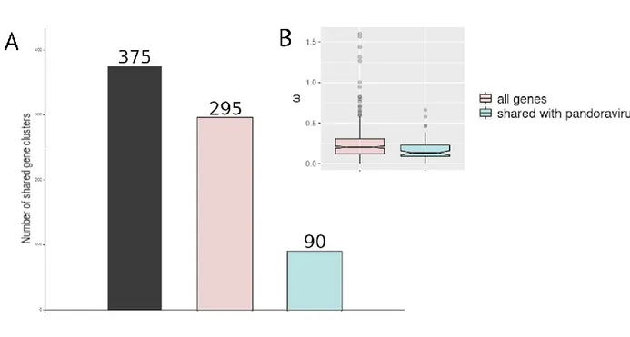
ribonucleoside-diphosphate reductase (present in pandoraviruses). The five M. kamchatka specific
194
proteins (Table 1) associated to functional motifs or domain signature correspond to:
195
- two proteins (mk_93 and mk_104) containing a type of zinc finger (Ring domain) mediating
196
protein interactions,
197
- one protein (mk_469) with similarity to the (BI)-1 like family of small transmembrane proteins,
198
- one predicted LexA-related signal peptidase (mk_166),
199
- one DNA methyltransferase (mk_92).
200
Evaluation of the selection pressure exerted on mollivirus genes 201
The availability of two distinct strains of mollivirus allows the first estimation of the selection 202
pressure exerted on their shared genes during their evolution. This was done by computing the 203
10 ratio ω=dN/dS of the rate of non-synonymous mutations (dN) over the rate of synonymous
204
mutations (dS) for pairs of orthologous genes. ω values much lesser than one are associated to 205
genes the mutation of which have the strongest negative impact on the virus fitness. The high 206
sequence similarity of proteins shared by M. sibericum and M. kamchatka allowed the generation 207
of flawless pairwise alignments and the computation of highly reliable ω values for most (i.e. 208
397/411) of their orthologous singletons. 209
Fourteen singleton pairs were not taken into account in the selection pressure analysis 210
because of their either identical or quasi-identical sequences (11 of them), or unreliable pairwise 211
alignments (3 of them). For the 397 gene pairs retained in the analysis, the mean ω value was 0.24
212
± 0.14 (Fig. 6). This result corresponds to a strong negative selection pressure indicating that most 213
of the encoded proteins greatly contribute to the molliviruses’ fitness. Together with the high level 214
of pairwise similarity (92%) of their proteins, this also indicates that M. kamchatka evolved very 215
little during the last 30,000 years and that the M. sibericum genome was not prominently damaged 216
during its cryostasis in permafrost. 217
The analysis restricted to the 244 pairs of ORFan-coding genes resulted in a very similar ω 218
value of 0.29± 0.15 (Fig. 6). This indicates that although homologs of these proteins are only found
219
in molliviruses, they have the same impact on the virus fitness than more ubiquitous proteins. This
220
confirms that they do encode actual proteins, albeit with unknown functions. In contrast, four
221
orthologous pairs (ms_160/mk_141; ms_280/mk_262; ms_171/mk_151; ms_430/mk_411; 222
ms_60/mk_48) exhibit ω value larger than one. Those ORFans either are under positive selection
223
for maintaining their functions, or newly created gene products undergoing refinement or
224
pseudogenization.
225
We further examined the selection pressure of proteins-coding genes with homologs in 226
pandoraviruses. We used their 10 sequenced genomes to generate the corresponding gene 227
clusters (Fig. 7). The 90 clusters shared by both virus groups included 64 singletons (single copy 228
11 gene present in all viruses), among which 55 were suitable for dN/dS computations. The mean ω 229
value (0.17 ± 0.1) was very low, indicating that these genes, forming a “super core” gene set
230
common to the molliviruses and pandoraviruses, are under an even stronger negative selection 231
pressure than those constituting the provisional (most likely overestimated) mollivirus core gene 232
set . 233
Genomic inhomogeneity 234
The original genome analysis of Lausannevirus (a member of the Marseilleviridae family) (17) 235
revealed an unexpected non-uniform distribution of genes according to their annotation. 236
“Hypothetical” genes (i.e. mostly ORFans) were segregated from “annotated” (i.e. mostly non-237
ORFans) in two different halves of the genome. In a more recent work, we noticed a similar bias in 238
the distribution of Pandoravirus core genes (13). The availability of a second mollivirus isolate gave 239
us the opportunity to investigate this puzzling feature for yet another group of Acantamoeba-240
infecting virus. In Fig. 8, we plotted the distribution of three types of genes: 1- those with 241
homologs in A. castellanii (n= 55 for M. sibericum and n= 51 for M. kamchatka), 2- those belonging 242
to the super core set shared by both molliviruses and pandoraviruses (n=64), 3- those unique to 243
either mollivirus strains (n=26 for M. sibericum, n=12 for M. kamchatka). These plots reveal a 244
strong bias in the distribution of the super core vs. ORFan genes (Fig. 8). The first half of the M. 245
sibericum genome exhibits 90% of its ORFans while the second half contains most of the members
246
of the super core gene set. In contrast, genes eventually exchanged with the host display a more 247
uniform distribution. The lack of an apparent segregation in the distribution of ORFans in the M. 248
kamchatka genome might be due to their underprediction as no transcriptome information is
249
available for this strain. Fig. 9 shows that there is also a strong bias in the distribution of single-250
copy genes vs. those with paralogs in either M. sibericum and/or M. kamchatka. Altogether, these 251
analyses suggest that the two genome halves follow different evolutionary scenario, the first half 252
12 concentrating the genomic plasticity (de novo gene creation, gene duplication), the other half 253
concentrating the most conserved, eventually essential, gene content. 254
255
Discussion 256
Following the discovery of their first representatives, each families of giant (e.g. Mimiviridae, 257
Pithoviridae, Pandoraviridae) and large (e.g. Marseilleviridae) viruses infecting acanthamoeba have 258
expanded steadily, suggesting they were relatively abundant and present in a large variety of 259
environments. One noticeable exception has been the molliviruses, the prototype of which 260
remained unique after its isolation from 30,000-year old permafrost. The absence of M. sibericum 261
relatives from the large number of samples processed by others and us since 2014, raised the 262
possibility that they might have gone extinct, or might be restricted to the Siberian arctic. Our 263
isolation of a second representative of the proposed Molliviridae family, M. kamtchatka, at a 264
location more than 1,500 km from the first isolate and enjoying a milder climate, is now refuting 265
these hypotheses. Yet, the planet-wide ubiquity of these viruses remains to be established, in 266
contrast to other acanthamoeba-infecting giant viruses (7). Even when present, mollivirus-like 267
viruses appear to be in very low abundance, as judged from the very small fraction of 268
metagenomics reads they represent in total sample DNA for M. kamchatka (about 0.02 part per 269
million) as well as for M. sibericum (about one part per million)(6). Another possibility would be 270
that the actual environmental host is not an acanthamoeba, the model host used in our laboratory. 271
However, evidences of specific gene exchanges with acanthamoeba (including a highly conserved 272
homolog major capsid protein) (6, 18, 19) make this explanation unlikely. We conclude that 273
members of the proposed Molliviridae family are simply less abundant than other acanthamoeba-274
infecting viruses, a conclusion further supported by the paucity of Mollivirus-related sequences in 275
the publicly available metagenomics data (data not shown). 276
13 As always the case, the characterization of a second representative of a new virus
277
representative opened new opportunities of analysis. Unfortunately, the closeness of M. 278
kamchatka with M. sibericum limited the amount of information that could be drawn from their
279
comparison. For instance, the number of genes shared by the two isolates is probably a large 280
overestimate of the “core” gene set characterizing the whole family. On the other hand, the 281
closeness of the two isolates allowed an accurate determination of the selection pressure 282
(ω=dN/dS) exerted on many genes, showing that most of them, including mollivirus ORFans, 283
encode actual proteins the sequence of which are under strong negative selection and thus 284
contribute to the virus fitness. Given the partial phylogenetic affinity (i.e. 90 shared gene clusters) 285
of the mollivirus with the pandoraviruses, we also assessed the selection pressure exerted on 55 of 286
these “super core” genes, and found them under even stronger negative selection (Fig. 7). This 287
suggests that this super core gene set might have been present in a common ancestor to both 288
proposed families. 289
If we postulate that M. sibericum underwent into a complete stasis when it became frozen in 290
permafrost while M. kamchatka remained in contact with living acanthamoeba, we could consider 291
the two viral genomes to be separated by at least 30,000 years of evolution (eventually more if 292
they are not in a direct ancestry relationship)(20). The high percentage of identical residues (92%) 293
in their proteins corresponds to a low substitution rate of 1.7 10-6 amino acid change/position 294
/year. This is an overestimate since the two viruses probably started to diverge from each other 295
longer than 30,000-year ago. This value is nevertheless comparable with estimates computed for 296
poxviruses (21) given the uncertainty on the number of replicative cycles occurring per year. The 297
high level of sequence similarity of M. kamchatka with M. sibericum also indicates that the later 298
did not suffer much DNA damage during its frozen stasis, even in absence of detectable virus-299
encoded DNA repair functions. 300
14 Horizontal gene transfers with the host were suggested by the fact that 51 proteins shared by 301
the two mollivirus strains exhibited a second best match in acanthamoeba. Because no homolog is 302
detected in other eukaryotes for most of them, these transfers probably occurred in the mollivirus-303
to-host direction. The clearest case is that of a major capsid protein homolog (mk_314, ml_347) 304
sharing 64% identical residues with a predicted acanthamoeba protein (locus: XP_004333827). 305
Two other genes encoding proteins that have also homologs in molliviruses flank the 306
corresponding host gene. However, the corresponding viral genes are not collinear in M. sibericum 307
or M. kamchatka and were probably transferred from a different, yet unknown mollivirus strain. 308
The presence of a 100% conserved major capsid protein homolog in the genome of M. kamchatka 309
and M. sibericum is itself puzzling. Such protein (with a double-jelly roll fold) is central to the 310
structure of icosahedral particles (22). Consistent with its detection in M. sibericum virions (6), its 311
conservation in M. kamchatka, suggests that it still plays a role in the formation of the spherical 312
mollivirus particles, while it has no homolog in the pandoraviruses. Inspired by previous 313
observations made on the unrelated Lausannevirus genome (17), we unveiled a marked 314
asymmetry in the distribution of different types of protein-coding genes in the Mollivirus genomes. 315
As shown in Fig. 8 the left half of the genome concentrates most of the genes coding for strain-316
specific ORFans while the right half concentrates most of super core genes shared with 317
pandoraviruses. This asymmetry is even stronger for the multiple copy genes while single-copy 318
genes are uniformly distributed along the genome (Fig. 9). The molliviruses thus appear to confine 319
their genomic “creativity” (de novo creation and gene duplication) in one-half of their genome, 320
leaving the other half more stable. An asymmetry in the distribution of the core genes was 321
previously noticed in the pandoravirus genomes (13). Such features might be linked to the 322
mechanism of replication that is probably similar for the two virus families. Further studies are 323
needed to investigate this process. The asymmetrical genomic distribution of pandoravirus core 324
genes and mollivirus super core genes might be a testimony of their past common ancestry. 325
15 Despite their differences in morphology, as well as in virion and genome sizes, the comparative 326
analysis of the prototype M. sibericum and of the new isolate M. kamchatka confirms their 327
phylogenetic affinity with the Pandoraviruses (Fig. 3, Fig. 10). However, it remains unclear whether 328
this is due to a truly ancestral relationship between them, or if it is only the consequence of 329
numerous past gene exchanges favored by the use of the same cellular host. From the perspective 330
of the sole DNA polymerase sequence, the two known molliviruses do cluster with the 331
pandoraviruses, albeit at a larger evolutionary distance than usually observed between members 332
of the same virus family (Fig. 10). In absence of an objective threshold, and pending the 333
characterization of eventual “missing links”, we thus propose to classify M. sibericum and M. 334
kamchatka as members of the proposed Molliviridae family, distinct from the Pandoraviridae.
335 336
Materials and Methods 337
Virus isolation 338
We isolated M. kamchatka from muddy grit collected near Kronotski Lake, Kamchatka (Russian 339
Federation N :54 32 59, E :160 34 55). The sample was stored for twenty days in pure rice medium 340
(23) at room temperature. An aliquot of the pelleted sample triggered an infected phenotype on a 341
culture of Acanthamoeba castellanii Neff (ATCC30010TM) cells adapted to 2,5µg/mL of 342
Amphotericin B (Fungizone), Ampicillin (100µg/ml), Chloramphrenicol (30µg/ml) and Kanamycin 343
(25µg/ml) in protease-peptone–yeast-extract–glucose (PPYG) medium after two days of incubation 344
at 32°C. A final volume of 6 mL of supernatant from two T25 flasks exhibiting infectious 345
phenotypes was centrifuged for 1 hour at 16,000xg at room temperature. Two T75 flasks were 346
seeded with 60,000 cells/cm² and infected with the resuspended viral pellet. Infected cells were 347
cultured in the same conditions as described below. We confirmed the presence of viral particles 348
by light microscopy. 349
Validation of the presence of M. kamchatka in the original sample 350
16 To confirm the origin of the M. kamchatka isolate from the soil of the Kronotsky river bank, 351
DNA was extracted from the sample and sequenced on an Illumina platform, leading to 352
340,320,265 pair-ended reads (mean length 150bp). These metagenomics reads were then 353
mapped onto the genome sequence of M. kamchatka. Seven matching (100 % Identity) pair-ended 354
reads (hence 14 distinct reads) were detected, indicating the presence of virus particles in the 355
original sample, although at very low concentration. However, the very low probability of such 356
matches by chance (p<10-63) together with the scattered distribution of these matches along the 357
viral genome, further demonstrate the presence of M. kamchatka in the original sample.
358 359
Virus Cloning 360
Fresh A. castellanii cells were seeded on a 12-well culture plate at a final concentration of 361
70,000cells/cm². Cell adherence was controlled under light microscopy after 45 minute and viral 362
particles were added to at a multiplicity of infection (MOI) around 50. After 1 h, the well was 363
washed 15 times with 3 mL of PPYG to remove any viral particle in suspension. Cells were then 364
recovered by gently scrapping the well, and a serial dilution was performed in the next three wells 365
by mixing 200μL of the previous well with 500μL of fresh medium. Drops of 0.5μL of the last 366
dilution were recovered and observed by light microscopy to confirm the presence of a unique A. 367
castellanii cell. The 0.5μL droplets were then distributed in each well of three 24-well culture plate.
368
Thousand uninfected A. castellanii cells in 500μL of PPYG were added to the wells seeded with a 369
single cell and incubated at 32°C until witnessing the evidence of a viral production from the 370
unique clone. The corresponding viral clones were recovered and amplified prior purification, DNA 371
extraction and cell cycle characterization by electron microscopy. 372
Virus mass production and purification 373
A total number of 40 T75 flasks were seeded with fresh A. castellanii cells at a final 374
concentration of 60,000 cells/cm². We controlled cell adherence using light microscopy after 45 375
17 minute and flasks were infected with a single clone of M. kamchatka at MOI=1. After 48h hours of 376
incubation at 32°C, we recovered cells exhibiting infectious phenotypes by gently scrapping the 377
flasks. We centrifuged for 10 min at 500×g to remove any cellular debris and viruses were pelleted 378
by a 1-hour centrifugation at 6,800xg. The viral pellet was then layered on a discontinuous cesium 379
chloride gradient (1,2g/cm²/ 1,3g/cm²/ 1,4g/cm²/ 1,5g/cm²) and centrifuged for 20h at 103,000xg. 380
The viral fraction produced a white disk, which was recovered and washed twice in PBS and stored 381
at 4 °C or −80 °C with 7,5% DMSO. 382
383
Infectious cycle observations using TEM 384
Twelve T25 flasks were seeded with a final concentration of 80,000 cells/cm² in PPYG medium 385
containing antibiotics. In order to get a synchronous infectious cycle eleven flasks were infected by 386
freshly produced M. kamchatka at substantial MOI=40. The A. castellanii infected flasks were fixed 387
by adding an equal volume of PBS buffer with 5 % glutaraldehyde at different time points after the 388
infection : 1h pi, 2h pi, 3h pi, 4h pi, 5h pi, 6h pi, 7h pi, 8h pi, 9h pi, 10h pi and 25h pi. After 45min of 389
fixation at room temperature, cells were scrapped and pelleted for 5min at 500xg. Then cells were 390
resuspended in 1ml of PBS buffer with 2,5 % glutaraldehyde and stored at 4°C. Each sample was 391
coated in 1mm³ of 2 % low melting agarose and embedded in Epon-812 resin. Optimized osmium-392
thiocarbohydrazide-osmium (OTO) protocol was used for staining the samples: 1h fixation in PBS 393
with 2 % osmium tetroxide and 1.5 % potassium ferrocyanide, 20 min in water with 1 % 394
thiocarbohydrazide, 30min in water with 2 % osmium tetroxide, overnight incubation in water with 395
1 % uranyl acetate and finally 30min in lead aspartate. Dehydration was made using an increasing 396
concentration of ethanol (50 %, 75 %, 85 %, 95 %, 100 %) and cold dry acetone. Samples were 397
progressively impregnated with an increasing mix of acetone and Epon-812 resin mixed with DDSA 398
0,34v/v and NMA 0,68v/v (33 %, 50 %, 75 % and 100%). Final molding was made using a hard 399
Epon-812 mix with DDSA 0,34v/v NMA 0,68v/v and 0,031v/v of DMP30 accelerator and hardened 400
18 in the oven at 60°C for 5 days. Ultrathin sections (90nm thick) were observed using a FEI Tecnai G2 401
operating at 200kV. 402
DNA Extraction 403
M. kamchatka genomic DNA was extracted from approximately 5 x 10⁹ purified virus particles
404
using Purelink Genomic extraction mini kit according to the manufacturer’s recommendation. Lysis 405
was performed with in a buffer provided with the kit and extra DTT at a final concentration of 406
1mM. 407
Genome sequencing and assembly 408
M. kamchatka genome was assembled using Spades (24) with a stringent K-mer parameter
409
using both various iteration steps (k = 21,41,61,81,99,127), the “-careful” option to minimize 410
number of mismatches in the final contigs, and the “-nanopore” option to use long reads. 411
Annotation of Mollivirus sibericum and Mollivirus kamchatka 412
A stringent gene annotation of M. sibericum was performed as previously described (13) using 413
RNA-seq transcriptomic data (6). Stranded RNA-seq reads were used to accurately annotate 414
protein-coding genes. Stringent gene annotation of M. kamchatka was performed w/o RNA seq 415
data but taking into account protein similarity with M. sibericum. Gene predictions were manually 416
curated using the web-based genomic annotation editing platform Web Apollo (25). Functional 417
annotations of protein-coding genes of both genomes were performed using a two-sided approach 418
as already previously described (13). Briefly, protein domains were searched with the CD-search 419
tool (26) and protein sequence searching based on the pairwise alignment of hidden Markov 420
models (HMM) was performed against the Uniclust30 database using HHblits tool (27). Gene 421
clustering was done using Orthofinder’s default parameters (28) adding the “-M msa -oa” option. 422
Strict orthology between pairs of proteins was confirmed using best reciprocal blastp matches. 423
Selection Pressure Analysis 424
19 Ratios of non-synonymous (dN) over synonymous (dS) mutation rates for pairs of orthologous 425
genes were computed from MAFFT global alignment (29) using the PAML package and codeml with 426
the « model = 2 » (30). A strict filter was applyied to the dN/dS ratio: dN > 0, dS > 0, dS ≤ 2 and 427
dN/dS ≤ 10. The computation of the Codon Adaptation Index (CAI) of both Mollivirus was 428
performed using the cai tool from the Emboss package (31). 429
Metagenome sequencing, assembly and annotation 430
All metagenomic sample were sequenced with DNA-seq paired-end protocol on Illumina HiSeq 431
platform at Genoscope producing 16 datasets of 2x150bp read length. Raw reads quality was 432
evaluated with FASTQC (32). Identified contaminants were removed and remaining reads were 433
trimmed on the right end using 30 as quality threshold with BBTools (33). Assemblies of filtered 434
data sets were performed using MEGAHIT (34) with the following options: “--k-list 33,55,77,99,127 435
--min-contig-len 1000”. All filtered reads were then mapped to the generated contigs using 436
Bowtie2 (35) with the “--very-sensitive” option. 437
Availability of data 438
The M. kamtchatka annotated genome sequence is freely available from the public through the 439
Genbank repository (URL://www.ncbi.nlm.nih.gov/genbank/) under accession number XXXXX. 440
441
Acknowledgements 442
We are deeply indebted to our volunteer collaborator Alexander Morawitz for collecting the 443
Kamchatka soil sample. We thank N. Brouilly, F. Richard and A. Aouane (imagery platform, Institut 444
de Biologie du Développement de Marseille Luminy) for their expert assistance. E Christo-Foroux is 445
the recipient of a DGA-MRIS scholarship (201760003). This project has received funding from the 446
European Research Council (ERC) under the European Union's Horizon 2020 research and 447
innovation program (grant agreement No 832601) and from the FRM prize “Lucien Tartois” to C. 448
20 Abergel. The funding bodies had no role in the design of the study, analysis, and interpretation of 449
data and in writing the manuscript. 450
Competing interests 451
The authors declare that they have no competing interests 452
21 Figures
454 455
456
Fig 1. Ultrathin section TEM image of a neo-synthetized M. kamchatka particle in the cell 457
cytoplasm 7h post infection. The structure of the mature particles appear identical to that of M. 458
sibericum.
459 460
22 461
462
Fig 2. Ultrathin section TEM image of A. castellanii cell 7 to 10 hours post infection by M. 463
kamchatka. (A) Viral factory exhibiting fibrils (F), a nascent viral particle (V), and surrounding
464
mitochondria (M). Fragments of the ruptured nuclear membrane are visible as dark bead strings. 465
(B) Details of a nuclear membrane rupture through which fibrils synthetized in the nucleus (N) are 466
shed into the cytoplasm (C). 467
23 469
470
471
Fig 3. Distribution of the best-matching NR homologs of M. kamchatka predicted proteins. Best-472
matching homologous proteins were identified using BLASTP (E value <10–5) against the non-473
redundant (NR) database (15) (after excluding M. sibericum). Green shades are used for 474
eukaryotes, red shades for viruses. 475
24 477
478 479
Fig 4. Eventual gene transfers from a pandoravirus to M. kamchatka. Both phylogenetic trees were 480
computed from the global alignments of orthologous protein sequences using MAFFT (29). IQtree 481
(36) was used to determine the optimal substitution model (options: « -m TEST » and « –bb 482
1000 »). (A) Protein mk_165 (no predicted function). (B) Predicted methyltransferase mk_92. Both 483
M. kamchatka protein sequences appear embedded within the pandoravirus trees. In (A), the long
484
branch leading to the M. kamchatka homolog suggests its accelerated divergence after an ancient 485
acquisition from a pandoravirus. In (B), the long branch leading to the P. dulcis homolog might 486
alternatively be interpreted as a non-orthologous replacement of the ancestral pandoravirus 487
version of the gene. 488
25 489
490 491
Fig 5. Genomic features of strain-specific ORFans. (A) Codon adaption index (CAI). (B) G+C content. 492
(C) Protein length. Box plots show the median, the 25th and 75th percentiles. P-value are 493
calculated using the Wilcoxon test. 494
26 495
496
Fig 6. Selection pressure among different classes of genes. Values of ω (i.e. dN/dS) were computed 497
from the alignments of homologous coding regions in M. kamchatka and M. sibericum. (A) 498
Distribution of calculated ω values (n=397). (B) Box plots of the ω ratio among ORFan genes 499
(n=243) and non ORFan genes (n=154). Box plots show the median, the 25th, and 75th percentiles. 500
All p-value are calculated using the Wilcoxon test. 501
27 503
504
Fig 7. Comparison of the mollivirus and pandoravirus core gene contents. (A) The distribution of 505
the protein clusters shared by all pandoraviruses (black), the two Molliviruses (pink), and by both 506
virus groups (super core genes) (blue). (B) Box plot of ω values calculated from the alignment of 507
molliviruses core genes (pink), and super core genes (blue). Box plots show the median, the 25th, 508
and 75th percentiles. 509
28 511
Fig 8. Distribution of different classes of genes along mollivirus genomes. (A) Variation of the gene 512
density as computed by the ggpplot2 “geom_density” function (37). Genes with best-matching 513
homologs in A. castellanii (in the NR database excluding mollivirus) are uniformly distributed (in 514
pink) in contrast to super core genes (in green) and strain-specific ORFans (in blue). (B) Cumulative 515
distribution of the above classes of genes using the same color code. 516
517 518
29 519
520
Fig 9. Distribution of single-copy vs. multiple copy genes along mollivirus genomes. Single copy 521
genes (in blue) in both strains are uniformly distributed in contrast to genes with paralogs (pink) in 522
at least one strain that cluster in the left half of the genomes. (A) M. sibericum (n=48). (B) M. 523
kamchatka (n= 46).
30 525
526
Fig 10. Phylogeny of DNA polymerase B of large and giant dsDNA viruses. This neighbor-joining tree 527
was computed (JTT substitution model, 100 resampling) on 397 amino acid positions from an 528
alignment of 42 sequences computed by MAFFT (29). Branches with boostrap values <60% were 529
collapsed. 530
31 Tables
531 532
Table 1. Status of the protein-coding genes unique to M. kamchatka 533
ORF ID Predicted function Putative evolutionary scenario
mk_25 None (ORFan) de novo creation or loss in M. sibericum
mk_92 DNA methyltransferase loss in M. sibericum (present in some pandoraviruses) mk_93 Ring domain loss in M. sibericum (present in some pandoraviruses) mk_104 Ring domain loss in M. sibericum (present in some pandoraviruses) mk_127 None (ORFan) de novo creation or loss in M. sibericum
mk_159 None loss in M. sibericum (present in some pandoraviruses)
mk_165 None HGT from Pandoravirus
mk_166 Peptidase loss in M. sibericum (present in some pandoraviruses) mk_172 None (ORFan) de novo creation or loss in M. sibericum
mk_182 None (ORFan) de novo creation or loss in M. sibericum
mk_231 None (ORFan) de novo creation or loss in M. sibericum
mk_313 None (ORFan) de novo creation or loss in M. sibericum
mk_369 None (ORFan) de novo creation or loss in M. sibericum
mk_415 None (ORFan) de novo creation or loss in M. sibericum
mk_441 None (ORFan) de novo creation or loss in M. sibericum
mk_466 None (ORFan) de novo creation or loss in M. sibericum
mk_467 None loss in M. sibericum (HGT to Acanthamoeba)
mk_469 B1-1 like loss in M. sibericum (present in Acanthamoeba) mk_476 None (ORFan) de novo creation or loss in M. sibericum
mk_478 None (ORFan) de novo creation or loss in M. sibericum
32 Table 2. Status of the protein-coding genes unique to M. sibericum
535
ORF ID Predicted function Putative evolutionary scenario
ms_1 None (ORFan) De novo creation or loss in M. kamchatka
ms_3 None (ORFan) De novo creation or loss in M. kamchatka
ms_5 None (ORFan) De novo creation or loss in M. kamchatka
ms_7 None (ORFan) De novo creation or loss in M. kamchatka
ms_8 None (ORFan) De novo creation or loss in M. kamchatka
ms_13 None (ORFan) De novo creation or loss in M. kamchatka
ms_14 None HGT to Acanthamoeba
ms_38 None (ORFan) De novo creation or loss in M. kamchatka
ms_42 None (ORFan) De novo creation or loss in M. kamchatka
ms_53 None (ORFan) De novo creation or loss in M. kamchatka
ms_64 None loss in M. Kamchatka (present in Noumeavirus)
ms_109 None (ORFan) De novo creation or loss in M. kamchatka
ms_120 None (ORFan) De novo creation or loss in M. kamchatka
ms_136 None (ORFan) De novo creation or loss in M. kamchatka
ms_138 None (ORFan) De novo creation or loss in M. kamchatka
ms_139 None (ORFan) De novo creation or loss in M. kamchatka
ms_144 None (ORFan) De novo creation or loss in M. kamchatka
ms_157 None (ORFan) De novo creation or loss in M. kamchatka
ms_159 None (ORFan) De novo creation or loss in M. kamchatka
ms_166 None (ORFan) De novo creation or loss in M. kamchatka
ms_172 None (ORFan) De novo creation or loss in M. kamchatka
ms_190 None loss in M. kamchatka (present in some pandoraviruses)
ms_193 None (ORFan) De novo creation or loss in M. kamchatka
ms_246 None (ORFan) De novo creation or loss in M. kamchatka
ms_258 None (ORFan) De novo creation or loss in M. kamchatka
ms_311 Zinc-finger domain loss in M. kamchatka (present in Gossypium hirsutum) ms_312 Zinc-finger domain loss in M. kamchatka (present in Cavenderia fasciculata)
ms_313 None loss in M. kamchatka (present in some pandoraviruses)
ms_464 None (ORFan) De novo creation or loss in M. kamchatka
ms_465 None (ORFan) De novo creation or loss in M. kamchatka
ms_479 DNA methyltransferase loss in M. kamchatka (present in some pandoraviruses) ms_494 None (ORFan) De novo creation or loss in M. kamchatka
ms_495 None (ORFan) De novo creation or loss in M. kamchatka
536 537
33 References
538
1. Shi T, Reeves RH, Gilichinsky DA, Friedmann EI, 1997. Characterization of Viable Bacteria from 539
Siberian Permafrost by 16S rDNA Sequencing. Microb Ecol 33:169-179. 540
https://doi.org/10.1007/s002489900019. 541
2. Vishnivetskaya T, Kathariou S, McGrath J, Gilichinsky D, Tiedje JM 2000. Low-temperature 542
recovery strategies for the isolation of bacteria from ancient permafrost sediments. Extremophiles 543
4:165-173. https://doi.org/10.1007/s007920070031. 544
3. Graham DE, Wallenstein MD, Vishnivetskaya TA, Waldrop MP, Phelps TJ, Pfiffner SM, Onstott TC, 545
Whyte LG, Rivkina EM, Gilichinsky DA, Elias DA, Mackelprang R, VerBerkmoes NC, Hettich RL, 546
Wagner D, Wullschleger SD, Jansson JK. 2012. Microbes in thawing permafrost: the unknown 547
variable in the climate change equation. ISME J 6:709-712. 548
https://doi.org/10.1038/ismej.2011.163. 549
4. Yashina S, Gubin S, Maksimovich S, Yashina A, Gakhova E, Gilichinsky D. 2012. Regeneration of 550
whole fertile plants from 30,000-y-old fruit tissue buried in Siberian permafrost. Proc Natl Acad Sci 551
U S A 109:4008-4013. https://doi.org/10.1073/pnas.1118386109. 552
5. Legendre M, Bartoli J, Shmakova L, Jeudy S, Labadie K, Adrait A, Lescot M, Poirot O, Bertaux L, 553
Bruley C, Couté Y, Rivkina E, Abergel C, Claverie JM. 2014. Thirty-thousand-year-old distant relative 554
of giant icosahedral DNA viruses with a pandoravirus morphology. Proc Natl Acad Sci U S A 555
111:4274-4279. https://doi.org/ 10.1073/pnas.1320670111. 556
6. Legendre M, Lartigue A, Bertaux L, Jeudy S, Bartoli J, Lescot, M, Alempic JM, Ramus C, Bruley C, 557
Labadie K, Shmakova L, Rivkina E, Couté Y, Abergel C, Claverie JM. 2015. In-depth study of 558
Mollivirus sibericum, a new 30,000-y-old giant virus infecting Acanthamoeba. Proc Natl Acad Sci U 559
S A 112:E5327-E5335. https://doi.org/10.1073/pnas.1510795112. 560
34 7. Abergel C, Legendre M, Claverie JM. The rapidly expanding universe of giant viruses: Mimivirus, 561
Pandoravirus, Pithovirus and Mollivirus. 2015. FEMS Microbiol Rev 39:779-796. 562
https://doi.org/10.1093/femsre/fuv037. 563
8. Levasseur A, Andreani J, Delerce J, Bou Khalil J, Robert C, La Scola B, Raoult D. 2016. Comparison 564
of a Modern and Fossil Pithovirus Reveals Its Genetic Conservation and Evolution. Genome Biol 565
Evol 8:2333-2339. https://doi.org/10.1093/gbe/evw153. 566
9. Andreani J, Aherfi S, Bou Khalil JY, Di Pinto F, Bitam I, Raoult D, Colson P, La Scola B. 2016. 567
Cedratvirus, a Double-Cork Structured Giant Virus, is a Distant Relative of Pithoviruses. Viruses 568
8:300. https://doi.org/10.3390/v8110300. 569
10. Bertelli C, Mueller L, Thomas V, Pillonel T, Jacquier N, Greub G. 2017. Cedratvirus lausannensis - 570
digging into Pithoviridae diversity. Environ Microbiol 19:4022-4034. 571
https://doi.org/10.1111/1462-2920.13813. 572
11. Quemin ER, Corroyer-Dulmont S, Baskaran A, Penard E, Gazi AD, Christo-Foroux E, Walther P, 573
Abergel C, Krijnse-Locker J. 2019. Complex membrane remodeling during virion assembly of the 574
30,000-year-old Mollivirus sibericum. J Virol 93(13). pii: e00388-19. 575
https://doi.org/10.1128/JVI.00388-19. 576
12. Philippe N, Legendre M, Doutre G, Couté Y, Poirot O, Lescot M, Arslan D, Seltzer V, Bertaux L, 577
Bruley C, Garin J, Claverie JM, Abergel C. 2013. Pandoraviruses: amoeba viruses with genomes up 578
to 2.5 Mb reaching that of parasitic eukaryotes. Science 341:281-286. 579
https://doi.org/10.1126/science.1239181. 580
13. Legendre M, Fabre E, Poirot O, Jeudy S, Lartigue A, Alempic JM, Beucher L, Philippe N, Bertaux 581
L, Christo-Foroux E, Labadie K, Couté Y, Abergel C, Claverie JM. 2018. Diversity and evolution of the 582
emerging Pandoraviridae family. Nat Commun 9:2285. 583
https://doi.org/10.1038/s41467-018-04698-4. 584
35 14. Legendre M, Alempic JM, Philippe N, Lartigue A, Jeudy S, Poirot O, Ta NT, Nin S, Couté Y,
585
Abergel C, Claverie JM. 2019. Pandoravirus celtis illustrates the microevolution processes at work 586
in the giant Pandoraviridae genomes. Front Microbiol 10:430. https://doi.org/ 587
10.3389/fmicb.2019.00430. 588
15. NCBI Resource Coordinators. 2018. Database resources of the National Center for 589
Biotechnology Information. Nucleic Acids Res 46:D8-D13. https://doi.org/10.1093/nar/gkx1095. 590
16. Heidel AJ, Lawal HM, Felder M, Schilde C, Helps NR, Tunggal B, Rivero F, John U, Schleicher M, 591
Eichinger L, Platzer M, Noegel AA, Schaap P, Glöckner G. 2011. Phylogeny-wide analysis of social 592
amoeba genomes highlights ancient origins for complex intercellular communication. Genome Res 593
21:1882-1891. https://doi.org/10.1101/gr.121137.111. 594
17. Thomas V, Bertelli C, Collyn F, Casson N, Telenti A, Goesmann A, Croxatto A, Greub G. 2011. 595
Lausannevirus, a giant amoebal virus encoding histone doublets. Environ Microbiol 13:1454-1466. 596
https://doi.org/10.1111/j.1462-2920.2011.02446.x. 597
18. Maumus F, Blanc G. 2016. Study of gene trafficking between acanthamoeba and giant viruses 598
suggests an undiscovered fof amoeba-infecting viruses. Genome Biol 599
Evol 8:3351-3363. https://doi.org/10.1093/gbe/evw260. 600
19. Chelkha N, Levasseur A, Pontarotti P, Raoult D, Scola B, Colson P. 2018. A phylogenomic study of 601
Acanthamoeba polyphaga draft genome sequences suggests genetic exchanges with giant viruses.
602
Front Microbiol 9:2098. https://doi.org/10.3389/fmicb.2018.02098. 603
20. Duchêne S, Holmes EC. 2018. Estimating evolutionary rates in giant viruses using 604
ancient genomes. Virus Evol 4:vey006. https://doi.org/10.1093/ve/vey006. 605
21. Hughes AL, Irausquin S, Friedman R. 2010. The evolutionary biology of poxviruses. Infect Genet 606
Evol 10:50-59. https://doi.org/10.1016/j.meegid.2009.10.001. 607
22. San Martín C, van Raaij MJ. 2018. The so far farthest reaches of the double jelly roll capsid 608
protein fold. Virol J 15:181. https://doi.org/10.1186/s12985-018-1097-1. 609
36 23. Arslan D, Legendre M, Seltzer V, Abergel C, Claverie JM. 2011. Distant Mimivirus relative with a 610
larger genome highlights the fundamental features of Megaviridae. Proc Natl Acad Sci U S A 611
108:17486-17491. https://doi.org/10.1073/pnas.1110889108. 612
24. Bankevich A, Nurk S, Antipov D, Gurevich A, Dvorkin M, Kulikov AS, Lesin V, Nikolenko S, Pham 613
S, Prjibelski A, Pyshkin A, Sirotkin A, Vyahhi N, Tesler G, Alekseyev MA, Pevzner P. 2012. SPAdes: A 614
New Genome Assembly Algorithm and Its Applications to Single-Cell Sequencing. J Comput Biol 615
19:455-477. https://doi.org/10.1089/cmb.2012.0021. 616
25. Dunn NA, Unni DR, Diesh C, Munoz-Torres M, Harris NL, Yao E, Rasche H, Holmes I, Elsik C, 617
Lewis S. 2019. Apollo: Democratizing genome annotation. PLoS Comput Biol 15: e1006790. 618
https://doi.org/10.1371/journal.pcbi.1006790. 619
26. Marchler-Bauer A, Derbyshire MK, Gonzales NR, Lu S, Chitsaz F, Geer LY, Geer RC, He J, Gwadz 620
M, Hurwitz DI, Lanczycki CJ, Lu F, Marchler GH, Song JS, Thanki N, Wang Z, Yamashita RA, Zhang D, 621
Zheng C, Bryant SH. 2015. CDD: NCBI's conserved domain database. Nucleic Acids Res 43:D222-622
226. https://doi.org/10.1093/nar/gku1221. 623
27. Remmert M, Biegert A, Hauser A, Söding J. 2011. HHblits: lightning-fast iterative 624
protein sequence searching by HMM-HMM alignment. Nat Methods. 9:173-175. 625
https://doi.org/10.1038/nmeth.1818. 626
28. Emms DM, Kelly S. 2015. OrthoFinder: solving fundamental biases in whole genome 627
comparisons dramatically improves orthogroup inference accuracy. Genome Biol 16:157. 628
https://doi.org/10.1186/s13059-015-0721-2. 629
29. Katoh K, Misawa K, Kuma K-I, Miyata T. 2002. MAFFT: a novel method for rapid multiple 630
sequence alignment based on fast Fourier transform. Nucleic Acids Res 30:3059-3066. 631
https://doi.org/10.1093/nar/gkf436. 632
30. Yang Z. 2007. PAML 4: phylogenetic analysis by maximum likelihood. Mol Biol Evol 24:1586-633
1591. https://doi.org/10.1093/molbev/msm088. 634
37 31 . Rice P, Longden I, Bleasby A. 2000. EMBOSS: the European molecular biology open software 635
suite. Trends Genet 16:276-277. https://doi.org/10.1016/s0168-9525(00)02024-2. 636
32. Andrews S. 2010. FastQC: a quality control tool for high throughput sequence data. Available 637
online at: http://www.bioinformatics.babraham.ac.uk/projects/fastqc . 638
33. Bushnell B, Rood J, Singer E. 2017. BBMerge – Accurate paired shotgun read merging via 639
overlap. PLoS ONE 12: e0185056. https://doi.org/10.1371/journal.pone.0185056. 640
34. Li D, Liu C-M, Luo R, Sadakane K, Lam T-W. 2015. MEGAHIT: An ultra-fast single-node solution 641
for large and complex metagenomics assembly via succinct de Bruijn graph. Bioinformatics 642
31:1674-1676. https://doi.org/10.1093/bioinformatics/btv033. 643
35. Langmead B, Salzberg SL. 2012. Fast gapped-read alignment with Bowtie 2. Nat Methods 644
9:357-359. https://doi.org/10.1038/nmeth.1923. 645
36. Nguyen LT, Schmidt HA, von Haeseler A, Minh BQ. 2015. IQ-TREE: A fast and effective stochastic 646
algorithm for estimating maximum likelihood phylogenies. Mol Biol Evol 32:268-274. 647
https://doi.org/10.1093/molbev/msu300. 648
37. Wickham H. ggplot2: Elegant Graphics for Data Analysis, p 33-74. 2016. Springer-Verlag New 649
York. 650