Materials Today: Proceedings 1S ( 2014 ) 221 – 224
2214-7853 © 2014 The Authors. Published by Elsevier Ltd. This is an open access article under the CC BY-NC-ND license (http://creativecommons.org/licenses/by-nc-nd/3.0/).
Selection and Peer-review under responsibility of Physics Department, University of Namur. doi: 10.1016/j.matpr.2014.09.026
Available online at www.sciencedirect.com
ScienceDirect
Living Light: Uniting biology and photonics – A memorial meeting in honour of Prof Jean-Pol
Vigneron
Multiscale replication of iridescent butterfly wings
M. Thomé
a, L. Nicole
b, S. Berthier
a,c*
aInstitut des NanoSciences de Paris, UMR 7588, 4 Place Jussieu, 75005 Paris, France.
bSorbonne Universités, UPMC Univ Paris 06, UMR 7574, Chimie de la Matière Condensée de Paris, F-75005, Paris, France. cUniversité Paris Diderot, 5 rue Thomas-Mann, 75013 Paris, France.
Abstract
Natural photonic structures have been extensively studied and have shown their interest as a source of inspiration for new bio-inspired devices in many areas. After these initial studies and characterization phases, we have now to reproduce these structures, mainly in inorganic materials, to exacerbate interesting effects or generate new ones. If we want to preserve the best of their multi-scale and more or less ordered structures, producing a molding seems more appropriate. Such prints can be achieved by physical or chemical means, the latter being a priori particularly suitable for three-dimensional structures.
© 2014 The Authors. Published by Elsevier Ltd.
Selection and Peer-review under responsibility of Physics Department, University of Namur. This is an open access article under the CC BY-NC-ND license (http://creativecommons.org/licenses/by-nc-nd/3.0/).
Keywords: molding ; Morpho ; PVD ; sol-gel ; photonic structure
1. Introduction
To manage the physical and chemical exchanges between the outside world and living organisms, and enable them to cope with the various constraints that apply to them, evolution has led to the development of many different structures at any scales [1, 2]. The general characteristics of these natural structures are: (a) Multifunctionality. Structures systematically assume several vital functions for the body and are optimized on average for all of these functions. (b) Unlike our artificial devices, natural structures use only very few elements of the periodic table. (c) These two characteristics, doing more with less, impose a multi-scaled complex structure, with a controlled disorder
* Corresponding author. Tel.: +33 1 44 27 40 85; fax: +33 1 44 27 39 82.
E-mail address: [email protected]
© 2014 The Authors. Published by Elsevier Ltd. This is an open access article under the CC BY-NC-ND license (http://creativecommons.org/licenses/by-nc-nd/3.0/).
222 M. Thomé et al. / Materials Today: Proceedings 1S ( 2014 ) 221 – 224
(Fig.1) [3-8]. Despite progresses recently reported [9], these latter characteristics are generally difficult to artificially reproduce using conventional techniques of nano-structuring. In the case of butterfly wings, various chemical and physical routes have been thus investigated for replicating natural architectures including physical vapor deposition (PVD), chemical vapor deposition, atomic layer deposition, and chemical solution deposition (CSD) [10-13]. These approaches give rise either to a thin positive replica, natural structures serving as scaffolds, or a negative one, natural structures being used as molds. At this stage, two general remarks concerning replica and deposition could be done: (a) Replica are rather brittle and present usually a limited size after biotemplate removal (a few tens of microns) which limit their further handling, use, and integration into more complex devices. (b) A given deposition technique could be more or less adapted to a natural structure depending on its periodicity dimension (1D, 2D, 3D). Indeed, physical deposition methods, rather directional are well suited to two-dimensional but could be less efficient for most of the three dimensional structures, unlike chemical solution deposition methods, which allow a good infiltration of the 3D structures. In this article, we compare two deposition techniques (PVD and CSD) to produce thick negative replica of multi-scale and three-dimensional structures of iridescent butterfly wings.
2. Butterfly wings replication
The two butterflies that we used as biotemplate are males of the Morphidae family: Morpho rhetenor (Fig. 1) and
Morpho menelaus. The wing are covered by different types of scales, cover and ground photonic scales
(approximately 100 ȝm x 50 µm), each scale being itself covered by a grating of ridges (1ȝm apart). The ridges are composed of a stack of lamellae (50 nm thick) which acts as a multilayered air – chitin film. Optical thicknesses of the layers are such that only blue waves interfere constructively [4, 7, 14].
Fig. 1. The different scales used to observe butterfly wings (M. rhetenor male) and their units of measure: (a) Dorsal side of the wing; (b) Organization of the scales (optical microscopy); (c) SEM view of a photonic scale; (d) TEM view of a transversal cut in a photonic scale. [7, 14]
Physical vapor deposition of SiO2 was prepared by RF sputtering using a diode module having a power supply
operating at 13.5 MHz, a disk-shaped electrode of 13 cm in diameter and a SiO2 target to substrate distance of 4 cm.
The base pressure of the vacuum chamber is 10-6 Torr and the sputtering takes place in an argon atmosphere at a pressure of 10 mTorr and at a temperature of 100°C. It is worth mentioning that the multiscale structure of butterfly wings is preserved in similar experimental conditions (blank test), i.e. low pressure and 200°C, since slight color variations of the wing are observed. This observation is confirmed by TGA analysis performed on butterfly wings which displays not significant loss below 200°C. The silica deposited layer on the wing of a Morpho rhetenor is about 2 microns thick after 13h.
Chemical solution deposition used in this study combines sol-gel chemistry, solution evaporation process and dip-coating. This method consists in the deposition of a solution of precursors containing titanium isopropoxide, acetylacetone (acac) and ethanol (EtOH) as solvent (molar ratio: 1 Ti : 2 acac : 10 EtOH). The titania-based film was deposited at withdrawal speed of 0.68 cm.s-1 in a relatively dry atmosphere (relative humidity RH = 15 %). In order to promote controlled hydrolysis-condensation reactions, samples were next aging 24 hours at 35°C and RH = 75%. The titania-based layer coated on the wing of a Morpho menelaus is about 2 microns thick.
SEM images were obtained with a SEM Hitachi S-3400N and a Zeiss Neon40 ESB CrossBeam SEM-FEG with FIB. TGA experiments were performed with a TGA Netzsch STA 409 PC. Ellipsometry measurements were done with M-2000U Woollam spectroscopic ellipsometer.
223 M. Thomé et al. / Materials Today: Proceedings 1S ( 2014 ) 221 – 224
3. Physical vapor deposition vs chemical solution deposition
With regard to the multiscale structure of butterfly wings, we have adopted the fourth-level observation method for analyzing our replica: wing, scales, ridges and lamellae [7, 14]. From a macroscopic point of view, wings appear white after silica PVD deposition and brown after titania-based CSD. Such difference could be explained by the scattering of light due to the micron-size of the silica dense layer and by the filling of the fine structure (lamellae) of wings by titania precursors. It is important to notice that the refractive index of the very slightly colored TiO2-based
layer is far lower than those of crystallized TiO2 species (anatase and rutile phases) since this layer is composed of
amorphous and partially condensed oxo – alkoxo – ȕ-diketonate – titanium species [15]. The brown color could thus correspond to the color of natural brown pigments on the inferior sides of the structural scales which are observable due to close refractive indexes of the chitineous matrix (n550=1.56) and the poorly condensed hybrid TiO2-based
layer (n550=1.73).
Fig. 2. (a, b) SEM images of M. rhetenor scales after physical deposition of SiO2; (c, d) SEM images of M. menelaus scales after sol-gel
deposition of TiO2.
At a microscopic scale, we observe that PVD preserves the scales and the ridges array (Fig. 2.a, 2.b). Concerning the CSD, cover scales are curved (Fig. 2.c) whereas ground photonic scales are not damaged by the deposited layer (Fig. 2.d). This difference of behavior between curved cover and ground scales towards CSD is most probably due to the absence of the melanin in the cover scales, compound known to improve rigidity of natural structures and to their difference of inter-ridges spacing. Ground scales inter-ridges spacing are twice smaller than cover scales ones which could favor their further deformation upon solvent evaporation and condensation of titania precursor. This tendency to curve has been already observed with CSD and has been overcome by clamping the biotemplate between two glass substrates [13, 16]. With the CSD, ridges are no more observed, meaning thus that a thick sol-gel layer is deposited on the scales. It explains also the presence of cracks on the scales (Fig. 2.d), a well-known phenomenon in sol-gel caused by important capillary stresses [17]. Unfortunately, cracks could contribute to the fragility of the whole replica limiting thus the further handling of negative replica. These cracks could be more or less important depending on the solution composition, processing conditions and film thickness. Cracks use to increase with the inorganic content in the coating, i.e. for low Ti / acac molar ratio, with the concentration in non-volatile species inside the solution and with the thickness of the coated layer.
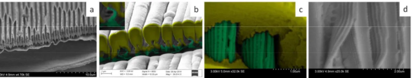
At the lower scale (ridges and lamellae), one can observe that many ridges of natural scales tend to collapse two by two (Fig. 3.a). This is a phenomenon often observed in natural wings which could dramatically impede the rate of impregnation and thus the quality of replication. After physical deposition (Fig. 3.b), one can easily observed the classical columnar growth with cathodic sputtering technique (Volmer-Weber growth). The ridges are bent and narrowed of about half of their initial height (~ 1ȝm instead of ~ 2ȝm for natural scales). This narrowing concern also lamellae which are atrophied (inset Fig. 3.b). The number of ridges apparently covered by the silica layer is half of the ridges number of the natural structure which could be inherent to sputtering-based techniques, i.e. joined growth between two adjacent ridges. Moreover, cathodic sputtering results in a partial impregnation of the biotemplate. On the contrary, with CSD, ridges are less damaged and deformed (Fig. 3.c). Besides, CSD allows a better impregnation of the fine structure (Fig. 3.d). Indeed, ethanol favors a high penetration of non-volatile species inside nanoscopic voids (inter-lamellae spaces) because of its high wetting property (low surface tension) with hydrophobic/hydrophilic surfaces. In addition to that, its high volatility at room temperature and ambient pressure is more respectful of the fragile organic nature of butterfly wings. And finally, it solubilizes a large number of components which may constitute the initial solution (metal alkoxides, organic inhibitors, surfactants, water ...) [18].
224 M. Thomé et al. / Materials Today: Proceedings 1S ( 2014 ) 221 – 224
Fig. 3. (a) SEM view of a cut across the ridges of Morpho rhetenor; (b) FIB cut of the deposit produced by PVD (silica in yellow, the remaining chitin is in green); (c) Crack view in the TiO2 layer (TiO2 in yellow, chitin in green); (d) Defects view in TiO2 layer.
4. Conclusion
We have presented two deposit methods that allow us to make a molding of the wing at any levels of their structures and that reproduce the finest of them: the lamellae. The CSD provides more suitable deposition conditions (room temperature and ambient pressure) relative to physical deposition ones, much more compatible with the intrinsic fragility of wings. Due to its liquid physical state, the CSD method also allows impregnating the three-dimensional structures more easily. Thus, a combined approach involving first sol-gel deposition allowing a high impregnation of the fine structure followed by cathodic sputtering deposition enabling the growth of dense cracks-free materials could be an interesting strategy. The deposits obtained are "inverse replica". Nevertheless, in the particular case of Morpho photonic scales structures, direct and inverse geometries are similar. It can therefore be interesting to recover reverse mold directly after deposition, without repeating a second molding. For this, the deposits must be consolidated and the remaining organic phase eliminated. This part will be presented elsewhere.
Acknowledgements
This work was supported by French state funds managed by the ANR within the Investissements d'Avenir programme under reference ANR-11-IDEX-0004-02, and more specifically within the framework of the Cluster of Excellence MATISSE. The Focused Ion Beam, facility of the Institut de Minéralogie et de Physique des Milieux Condensés, is supported by Région Ile de France grant SESAME 2006 N°I-07-593/R, INSU-CNRS, INP-CNRS, UPMC - Paris 6, and by the French National Research Agency (ANR) grant no. ANR-07-BLAN-0124-01. We thank S. Chénot (INSP) for his very active technical participation to the PVD and I. Estève for FIB-SEM pictures.
References
[1] P. Fratzl, R. Weinkamer, Progress in Materials Science 52/8 (2007) 1263-1334.
[2] B. Bhushan, Phil. Trans. R. Soc A: Mathematical Physical and Engineering Sciences 367/1893 (2009) 1445-1486. [3] A.R. Parker, Journal of Optics A: Pure and Applied Optics 2/6 (2000) R15-R28.
[4] L.P. Biro, J.P. Vigneron, Laser & Photonics Reviews 5/1 (2011) 27-51.
[5] J. Boulenguez, S. Berthier, F. Leroy, Applied Physics A: Materials Science & Processing 106/4 (2012) 1005-1011. [6] A.L. Ingram, A.R. Parker, Phil. Trans. R. Soc B-Biological Sciences 363/1502 (2008) 2465-2480.
[7] S. Berthier, Photonique des Morphos, Springer-Verlag, Paris, 2010. [8] S. Kinoshita, S. Yoshioka, Chemphyschem 6/8 (2005) 1442-1459.
[9] I.B. Burgess, J. Aizenberg, M. Loncar, Bioinspiration & Biomimetics 8/4 (2013) 045004. [10] M.R. Jorgensen, M.H. Bartl, Journal of Materials Chemistry 21/29 (2011) 10583-10591. [11] D.P. Pulsifer, A. Lakhtakia, Bioinspiration & Biomimetics 6/3 (2011) 031001.
[12] D. Zhang, Morphology Genetic Materials Templated from Nature Species, Zhejiang University Press - Springer Berlin Heidelberg, 2012. [13] L. Nicole, C. Laberty-Robert, L. Rozes, C. Sanchez, Nanoscale 6 (2014) 6267-6292.
[14] S. Berthier, Iridescences: The Physical Colors of Insects, Springer, Paris, 2007. [15] J. Livage, M. Henry, C. Sanchez, Progress in Solid State Chemistry 18/4 (1988) 259-341. [16] W. Zhang, D. Zhang, T.X. Fan, J. Ding, Q.X. Gu, H. Ogawa, Nanotechnology 17/3 (2006) 840.
[17] C.J. Brinker, G.W. Scherer, Sol-Gel Science, The Physics and Chemistry of Sol-Gel Processing, Academic Press, San diego, 1990. [18] C. Sanchez, C. Boissiere, D. Grosso, C. Laberty, L. Nicole, Chemistry of Materials 20/3 (2008) 682-737.