Chemical and enzymatic tools to study proteins in their native
cellular environment
By
Jennifer Zhengzheng Yao B.S. in Chemical Biology (2008) University of California, Berkeley
MASSACHUSTS INS'Tf OF TECHNOLOGY WE1
JUL
01 2013
LIBRARIES
Submitted to the Department of Chemistry in Partial Fulfillment of the Requirements for the
Degree of Doctor of Philosophy at the
Massachusetts Institute of Technology June 2013
0 2013 Massachusetts Institute of Technology All rights reserved
Signature of the Author:
Department of Chemistry May 24, 2013
Certified by:
f-1 Iice Y. Ting Ellen Swallow Richards Associate Professor of Chemistry Thesis supervisor
Accepted by:
Robert W. Field Chairman, Departmental Committee on Graduate Students
This doctoral thesis has been examined by a committee of the Department of Chemistry as follows:
JoAnne Stubbe Novartis Professor of Chemistry Professor of Biology Thesis committee chair
Lfice Y. Ting Ellen Swallow Richards Associate Professor of Chemistry Thesis advisor
Alexander M. Klibanov Firmenich Professor of Chemistry and Bioengineering Thesis committee member
Chemical and enzymatic tools to study proteins in their native
cellular environment
By
Jennifer Zhengzheng Yao
Submitted to the Department of Chemistry on May 24, 2013 in Partial Fulfillment of the Requirements for the Degree of Doctor of Philosophy
ABSTRACT
A detailed understating of living systems requires methods to probe molecular processes in cells and whole organisms. A set of technologies that combines chemical and genetic probes have been developed to address the need for dynamic and noninvasive assay of biological processes. In addition to be able to visualize the localization, trafficking, and turnover of individual proteins, strategies that allow the tagging, and imaging, and identification of entire proteomes have also offered valuable insights into disease biology.
Since protein visualization serves as a complement to protein identification, this thesis first describes the development of a protein labeling technique that is able to specifically target diverse fluorophores to proteins inside live cells. The methodology uses the E. coli lipoic acid ligase (LplA) that we have engineered to accept and ligate an azide functional handle onto a 13-amino acid LplA acceptor peptide (LAP). Subsequent derivatization of the azide with fluorophores functionalized with cyclooctyne via strain-promoted azide-alkyne cycloaddition allowed us to target many bright and photostable fluorophores that could be used in super resolution imaging. Due to the numerous applications to which cyclooctynes are being applied, our observation of the behavior of different cyclooctynes inside cells should also prove useful to the protein labeling community and beyond.
For protein identification, we describe our work of engineering and using LplA to site-specifically target a benzophenone photocrosslinker. Our observations led us to the conclusion that although benzophenone is generally regarded as the more efficient and specific photocrosslinker than aryl azide and diazirine, its high geometric constraint to its proximal crosslinkable C-H bonds may decrease its crosslinking yield. Knowing the protein structure and amino acid environment surrounding benzophenone could help in choosing the most optimal position for the photocrosslinker. Finally, in a different crosslinking approach, we discuss our effort towards using a promiscuous peroxidase enzyme that generates biotin-phenol radicals to study membrane protein topology.
Acknowledgements
I would like to thank my advisor Professor Alice Ting for her support and inspiration over the past five years. Her passion, relentlessness, and perseverance towards tackling some of the most challenging problems in science have always been a source of motivation during my PhD study. I would like to thank her for her patience and encouragement during times when nothing was working. Her creative ideas and solutions have always inspired and encouraged me to think outside of the box to gain different perspectives.
I am thankful to the members of my thesis committee: Professor Alex Klibanov and Professor JoAnne Stubbe. I've learned a lot from Alex's biological tutorial class during my first year as a graduate student. I appreciate his encouragement during my PhD study. As for my thesis committee chair Professor JoAnne Stubbe, her deep passion and dedication to science are truly inspiring. I enjoyed our annual thesis meetings where she provided me not only with insightful scientific advice on my projects, but also guidance in my career development. During the development of this thesis, JoAnne provided very detailed comments and careful edits. This thesis would not be possible without her support and contribution.
I would also like to thank my undergraduate research advisor Professor Dirk Trauner (University of Munich) and the graduate student I worked with in his lab, Dr. Matthew Banghart. It was in Dirk's lab where I first became interested in developing chemical tools to study biological events. It was Dirk and Matt who first instilled in me the notion of carrying out organic chemical reactions inside a live cell or animal and what implications it could bring. Throughout my PhD career, I was able to keep contact with Matt who is doing a postdoc at Harvard Medical School. I enjoyed to our celebratory dinner when we published at the same time, I, for my first paper during graduate school and he, for his first publication during postdoc. I am thankful to Matt who is always understanding and encouraging.
For the members of the Ting lab, past and present, I am lucky and honored to have the opportunity to work with such a talented and diverse group of scientists. I would like to thank Dan, for being a source of entertainment that keeps us together and for always offering insightful advice; Tao, for training me when I first started working, for our enjoyable collaboration on the fluorophore targeting project, and for being a wonderful bench mate; Katie, for being the lab mother; Stephanie, for our scientific and non-scientific conversations and for being my lab rock. To all other members of the Ting lab: Jeff, Ken, Vicky, Kurt, Chai, Wenjing, Philipp, Jake, Carolyn, Oom, Monica, Kayvon and Betty Lou, thank you for making lab such a special and enjoyable work place, for supporting me through difficult times, and for sharing laughter and tears.
During the last two years of my PhD journey I feel blessed to have Philip by my side. I would like to thank him for always being supportive and encouraging, and for keeping me sane with his often insane ideas and fun projects. I am excited and look forward to many adventures in the future together. At last I am deeply grateful to my parents, who
have supported me in every way they could and have sacrificed a lot so I can study worry free. I know I may not have called home as often as I should, but they were the ones on my mind when I had to work late into the night. This thesis is dedicated to my parents, without whom I would not be the person I am today.
Table of Contents Title Page ... 1 Signature Page ... 2 Abstract ... 3 Acknowledgem ents... 4 Table of Contents... 6 List of Figures ... 9 List of Tables ... 14 List of Abbreviations ... 15
Chapter 1. Introduction: specific and non-specific enzyme-mediated small molecule labeling m ethods ... 18
Introduction...19
Enzymes and peptides as protein tags to target small molecule probes ... 20
Using bioorthogonal chemistries to label proteins with small molecules...24
Site-specific targeting and crosslinking with photocrosslinkers to identify protein-protein interactions...26
Prom iscuous enzym es... 28
Conclusion ... 31
References...32
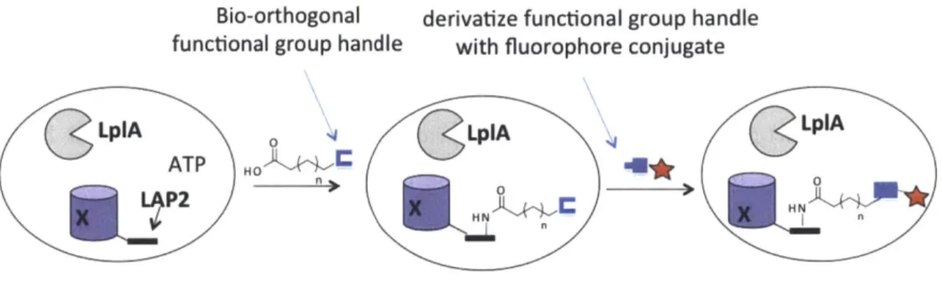
Chapter 2. Development of intracellular protein labeling with diverse fluorophores using enzyme-mediated azide ligation and strain-promoted cycloaddition ... 37
Screening for an efficient LplA ligase and azide pair for site-specific protein labeling in sid e liv e cells...4 2
Characterization of an improved LplA enzyme and azide probe pair ... 48
Exploring strain-promoted azide-alkyne cycloaddition reaction inside live cells using alternative source of azide ... 57
C on clu sion ... 6 1 Experimental methods ... 62
R eferen ces...6 7 Chapter 3. Optimization and demonstration of diverse fluorophore targeting inside live cells ... 70
In tro d u ction ... 7 1 Comparison of LplA mutant enzymes and azide pairs using a live-cell labeling assay ... 7 2 Comparison of cyclooctyne structures... 77
Demonstration of intracellular protein labeling with ADIBO-fluorescein diacetate... 84
Challenges and optimization for targeting large fluorophores inside live cells... 87
Extension of site-specific two-step labeling method to diverse fluorophore structures with labeling inside cells and on the cell surface... 97
Measurement of overall two-step ligation yield in cells... 100
C on clu sio n ... 10 2 E xperim ental m ethods ... 107
R eferen ces...1 16 Chapter 4. Development of a photocrosslinker ligase for protein-protein interaction detection...120
Introduction to methods of protein-protein interaction detection... 121
Classes of photocrosslinkers ... 126
Screening for a benzophenone ligase... 130
In vitro photocrosslinking of LAP-tagged FKBP and FRB proteins using benzophenone p ro b e ... 14 2
Optimizations to improve in vitro photocrosslinking yield ... 147
Synthesis and LplA(W37G) incorporation of alkyl benzophenone...153
In vitro photocrosslinking with new alkyl benzophenone probe ... 157
Quantify benzophenone ligation yield inside live mammalian cells ... 162
Photocrosslinking inside live mammalian cells...166
LAP-actin photocrosslinking with drug treatment inside live mammalian cells...177
C on clu sion ... 18 1 E xperim ental m ethods ... 183
R eferen ces...192
Chapter 5. Effort towards membrane protein topology mapping with an ascorbate peroxidase enzyme ... 199
Introdu ction ... 200
EF-hand domain-containing protein 1 -LETM1...205
Restricted and diffusive labeling patterns of localized APEX...208
Biotin-tyramide labeling patterns of APEX-fused LETMI...211
Controlling expression levels of LETM 1 fusion proteins...218
Conclusion and future direction...222
E xperim ental m ethods ... 224
R eferen ces...22 8 Curriculum Vitae...231
List of Figures
Chapter 1: Introduction: specific and non-specific enzyme-mediated small molecule labeling methods
Chapter 2. Development of intracellular protein labeling with diverse fluorophores using enzyme-mediated azide ligation and strain-promoted cycloaddition
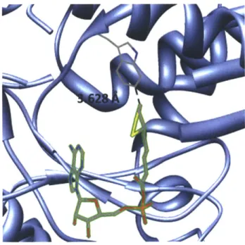
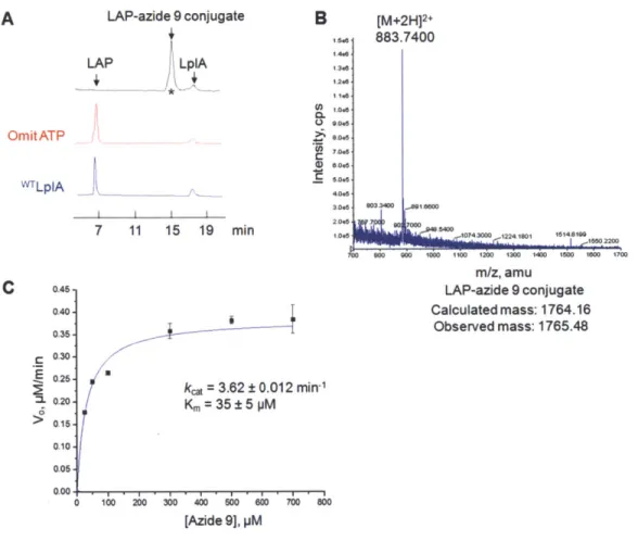
Figure 2-1. Natural ligation of lipoic acid catalyzed by wild-type LplA...38 Figure 2-2. Small and large probes to be incorporated by LplA ... 40 Figure 2-3. Two-step LplA ligation using functional group handle and chemoselective bioorthogonal reaction inside live cells ... 41 Figure 2-4. Small molecule substrate binding pocket of K coli lipoic acid ligase (LplA) w ith bound lipoyl-A M P ... 45 Figure 2-5. Screening to identify the best LplA mutant/azide substrate pair ... 47 Figure 2-6. In vitro characterization of azide 9 ligation catalyzed by LplA(W371) .... 49 Figure 2-7. Fixed cell labeling on nuclear localized LAP-YFP with cy5-alkyne via
C u A A C ... 5 1
Figure 2-8. Characterization of intracellular azide ligation yields by native gel-shift assay
... 5 3
Figure 2-9. Schematic representation of streptavidin gel-shift assay to measure azide
ligation y ield ... 54
Figure 2-10. Characterization of intracellular azide ligation yields by streptavidin gel-sh ift assay ... 5 5 Figure 2-11. Fixed cell detection of metabolically incorporated azido sugar analog
azido-GalNAc with cy3-alkyne via CuAAC ... 58
Figure 2-12. Live cell detection of metabolically incorporated azido sugar analog azido-GalNAc with strain-promoted azide-alkyne cycloaddition ... 60
Chapter 3. Optimization and demonstration of diverse fluorophore targeting inside
live cells
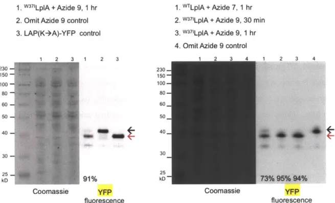
Figure 3-2. Identification of the best LplA mutant/azide substrate pair for intracellular protein labeling ... 74 Figure 3-3. Quantitation of data shown in Figure 3-2 ... 75
Figure 3-4. Immunofluorescence with anti-FLAG to compare labeling against expression of all enzym es ... 76 Figure 3-5. Evaluation of various cyclooctyne structures for site-specific intracellular protein labeling ... 80
Figure 3-6. Analysis of background labeling by cyclooctyne-fluorescein conjugates ... 8 2 Figure 3-7. Two-dimensional optimization of ADIBO-fluorescein diacetate loading concentration and washout tim e... 84 Figure 3-8. Two-step fluorophore targeting via strain-promoted azide-alkyne
cy cload dition ... 85 Figure 3-9. ADIBO-fluorescein labeling of three localized LAP-BFP fusions, LAP-p-actin, and LA P-M A P2 ... 86
Figure 3-10. Comparison of MOFO-ATTO 647N and ADIBO-ATTO 647N nuclear labeling specificities... 88 Figure 3-11. Comparison of two different linker structures for MOFO-ATTO 647N
... 8 9 Figure 3-12. Suppression of mitochondrial background labeling with FCCP...91 Figure 3-13. Suppression of mitochondrial background labeling with QSY21...92 Figure 3-14. Suppression of mitochondrial background labeling with FCCP and QSY21
... 9 4 Figure 3-15. Comparison of ADIBO-ATTO 647N labeling of LAP-p-actin with and without suppression of mitochondrial background using QSY21 quencher ... 96 Figure 3-16. Intracellular protein labeling with diverse fluorophore structures...97 Figure 3-17. Cell surface and intracellular protein labeling with DIBO-Alexa Fluor 647 and D IB O -biotin ... 99 Figure 3-18. Streptavidin gel-shift analysis of two-step labeling yield in live cells.... 101
Chapter 4. Development of a photocrosslinker ligase for protein-protein interaction detection
Figure 4-1. Three classes of photocrosslinkers...126 Figure 4-2. Lipoic acid ligase (LplA) with either bound lipoic acid or a benzophenone unnatural small molecule docked into the enzyme active site... 130 Figure 4-3. Synthetic scheme of benzophenone photocrosslinkers... 131 Figure 4-4. Screening to identify LplA mutant/benzophenone substrate pair...132 Figure 4-5. Unsuccessful attempt to detect in vitro ligation of benzophenone to E2p protein using biotin-hydrazide ... 134 Figure 4-6. Hydrolysis of hydrazones and oximes ... 135 Figure 4-7. Reaction between benzophenone and biotin-hydroxylamine to form oxime p ro d u ct. ... 13 7 Figure 4-8. Detection of in vitro ligated benzophenone using AF488-hydroxylamine ... 1 3 9 Figure 4-9. Detection of benzophenone chemically conjugated to FRB-YFP using biotin-hydroxylam ine ... 14 1 Figure 4-10. Crystal structure of FKBP-rapamycin-FRB ternary complex... 142 Figure 4-11. In vitro photocrosslinking with purified proteins FKBP-LAP and FRB-YFP ... 14 6 Figure 4-12. Crystal structure of FRB-rapamycin-FKBP ternary complex with methionine residues in ball-and-stick ... 148 Figure 4-13. In vitro photocrosslinking with methionine to alanine mutant proteins of F K B P -L A P ... 149 Figure 4-14. In vitro photocrosslinking of myc-FKBP-LAP and HA-FRB ... 151 Figure 4-15. Scheme of benzophenone insertion into nearby C-H bond after UV
irradiation ... 153 Figure 4-16. Benzophenone and benzophenone derivatives with different functional groups attached to the fourth position of the benzophenone core ... 155 Figure 4-17. Synthetic scheme of alkyl benzophenone probes with methylene unit attached to the benzophenone core ... 155
Figure 4-18. In vitro characterization of para alkyl benzophenone ligation by
L plA (W 37G )... 156
Figure 4-19. In vitro photocrosslinking comparison between alkyl benzophenone and benzophenone ... 159 Figure 4-20. Reducing concentrations of FKBP-LAP(F) and FRB helps in eliminating background photocrosslinking ... 161 Figure 4-21. Gel-shift analysis of benzophenone ligation yield in cells...163 Figure 4-22. Native gel-shift assay to measure benzophenone ligation yield over time ... 16 4 Figure 4-23. Expression of FKBP-LAP and FRB in HEK 293T cells ... 167 Figure 4-24. Live cell benzophenone ligation and photocrosslinking of myc-FKBP-LAP and H A -F R B ... 168 Figure 4-25. Benzophenone photocrosslinker labeling scheme of LAP-actin in live cells ... 16 9 Figure 4-26. Benzophenone ligation and photocrosslinking on HA-LAP/LAP(F)-actin in live m am m alian cells ... 170 Figure 4-27. Western blot stained with anti-FLAG antibody to detect FLAG-tagged LplA(W37G) after live cell photocrosslinking of HA-LAP/LAP(F)-actin ... 172 Figure 4-28. Live cell photocrosslinking of HA-LAP-actin with alkyl benzophenone incubation tim e course ... 174 Figure 4-29. Benzophenone ligation and photocrosslinking performed on HA-LAP-M A P2 in live m am m alian cells...175 Figure 4-30. Schematic representations of different LAP-actin crosslinked complexes with and without depolymerization drug treatment...177 Figure 4-31. Live cell photocrosslinking of LAP-actin with various drug treatments ... 17 9
Chapter 5. Effort towards membrane protein topology mapping with an ascorbate peroxidase enzyme
Figure 5-1. Scheme depicting labeling with APEX fused to either matrix or IMS side of an IM M protein ... 204 Figure 5-2. Amino acid sequence of human LETMI ... 206 Figure 5-3. Restricted and diffusive biotin-tyramide labeling patterns in HEK 293T and C O S -7 cells ... 209 Figure 5-4. Constructs with APEX fused to different regions of LETMI...211 Figure 5-5. APEX 115-LETMI fusion protein shows cleavage products on Western blot ... 2 12 Figure 5-6. Biotin-tyramide labeling with APEX-LETM1 fusion constructs in HEK 293T cells ... 2 17 Figure 5-7. Western blot analysis of APEX-LETMI fusion proteins expressed in HEK 2 9 3T cells...2 19 Figure 5-8. Western blot analysis of COS-7 cells lipofected (L2k) or viral infected with LETMI or LETM1-mCherry construct ... 220
List of Tables
List of Abbreviations
aa ... am in o acid s A C P ... acyl-carrier protein A TP ... adenosine triphosphate azide 7...8-azidooctanoic acid azide 8...9-azidononanoic acid azide 9...1 0-azidodecanoic acid azide 10...11 -azidoundecanoic acid BFP...blue fluorescent protein B SA ... bovine serum album in B T ... biotin -tyram ide CFD A ... carboxyfluorescein diacetate CH O ... Chinese ham ster ovarian cells CuAAC...copper-catalyzed azide-alkyne cycloaddition D A B ... diam inobenzidine D C M ... dichlorom ethane D H FR ... dihydrofolate reductase D IC ... differential interference contrast D IFO ... difluorinated cyclooctyne DIMAC...6,7-dimethoxyazacyclooct-4-yne DMEM...Dulbecco's modified eagle medium D M F...N ,N -dim ethylform am ide D M SO ... dim ethylsulfoxide D N A ... deoxyribonucleic acid DPBS...Dulbecco's phosphate buffered saline D T T ... dith iothreito l E2p...9 kDa hybrid lipoyl domain derived from E.coli PDH complex E . coli...E scherich ia coli EDTA...ethylenediaminetetracetic acid ER ... endoplasm ic reticulum F P ... fluorescent protein E R ... endoplasm ic reticulum E SI...electrospray ionization F -actin ... filam entous actin FK B P12 ... FK 506 binding protein 12 FlAsH... fluorescein arsenical hairpin binder FRB...FKBP-rapamycin binding domain of the mTOR protein FRET...fluorescence resonance energy transfer FP ... fl uorescent protein G -actin ... globular actin G FP ... green fluorescent protein hAGT...06 -alkylguanine-DNA alkyltransferase
HA...human influenza hemagglutinin epitope H alo-tag...haloalkane dehalogenase
HDDA... . . . ... N,N'-dimethyl- 1,6-diaminohexane HEK 293T...human embryonic kidney cells 293T HEPES...4-(2-hydroxyethyl)-1-piperazineethanesulfonic acid H P 1...heterochrom atin protein 1 HPLC...high-performance liquid chromatography IMM... inner mitochondrial membrane IM S ... interm em brane space IPTG...Isopropyl
p-D-1-thiogalactopyranoside
LAPI...22-amino acid rationally designed LplA acceptor peptide LAP...the best 13-amino acid Lp1A acceptor peptide evolved from yeast display LAP(F)...LAP peptide with Trp downstream of catalytic Lys residue changed to Phe LC-MS/MS...liquid chromatography, tandem mass spectrometry LplA...Escherichia coli lipoic acid ligase MAP2...microtubule-associated protein 2 M eC N ... acetonitrile M EM ... m inim al eagle m edium MOFO...monofluorinated cyclooctyne M S ... m ass spectrom etry nA v ... neutravidin N E S ... nuclear export signal N H S...N -hydroxysuccinim ide N LS...nuclear localization sequence N M R ... nuclear m agnetic resonance OMM...outer mitochondrial membrane PAGE...polyacrylamide gel electrophoresis PA L ... photoaffinity labeling PALM...photoactivated localization microscopy PB S...phosphate buffered saline PCR ... polym erase chain reaction P D B ... protein databasePFA ... paraform aldehyde PK C ... phosphokinase C PPTase...phosphopantetheine transferase Q Y ... quantum yield R O S ... reactive oxygen species S A ... strep tav id in SA-HRP...streptavidin-conjugated horseradish peroxidase s.d ... standard deviation SD S...so dium dodecyl sulfate STED...stimulated emission depletion microscopy STORM...stochastic optical reconstruction microscopy T A ... transient absorption TBS-T...Tris-buffered saline supplemented with Tween-20 TBTA...tris-(benzyltriazolylmethyl)amine T E A ... triethylam ine TLC ... thin-layer chrom atography
TM ... transmembrane TM R... tetramethyrhodamine Tris...tris(hydroxymethyl)aminomethane UAA...unnatural amino acid W B...western blotting W CL...whole cell lysate YFP...yellow fluorescent protein
Chapter 1. Introduction:
Introduction
Scientists have long adapted enzymes for fundamental biochemistry research. The unifying theme of this thesis is to utilize the natural and engineered abilities of enzymes as robust biochemical tools that can be applied to study biological processes. This is accomplished in parallel with the design and synthesis of organic small molecule probes, such as fluorophores or probes baring a chemoselective functional handle, to be recognized and incorporated by the enzymes.
Green fluorescent protein (GFP) and its multicolored variants (1) are perhaps the most famous and widely used biochemical tools that have revolutionized cell biologists' ability to gain insight into cellular processes in living cells. Fluorescent proteins (FPs) serve as imaging protein reporter tags to visualize protein movement and localization. FPs offer perfect targeting specificity through genetic fusion and are generally stable upon chromophore maturation in all cellular compartments. Aside from routine imaging applications, scientists have engineered FPs for various applications such as photoactivatable FPs for super-resolution imaging (2) and split-GFP (3) to monitor protein trafficking, interaction and maturation. However, despite the many advantages and utilities of FPs, they carry a payload of 27 kDa when fused to the protein of interest, and are generally less bright and photostable compare to small organic fluorophores (4). We and others have observed interference caused by FP fusion to protein of interest (5). For example, mCherry-tagged actin is excluded from the nucleus of the cell, likely because the mCherry is interfering with the binding of cofilin, a nuclear importer of actin. Small molecule organic fluorophores are brighter, more photostable, and far smaller than FPs (4). However, they do not have the ease and perfect specificity of genetic targeting offered by FPs. Scientists have therefore came up with strategies to target small molecules fluorophores via the use of enzymes.
Enzymes and peptides as protein tags to target small molecule probes
Covalent enzymatic tags (self-modifying enzymes) and non-covalent enzymatic tags To exploit using enzymes as protein tags to target small organic molecules, the group of Kai Johnsson employed the ubiquitous human DNA repair enzyme O6 alkylguanine-DNA alkyltransferase (hAGT) (6) which transfers alkyl groups from the 06-position of guanine onto a cysteine residue in its active site in a one-turnover self-modification reaction. The Johnsson group has derivatized the para position of the benzyl group of 06-benzyl guanine with a variety of probes, like fluorophores and biotin, to
achieve specific targeting (7,8) of these molecules by fusing hAGT to protein of interest. Further evolution based on phase display led to a more active quadruple mutant hAGT enzyme whose activity is 20 times faster than the wild-type hAGT, which eliminated the need for hAGT deficient cell lines (9). This more active hAGT, termed SNAP-tag, has been used to target benzyl guanines derivatized with fluorescein, biotin, and digoxigenin probes. Later, the Johnsson group expanded the substrate specificity of hAGT through directed evolution to report another hAGT mutant that exhibits 100 times greater preference for cytosine over guanine derivatives. The evolved cytosine-specific enzyme, termed CLIP-tag (10), has been applied together with SNAP-tag to orthogonally label two populations of proteins with different fluorophores inside the same cell.
Another example of self-modifying enzyme that has been adapted as enzymatic tag is a mutant form of the enzyme haloalkane dehalogenase (11). The wild-type haloalkane dehalogenase hydrolyzes haloalkanes by forming a covalent bond between the asparagine residue in its active site and the alkyl group in an SN2 reaction. A nearby
histidine residue in the active site then hydrolyzes the covalent bond to recycle the enzyme. In an attempt to convert this enzyme to a covalent labeling tag, researchers at Promega installed a His289 mutation that eliminated the second hydrolysis step (12). This mutant form of haloalkane dehalogenase is named HaloTag. Substrates for HaloTag include haloalkane conjugated fluorophores and functionalized agarose surface resin for protein purification (11). The HaloTag labeling technology possesses high specificity and low background labeling due to the lack of endogenous dehalogenases in eukaryotic cells. Recently it has been applied in purification of the protein cannabinoid receptor CB2 (13), fused to p75 neurotrophin receptor and tubulin to assess the retrograde axonal transport
of the receptor (14), as well as specific labeling of xenograft tumors in living animals
(15).
In addition to covalent modification on self-labeling enzymatic tags, several proteins with tight binding to their substrates have also been exploited as non-covalent labeling methods. One of the first examples of non-covalent tagging is the F. coli. dihydrofolate reductase (eDHFR), which has high affinity to its antibiotic small molecule ligand trimethoprim (TMP) with KD as low as 1 nM (16). TMP conjugates to various fluorescent probes can be synthesized by introducing the fluorophores to the para-methoxy position of TMP. Modifications introduced at this position of TMP raises the KD
only slightly to ~35 nM, which is still a lot lower than the affinity between TMP and mammalian DHFR. The eDHFR-TMP receptor-ligand pair has been demonstrated in labeling proteins in the nucleus, plasma membrane, and cytosol in various cell types (16,17).
Another example of non-covalent protein tag based on the high binding affinity between enzyme and its ligand is the FK506 binding protein 12 (FKBP12), and its natural ligand rapamycin, a system pioneered by the Schreiber group (18,19). Clarkson and coworkers developed a nonnative FKBP12-rapamycin pair by engineering a FKBP12(F36V) mutant which has high affinity (KD~0.094 nM) to a synthetic derivative of rapamycin that can be further conjugated to fluorophores and other probes (20,21). Robers et al demonstrated labeling of numerous intracellular proteins fused to FKBP12(F36V) with different rapamycin derivative conjugates (21). However, even though the two non-covalent enzymatic tagging strategies discussed do have very high affinities between receptor and ligand, they are not suitable for long-term studies compared to covalent enzymatic tags.
Overall, enzymatic protein tags, either based on covalent self-labeling or high-affinity non-covalent interaction, provide a way to target small molecule organic fluorophores inside live mammalian cells with high specificity. A significant drawback is that most enzymatic tags are still fairly large. The smallest enzymatic tag that has been demonstrated for specific probe targeting inside live cells mentioned above is the 12 kDa FKBP12(F36V) enzyme but its non-covalent binding limits its application. In the next
section we review enzymatic labeling techniques that are based on small peptide tag fusions to address this issue.
Enzymatic labeling ofpeptide tags
The ideal labeling method would combine the exquisite specificity conferred by enzymes, the superior photophysical property of small molecule fluorophores, together with minimal perturbation to the native protein. While specific targeting to endogenous proteins is an extremely challenging feat, scientists have developed enzyme-based techniques that only require fusing a small peptide to the protein of interest. Enzyme-mediated small peptide tags are recognized by specific enzymes, which then selectively modify the tags with small molecule substrates.
Zhou et al. used phage display evolution to evolve two orthogonal 12-amino acid peptides Al and S6 as efficient substrates for Sfp and AcpS phosphopantetheinyl transferases (PPTases) (22). The PPTases catalyze the covalent linkage of a coenzyme A-derivatized probe to a serine residue in the peptide substrates. However, the charged phosphopantetheine arm makes the probe conjugates difficult to cross the cell membranes, limiting the method to labeling only on the cell surface (22,23).
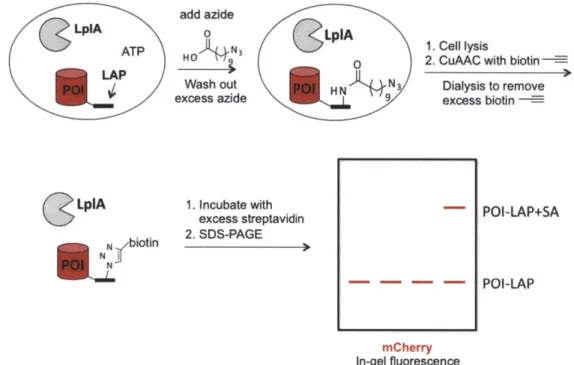
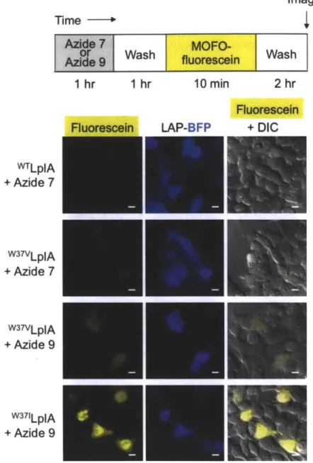
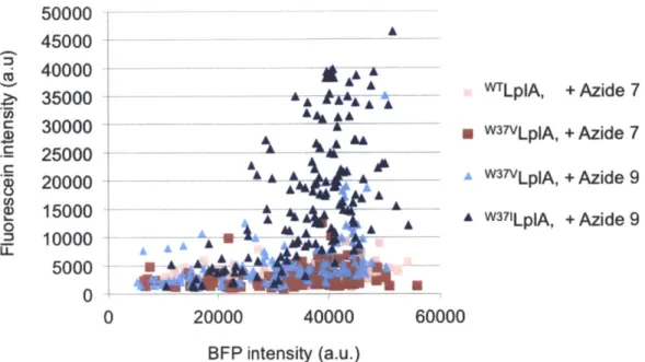
Our lab has been a pioneer in this area of enzyme-mediated peptide tag labeling methods. We developed probe targeting techniques based on lipoic acid ligase (LplA) and biotin ligase (BirA) together with their respective peptide tags. We engineered the E. coli enzyme lipoic acid ligase (LplA) though site-directed mutagenesis to incorporate unnatural small molecules such as fluorophores (5,24,25), functional groups (26-28), and a photocrosslinker (29) site-specifically onto the lysine residue in a 13-amino-acid LplA acceptor peptide (LAP). To develop this technology we now call PRIME (PRobe Incorporation Mediated by Enzymes), we explored the plasticity of the small molecule binding pocket of LplA and also reduced the size of its protein substrate from the 9 kDa E2p subunit of pyruvate dehydrogenase to the 13- amino acid LAP through yeast-display evolution (30). By examining the crystal structure of E. coli LplA with bound lipoyl-AMP, we engineered LplA to accept unnatural probes through alanine scanning of amino acid residues in the active site (29) and identifying a key "gatekeeper" residue, Trp37, that when mutated to smaller amino acids can enlarged the LplA active site to enable it to
bind to larger unnatural probes, such as different derivatives of coumarin (5,24,25), aryl azide (29), alkyl azide (26,27), and trans-cyclooctene (28). Dr. Chayasith Uttamapinant and coworkers demonstrated specific labeling of coumarin fluorophore by LplA(W37V) onto many LAP-tagged proteins inside live mammalian cells (5). More importantly, through this peptide-tagging method, they were able to detect the presence of actin in the nucleus, which was shown to preclude actin when it was fused to a large fluorescent protein, further highlighting and confirming the importance of a small tag.
The K coli biotin ligase (BirA) covalently attaches biotin to a lysine residue of a 15-amino acid biotin acceptor peptide (AP). A ketone derivative of biotin was synthesized for subsequent chemoselective reaction with hydrazide or hydroxylamine-conjugated biophysical probes in a two-step process (31). Dr. Sarah Slavoff and coworkers also explored BirA from yeast and Pyrococcus horikoshii to label azide- or alkyne-derivatized biotin analogs, which allowed for downstream detection using click chemistry (32). However, one limitation of the BirA labeling method is that since endogenous biotin is still preferred as the substrate, the labeling method either requires biotin starvation of the cells prior to BirA intracellular labeling or is restricted to the cell surface.
The Ploegh group harvested the enzyme sortase A from Staphylococcus aureus which recognizes a only 5-amino acid peptide sequence LPXTG, the shortest peptide tag available (33,34). The sortase A enzyme performs a transpeptidation reaction by first cleaving the peptide bond between the T and G residues, and at the same time forming a new peptide bond to a polyG-derivatized small molecule. However, this method is also restricted to the cell surface because the polyG substrates are not cell permeable.
Using bioorthogonal chemistries to label proteins with small molecules
So far we have discussed using enzymes that are able to target fluorophores functionalized with ligands they recognize - haloalkane for Halo-Tag, guanine or cytosine for hAGT and its variant, TMP for eDHFR, so the labeling reaction is complete in a single step by the specific enzyme. In another approach, bioorthogonal chemistries between two functional groups that are not found inside mammalian cells have been extensively explored as a way to target small molecules with high specificity. Chemical labeling techniques that utilize bioorthogonal reactions (35) to target small organic fluorophores inside live cells have seen much advancement over the years. Aside from harnessing specificity from Nature, chemists have began to develop reactions and engineer functional group handles that can specifically recognize and react with each other not only in the exogenous environment of an isolated flask, but inside the complex milieu of cells and living organisms. To develop bioorthogonal reactions as a way to target fluorophores in a biological environment, the functional groups, or reactive "handles" involved in the bioorthogonal reaction must stay inert until they meet to selectively to react with each other; the reaction condition must be biocompatible in terms of pH and temperature and nontoxic to the biological system under study; the reaction must have fast kinetics so that the product is formed at a reasonable rate on the scale of protein turnover and the progression of biological processes, even when the reactant concentrations are preferably low. The bioorthogonal product formed must be stable to proteases and not be degraded so to have a consistent readout to be useful as chemical reporters. Excess reactants should be removed from the system to minimize unwanted side reaction, or masked to reduce possible background signal. The versatility of bioorthogonal reactions has been demonstrated by their ability to tag proteins and glycans. The Staudinger ligation between azide and triarylphosphine was the first bioorthogonal reaction used to label cell-surface azide-derivatized glycans on live cells (36), and subsequently in live animals (37). Despite much effort in mechanistic modifications to increase the reaction kinetics, reduce phosphine oxidation, and aza-ylide hydrolysis, its slow second-order rate constant (10- M's 1) leaves much more to be desired (38,39).
Even though the Staudinger ligation is slow, it brought to chemists' attention the potential of organic azide as an excellent functional group handle. The small size of the azide, its absence from living organisms, its kinetic stability inside cells, and its nontoxic profile as demonstrated by early approved drugs like azidothymidine all hinted to the potential of organic azide as an excellent functional group handle. The copper-free or strain-promoted azide-alkyne cycloaddition was adapted from the Huisgen [3+2] cycloaddition that initially used heat and pressure to drive the reaction (40), as well as the later developed Cu(I)-catalyzed azide-alkyne cycloaddition demonstrated by Sharpless and Meldal (41). Instead of using heat, pressure, or Cu(I) which are incompatible inside cells, the strain-promoted azide-alkyne cycloaddition uses energy stored in the ring strain of cyclooctyne to drive the reaction. Since 2000, numerous generations of cyclooctynes were synthesized with improved reactivity, kinetics, hydrophilicity, and overall biocompatibility. The highest second-order rate constant of a strain-promoted azide-alkyne cycloaddition reaction that has been demonstrated on live cell-surface was reported to be 0.96 M-1s-1 (42), twenty times higher than the initial Staudinger ligation. In the first part of this thesis, strain-promoted azide-alkyne cycloaddition is used for the targeting of diverse fluorophores to specific proteins inside live cells.
Yet another recent addition to the toolbox of bioorthogonal reactions is the reaction between tetrazine and trans-cyclooctene (43). Developed by the Fox group, this inverse Diels-Alder reaction is the fastest bioorthogonal reaction known to date, optimized to achieve an impressive rate constant of 22,000 M' s4 . Our lab has demonstrated two-step fluorophore targeting using a lipoic acid ligase enzyme in combination with Diels-Alder cycloaddition to perform intracellular protein labeling (28). The very fast cycloaddition kinetics yields substantial improvements in signal to background ratio. Furthermore, the tetrazine-fluorophore conjugate is also fluorogenic, which is an additional benefit that masks the background caused by unreacted reagents.
Site-specific targeting and crosslinking with photocrosslinkers to identify protein-protein interactions
So far we have discussed a few probe labeling techniques that capitalize on the specificity of enzymes and bioorthogonal reactions to target small molecules to protein of interest inside cells. We have seen many enzyme-based fluorophore or probe targeting methods that have been demonstrated to label a specific protein among the plethora of proteins inside live mammalian cells. High specificity is a critical requirement since the target is often a single protein. In a different approach, scientists sought methods to label and identify multiple proteins associated with a protein of interest or within a particular cellular compartment. Identifying the many proteins of a certain proteome, within a certain compartment of the cell, or under an altered cellular state requires a different labeling approach.
One of the conventional approaches to capture groups or complexes of proteins is affinity-based methods. Methods such as co-immunoprecipitation (44) that uses an antibody against the protein of interest requires first lysing the cells, then immunocapture the protein of interest together with its interacting proteins. Subsequent Western blots or mass spectrometry (MS) analysis is used to identify proteins that are pulled down. However, using cell lysates as starting material already introduces numerous artifacts. First, the proteins are removed from their native environment. In cell lysate, without cellular compartmentalization that maintains various pHs and effective concentrations of biomolecules, complexes that form depending on those factors could dissociate or aggregate with other proteins. Second, disruption of protein complexes during cell lysis also presents a serious problem. Weakly bound proteins could be lost during washing, or non-specific binding of proteins that are not endogenous interacting partners could wrongly associate. Last but not least, the limited availability of high quality of antibodies introduces additional constraint.
As an alternative, chemical crosslinking prior to immunocapture is employed to covalently link interacting proteins while the proteins are in their endogenous states. Covalent linkage allows more stringent washing conditions to remove non-specifically interacting proteins without fear of losing weakly bound proteins (45). Traditional chemical crosslinking reagents include formaldehyde or amine-reactive moieties such as
N-hydroxysuccinimide (NHS) esters. Formaldehyde can readily react with lysines, tryptophans, or N-termini of proteins after minutes of exposure; however, it does not have any spatial resolution or protein specificity once it permeates cell membranes. To address this issue, photoaffinity labeling (PAL) (46) that uses site-specifically conjugated photoreactive crosslinkers, or photocrosslinkers, enables identifying interacting proteins specific to a protein of interest. Photocrosslinkers such as aryl azides, benzophenones, and diazirines can be site-specifically introduced to proteins via in vitro cysteine-maleimide chemistry or incorporated as unnatural amino acids using the unnatural amino acid (UAA) mutagenesis technique developed by the Schultz group (47). Crosslinking with site-specific photocrosslinkers offers high temporal and spatial resolution to capture proteins in their native states and has been demonstrated inside live mammalian cells. However, as discussed previously, techniques to site-specifically target these small molecule photocrosslinkers are still limited. In Chapter 4, we introduce an enzyme-based ligation technique for the benzophenone photocrosslinker.
Promiscuous enzymes
Achieving location specificity of photocrosslinkers or other reactive species to form covalent linkage with proteins inside live cells is a challenging task. By site-specifically attaching a photocrosslinker to a protein of interest allows us to pull down one or multiple interacting proteins, but would not enable us to identify, for example, the entire proteome of the mitochondrial matrix under drug treatment. To identify the proteome of an entire cell or a specific organelle under its native environment without contamination from cell lysate or organelle purification, scientists have yet again looked to enzymes for solutions. One approach is to use enzymes that can convert small molecule probes to diffusive reactive intermediates that can form covalent bonds with its nearby proteins. Having diffusive reactive species versus site-specifically attached crosslinkers also obviates the bottleneck of crosslinker length and shape, which limits its range of reactivity. Several enzymes that are able to take a chemical probe and generate a reactive intermediate have been applied in this type of promiscuous protein labeling. Horseradish peroxidase (HRP) was first applied as a reporter enzyme for electron microscopy (EM) (48) by reacting with diaminobenzidine (DAB) in the presence of H202 to form an insoluble tar-like product that is electron-opaque and can serve as an EM contrast agent. Later, HRP was adapted by Kotani et al. to catalyze the generation of nitrenes from aryl azides (49). In this enzyme-mediated activation of radical source (EMARS) method, cell membrane-targeted HRP generated radicals of aryl azide-biotin conjugate that crosslinked to nearby proteins, which can be pulled down and analyzed using either streptavidin-coated beads or membrane protein specific antibody. EMARS labeling has been applied to study cell-surface molecular clusters of
p1-integrin,
epidermal growth factor receptor (EGFR), insulin-like growth factor-I receptor (IGF1R), and EphA2 receptor (50,51). Immunoelectron microscopy of the EMARS-labeled HRP-conjugated antibody showed 96% of biotinylated proteins were within 300 nm of the antibody-coated particles (50). However, one limitation of HRP is that the enzyme is not active in the cytosol, perhaps because its disulfide bonds were reduced under the reducing environment.
Promiscuous biotin ligase is another example of an enzyme that can generate reactive small molecules. Through a point mutation (RI 18G) (52) in the wild-type biotin
ligase which has exquisite specificity towards its endogenous protein substrate, promiscuous biotin ligase is able to prematurely release the adenylated biotinoyl-5'-AMP from its active site without the protein substrate present. Once released, biotinyl-5'- AMP can react with amine functional groups on nearby proteins forming an amide bond. Biotinylated proteins are then enriched using streptavidin binding and analyzed on Western blot. However, the time of the reaction is especially long, requiring longer than 6 hours of biotin incubation to saturated labeling. Even though the labeling is performed in
intact live cells, but the prolonged labeling time could cause artifacts in endogenous protein interactions. Biotinylated proteins could diffuse away leading to poor spatial resolution or even cause subsequent unwanted cellular changes. Furthermore, the lack of temporal resolution would not allow differentiation and detection of rapid changes in proteomes. Labeling with promiscuous biotin ligase was demonstrated by tagging the
enzyme to nuclear lamina protein, lamin A, and the construct was expressed and labeled in HEK 293 cells (53). Roux et al identified 122 proteins, some of which are known proteins associated with the nuclear envelope, while others are novel, such as the protein FAM169A (later called SLAP75) which does not have any predicted transmembrane region or any sequence motifs that would hint to its localization. FAM169A was also confirmed by immunostaining to be located in the nuclear envelope.
To develop an enzyme that can be specifically targeted to a region within the cell, works in different organelle environments, and has high temporal and spatial resolution, our lab has engineered a plant ascorbate peroxidase called APEX to catalyze the formation of biotin-tyramide radicals in the presence of H202 (54). The short lifetime (less than 1 pts) of the radical together with its membrane-impermeant nature ensure tight labeling radius and high temporal resolution. Rhee et al. targeted APEX to the mitochondrial matrix and the inner mitochondrial space (IMS), two regions that are especially difficult to purify through subcellular fractionation and organelle purification (54). Using matrix-targeted APEX, biotin-tyramide and H202 labeling, followed by streptavidin enrichment and tandem mass spectrometry analysis, 495 proteins were identified in the matrix proteome, with 464 proteins having prior mitochondrial annotation (54). Notably, proteins such as CPOX and PPOX which were previously assigned to the mitochondrial intermembrane space were detected in the matrix proteome,
leading to novel insights to their trafficking and functional roles in the heme biosynthesis pathway. Currently APEX is being applied towards identifying the proteome of especially challenging regions of the cell that are impossible to purify through traditional means, such as the synaptic space or the junction between mitochondria and the endoplasmic reticulum (ER). In the final chapter of this thesis, we describe extending APEX labeling to probe the topology of membrane proteins.
Conclusion
Despite the advances in cell biology brought by the fluorescent proteins, their large size and poor photophysical properties prompted scientists to look for alternatives. Compared to fluorescent protein tags, chemical tags such as small molecule fluorophore or other biophysical probes are smaller, brighter and more versatile. To target small molecules site-specifically to a protein of interest inside the cell, scientists have utilized the exquisite specificity of enzymes as well as developed bioorthogonal chemical reactions that can work efficiently within the complex environment inside the cell. In
Chapter 2 and 3, we describe a fluorophore targeting method that uses only a small peptide tag in combination with enzymatic labeling and azide-alkyne cycloaddition to
label numerous proteins inside cells with various fluorophores.
While being able to visualize the localization, movement, and interaction of a specific protein inside its native environment has brought tremendous insights to cell biology regarding the how a cell functions, the ability to identify with high temporal resolution pools or complexes of proteins associated with a single protein, of a particular cellular organelle, or even of the entire cell under a specific cellular condition has also revolutionized the way scientists study and understand biological processes at the molecular and cellular level. In Chapter 4, we present a method to site-specifically ligate a photocrosslinker to identify protein-protein interactions. Aside from targeted photocrosslinkers, scientists have developed enzymes that can generate reactive species, such as radicals, which then react nonspecifically with proximal proteins. In Chapter 5, we discuss current effort towards developing two parallel but distinct approaches to use enzyme-generated radical species to study membrane protein topology.
References
1. Sample V, Newman RH, Zhang J. The structure and function of fluorescent proteins. Chem Soc Rev. 2009 Oct;38(10):2852-64.
2. Wiedenmann J, Gayda S, Adam V, Oswald F, Nienhaus K, Bourgeois D, et al. From EosFP to mIrisFP: structure-based development of advanced photoactivatable marker proteins of the GFP-family. J Biophotonics. 2011 Jun;4(6):377-90.
3. Ozawa T. Protein reconstitution methods for visualizing biomolecular function in living cells. Yakugaku Zasshi. 2009 Mar;129(3):289-95.
4. Lavis LD, Raines RT. Bright ideas for chemical biology. ACS Chem. Biol. 2008 Mar 20;3(3):142-55.
5. Uttamapinant C, White KA, Baruah H, Thompson S, Femindez-Suirez M,
Puthenveetil S, et al. A fluorophore ligase for site-specific protein labeling inside living cells. PNAS. 2010 Jun 15;107(24):10914-9.
6. Keppler A, Gendreizig S, Gronemeyer T, Pick H, Vogel H, Johnsson K. A general method for the covalent labeling of fusion proteins with small molecules in vivo. Nat Biotech. 2003 Jan;21(1):86-9.
7. Keppler A, Pick H, Arrivoli C, Vogel H, Johnsson K. Labeling of fusion proteins with synthetic fluorophores in live cells. Proc. Natl. Acad. Sci. U.S.A. 2004 Jul
6;101(27):9955-9.
8. Keppler A, Kindermann M, Gendreizig S, Pick H, Vogel H, Johnsson K. Labeling of fusion proteins of 06-alkylguanine-DNA alkyltransferase with small molecules in vivo and in vitro. Methods. 2004 Apr;32(4):437-44.
9. Juillerat A, Gronemeyer T, Keppler A, Gendreizig S, Pick H, Vogel H, et al. Directed evolution of 06-alkylguanine-DNA alkyltransferase for efficient labeling of fusion proteins with small molecules in vivo. Chem. Biol. 2003 Apr; 10(4):313-7.
10. Gautier A, Juillerat A, Heinis C, Correa IR Jr, Kindermann M, Beaufils F, et al. An engineered protein tag for multiprotein labeling in living cells. Chem. Biol. 2008 Feb;15(2):128-36.
11. Los GV, Encell LP, McDougall MG, Hartzell DD, Karassina N, Zimprich C, et al. HaloTag: a novel protein labeling technology for cell imaging and protein analysis. ACS Chem. Biol. 2008 Jun 20;3(6):373-82.
12. Pries F, Kingma J, Krooshof GH, Jeronimus-Stratingh CM, Bruins AP, Janssen DB. Histidine 289 is essential for hydrolysis of the alkyl-enzyme intermediate of
13. Locatelli-Hoops S, Sheen FC, Zoubak L, Gawrisch K, Yeliseev AA. Application of HaloTag technology to expression and purification of cannabinoid receptor CB2. Protein Expr. Purif. 2013 May;89(1):62-72.
14. Mok S-A, Lund K, Lapointe P, Campenot RB. A HaloTag@ method for assessing the retrograde axonal transport of the p75 neurotrophin receptor and other proteins in compartmented cultures of rat sympathetic neurons. J. Neurosci. Methods. 2013 Mar 30;214(1):91-104.
15. Tseng J-C, Benink HA, McDougall MG, Chico-Calero I, Kung AL. In Vivo
Fluorescent Labeling of Tumor Cells with the HaloTag@ Technology. Curr Chem Genomics. 2012;6:48-54.
16. Miller LW, Cai Y, Sheetz MP, Cornish VW. In vivo protein labeling with
trimethoprim conjugates: a flexible chemical tag. Nat Meth. 2005 Apr;2(4):255-7.
17. Calloway NT, Choob M, Sanz A, Sheetz MP, Miller LW, Cornish VW. Optimized fluorescent trimethoprim derivatives for in vivo protein labeling. Chembiochem. 2007 May 7;8(7):767-74.
18. Bierer BE, Somers PK, Wandless TJ, Burakoff SJ, Schreiber SL. Probing
immunosuppressant action with a nonnatural immunophilin ligand. Science. 1990 Oct 26;250(4980):556-9.
19. Spencer DM, Wandless TJ, Schreiber SL, Crabtree GR. Controlling signal transduction with synthetic ligands. Science. 1993 Nov 12;262(5136):1019-24.
20. Clackson T, Yang W, Rozamus LW, Hatada M, Amara JF, Rollins CT, et al. Redesigning an FKBP-ligand interface to generate chemical dimerizers with novel specificity. Proc. Natl. Acad. Sci. U.S.A. 1998 Sep 1;95(18):10437-42.
21. Robers M, Pinson P, Leong L, Batchelor RH, Gee KR, Machleidt T. Fluorescent labeling of proteins in living cells using the FKBP12 (F36V) tag. Cytometry A. 2009 Mar;75(3):207-24.
22. Zhou Z, Cironi P, Lin AJ, Xu Y, Hrvatin S, Golan DE, et al. Genetically encoded short peptide tags for orthogonal protein labeling by Sfp and AcpS
phosphopantetheinyl transferases. ACS Chem. Biol. 2007 May 22;2(5):337-46.
23. Yin J, Straight PD, McLoughlin SM, Zhou Z, Lin AJ, Golan DE, et al. Genetically encoded short peptide tag for versatile protein labeling by Sfp phosphopantetheinyl transferase. Proc. Natl. Acad. Sci. U.S.A. 2005 Nov 1;102(44):15815-20.
24. Jin X, Uttamapinant C, Ting AY. Synthesis of 7-Aminocoumarin by Buchwald-Hartwig Cross Coupling for Specific Protein Labeling in Living Cells.
25. Cohen JD, Thompson S, Ting AY. Structure-Guided Engineering of a Pacific Blue Fluorophore Ligase for Specific Protein Imaging in Living Cells. Biochemistry. 2011 Sep 27;50(38):8221-5.
26. Fernandez-Suairez M, Baruah H, Martinez-Hernandez L, Xie KT, Baskin JM, Bertozzi CR, et al. Redirecting lipoic acid ligase for cell surface protein labeling with small-molecule probes. Nature Biotechnology. 2007;25(12):1483-7.
27. Yao JZ, Uttamapinant C, Poloukhtine A, Baskin JM, Codelli JA, Sletten EM, et al. Fluorophore Targeting to Cellular Proteins via Enzyme-Mediated Azide Ligation and Strain-Promoted Cycloaddition. J. Am. Chem. Soc. 2012 Feb 29;134(8):3720-8.
28. Liu DS, Tangpeerachaikul A, Selvaraj R, Taylor MT, Fox JM, Ting AY. Diels-Alder Cycloaddition for Fluorophore Targeting to Specific Proteins inside Living Cells. J. Am. Chem. Soc. 2012 Jan 18;134(2):792-5.
29. Baruah H, Puthenveetil S, Choi Y-A, Shah S, Ting AY. An Engineered Aryl Azide Ligase for Site-Specific Mapping of Protein-Protein Interactions through Photo-Cross-Linking. Angewandte Chemie International Edition. 2008;47(37):7018-21.
30. Puthenveetil S, Liu DS, White KA, Thompson S, Ting AY. Yeast Display Evolution of a Kinetically Efficient 13-Amino Acid Substrate for Lipoic Acid Ligase. J. Am. Chem. Soc. 2009 Nov 18;131(45):16430-8.
31. McNeill E, Chen I, Ting AY. Synthesis of a Ketone Analogue of Biotin via the Intramolecular Pauson-Khand Reaction. Org. Lett. 2006 Sep 1;8(20):4593-5.
32. Slavoff SA, Chen I, Choi Y-A, Ting AY. Expanding the Substrate Tolerance of Biotin Ligase through Exploration of Enzymes from Diverse Species. J. Am. Chem. Soc. 2008 Jan 1;130(4):1160-2.
33. Popp MW, Antos JM, Grotenbreg GM, Spooner E, Ploegh HL. Sortagging: a versatile method for protein labeling. Nat. Chem. Biol. 2007 Nov;3(11):707-8.
34. Popp MW-L, Antos JM, Ploegh HL. Site-specific protein labeling via sortase-mediated transpeptidation. Curr Protoc Protein Sci. 2009 Apr;Chapter 15:Unit 15.3.
35. Sletten EM, Bertozzi CR. Bioorthogonal Chemistry: Fishing for Selectivity in a Sea
of Functionality. Angewandte Chemie International Edition. 2009;48(38):6974-98.
36. Saxon E, Bertozzi CR. Cell surface engineering by a modified Staudinger reaction. Science. 2000 Mar 17;287(5460):2007-10.
37. Prescher JA, Dube DH, Bertozzi CR. Chemical remodelling of cell surfaces in living animals. Nature. 2004 Aug 19;430(7002):873-7.
38. Lin FL, Hoyt HM, Van Halbeek H, Bergman RG, Bertozzi CR. Mechanistic investigation of the staudinger ligation. J. Am. Chem. Soc. 2005 Mar 2; 127(8):2686-95.
39. Agard NJ, Baskin JM, Prescher JA, Lo A, Bertozzi CR. A Comparative Study of Bioorthogonal Reactions with Azides. ACS Chem. Biol. 2006 Nov 1;1(10):644-8.
40. Huisgen R. Centenary Lecture -1,3-Dipolar Cycloadditions. Proc. Chem. Soc. London. 1961;(OCT):357-96.
41. Meldal M, Tornee CW. Cu-catalyzed azide-alkyne cycloaddition. Chem. Rev. 2008 Aug;108(8):2952-3015.
42. Jewett JC, Sletten EM, Bertozzi CR. Rapid Cu-Free Click Chemistry with Readily Synthesized Biarylazacyclooctynones. J Am Chem Soc. 2010 Mar
24;132(11):3688-90.
43. Blackman ML, Royzen M, Fox JM. The Tetrazine Ligation: Fast Bioconjugation based on Inverse-electron-demand Diels-Alder Reactivity. J Am Chem Soc. 2008 Oct 15;130(41):13518-9.
44. Co-immunoprecipitations revisited: an update on experimental concepts and their implementation for sensitive interactome investigations of endogenous proteins -Springer. [cited 2013 Apr 19]; Available from:
http://link.springer.com/article/l0.1007/s00216-007-1385-x/fulltext.html
45. Phizicky EM, Fields S. Protein-protein interactions: methods for detection and analysis. Microbiol. Rev. 1995 Mar 1;59(1):94-123.
46. Hatanaka Y, Sadakane Y. Photoaffinity Labeling in Drug Discovery and
Developments: Chemical Gateway for Entering Proteomic Frontier. Current Topics in Medicinal Chemistry. 2002 Mar 1;2(3):271-88.
47. Liu W, Brock A, Chen S, Chen S, Schultz PG. Genetic incorporation of unnatural amino acids into proteins in mammalian cells. Nat Meth. 2007 Mar;4(3):239-44.
48. Hopkins C, Gibson A, Stinchcombe J, Futter C. Chimeric molecules employing horseradish peroxidase as reporter enzyme for protein localization in the electron microscope. In: Jeremy Thorner SDE and JNA, editor. Methods in Enzymology [Internet]. Academic Press; 2000 [cited 2013 Mar 31]. p. 35-45. Available from: http://www.sciencedirect.com/science/article/pii/S0076687900272650
49. Kotani N, Honke K. [Novel approach for the cell surface molecular interactome using enzyme-mediated activation of radical sources (EMARS) reaction]. Seikagaku. 2011 Aug;83(8):754-8.
50. Kotani N, Gu J, Isaji T, Udaka K, Taniguchi N, Honke K. Biochemical visualization
[cited 2013 Mar 31]; Available from:
http://www.pnas.org/content/early/2008/05/20/0710346105
51. Honke K, Kotani N. Identification of Cell-Surface Molecular Interactions under
Living Conditions by Using the Enzyme-Mediated Activation of Radical Sources (EMARS) Method. Sensors. 2012 Nov 22;12(12):16037-45.
52. Kwon K, Streaker ED, Ruparelia S, Beckett D. Multiple disordered loops function
in corepressor-induced dimerization of the biotin repressor. J. Mol. Biol. 2000 Dec 15;304(5):821-33.
53. Roux KJ, Kim DI, Raida M, Burke B. A promiscuous biotin ligase fusion protein
identifies proximal and interacting proteins in mammalian cells. J Cell Biol. 2012 Mar 19;196(6):801-10.
54. Rhee H-W, Zou P, Udeshi ND, Martell JD, Mootha VK, Carr SA, et al. Proteomic mapping of mitochondria in living cells via spatially restricted enzymatic tagging.
Chapter 2. Development of intracellular protein labeling with diverse fluorophores using enzyme-mediated azide ligation and strain-promoted cycloaddition Part of the work discussed in this chapter has been published in: J. Yao, C. Uttamapinant, A. Poloukhtine, J.M. Baskin, J.A. Codelli, E.M. Sletten, C.R. Bertozzi, V.V. Popik, A.Y. Ting, "Fluorophore targeting to cellular proteins via enzyme-mediated azide ligation and strain-promoted cycloaddition", JAm Chem Soc 2012 (134), 3720-3728.
Introduction
In the previous chapter, we described various enzyme-based methods of fluorophore targeting. We seek to develop a fluorophore targeting method that combines high labeling specificity with small noninvasive peptide tag, works for intracellular protein labeling inside live cells, and is generalizable towards a wide array of fluorophores with different photophysical functionalities. The Ting lab has developed enzyme-based fluorophore labeling techniques based on the Escherichia coli enzyme lipoic acid ligase (LplA) (1,2). The natural function of LplA in E. coli is to catalyze the ATP-dependent ligation of the cofactor lipoic acid onto the lysine residue in one of its three substrates: E2p subunit of pyruvate dehydrogenase complex, E3 subunit of 2-oxoglutarate dehydrogenase complex, and H-protein of glycine cleavage system (Figure 2-1) (1). The mechanism of lipoic acid transfer proceeds in two steps. First, the enzyme activates the carboxylic acid of the lipoic acid cofactor using ATP and releasing pyrophosphate in the process. In the second step, the reactive adenylate ester intermediate, lipoyl-AMP, is attacked by the s-amino group of a lysine residue in the lipoate-acceptor protein (e.g., E2p) to effect lipoic acid transfer.
E. Coli lipoic acid ligase (LpLA)
QNH3
H lipoic acid HNX(<)
iH + ATP LplAelipoyl-AMP
E2p (9 kDa) lipoyl-E2p
Figure 2-1. Natural ligation of lipoic acid catalyzed by wild-type LplA.
The enzyme has high specificity towards its protein substrates, but exhibits much more promiscuity for its small molecule substrate (2). Over the years, the Ting lab has exploited this small molecule plasticity of LplA and engineered LplA, as well as its protein substrate for the application of protein labeling over several stages. First, in the work of Dr. Sujiet Puthenveetil, he used iterative cycles of rational design (3) and later