HAL Id: hal-03004808
https://hal-cnrs.archives-ouvertes.fr/hal-03004808
Submitted on 20 Nov 2020
HAL is a multi-disciplinary open access
archive for the deposit and dissemination of
sci-entific research documents, whether they are
pub-lished or not. The documents may come from
teaching and research institutions in France or
abroad, or from public or private research centers.
L’archive ouverte pluridisciplinaire HAL, est
destinée au dépôt et à la diffusion de documents
scientifiques de niveau recherche, publiés ou non,
émanant des établissements d’enseignement et de
recherche français ou étrangers, des laboratoires
publics ou privés.
Crystal Structure of Escherichia coZi TEMl
P-Lactamase at 1.8 A Resolution
Christian Jelsch, Lionel Mourey, Jean-Michel Masson, Jean-Pierre Samama
To cite this version:
Christian Jelsch, Lionel Mourey, Jean-Michel Masson, Jean-Pierre Samama. Crystal Structure of
Escherichia coZi TEMl P-Lactamase at 1.8 A Resolution. Proteins: Structure, Function, and Genetics,
Wiley, 1993, 16 (4), pp.16364 - 383. �10.1002/prot.340160406�. �hal-03004808�
Crystal Structure
of Escherichia coZi
TEMl
P-Lactamase at
1.8
A
Resolution
Christian Jelsch,'*2 Lionel Mourey,'* Jean-Michel Masson? and Jean-Pierre Samama'V2
'Groupe de Cristallographie Biologique, Laboratoire de Pharmacologie et de Toxicologie Fondamentales, 31077 Toulouse, France; 'UPR cle Biologie Structurale, Znstitut de Biologie Molkculaire et Cellulaire du CNRS, 67084 Strasbourg, France; and 3Groupe Zngknierie des Protdines, Laboratoire cle Pharmacologie et de Toxicologie Fondamentales, 31 077 Toulouse, France
ABSTRACT
The X-ray structure of E8-cherichia coZi TEMl p-lactamase has been re- fined to a crystallographic R-factor of 16.4% for
22,510 reflections between 5.0 and 1.8
A
resolu- tion; 199 water molecules and 1 sulphate ion were included in refinement. Except for the tips of a few solvent-exposed side chains, all protein atoms have clear electron density and refinedto
an average atomic temperature factor of 11A2.
The estimated coordinates error is 0.17A.
The substrate binding site is located at the in- terface of the two domains of the protein and contains 4 water molecules and the sulphate an- ion. One of these solvent molecules is found at hydrogen bond distance from S70 and E166. S70 and 5130 are hydrogen bonded to K73 andK234, respectively. It was found that the E. coZi
TEMl and Staphylococcus aureus PC1 p-lacta-
mases crystal structures differ in the relative orientations of the two domains composing the enzymes, which result in a narrowed substrate binding cavity in the TEMl enzyme. Local but significant differences in the vicinity of this site may explain the occurrence of TEMl natural mutants with extended substrate specificities. 0 1993 Wiley-Liss, Inc.
Key words: X-ray structure, TEM1, p-lactam- ase, antibiotics, bacterial resis- tance, serine hydrolase
INTRODUCTION
p-lactamine molecules are the most commonly used antibiotics in antibacterial therapy. Their tar-
gets are the penicillin-binding proteins (PBPs), in- ner-membrane carboxy-transpeptidase enzymes in- volved in the biosynthesis and repair of the bacterial cell wall. However, the therapeutic effectiveness of p-lactam antibiotics is constantly diminishing. This bacterial resistance is mainly due to the p-lacta- mase enzymes that protect the bacterial carboxy- transpeptidases by hydrolyzing the p-lactam ring before the antibiotics reach their targets.
Genes coding for p-lactamases are proliferating among pathogenic bacterial strains in hospitals. For instance, the TEM enzyme, encoded on a plasmid in
0 1993 WILEY-LISS. INC.
Escherichia coli, is widespread among numerous species of Gram- bacteria. Newly introduced anti- biotics rapidly lose their therapeutic effectiveness as p-lactamases acquire the ability to hydrolyze them. It appears that these enzymes, under intense anti- biotic pressure on bacteria, extend their hydrolysis spectrum through point mutations of amino acids. Several TEM and SHY derived p-lactamases, which confer bacterial resistance to the third generation cephalosporins, notably cefotaxime and ceftazidime, have been recently identified.
'-'
Various classifications of p-lactamases have been proposed. The sequence alignment approach divides p-lactamases in 4 classes: A, B, C, and D.435 Many class A p-lactamases from
Gram+
organisms are penicillinases. The TEM and SHY class A p-lacta- mases, disseminated in Gram- bacteria, hydrolyze both penicillins and cephalosporins.6 Class C p-lac- tamases have a cephalosporinase substrate profile. Enzymes hydrolyzing oxacillin more efficiently than penicillin have been grouped in class D. Various crystal structures of wild-type class A p-lactamases have been reported: Streptomyces albus G,' Staphy-lococcus aureus PC1,' Bacillus licheniformis 749/C,'
E . coli TEM1,1° as well as protein mutant struc- tures: D179N of S. aureus" and E166A ofB. licheni- formis.l2 Independently of our work, X-ray analysis of the TEMl E166N mutant-PenG complex has re- cently appeared."
Despite their high sequence homologies and the similarities in their three-dimensional structures, the class A p-lactamases demonstrate different cat- alytic efficiencies toward penicillin and cephalospo- rin antibiotics14 which should be explained by dif- ferences in the protein structures. Hydrogen bond networks, which provide long-range interactions, are observed in the 1.8
A
resolution TEMl struc- ture. The significant differences observed betweenReceived January 7,1993; revision accepted March 19,1993. Address reprint requests to Pr. J.P. Samama, Groupe de Cri- stallographie Biologique, Laboratoire de Pharmacologie et de Toxicologie Fondamentales, CEMESCNRS, 29 rue Jeanne Marvig, BP4347, 31055 Toulouse, France.
1.8
A
RESOLUTION STRUCTURE OF TEMl p-LACTAMASETABLE
I. Fast Rotation Function as a Function of the Resolution Range*365
Upper resolution limit
3.5
A
411 4.5A
5 A 6 A 7 ALower resolution limit
20
A
1.54 1.33 1.00 0.86 1.07' 1.13*15
A
0.92 0.67 0.48 0.40 0.55* 0.72*12
A
0.65 0.67 0.30 0.16 0.34* 0.50*10
A
0.10 0.01 2nd position 2nd position 4th position Absent8 A Absent Absent Absent Absent Absent
-
*Normalized structure factors E were used; the Patterson cutoff was 4-30 A.
+The value 6/u representa the difference between the right peak and the next highest peak divided by the standard deviation. The
first peak corresponds to the right solution unless stated.
*The maximum of the rotation function duplicates in two near maxima. Euler angles of the rotation function solution are: a = 171",
p = 75", y = 168".
TEMl and PC1 are certainly linked to the different catalytic behaviors and to the evolution of substrate specificity capability.
MATERIALS AND METHODS
Crystallization, Data Collection, and Structure Determination
Crystals of E. coZi p-lactamase TEMl were pre-
pared by the hanging drop method a t 6°C. A solution of protein (20 mg/ml) in 10% saturated ammonium sulphate solution, 60 mM phosphate buffer a t pH 7.8, was equilibrated against the same buffer con- taining 46% ammonium sulphate and 4% (v/v) ace- tone. The crystals diffract up to 1.8
A
resolution and belong to space group P2,2,2, with cell dimensionsa = 43.1
A,
b = 64.4A,
c = 91.2A.
There is one molecule per asymmetric unit.15Diffraction data were measured on a Siemens/ Nicolet area detector with monochromated CuKa X-rays supplied by a Rigaku anode generator. Five crystals were used and a unique set of structure fac- tors containing 98% of the possible reflections a t 1.8
A
resolution was obtained. Ninety-eight percent of the reflections have a signalhoise ratio (F/uF)greater than 5.0.
Structure determination using the molecular re- placement method with a polyalanine model derived from the Ca coordinates of PC1 p-lactamase16 (Pro-
tein Data Bank entry: 1BLM) turned out to be pos- sible. The best signal of the fast rotation function" was obtained using a large resolution range (20-3.5
A).
Inclusion of low resolution data (20-12A)
was essential to observe the right peak in the first posi- tion (Table I). The solutions from the translation function of Crowther and Blow,'" performed be-tween 4-20
A
resolution, were checked by visual inspection on computer graphics of the resulting crystal packing. The right translation was definitely discriminated by rigid-body refinement between 10 and 7.0A
resolution using the program CORELSl' and was best identified according to the correlation coefficient computed between Fobs and Fcalc. Accord- ing to this criteria, the difference between correctand wrong translation solutions amounted to 9% but was only 1.5% according to the R value calculation. The molecular replacement model, including side chain atoms, was partly refined using CORELS and energy minimization with X-PLORZo to a crystallo- graphic R-factor of 0.39 between 10 and 3.5
A
reso- lution. The set of phases calculated from this model was usedto
interpret the four heavy atom deriva- tives that were found a t that time: K,PtCl,, para- chloromercury benzene sulfonic acid (PCMBS), K,Hg I,, and KAu(CN),. The multiple isomorphous re-
placement (MIR) method provided an electron den- sity map of good quality a t 3.5
A
resolution (f.0.m. =0.711, which was further improved by solvent flat- tening. The protein was built in this electron density map. The crystal structure determination and re-
finement a t 2.5
A
resolution has been previously reported."Crystallographic Refinement
Molecules and electron density maps were dis- played on an Evans and Sutherland ESV3+ inter- active graphic system using the program FRODO.'l
The X-PLOR package version 2.1 was used for re- finement in reciprocal space. Simulated annealing, with molecular dynamics, was run using the slow cooling protocol for 2.7 ps (time step of 0.5 fs) with decreasing temperature from 3,000"K to 300°K.
Mo-
lecular dynamics simulations were followed by 150 cycles of energy minimization. Temperature factors were refined using the BGROUP option (two B fac- tors per residue) in the first reciprocal space refine- ment a t 2.9A
resolution. In the following refine- ment cycles, individual atomic B factors were refined.The resolution was extended in five steps from 2.9
A
to 1.8A.
Manual corrections of the structure were done after each step, and eventually dealt with in- appropriate atom shifts occuring during the compu- tation. The unambiguous positioning of loop regions and the assignment of most amino acids were done,after the first refinement cycle (R = 0.267), in a
366
R-factor
(%) $ 5 16 0
5 040
30
20
1 0 0 16,9 17 16,9 16,4 4a
4 r? N 4 9 N Y-
B
5:
6
Y IB
5:
2
10 45:
w
Y-
$
Refinement
0 0 CIFig. 1. Evolution of the crystallographic R-factor during refinement. The resolution shell and the number of solvent molecules included at each step are indicated.
puted at 2.9
A
resolution. After refinement, one se- quence shift was corrected a t 2.5w
resolution, in a(3FOb, - 2Fcalc,@ca,c) map. The proper orientation of some carbonyl groups and of the side chains of thre- onine and leucine residues were defined in the 2.3
w
and 2.0
A
resolution electron density maps. The pro- tein structure was in excellent agreement with the(mobs
- Fcalc, QCalc) electron density map computeda t 1.8
A
resolution after the fifth refinement cycle, and solvent molecules handling was started at that stage.Inclusion of
Water
MoleculesWater molecules were included so as to account for the largest positive peaks in the (Fobs - Fcalc, @talc) difference Fourier map, drawn at four standard de- viations above the mean electron density value, pro- vided they were a t hydrogen bond distance from pro- tein or other solvent atoms. Isolated electron density peaks were ignored. Water molecules were intro- duced in seven steps, with initial occupancy and B
factor respectively fixed to 0.8 and 20
b".
Molecular dynamics was not performed after incorporation of solvent molecules and atomic positions were only refined by energy minimization. Atomic tempera- ture factor refinement was performed using the BREFINEMENT procedure of the X-PLOR package and the water occupancies were refined using the program PROLSQ.23 During this process, water molecules were discarded when their temperature factors refined to more than 40A'
or when their occupancies were less than 0.3.Refinement converged as the number of solvent
molecules reached 200. Occupancies and tempera- ture factors were finally refined five times alterna- tively and successively. The water molecules have been sorted in descending order of reliability, where the reliability is defined by the quality factor: (oc-
cupancy)"/(temperature factor)."*
As a final stage of the refinement process, the his- tidine, asparagine, and glutamine side chains were examined and eventually reoriented in the electron density in order to satisfy appropriate and reason- able hydrogen bonding patterns.
RESULTS Refinement
The evolution of the crystallographic R-factor dur- ing refinement is described in Figure 1. The R-factor of the initial model built in the solvent flattened MIR electron density map was 0.51. It dropped to
0.34 after energy minimization and reached 0.26 af-
ter molecular dynamics refinement, including data to 2.9
A
resolution. The resolution was extended in five steps to 1.8A
and the R-factor did not cross the0.20 barrier as long as water molecules were not included in the refinement.
After inclusion of the first set of 97 water mole- cules, the (Fobs - Fcalc, (Pcalc) electron density map displayed four positive peaks in a regular tetrahe- dral arrangement around an already present water molecule. This arrangement of atoms was inter- preted as a sulphate anion, consistent with the fact that crystals were obtained in 46% saturated ammo- nium sulphate. Its position was assumed reasonable,
1.8
A
RESOLUTION STRUCTURE OF TEMl P-LACTAMASE 367Fig. 2. Electron density map (2F,, - Fmb, @&) at 1.8 A resolution of residue Ser 82. The contour level is 1.0 u.
in the vicinity of S130 and R244 side chains. Its occupation and temperature factor are 0.7 and 25
A2,
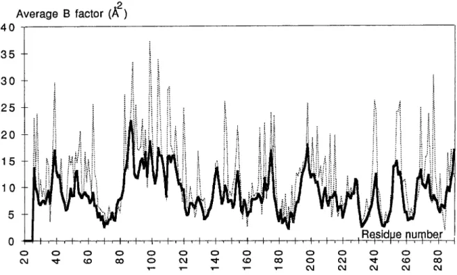
respectively. Another feature of the difference Fourier map, was the presence of positive peaks near each hydroxyl group of S82 and S285. Both peaks were too close to the CP atom to be assigned to water molecules and were interpreted as the oc- curence of two alternate conformations of the serine side chains (Fig. 2).The electron density is well defined for the whole protein except a t the end of a few long side chains exposed to solvent. Consistently, those residues: K55, R83, Q88, Q90, Q99, E104, K111, K146, K256, and R277 have relatively high temperature factors (30 to 40
A2).
After convergence of the refinement a t 1.8
A
res- olution, all 263 amino acids of TEMl @-lactamase, 199 water molecules, and a sulphate anion were as- signed. The crystallographic R-factor is 0.164 for 22,510 reflections between 5 and 1.8A
resolution. Average temperature factors and deviations from ideal geometry are given in Table 11. The distribu- tion of the B factors along the polypeptide chain is shown in Figure 3. The essential residues for catal- ysis (S70, K73, S130, E166, K234) have temperature factors below the average value computed for allprotein atoms. The total water content in the crystal amounts to 43%. The Luzatti plot25 gives an esti- mate of the coordinates error of 0.17
A
(Fig. 4). The Ramachandran plot (Fig. 5) reveals a few residues in medium to highly strained conformations: M69, S130, N175, D179, and L220. Their structural im- plications will be discussed.Description of the Structure
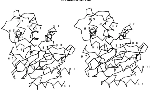
The TEMl enzyme (Fig. 6 ) is a globular protein of ellipsoidal shape with dimensions 30
A
x 40A
x 50A.
Its secondary structure elements were assigned using the program DSSF6 (Table III). Seventeen percent of the residues are involved in p-strands, 5%in 3,, helices, and 39% in a-helices. The molecule can be described as a two domains protein. The first one includes a five-stranded p-sheet onto which he- lices H1, H10, and H11 are packed, toward the sol- vent interface. The interface between this P-sheet and the 3 helices is a large hydrophobic core. The second domain is made of eight helices (H2 to H9) located on the other side of the pleated sheet. Two
hinge regions are connecting these domains which generate a large depression at their interface, cor- responding to the substrate binding site. These hinge regions are held by hydrogen bonds and salt-
bridges which should prevent any large conforma- tional change to occur.
The main p-sheet is formed by five antiparallel strands (Fig. 7). The residues of the two first strands
(S1 and S2) belong to the N-terminal part of the chain (43-601, and the three others (S3, S4,
S5)
to the C-terminal moiety (230-266). A @-bulge occurs, between P-strands S1 and S2, a t position 58; accord- ing to the classical @-bulge nomenclature, L57, E58, and E48 are, respectively, a t positions 1,2, and X of that motif.27 In addition to the main P-sheet, there are two small two-stranded antiparallel sheets(SB
and SC). Each strand is made of two residues. Strand S2 (56-60) is made of five residues, while its adjacent strand Sl(43-50) is eight residues long.
TABLE 11. Refinement Statistics and Deviations From Ideal Geometrv
Resolution range 5-1.8
A
R-factor for 22,510 reflections 16.4% Average temperature factors
8.8
A2
Main chain atoms (Ca, N, C, 0)Side chain atoms 13.1
A"
Water molecules 15.1
A2
Whole molecule (including solvent) 11.3
A2
Root-mean-square deviations fromideal geometry
Bond lengths 0.019
A
Improper dihedral angles 1.12"
Angles 2.66"
Dihedral angles 23.1"
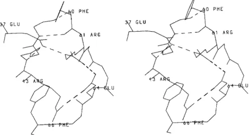
As a consequence, residues 43 and 44 at the N-ter- minal end of S1 have no hydrogen bonding partners. This is compensated by the hydrogen bond, also found in the B. licheniformis enzyme, between V44 main chain nitrogen atom and E37 side chain and by the salt-bridge R43-E64 (Fig. 8). These interactions, together with the salt-bridge between R61 and E64 and the hydrogen bond between E37 and R61 side chains, form the first hinge region. It locks the N-terminal part of the interdomain crossover loop, made of residues 60-68, which links strand S2 to the catalytic helix H2.
The conformation of the second hinge region (res- idues 212-222, Fig. 9) is determined by the salt-
bridge between R222 and the conserved and buried D233. A structural water molecule, WAT294, stabi- lizes the loop 217-220 and provides the missing hy- drogen bond partners of main chain nitrogen atoms L221 and R222 a t the N-terminal part of 3,, helix H10.
Topology of the Substrate Cavity
Several regions define this cavity shown in Figure 10: on one side, residues 234-237 belong to strand
S3 while R244 is found on the adjacent strand S4. Residues El04 and Y105 are located in a solvent exposed loop before H3. The electron density of Y105 is weak and its side chain has relatively high atomic temperature factors (= 25
A").
S130, D131, N132, which form a short connection between helices H4 and H5, are on the opposite side of the cavity with respect to strand S3. D131 side chain is buried and interacts with the main chain nitrogen atoms of V108, T109, N132, T133, and A134, bringing strong constraints on the respective positions of the N ter- mini of H3 and H5 to which these residues belong. In addition, it forms a hydrogen bond with T1090G1. This strong anchoring of D131 might insure the cor- rect positioning of S130 and explains its slightly strained conformation. V216 side chain is pointing toward the cleft where the acylating S70 is located. According to the DSSP secondary structure assign-ment, this serine belongs to a one turn 3,, helix (69-71) continued by an a-helix (72-85) which brings K73 side chain a t 2.9
A
from S700G.The R-loopZ8 (residues 161-179) (Fig. 11) forms one edge of the substrate binding side. The two ex- tremities of this peptide stretch are distant by only 3.5
A.
It is an important structural feature ofclass Ap-lactamases which carries the essential residue El66 and contains the short 3,, helix H7 (168-170). The conformations of two distorted type I1 p-turns (173-176) and (177-180) are stabilized by a set of electrostatic interactions consisting of two salt- bridges: R164-D179 and D176-Rl78. The third resi- dues of each turn (N175 and D179) have strained conformations with respective (@, q) values of (67", 8O) and (61°, 39"). The main chain oxygen atom of residue El77 is not hydrogen bonded to the main chain nitrogen of residue 180 but to T1800G1, a stkictly invariant residue. The N and C extensions of the R-loop are held together by three hydrogen bonds which involve p-sheet SB (Fig. 12).
The R-loop is located a t the protein-solvent inter- face but is strongly linked to the rest of the mole- cule. Besides one direct hydrogen bond (N1700 to E240N), interactions involve 8 buried water mole- cules and main chain polar atoms of the protein (Fig. 13).
Proper orientation of the catalytic El66 side chain is provided by the cis peptide bond formed with P167 and stabilized by a hydrogen bond with N170 side chain and by van der Waals contacts with the buried L169. In addition, its main chain carbonyl and ni- trogen atoms are in interaction with the amide group of N136 (Fig. 14).
Four water molecules and a sulphate anion have been identified in the substrate cavity (Fig. 15). They contribute to a complex polar interactions net- work (Fig. 16) from which only the strong interac- tions are given in Table
IV.
The water molecule (WAT297) hydrogen bonded to E1660E1 (2.7A)
is very likely the one involved in catalysis. WAT323, a t 2.8A
from A2370 and N, occupies the oxyanion hole." The sulphate ion is, interestingly, much closer to S1300G (2.9A)
than to R244 (3.2A)
which would be accessible for strong salt-bridge interac- tion. E1660E1 is separated from K73NZ and S700G by, respectively, 3.4A
and 4.2A.
S700G and S1300G are 3.5A
apart.Hydrophobic Clusters and Domains Interface The contacts between helices H1, H10, and H11 and strands S1, S4, and S5 of the first domain stand on hydrophobic interactions and involve almost all amino acids from one side of those strands. The sin- gle polar interaction found within this core arises from Y46: its phenolic group is hydrogen bonded, through the buried WAT369, to one oxygen atom of the solvent shielded side chain of E48. The second
1.8 A RESOLUTION STRUCTURE OF TEMl p-LACTAMASE 369
-2
Average B factor (A )
3 5
. .30
25
2 0
1 5
1 05
0
Fig. 3. Average Sfactors of main chain (thick line) and side chain (dotted line) atoms of TEMl.
oxygen atom of the carboxylate forms a salt-bridge with R259.
The core of the helical domain is also remarkably hydrophobic. Helix H2, the inner element of this do- main, contains four polar residues which have a ded- icated function: S70, T71, K73, and C77. Helices H2, H8, and H9 are held by an hydrophobic cluster formed by residues V74, G78, and L81 on H2, L190 and L193 on H8, and L207 and M211 on H9. L199 on the connecting loop between H8 and H9 is also pointing toward this interface (Fig. 17). Helix H8 is amphiphilic and each third of its surface, considered along its axis, has a special function: one third is facing H2, one third is solvent oriented, and one third is involved in the two domains interface. In- deed, H8 runs against S1, S4, and S5 providing strong hydrophobic interactions (M186, A187, L190) with residues from those strands (147, L49, 1247, 1260, V262). These contacts represent about one half of the whole interface between the two domains. The second half, protected from solvent by the salt- bridge R43-E64, involves residues F66, P67, M69, T71, from the crossover loop, and residues G45, G236, G238, G245, Y264, from S1, S3, S4, and S5 (Fig. 18). In addition, five hydrogen bonds are found. They occur between P670, WAT321, and T2650, and between Y2640H, T710G1, M680, and T71N.
Charge-Charge Interactions and Buried Charged Residues
There are 13 intramolecular and 1 intermolecular salt-bridges in the TEMl p-lactamase crystal struc-
ture (Table V), most of them being located on the protein surface and four of these occurring in the Q-loop. The aspartate and glutamate residues of salt-bridges E48-R259, R164-D179, and R222-D233 are buried in the protein structure.
Aspartic acids 85, 131, 157, and 214 are also bur- ied. D85, a t the C terminus of H2 has its side chain oriented such as to form hydrogen bonds to T200N and S2030G and seems to contribute to the position- ing of the N terminus of H9. A special orientation is found for D157 acidic group, whose OD1 is hydrogen bonded to T1600G1, which is itself hydrogen bonded
to T1810G1. This buried aspartic acid side chain seems to determine the proper orientation of the 2
threonine side chains in order to orient T181CG2 in the hydrophobic interface between the two protein domains, at van der Waals distance of Y264 aro- matic ring. The role of D131 in the topology of the substrate cavity has already been described. D214 belongs to the peptide stretch forming the second hinge region. Its carboxylic group is hydrogen bonded to D2330D1 and to the buried WAT309 of the active site (Figs. 9, 16).
Solvent Structure
A total of 199 water molecules, as well as a sul- phate anion in the active site, have been identified in the structure of p-lactamase TEM1. The average occupancy and the average temperature factor of water molecules are 0.61 and 15.1
A',
respectively. There is a layer of water molecules a t the contactR factor
1/d
0.2 0.25 0.3 0.35 0.4 0.45 0.5 0.55
Fig. 4. Evolution of the crystallographic R-factor as a function of the inverse of the resolution, superim- posed on theoretical R-factors deduced from mean coordinate errors.23
areas of symmetry-related protein molecules. It rep- resents, together with van der Waals contacts, the major crystal packing interactions. Indeed, there are only a dozen direct hydrogen bonds and one salt- bridge between atoms of symmetry-related mole- cules.
DISCUSSION
The structure of the TEMl enzyme was solved by multiple isomorphous replacement, although molec- ular replacement using the PC1 structure provided the right solution. The low rotation and translation functions signals are likely related to the different domain-domain orientations that are described in details in a later paragraph.
The general fold of the TEMl enzyme is similar to the one found in previously determined class A
p-lactamase structures. The protein is formed of two domains, connected by two hinge regions, and dis- plays remarkable long-range polar interaction net- works whose analysis is relevant with respect to the
following points: conformational change during ca- talysis, response to mutations leading to extended substrate specificity within the TEM family, and dif- ferent catalytic efficiencies within class A enzymes. Class A p-lactamases from different sources ex- hibit different substrate specificities but all essen- tial residues, except R244, implicated in catalysis, are conserved. The TEMl enzyme is peculiar, as it generates by point mutations a set of naturally oc- curing proteins with extended substrate specifici- ties.2 It thus seems that this enzyme is an excellent candidate to possibly unravel the molecular mecha- nism by which a protein could exhibit full enzyme efficiency3' while changing substrate specificity. This goal should be achieved by accurate kinetic measurements on TEMl enzyme and site-directed mutants of the protein, with a given set of enzyme substrates, coupled with X-ray structure deterrnina- tions.
The amount of solution data on class A p-lacta- mases and on their site-directed mutants is quite
1.8
a
RESOLUTION STRUCTURE OF TEMl P-LACTAMASE371
1
PSI
.80°
O0
-
180"
-180°
0"
180"
PHI
Fig. 5. Ramachandran plot of TEMl . Non-glycine residues in unfavorable conformations are labeled.
significant. However, it sometimes turns out that data on enzymes from different sources are merged with the aim of providing a consensus view of some properties. Such approach, which assumes struc- tural identities between those enzymes and antici- pates the conformational effects of amino acid sub- stitution, may lead to a confusing picture. The analysis and discussion of the TEMl structure will, thus, often refer to the available 5'. aureus PC1 crys-
tallographic structure (Protein Data Bank entry: 3BLM), sequence alignments within the class A
family, and exclusively, to site-directed TEMl mu- tants.
The Central Helix H2
H2 is one helix turn longer in TEMl compared to
PC1. D85, a specific residue of Gram- enzymes, is located in this turn and there is no equivalence in PC1 of its interaction with S2030G. This helix does not have the same sequence in both p-lactamases. This is especially reflected in the polarity of the cav-
ity found in the vicinity of El66. In TEM1, this area is quite hydrophobic and contains F72, L76, A135, L139, P145, L148, and L162 whose side chains dis- play optimal van der Waals interactions (Fig. 19). In PC1, the replacement on F72 and L162 by, respec- tively, serine and proline, empties the cavity which is filled by the side chains of residues N76 and N135. It results in a totaly different character of the cavity in which water molecules have been located.'
T71 was shown to be essential to proper structural stability of TEM1.31 The side chain of threonine 71 extends from helix H2 toward the domains interface of the protein. The hydroxyl group forms two impor- tant hydrogen bonds with the hydroxyl group of
Y264 and with M68 main chain oxygen atom, while T71CG.2 is a t about 4.5
A
from residues 235-236 main chain atoms.The disulfide bridge (C77-Cl23) connects helices H2 and H4 and is found in 7 out of 20 class A p-lac- t a m a ~ e s . ~ Removal of this covalent link by site-di- rected mutagenesis ((277s) led to an enzyme with the same activity but reduced thermostability a t
Fig. 6. Stereo view of the Ca-backbone of TEM1 p-lactamase. The secondary structure elements are labeled according to Table 111.
TABLE
111. Secondary Structural Elements in TEMl @-Lactamase* Helices H1 H2 H3 H4 H5 H6 H7 H8 H9 H10 H11 a 310 310 a a a a 3,0 310 a a a 26-40 69-71 72-85 109-1 11 119-128 132-142 145-154 168-170 183-195 201-212 221-224 272-288 Antiparallel B-sheets Main P-sheets1
43-50 s 2 56-60 s 3 230-237 s 4 244-251 Sheet B s 5 259-266 SB1 66-67 SB2 180-181 Sheet Csc
1 94-95 s c 2 117-118 Disulfide bridge Cys 77-Cys 123P-bulge 57, 58, 48
*Residue numbering according to Ambler et a1.5
40"C, while the enzyme is unstable upon mutations of both C77 and T71.32
Hinge Regions and Domains Interface
The two hinge regions are held by charge-charge interactions. Two glutamic acids (37 and 64) and two arginine residues (43 and 61), which are not all strictly conserved within p-lactamases, are involved in the first one. In the PC1 enzyme, the R43H,
R61N, and E64K mutations lead to much weaker interactions between domains. The salt-bridge be- tween residues R222 and D233 determines the con- formation of the second hinge region and explains the strained (-103", -117") dihedral angles of L220. Consistently, replacement of D233 in TEMl by other amino acids reduces strongly the catalytic ac- t i ~ i t y . ~ ~
In KlebsPella pneumaniue, PIT-2, Pseudomonas
aeruginosa, and TEM p-lactamases, position 214 is
occupied by an aspartic acid. In the TEMl structure, the buried D214 has its side chain hydrogen bonded to one of the carboxylic oxygens of D233 and to wa-
ter molecule 309 which interacts with K234 in the active site (Fig. 16). Such proximity of aspartic res- idues should increase the pKa of one of the acidic groups. D233, which forms a salt-bridge with R222, is invariant whereas D214 is only found in 4 out of 20 class A p-lactamases. As the overall confor- mations in this area are quite similar in TEMl and PC1 enzymes, where an asparagine is found at po- sition 214, it is likely that the protonated residue is D214.
At the interface between domains, G45 is facing P183 and F66. These residues are strictly conserved and no other residue than G45 could be sterically accomodated. In contrast to B. lichaniformis p-lac-
1.8
A
RESOLUTION STRUCTURE OF TEMl P-LACTAMASE373
Fig. 7. Stereo view of the main chain atoms of the five-stranded antiparallel sheet. Hydrogen bonds are indicated by hatched lines.
Fig. 8. Hydrogen bonds and charge-charge interactions (hatched lines) at the first hinge region.
tamase s t r u ~ t u r e , ~ there is no water molecule buried in this area. G245 is replaced by N245 in PC1. This significant increase in sterical volume, in such an important area, is compensated by the M69A muta- tion which creates a small cavity which is filled by the methyl group of A238. In this position, a glycine residue is almost always found in p-lactamases. Res- idue 69 is quite variable in size and character among class A enzymes but is always found in a high con- formational e n e ~ - g y . ~ , ~ Although it seems that its mutation could be accomodated without major rear-
rangement in the TEMl structure, the M69L mu- tant displays marginally perturbed kinetic behavior but shows dramatically decreased irreversible inac- tivation by both clavulanic acid and ~ u l b a c t a m . ~ ~
The D157-Tl60-Tl81 interaction found in TEMl seems to provide the proper orientation of T181CG2 toward Y264 aromatic ring. A similar hydrogen bonds pattern is found in PC1 but the mutation T181S creates a cavity delineated by residues F66, P183, F186, and F264. F66 and P183 are conserved between the two enzymes, but small movements of
Fig. 9. Stereo view of the second hinge region showing hydrogen bonds and charge-charge interactions (hatched lines): D214 and D233 are buried. WAT294 stabilizes the conformation of the loop 217-222.
ASP 2 1 4
%
ASP 2 1 1%
Fig. 10. Stereo view of the substrate binding cavity
their side chains and the important mutation M186F in PC1, compensate for the T181S substitu- tion. The mutation Y264F, however, results in the loss of the hydrogen bond with T710G1, which, as earlier mentioned, seems important for TEMl sta-
bility.
hydrogen bond partner, a hydroxyl group on the side chain of the fourth residue of this turn. This ex- plains that serine or threonine are always, except twice, found a t position 109.
Residue 167 is a proline in 13 out of the 20 class A p-lactamases. However, a cis peptide bond has also
The Substrate Binding Site been observed between El66 and I167 in PCl tal structure, which emphasizes its role for a proper crys-
-
-P107 is a strictly conserved residue in class A
p-Iactamases. In TEMl it appears as the second res- idue of a distorted type I turn. The conformation of this motive is strongly influenced by the hydrogen bond between T109N and the acidic group of the invariant and buried D131 which also requires, as
location and orientation of E166.
Strand S3 forms one side of the substrate binding cavity and bears residues which are important for enzyme stability or activity. S235 is a potential hy- drogen bond partner and was shown to interact with the carboxylate of PenG.ll Any other residue than
1.8
A
RESOLUTION STRUCTURE OF TEMl P-LACTAMASE 375Fig. 11. Stereo view of the 0-loop and of the four salt-bridges of TEMl enzyme.
Fig. 12. Stereo view of the N and C termini of the 0-loop. Residues 66-67 and 180-181 form sheet B.
G236 would have its side chain directed toward M69CB, main chain atoms of residues 70-71 and T71CG2, which might explain its invariance in p-lactamases. It has, nevertheless, been observed that phenotypes containing G236 TEMl mutants can resist to ampicillin a t very low concentrations of the a n t i b i o t i ~ . ~ ~
A237CB is pointing toward the substrate binding site, at 4
A
distance between M272 and R244 side chains, and should provide the main steric con- straint for the substrate position on this side of the cavity. Residue 237 is usually an alanine or a gly- cine in other class A p-lactamases. Bulkier amino acids are, however, also found a t that position and a threonine residue is even found in TEM5 natural mutant. Its side chain could either be oriented to-ward the substrate binding site, impairing binding,
or partially oriented toward R244, M272, and R275 generating steric conflicts that can only be released through displacement of some residues. In any case, substrate binding and/or local structure should be affected. Interestingly, the A237N TEMl mutant displays a lower kcat toward penam substrates but a higher activity with cephem a n t i b i o t i ~ s . ~ ~
The significant mutation A237Q occurs in the PC1 enzyme. This residue cannot be simply accomo- dated in the TEMl structure because Q237 and M272 side chains would collide. This observation points to the important fact that the region 265-276 is quite different in these enzymes, with respect to
sequences, conformations, and locations. Those dif- ferences have many consequences and arise because helix H11 is one turn longer in TEMl (residues 272- 274) than in PC1. As a consequence, P274 in PC1 is
. A S N 170 . A S N 17
4
1 3 &169
ASN 136
%
Fig. 14. Stereo view of the cis peptide bond 166-167 and of the surrounding hydrogen bonds (hatched lines) that maintain the conformation of E166.
found a t the same spatial location as the R275 side chain in TEM1, a residue only found in Gram- en- zymes, and the distance between CA atoms of resi- dues 271 from the two proteins amounts to 9
A.
These observations prompted us to analyze the se- quences of p-lactamases of known three-dimen- sional structures a t positions 246,220,244, and 276, taken in this order to illustrate the pairwise spatial proximities.In TEMl (1246, L220, R244, N276), R244 side chain has restricted movements as it is hydrogen
bonded to N276 and a t van der Waals distances of L220, A237, and M272 side chains. Residue 246 is buried and at van der Waals distance of residue 220. In PC1 (D246, L220, R244, and D276) the avail- ability of the positive charge of R244, implicated in the enzyme mechanism, should be reduced by the salt-bridge R244-D276 as in the B. Zicheniformis
p-lactamase (Protein Data Bank entry: 4BLM). Those two enzymes are identical with respect to the conformation of residues 265-276 and to the amino acids occurence a t all four positions mentioned
1.8 A RESOLUTION STRUCTURE OF TEMl B-LACTAMASE 377
u4 LYS 2-34 L Y S
Fig. 15. Electron density map (2F- - Fcalc, aCa,J of the catalytic region. The contour level is 1.0 u. Crosses indicate the water molecules.
above. The expected decrease in electrostatic poten- tial normally produced by R244 is consistent with the fact that the cacodylate ion, found in the B. li-
cheniformis p-lactamase crystal structure, is at hy- drogen bond distance from S1300G and S700G but
at 5.9
A
from the R244 guanidinium group. In S.albus G (D246, R220, N244, D276), R220 has been postulated to replace R244 function.36 Nevertheless, the buried D246 in the vicinity of R220, and the presence of D276, may reduce the effective positive charge of the guanidinium group. It is only in B.
cereus 56937 (D246, L220, R244, N276) and in E. coli
TEMl p-lactamases that R244 has no negative charge in its neighborhood.
G238, as already mentioned, is imposed by the presence of M69 side chain and N1700. Its mutation to serine, however, occurs in TEM3 and TEM4 p-lac- tamases. The minimum displacement possible to ac- comodate this side chain would be to move N170 which belongs to the R loop.
The insertion of residue I239 occuring in PC1, in the turn between p-strands S3 and S4 (238-243), also creates local differences between PC1 and TEMl p-lactamases a t this edge of the catalytic cleft. The conformations of the loops in both enzymes are different." As a consequence, the important main chain atoms of residue 237, whose nitrogen atom is part of the oxyanion hole, occupy slightly different positions in PC1 and TEMl enzymes.
Stability and Conformational Aspects of the &Loop
Part 171-173 of the 0 loop is in close vicinity to regions 66-67 and 238-241 of the protein. The crossover loop (60-68) has identical conformations in the two enzymes, although the sequences display significant differences. From TEMl to PC1 enzymes, the following mutations are observed P67A, E171Y, A172Y, I173S, and G238A, E240T, R241Y. They change the marked polar character of this region in
TEMl to a rather hydrophobic one in PC1. This could be related to the evolutionary capacity of the TEMl enzyme because mutations at some of these positions are responsible for altered substrate spec- ificity.
A172 is in aL conformation and the four next res-
idues (I, P, N, D) form a distorded type I1 p-turn. The
(@, dihedral angles of N175 are strained in order to release the steric conflict between its CB atom and P174 main chain oxygen atom. Actually, in most p-lactamases sequences, a glycine residue is found at position 175.
The salt-bridge R164-Dl79 is strictly conserved among class A p-lactamases, and links the two ends of the 0-loop. The P54 mutant D179N of the p-lac- tamase PC1 has a drastically reduced catalytic ac- tivity. A crystallographic study of this PC1 mu- tant" showed that removal of this link leads to a substantially disordered 0-loop. It should, however, be mentioned that among the four salt-bridges of the R-loop observed in TEM1, only one (R164-Dl79) re- mains in the PC1 p-lactamase. The 0-loop in the TEM enzyme is thus much more constrained.
Catalytic Residues
Except for R244, all residues involved in catalysis are invariant among class A p-lactamases. S70,
K73, S130, E166, and K234 have clear electron den- sity (Fig. 15) and refined to low temperature factors in the TEMl structure. Together with water mole- cules and a sulphate ion, they are involved in com- plex hydrogen bond interactions (Fig. 16). "he anal- ysis of these interactions needs care because hydrogen bond distances range from 2.7 to 3.3
A
and their description as strong, medium, or weak, espe- cially with respect to their significance in the cata- lytic mechanism, should also take into account the side chain mobilities. The flexibility of K73 side chain, located between S70 and E166, is large and tenths of an angstrom are easily gained or lost uponFig. 16. The interactions network in the catalytic site. Hatched lines indicate hydrogen bond distances less than 2.9 A. Dotted lines are used when the distance is above that value. Wavy lines are for special cases (see text).
variation of the x1 dihedral angle of S70 and S130. For instance, the distance between K73 and S130 side chains was reported to be 3.7 in the TEMl structure and 2.8
A
in the acylated complex." In our refined TEMl structure, the S130OG-K73NZ dis- tance is 4.2A
whereas it amounts 3.2A
and 3.7A
inB. lichenformis and S. aureus p-lactamases, respec- tively. This difference, illustrative of the already mentioned flexibility of the K73 side chain, might be a consequence of the electrostatic effect produced by the sulphate ion, found a t 2.9
A
from S1300G anda t 3.2
A
from the accessible guanidinium group ofR244.
According to the closest proximities observed, the active site could, tentatively, be divided into two ar-
eas. There are consecutive hydrogen bonds between, on one hand, the sulphate ion, S130, K234, WAT309, D214, D233, and R222 (WAT309 is also at
2.9
A
from S2350G) and on the other hand, K73, S70, WAT297, and El66 (Fig. 16). These two net- works are connected through the WAT297-WAT323 sulphate ion interactions. WAT297, between S701.8 A RESOLUTION STRUCTURE OF TEMl p-LACTAMASE 379
TABLE
IV.
MajorInteractions Within
the ActiveSite
of the EnzymeDistances Interactions
(A,
S700G K73NZ 2.9 S700G WAT323 2.9 S700G WAT297 2.9 E1660E 1 WAT297 2.7 E1660E2 N170ND2 3.0 N 1700D 1 WAT297 2.8 A2370 WAT323 2.8 A237N WAT323 2.8 S1300G SO4 2.9 R244NH1 SO4 3.2 K234NZ S1300G 2.9 K234NZ WAT309 2.9 S2350G WAT309 2.9 D2140D1 WAT309 2.9 WAT297 WAT391 2.8 WAT323 SO4 2.8-?
TABLE
V.
Salt-Bridges in TEMl B-Lactamase*Distance Salt-bridge
(A,
Lys32 Asp35 3.0 Lys34 Asp38 3.1 2.9 2.9 Glu37 Argsl Arg43 Glu64 Glu48 Arg259 2.8 Arg61 Glu64 3.0 Lys73 Glu166 3.4 Glu89 Art93 3.1 Argl6l Asp163 2.9 Arg164 Glu171 2.7 Arg164 Asp179 3.0 Asp176 Arg178 3.0 Arg222 Asp233 3.1Intermolecular crystal contacts
Lys32 Glu197 3.0
*Invariant residues are in bold type.
-
Fig. 17. Hydrophobic core between H2, H8, and H9. N-Ca-C backbone, thin lines; side chain atoms of
residues involved in non-polar interactions, thick lines.
38 SLY
I'
Fig. 18. Hydrophobic interactions at the interface of the two protein domains which involve the polypeptide stretch 66-71 (N-Ca-C backbone).
Fig. 19. The hydrophobic cavity in the vicinity of the residue El66 of TEMl enzyme.
and E166, is in proper position as partner of the deacylation step and WAT323, 2.8
A
from A237 main chain atoms and 3.3A
from S70N, occupies the oxyanion hole.A number of active site mutants of TEMl have been studied. El66 mutant enzymes
demonstrated the role of this glutamic acid in the deacylation step, and similar conclusions were drawn from the X-ray study of the E166A mutant of
B. licheniformis p-lactamase." Acylation by S70 on the rt! face of the p-lactam ring was shown in the crystal structure complex of the E166N mutant with PenG.ll It was then proposed that in the enzyme mechanism, K73NZ, located at 3.4
A
from E1660E1, acts as a general base in the acylation step.K234 is non-accessible to bulk water and the importance of the positive charge of its side chain, found at 2.9
A
from S1300G, has been analyzed by site-directed mutagenesis in K234R and K234T TEMl mutants.40 The drastic opposite kinetic effects resulting from these mutations led to the conclusion that K234 is involved in substrate binding and transition state stabilization. The evaluated contribution of the K234 side chain to apparent binding energy in the transition state is 4.6 kcal/mole. The R244Q or T and R244K or Smutants of TEMl have been produced and stud- ied.34.41 Kinetic results from these independent investigations show that kcat values for PenG substrate are nearly unaffected by the nature of the residue a t position 244, the apparent binding energy of R244 side chain to the transition state being around 2 KcaYmole. These data, obtained in solution with site-directed mutants of the TEMl enzyme would support a stronger interaction of the carboxylate group a t C-3 of the substrate with K234 than with R244 in the transition state, whereas the interaction with R244 would dominate after acyla- tion."
Superposition of TEMl and PC1 Crystal Structures
The Ca coordinates of p-lactamases PC1 and TEMl have been superposed by least-squares fit- ting, after omission of the largest differences (resi- dues 26-30,50-58,84-91,238-242,250-258, and 265-275). The root-mean-square deviation for the corresponding Ca atoms is 1.86
A
(without omission: r.m.s. = 2.49A).
This is to be compared to the lower r.m.6. deviation of 1.37A
found between PC1 and B.licheniformis enzymes!'
Inspection of the superimposed structures showed that rigid body handling of the whole protein struc- tures was not satisfactory. Indeed, the two enzymes are best superimposed when their two domains are handled independently and rotated in opposite di- rections along nearly parallel axes running through each domain (Fig. 20). Overall, these rotations amount to 7" and result in a wider opening of the substrate cavity in PC1 compared to TEM1. The cat- alytic properties and the substrate specificities of the TEMl and PC1 enzymes are probably influ- enced by this different aperture of the substrate binding cleft as well as by the many local structural and sequence differences already pointed out.
Mutated Residues in Extended Spectrum
"EM
P-LactamasesThe TEMl p-lactamase hydrolyzes penicillins and some first and second-generation cephalosporins. One to four point mutations can extend the hydrol- ysis spectrum of the TEM p-lactamase to some third- generation cephalosporins, especially ceftazidime and cef~taxime.'-~ The location of the mutated res- idues in the TEMl structure is displayed in Figure 21.
The extended spectrum TEM p-lactamases derive from TEMl or TEM2, two enzymes with identical enzymatic properties and only differing by the Q39K mutation. Residue Q39 is located a t the C terminus
1.8 A RESOLUTION STRUCTURE OF TEMl p-LACTAMASE 381
a
b
Fig. 20. Superposition of TEMl (thick line) and PCI (thin line) p-lactamases. a: Ca atoms of the B-domains (except loops and
INDEL regions) from both proteins were superimposed by least-
square procedures and the resulting transformation matrix applied to all Ca atoms. b: Ca atoms from each domain (except loops and
INDEL regions) were handled independently.
Fig. 21. Stereo view of the Ca-backbone of TEMl enzyme and van der Waal surfaces of the residues that confer, upon mutation of the TEM enzyme, extended substrate specificities.
of helix H1 at the protein-solvent interface, far from the active site. Its side chain amide group interacts with D35 carboxylic acid through 2 water molecules. Additionally, D35 forms a salt-bridge with K32 which also interacts with Q278 from H11. The E104K, R164S, and G238S mutations are found in natural protein mutants which derive from TEMl or
TEMP enzymes. The X-ray structure of TEMl does not provide a n explanation of whether Q or K a t position 39 might favor the occurence of further mu- tations.
E l 0 4 and E240 are a t the protein-solvent inter- face, a t the entrance of the substrate binding cavity, and their side chains are not involved in specific
interactions. However, E104, in the neighborhood of P167, could interact with the C-6 side chain of pe- nam substrates, and its substitution to a lysine would affect the kinetic properties of the resulting mutant. E240 is part of the tight loop connecting S3
and S4. Further crystallographic investigations seem necessary in order to explain the higher cata- lytic efficiency of the E240K mutant enzyme toward cephalosporines compared to the wild type p r ~ t e i n . ~ However, the proximity of El71 and El68 should be mentioned because interactions between K240 and those residues would affect the conformation of the Two of the four charge-charge interactions that stabilize the R loop in TEMl are formed between R164 and D179, E171. Their disruption upon R164S or H mutation will certainly destabilize this region but the redistribution of charges cannot be antici- pated.
R244 and T265 are adjacent residues on strands S4 and S5. One buried water molecule bridges T2650G1 to G2420 and S268N. T265CG2 is ori- ented toward R244 side chain. The mutation T265M would disrupt this buried hydrogen bond pattern and might lead to structural modifications in this region.
CONCLUSIONS
The molecular structure of the TEMl enzyme al- lows the first comparison of Gram- and G r a d p-lactamases. This analysis gives some insights into the structural reasons that may account for the dif- ferent kinetic properties, substrate specificities, and evolutionary capabilities encountered for TEMl compared to the PC1 enzyme. The TEMl wild-type structure cannot provide definite explanations about the structural events occuring upon the mu- tations which lead from TEMl or 2 to TEM3- TEM15 enzymes43 and further crystallographic studies are required. The density of strong interac- tions throughout the molecule would seem prohibi- tive to large conformational changes but, on the other hand, some of them could easily be reshuffled in order to accomodate local movements. The area, interestingly defined by loops 103-106, 238-241, 266-275, and R, deserves special attention. There are, in many respects, differences between TEMl and PC1 enzymes which impair the structural ex- planations on TEMl mutants based on the PC1 en- zyme structure and the generalization of the conclu- sions drawn from one enzyme to the other. The X-ray coordinates of the TEMl enzyme will be de- posited in the Brookhaven Data Bank.
ACKNOWLEDGMENTS
We thank Dr. Din0 Moras and collaborators at the UPR de Biologie Structurale in Strasbourg for all facilities placed a t our disposal throughout this
work.. C.J. was e m p l o y e d during his Ph.D. by Bio-
R loop.
structure SA,
Les
Algorithmes, Parc d’Innovation, 67400 Illkirch-Graffenstaden, France. This research was funded, in part, by the French Ministry of Re-search and Technology (Grant 891‘0840).
1. 2. 3. 4. 5. 6. 7. 8. 9. REFERENCES
Phillippon, A., Labia, R., Jacoby, G. Extended-spectrum p-lactamases. Antimicrob. Agents Chemother. 33:1131- 1136, 1989.
Collatz, E., Tran Van Nhieu, G., Billot-Klein, D., William- son, R., Gutmann, L. Substitution of serine for arginine in position 162 of TEM-type (3-lactamases extends the sub- strate profile of mutants enzymes, TEM-7 and TEM-101, to
ceftazidime and aztreonam. Gene 78:349-354, 1989. Sowek, J.A., Singer, S.B., Ohringer, S., Malley, M.F., Dougherty, T.J., Gougoutas, J.Z., Bush, K. Substitution of Lysine at position 104 or 240 of TEMl,,,, p-lactamase enhances the effect of serine 164 substitution on hydrolysis or affinity for cephalosporins and the monobactam aztreo- nam. Biochemistry 30:3179-3188,1991.
Joris, B., Ghuysen, J.M., Dive, G., Renard, A,, Dideberg,
O., Charlier, P., Frhre, J.M., Kelly, J.A., Boyington, J.C., Moews, P.C., Knox, J.R. The active-site serine penicillin- recognizing enzymes as members of the Streptomyces R61 DD-Peptidase family. Biochem. J. 250:313-324, 1988. Ambler, R.P., Coulson, A.F.W., FrBre, J.M., Ghuysen, J.M., Joris, B., Forsman, M., Levesque, R.C., Tiraby, G., Waley, S.G. A standard numbering scheme for the class A p-lactamases. Biochem. J. 276269-272, 1991.
Bush, K. Classification of p-lactamases: Groups 1, 2a, 2b and 2b‘. Antimicrob. Agents Chemother. 33:264-271, 1989.
Dideberg, O., Charlier, P., Wery, J.P., Dehottay, P., Dusart, J., Erpicum, T., FrBre, J.M. Ghuysen, J.M. The crystal structure of the p-lactamase of Streptomyces albus G a t 0.3 nm resolution. Biochem. J. 245:911-913, 1987. Herzberg, 0. Refined crystal structure of p-lactamase from Staphylococcus aureus PC1 a t 2.0 A resolution. J. Mol. Biol. 217:701-719, 1991.
Knox. J.R.. Moews. P.C. B-lactamase of Bacillus licheni- formis 749jC. Refinement a t 2 A resolution and analysis of hydration. J. Mol. Biol. 220:435-455, 1991.
10. Jelsch, C., Lenfant, F., Masson, J.M., Samama, J.P. p-lac- tamase TEMl of E. coli: Crystal structure determination a t 2.5 A resolution. FEBS Lett. 299:135-142, 1992. 11. Henberg, O., Kapadia, G., Blanco, B., Smith, T.S., Coul-
son, A. Structural basis for the inactivation of the P54 mutant of p-lactamase from S. aureus PC1. Biochemistry 12. Knox, J.R., Moews, P.C., Escobar, W.A., Fink, A.L. A cat- alytically-impaired class A p-lactamase: 2
a
crystal struc- ture determination and kinetics of the Bacillus licheni- formis E166A mutant. Protein Eng. 6:ll-18, 1993. 13. Strynadka, N.C.J., Adachi, H., Jensen, S.E., Johns, K.,Sielecki, A., Betzel, C., Sutoh, K., James, M.N.G. Molecu- lar structure of the acyl-enzyme intermediate in p-lactam hydrolysis a t 1.7 A resolution. Nature (Lond.) 359700- 705, 1992.
14. Matagne, A,, Misselyn-Bauduin, A.M., Joris, B., Erpicum, T., Granier, B., F r h e , J.M. The diversity of the catalytic properties of class A p-lactamases. Biochem. J . 2653131- 146, 1990.
15. Jelsch, C., Lenfant, F., Masson, J.M., Samama, J.P. Crys- tallization and preliminary crystallographic data on E. coli TEMl p-lactamase. J. Mol. Biol. 223:377-380, 1992. 16. Henberg, O., Moult, J . Bacterial resistance to p-lactam
antibiotics: Crystal structure of p-lactamase from Staphy- lococcus aureus PC1 a t 2.5 A resolution. Science 236694- 701,1987.
17. Crowther, R.A. The fast rotation function. In: “The Molec- ular Replacement Method.” Rossmann M.G. (ed) New York; Gordon and Breach, 1972: 173-178.
18. Crowther, R.A., Blow, D.M. A method of positioning a known molecule in an unknown crystal structure. Acta 19. Sussman, J.L. Constrained-restrained least-squares (CORELS) refinement of proteins and nucleic acids. Meth-
ods Enzymol. 115:271-303, 1985. 309503-9509,1991.
1.8
A
RESOLUTION STRUCTURE OF TEMl p-LACTAMASE 38320. Briinger, A.T. X-PLOR Manual, v. 2.1. The Howard Hughes Medical Institute and Department of Molecular Biophysics and Biochemistry, Yale University, New Ha- ven, CT, 1990.
21. Jones, T.A. A graphics model building and refinement sys- tem for macromolecules. J. Appl. Crystallogr. 11:268-272, 1978.
22. Bricogne, G. Methods and programs for direct-space ex- ploitation of geometric redundancies. Acta. Crystallogr. 23. Hendrickson, W.A. Stereochemically restrained refine- ment of macromolecular structures. Methods Enzymol. 115:252-270, 1985.
24. James, M.N.G., Sielecki, A.R. Structure and refinement of penicillopepsin at 1.8 A resolution. J. Mol. Biol. 163299- 361, 1983.
25. Luzatti, V. Traitement statistique des erreurs dans la d6- termination des structures cristallines. Acta Crystallogr. 5:802-810, 1952.
26. Kabsch, W., Sanders, C. Dictionary of protein secondary structure: Pattern recognition of hydrogen bonded and geometrical features. Biopolymers 22:2577-2637, 1983. 27. Richardson, J.S. The anatomy and taxonomy of protein
structure. Adv. Protein Chem. 34:167-339, 1981. 28. Leszinski, J.F., Rose, G.D. Loops in globular proteins: A
novel category of secondary structure. Science 234849- 855, 1986.
29. Murphy, B.P., Pratt, R.F. Evidence for an oxyanion hole in serine p-lactamases and DD-peptidases. Biochem. J. 256: 669-672, 1988.
30. Christensen H., Martin, M.T., Waley, S.G. p-lactamases are fully efficient enzymes. Biochem. J. 266853-861, 1990.
31. Dalbadie-McFarland, G., Neitzel, J.J., Richards, J.H. Ac- tive site mutants of p-lactamase: Use of an inactive double mutant to study requirements for catalysis. Biochemistry 32. Schulz, S.C., Dalbadie-McFarland, G., Neitzel, J.J., Rich- ards, J.H. Stability of wild type and mutant RTEM-1 p-lac- tamases: Effect of disulfide bond. Proteins 2290-297, 1987.
A32832-847,1976.
25~332-338, 1986.
33. Palzkill, T., Botstein, D. Probing p-lactamase structure and function using random replacement mutagenesis. Pro- teins 1429-44,1992.
34. Delaire, M., Labia, R., Samama, J.P., Masson, J.M. Site- d i dmutagenesis ofE. coli TEM-1 p-lactamase. J . Biol. Chem. 267:20600-20606, 1992.
35. Healey, W.J., Labgold, M.R., Richards, J.H. Substrate specificities in class A p-lactamases: Preference for pe- nams vs. cephems. The role of residue 237. Proteins 6:275- 283, 1989.
36. Jacob-Dubuisson, F., Lamotte-Brasseur, J., Dideberg, O., Joris, B., Fr6re, J.M. Arginine 220 is a critical residue for the catalytic mechanism of the Streptomyces albus G p-lac- tamase. Protein Eng. 4:811-819, 1991.
37. Samraoui, B., Sutton, B.J., Todd, R.J., Artymiuk, P.J., Wa- ley, S.G., Phillips, D.C. Tertiary structural similarity be- tween a class A p-lactamase and a penicillin-sensitive D-alanyl carboxypeptidase-transpeptidase. Nature (Lond.) 320378-380,1986.
38. Adachi, H., Ohta, T., Matauzawa, H. Site-directed mu- tants. at wsition 166. of RTEM-1 B-lactamase that form a stable acyl-enzyme intermediate ‘ h t h penicillin. J. Biol. Chem. 2663186-3191,1991,
39. Delaire, M., Lenfant, F., Labia, R., Maason, J.M. Site-di- rected mutagenesis on TEMl p-lactamase: Role of Glu166 in catalysis and substrate binding. Protein Eng. 4:805- 810,1991.
40. Lenfant, F., Labia, R., Masson, J.M. Replacement of lysine 234 affects transition state stabilization in the active site of p-lactamase TEM1. J. Biol. Chem. 26617187-17194, 1991.
41. Zafarella, G., Manavathu, E.K., Lerner, S.A., Mobashery, S. Elucidation of the role of Arg244 in the turnover pro- cesses of class A p-ladamases. Biochemistry 31:3847- 3852, 1992.
42. Moews, P.C., Knox, J.R., Dideberg, O., Charlier, P., F r h , J.M. p-lactamase of Bacillus licheniformis 749lC a t 2 A
resolution. Proteins 7:156-171. 1990.
43. Neu, H.C. The crisis in antibiotic resistance. Science 257: 1064-1073, 1992.