Characterization of CPG15 During Cortical Development and Activity
Dependent Plasticity
By Corey Harwell B.S., Chemistry
Tennessee State University, 2000
Submitted to the Department of Brain and Cognitive Sciences in Partial Fulfillment of the Requirements for the Degree of
Doctor of Philosophy in Neuroscience at the
Massachusetts Institute of Technology September 2006
© 2006 Massachusetts Institute of Technology. All rights reserved.
'0) , Signature of Author:
Department of Brain and Cognitive Sciences September 1, 2006
Certified by:
I,
-SElly Nedivi
Associate Professor of Neurobiology P Thesis Supervisor Accepted by:
"Matthew Wilson Picower Professor of Neuroscience Chairman, Department Graduate Committee
MASSACHUSETTS INSETTWJTj OF TECHNOLOGY
SEP 1
3
2006
LIBRARIES
_ _ _ · ----
C~-J-1ý
Characterization of CPG15 During Cortical Development and Activity
Dependent Plasticity
by
Corey C. Harwell
Submitted to the Department of Brain and Cognitive Sciences On September 1, 2006 in Partial Fulfillment of the
Requirements for the Degree of Doctor of Philosophy in Neuroscience
ABSTRACT
Regulation of gene transcription by neuronal activity is thought to be key to the translation of sensory experience into long-term changes in synaptic structure and function. Here we show that cpgl5, a gene encoding an extracellular signaling molecule that promotes dendritic and axonal growth and synaptic maturation, is regulated in the somatosensory cortex by sensory experience capable of inducing cortical plasticity. Using in situ hybridization, we monitored cpgl5 expression in 4-week-old mouse barrel cortex after trimming all whiskers except D1. We found that cpgl5 expression is depressed in the deprived barrels and enhanced in the barrel column corresponding to the spared D1 whisker. Induction of cpgl5 expression is significantly diminished in adolescent as well as adult CREB knockout mice. cpgl5's spatio-temporal expression pattern and its regulation by CREB are consistent with a role in experience-dependent plasticity of cortical circuits. Our results suggest that local structural and/or synaptic changes may be a mechanism by which the adult cortex can adapt to peripheral manipulations.
The balance between proliferation and apoptosis is critical for proper development of the nervous system. Yet, little is known about molecules that regulate apoptosis of proliferative neurons. Here we identify a soluble, secreted form of CPGl5 expressed in embryonic rat brain regions undergoing rapid proliferation and apoptosis, and show that it protects cultured cortical neurons from apoptosis by preventing activation of caspase 3. Using a lentivirus-delivered small hairpin RNA, we demonstrate that endogenous CPG15 is essential for the survival of undifferentiated cortical progenitors in vitro and in vivo. We further show that CPGl5 overexpression in vivo expands the progenitor pool by preventing apoptosis, resulting in an enlarged, indented cortical plate and cellular heterotopias within the ventricular zone, similar to the phenotypes of mutant mice with supernumerary forebrain progenitors. CPGl5 expressed during mammalian forebrain morphogenesis may help balance neuronal number by countering apoptosis in specific
neuroblasts subpopulations, thus influencing final brain size and shape. Thesis Supervisor: Elly Nedivi
Acknowledgements
First I would like to thank my advisor Elly Nedivi for taking time to be a mentor to me and for teaching me how to be a scientist. Without her faith in me I would not be where I am today, and I cannot put into words my gratitude.
I would also like to thank the past and present members of the Nedivi Lab, everyone has taught me so such in my time here. Just as important as the scientific lessons I learned are the friendships forged with lab members that will last a lifetime.
I would next like to thank my committee Martha Constantine-Paton, Rosalind Segal and especially Carlos Lois for being a second mentor, and his helpful technical advice.
I would like to thank my entire family especially my mother Regina Harwell, my father Willie Green and my brother Reggie Harwell for raising me and teaching me how to be a man. I certainly would not have made it this far without your guidance and love.
Finally, this would not have been possible without the love and unwavering support of my wife Lynn. She is by far the best thing that has happened to me since moving to Boston, and I am grateful everyday for having her love and support.
Contents
Abstract ... 2
Acknowledgments ... 3
Table of Contents ... 4
Chapter 1: Introduction ... 5
Chapter 2: Regulation of cpgl5 Expression during Single Whisker Experience in the Barrel Cortex of Adult Mice...17
Chapter 3:Soluble CPG15 expressed during early development rescues cortical progenitors from apoptosis...40
Chapter 4: Conclusions...79
Appendix...83
References ... 88
Chapter 1
Introduction
Cell death is an essential component of neural development.
A central question in developmental neurobiology is how does the cerebral cortex
go from a thin layer of neuroepithelial cells lining the ventricle to create the diversity of cell types arranged in a stereotypical pattern to form the circuitry of the mature functional brain (Stern, 2001). It is now known that formation of the cerebral cortex involves the proliferation of neural progenitor cells along the anatomically defined ventricular zone (1970). Traditionally it was believed that neural progenitors lining the ventricle would divide symmetrically to produce two progenitor cells, thereby expanding the progenitor pool, and divide asymmetrically to produce a progenitor cell and a newly generated neuron (Chenn and McConnell, 1995; Takahashi et al., 1996). It is now known that in addition to the traditionally viewed neuron/progenitor producing asymmetric division, neurons are also generated by asymmetric divisions of progenitors that produce a radial glial progenitor cell, and an intermediate progenitor that moves up into the subventricular zone and divides symmetrically to produce two neurons (Noctor et al., 2004). The next step in the development of the brain is migration of these newly postmitotic neurons to the cortical plate where they continue to differentiate and eventually make synaptic connections (Bayer et al., 1991). Traditionally, after cell division a cell was thought to choose one of two fates, either continued proliferation or differentiation into a neuron or glial cell (Bayer and Altman, 1991). This view highlights the assumption that the major factor governing the size of the final neuronal population in the mature brain is the
magnitude and rate progenitor of cell proliferation. It was previously assumed that cell death had a minor role during embryonic cortical development, and that the major wave of cell death in the CNS occurred during target selection (Oppenheim, 1991). However, fairly recently it has been shown that programmed cell death plays an important role in the development of the cerebral cortex(Blaschke et al., 1996; Blaschke et al., 1998; Kuan et al., 2000). While an established role for programmed cell death in cortical development is a relatively new concept, the importance of cell death during the course of normal embryonic development had been known for quite some time (Clarke and Clarke, 1995; Clarke and Clarke, 1996; Jacobson et al., 1997). First observed during amphibian metamorphosis, it was found that stereotyped, and predictable cell death was observed during the development of both invertebrate and vertebrate animals (Truman et al., 1990). Later, it was discovered that not all forms of cell death were created equally and there is a distinction between the cell death observed during development and tissue homeostasis, and the pathological cell death that occurs as a result of trauma or ischemia (Kerr et al., 1974; Kerr et al., 1972). The latter form came to be known as necrosis and is characterized by the swelling and rupturing of the damaged cells causing them to leak their contents into the surrounding environment inducing an inflammatory response (Kerr et al., 1972). In contrast, when cells die during normal development or during tissue homeostasis they usually shrink and condense while maintaining the integrity of the plasma membrane, thus allowing the dying cell to be engulfed by surrounding cells before any of its contents are leaked into the surrounding environment. One of the hallmarks of cells undergoing this controlled cell death is the fragmentation of its nuclear DNA as the cell is packaged for disposal (Kerr et al., 1972; Vaux and Korsmeyer, 1999;
Wyllie et al., 1980; Yuan et al., 1993). This process came to be known as apoptosis, coined from the ancient greek word that describes the 'falling off of flower petals or leaves from trees (Jacobson et al., 1997; Kerr, 1995; Kerr et al., 1972).
In a study using an optimized in situ end labeling plus technique (ISEL+) that labels fragmented nuclear DNA of dying cells, it was shown that cortical neuroblasts in the proliferative ventricular zone undergo programmed cell death (Blaschke et al., 1996). In fact using this particular technique it was observed that in the developing telencephalon as many as 70% of all cells are undergoing apoptosis at embryonic day 14, the rate of apoptosis declines to approximately 50% by embryonic day 18. The finding of such a high degree of cell death was highly controversial, and it remains unclear whether the ISEL+ is detecting cells in the early phase of apoptosis or, transient DNA breaks in cells not committed to apoptosis. While the accuracy of the ISEL+ and its ability to detect cells that were actually undergoing programmed cell death was highly controversial, with the magnitude of apoptosis in the developing cortex still subject to debate (Kuan et al., 2000; Voyvodic, 1996), this study did help shed light upon the idea that programmed cell death may be playing an important role during cortical development.
The Genetics of Programmed Cell Death
Genetics studies in the roundworm Caenorhabditis elegans demonstrated that animal cells have a built in cell suicide program, and that certain genes appear to be dedicated to the death program and its control. During the development of an adult worm 131 out of the 1090 total cells that make up the organism undergo programmed cell death in a lineage specific and mostly cell autonomous manner (Kuan et al., 2000; Metzstein et
al., 1998). Through genetic screening in the worms three groups of cell death genes have been identified. The first group of genes including egl-1, ced-4, and ced-3 and ced-9 are required for all somatic programmed cell death to occur (Conradt and Horvitz, 1998; Ellis and Horvitz, 1986; Trent et al., 1983). The next group includes the genes ces-1 and ces-2 and is necessary for cell death of specific types of cells (Yuan and Horvitz, 2004; Yuan and Horvitz, 1990). The last group of genes which includes ced-l, ced-6, ced-7, ced-2, ced-5, ced-10 and nuc-l, are involved in degradation of DNA and phagocytosis of the remains of the apoptotic cell (Metzstein et al., 1998). The finding that genes in each of these groups had mammalian homologs raised the possibility that the genetic program for cell suicide and the necessity for its function has been conserved throughout evolution.
The strongest evidence for the necessity of cell death genes during cortical development came from mutant mice deficient in the pro-apoptotic ced-3 homologues that comprise a family of cysteine-containing, apartate-specific proteases called caspases. All of the caspase deficient mice that have been examined (Caspases 1, 2, 3, 8, 9, and 11) appear to have a cell type specific death defects, while none appears to have global defects in apoptosis (Cecconi et al., 1998; Hakem et al., 1998; Kuan et al., 2000; Kuida et al., 1998; Kuida et al., 1996; Yoshida et al., 1998). The result from these caspase mutants indicates that the individual members of the caspase family appear to regulate cell death in a tissue specific or stimulus specific manner (Kuan et al., 2000). Surprisingly Casp3 and Casp9 null mutants had severe programmed cell death defects in the brain that lead to a malformed cortex with multiple indentations and periventricular masses consisting of supernumerary neurons(Hakem et al., 1998; Kuida et al., 1998; Kuida et al., 1996). The mutants are embryonic or perinatal lethal with a reduction in pyknotic cells observed
specifically in the brain. These mutants thus provided the strongest evidence for the necessity of programmed cell death programs for proper cortical development. The similarity of the phenotypes of Casp3 and Casp9 mutants raised the possibility that they may be functioning within the same pathway in the developing brain. In fact biochemical studies would reveal that caspase 9 functions as an upstream activator of caspase 3 in the cell death pathway (Kuan et al., 2000; Li et al., 1997). It still remains unclear what factors lie downstream of caspase 3 and leads to apoptosis in during embryonic cortical development. One known downstream target of caspase 3 is DNA fragmentation factor
45 (DFF45), which is responsible for causing the fragmented DNA that characterizes apoptotic cells. However, mice with homozygous mutations in DFF45 are viable with no discernable defects in brain development (Zhang et al., 1998). The finding that there are a small number of Casp3 null mice that survive to adulthood without obvious defects (Kuan et al., 2000), would indicated that there are additional compensatory pathways for developmental apoptosis that remain to be identified.
Neurotrophins are Extrinsic Factors Regulating Cell Death
Over fifty-five years ago Viktor Hamburger and Rita Levi-Montalcini made the observation that the sensory ganglia of the neck are small when compared to the brachial ganglia which are larger because they innervate the additional mass of the limb in the embryonic chick (Levi-Montalcini, 1987). In addition they found that if the embryonic chick limb bud is removed early on, neuroblasts in the corresponding spinal ganglia will degenerate. These results suggested that neurons were competing for a limited supply of peripherally derived trophic factors and their death is the result of the
competition of surplus neurons for limited amounts of these factors. This 'neurotrophic hypothesis' went on to become one of the most influential concepts in developmental neurobiology (Aloe, 2004; Kuan et al., 2000; Levi-Montalcini, 1987). Levi-Montalcini and colleagues would eventually go on to discover the first diffusible survival factor that would eventually become known as Nerve Growth Factor (NGF) (Cohen and Levi-Montalcini, 1956). Eventually a larger family of molecules structurally related to NGF would be identified. Four members of the family have been found to be expressed in birds and mammals: NGF, brain derived neurotrophic factor (BDNF), neurotrophin-3 (NT-3) and neurotrophin-4 (NT-4). The characterization of NGF and later brain derived neurotrophic factor (BDNF), neurotrophin-3 (NT-3), and neurotrophin-4 (NT-4) revealed the essential role for cellular interactions in numerous phases of neural development (Huang and Reichardt, 2001). The expression of these molecules in the developing cortex also gave further evidence for the necessity of apoptosis for proper cortical development, and that cell survival could potentially be regulated by an extrinsic trophic factor in a manner similar to that observed in the spinal cord ganglia (Emrnfors et al., 1992). Studies of the spatio-temporal distributions of NT-3 and BDNF, shows that these transcripts are expressed in the cortical ventricular/subventricular zones, coinciding with the onset of neurogenesis (Maisonpierre et al., 1990), along with their preferred receptors tyrosine kinase receptors TrkB and TrkC (Fukumitsu et al., 1998). The onset of
neurotrophin expression may mark the time period when they first become generally required to maintain survival of neural precursors, and postmitotic neurons and their localization supports the notion that the mechanisms proposed in the neurotrophic hypothesis extends into early development of the brain where neurotrophins play
developmental roles of particular importance to brain morphogenesis. Survival factor dependence in a proliferating CNS population has previously been demonstrated in the generation of oligodendrocytes, where 50% of the cells die soon after they leave the cell cycle and begin to differentiate (Barres et al., 1992). In this case cell death appears to be functioning to regulate the number of oligodendrocytes to match the size of the axon population that needs to be myelinated. For CNS neural progenitors the largest body of evidence to support the existence of an exogenous factor has been observed in vitro. Multiple studies using cultures of neural precursors at the onset of neurogenesis have been used to demonstrate that NT-3 and BDNF are essential for progenitor cell survival. Studies in cell culture showed that the survival of CNS precursors is regulated by neurotrophins (Poser et al., 2003) and the use of function blocking antibodies against BDNF and NT-3 in cortical neural progenitor cultures causes a drastic reduction in their survival and proliferation and an inhibition of neurogenesis (Barnabe-Heider and Miller, 2003).
While in vitro evidence would suggest that neurotrophins regulate the size of the precursor pool and neurogenesis, analyses of various neurotrophin and Trk receptor knockouts has provided little evidence to suggest that neurotrophin function is essential for normal development in these cell populations during embryonic cortical development (Huang and Reichardt, 2001). Studies utilizing gene targeting technology revealed mice with deletions in several genes encoding neurotrophins displayed varying deficits in the CNS and PNS (Brady et al., 1999; Fan et al., 2000; Fundin et al., 1997; Kernie et al., 2000; Minichiello and Klein, 1996; Stucky et al., 1998), but, interestingly, mutations in the most well known neurotrophins BDNF, NT-3, and NT-4 don't exhibit severe
perturbations in normal embryonic cortical development (Snider, 1994), possibly due to complex compensation mechanisms and redundancy of other neurotrophic molecules (Brady et al., 1999; Minichiello et al., 1998). Another possibility is that although the classical neurotrophins are important for survival in the PNS, they may regulate other aspects of neuronal development, and survival in the CNS is regulated by the ever widening spectrum newly discovered molecules that have been shown to have roles in survival (Henderson, 1996).
Additional Functions of Neurotrophins During Cortical Development
In addition to their proposed roles for promoting survival of specific neuron and progenitor populations neurotrophins have also been shown to effect the differentiation and growth of neurons in the developing brain. As discussed previously several neurotrophins are expressed in the neocortex and hippocampus during development, and this expression continues in the adult animal (Ernfors et al., 1992; Emrnfors et al., 1990; Maisonpierre et al., 1990). It is known that cortical progenitor cells express BDNF and NT-3 as well as their receptors TrkB and TrkC. It has also been shown that these neurotrophins play an important role in regulating cell cycle exit of cortical progenitors, and promoting a neural cell fate upon exiting the cell cycle (Bamrnabe-Heider and Miller, 2003; Ghosh and Greenberg, 1995). In cell culture BDNF, NT-3 and NT-4 have been shown to promote differentiation of hippocampal neuron precursors, and NT-3 has been shown to promote differentiation of rat cortical precursors.
In situ hybridization studies of neurotrophins and their receptors have shown that
they are developmentally regulated, where mRNA levels for TrkB and TrkC transiently peak between postnatal day 1 (P1) and P14 correlating with times of maximal neuronal growth, differentiation and synaptogenesis (Emrnfors et al., 1990). In the adult cortex BDNF and NGF mRNA have been shown to be regulated by epileptiform activity in the hippocampus and the cerebral cortex (Ernfors et al., 1991; Gall and Isackson, 1989). Changes in the levels of sensory stimuli such as light, or whisker activity also regulate the levels of BDNF and TrkB (Castren et al 1992, Schoups et al 1995, Rocamora et al., 1996). These activity dependent spatiotemporal patterns of expression are consistent with a role for neurotrophins during developmental and adult plasticity.
It is believed that neurotrophins may exert control over developmental and adult changes in cortical circuitry through control of dendritic and axonal arbor growth, as it was NGF's dramatic effects on neurite outgrowth that initially caught the attention of Levi-Montalcini and colleagues (Cohen and Levi-Montalcini, 1956; Levi-Montalcini, 1987). Neurotrophins are believed to exert both tropic and trophic influences on the growth of axons (McAllister et al., 1999). In vitro experiments have shown that neurotrophin gradients can mediate chemotropic effects on growth cones of specific populations of dorsal root ganglion cells (McAllister et al., 1999). Local application of BDNF, or application of an antagonist has been shown to increase and decrease respectively the complexity of Xenopus retinal ganglion cell dendritic arbors (Cohen-Cory, 1999; McAllister et al., 1999). Applications of BDNF, NT-3 or NT-4 to slices of neonatal cortex have also been shown to regulate the dendritic morphology of pyramidal cells over comparatively short time spans (Horch et al., 1999). In addition to their effect
on structure, neurotrophin can also have effects upon the synaptic properties of neurons. BDNF and NT-4 have been shown to rapidly enhance spontaneous synaptic activity in dissociated hippocampal neurons (Levine et al., 1998). In hippocampal slices acute applications of BDNF or NT-3 potentiates synaptic transmission at Schaffer collateral/CAl synapses (Kang and Schuman, 1995). Recent studies have shown that BDNF may function to promote synaptic maturation of GABAergic cells and thereby regulate the onset of the visual critical period for ocular dominance plasticity (Huang et al., 1999).
Although they were originally discovered for their survival and growth promoting activities, neurotrophins appear to function in numerous capacities throughout development, and even into the adult animal, by promoting growth and differentiation and even synaptic plasticity of surviving neuronal populations.
CPG15 as a putative Neurotrophic Factor
In order to isolate activity dependent effector genes, Nedivi and colleagues used a highly sensitive subtractive and differential cloning procedure (Nedivi et al., 1993). This
screen isolated over 300 candidate plasticity genes (CPGs). Sequence analysis showed that 120 of the cloned CPGs encode known proteins, and approximately 70 correspond to expressed sequence tags, while more than 100 are novel. The known CPGs can be classified into distinct functional categories such as, transcription factors, components of second messenger pathways, growth factors, and structural proteins. Amongst the novel CPGs a potential growth factor named Candidate Plasticity Gene 15 (CPGl15) was discovered. cpgl5 encodes a small highly conserved protein with a secretion signal and a
consensus sequence for membrane attachment via a glycosylphosphatidylinositol (GPI) link [Nedivi, 1993 #4; Hevroni, 1998 #48 Naeve, 1997 #156; Nedivi, 1998 #219]. CPG15 has been shown to function non-cell autonomously to coordinately regulate growth of apposing dendritic and axonal arbors, and to promote synaptic maturation in the developing Xenopus optic tectum (Cantallops et al., 2000; Hevroni et al., 1998). As an activity-regulated gene, late cpgl5 expression is contemporaneous with critical periods for activity-dependent plasticity and requires action potential activity. However, cpgl5 is also expressed in an activity-independent manner during early brain development prior to circuit formation and maturation, (Corriveau et al., 1999; Lee and Nedivi, 2002), suggesting that it may play an additional role at this stage.
Summary
While neurotrophins are the most widely studied molecules with important roles in brain morphogenesis and synaptic development, there are obviously numerous other extrinsic factors that regulate neuron development and function. CPGl15 may be a molecule with multiple roles in nervous system function similar to those attributed to the classical neurotrophins. In Chapter 2, I characterize the activity dependent expression of
cpgl5 in the rodent somatosensory cortex. I will demonstrate that cpgl5 expression is
regulated by sensory activity that has been shown induce receptive field plasticity in the mouse barrel cortex. I further demonstrate that the activity-dependent expression of
cpgl5 is at least partially regulated by the transcription factor CREB. In Chapter 3, I
characterize the activity independent expression of cpgl5 in the embryonic brain, and show that a soluble form of CPG15 functions to promote survival of neurons and neural
progenitors in vivo. In Chapter 4, I discuss the role of CPG15 as a molecule with pleiotropic functions in development and plasticity that are dependent upon the cellular context in which it is expressed.
Chapter
2
Regulation of cpgl5 Expression during Single Whisker Experience in the Barrel Cortex of Adult Mice
Abstract
Regulation of gene transcription by neuronal activity is thought to be key to the translation of sensory experience into long-term changes in synaptic structure and function. Here we show that cpgl5, a gene encoding an extracellular signaling molecule that promotes dendritic and axonal growth and synaptic maturation, is regulated in the somatosensory cortex by sensory experience capable of inducing cortical plasticity. Using in situ hybridization, we monitored cpgl5 expression in 4-week-old mouse barrel cortex after trimming all whiskers except D1. We found that cpgl5 expression is depressed in the deprived barrels and enhanced in the barrel column corresponding to the spared Dl whisker. Changes in cpg]5 mRNA levels first appear in layer IV, peak 12 h after deprivation, and then decline rapidly. In layers II/III, changes in cpg]5 expression appear later, peak at 24 h, and persist for days. Induction of cpgl5 expression is significantly diminished in adolescent as well as adult CREB knockout mice. cpgl5's spatio-temporal expression pattern and its regulation by CREB are consistent with a role in experience-dependent plasticity of cortical circuits. Our results suggest that local structural and/or synaptic changes may be a mechanism by which the adult cortex can adapt to peripheral manipulations.
Introduction
The rodent vibrissae system is a useful model for studying activity dependent plasticity in the adult cortex. Each vibrissa projects to a discrete aggregate of neurons in
layer IV of the somatosensory cortex, known as a barrel (Woolsey and Van der Loos, 1970). Neurons in a particular barrel column are maximally excited by a single principal whisker and to a weaker extent by surround whiskers (Armstrong-James and Fox, 1987; Simons, 1978). Neuronal receptive fields in the barrel cortex are plastic and change in response to sensory experience even in the adult brain (Fox, 2002). Simply trimming whiskers on the mystacial pad results in a profound depression of responses in the contralateral barrel cortex corresponding to the deprived whiskers (Armstrong-James et al., 1994; Diamond et al., 1993). When all the large whiskers are trimmed except a single ("spared") whisker, responses to the spared vibrissa are potentiated in the neighboring, deprived barrels following the initial depression caused by trimming of their primary whiskers ((Glazewski et al., 1996). Both synapse formation and elimination (Trachtenberg et al., 2002) and long term potentiation (LTP)-like mechanisms (Hardingham et al., 2003) have been implicated in barrel receptive plasticity. Yet the molecular mechanisms underlying cortical receptive field plasticity are largely unknown.
Molecules involved in learning and memory, LTP, long term depression (LTD) and in visual developmental plasticity have also been implicated in receptive field plasticity of barrel cortex (Glazewski et al., 1999; Glazewski et al., 1996). One such molecule is cyclic-AMP (cAMP) responsive element binding protein (CREB), a transcription factor that regulates gene expression by binding to Ca2÷/cAMP-responsive
element (CRE) promoter sites (Glazewski et al., 1999; Glazewski et al., 1996). In response to single whisker experience, potentiation of the spared vibrissa response in
adult a, 6 CREB mice is reduced relative to wild-type animals (Glazewski et al., 1999). In transgenic mice carrying a lacZ reporter gene driven by a promoter containing CRE
sites, single whisker experience induces lacZ expression in the barrel corresponding to the spared whisker (Barth et al., 2000). CREB activation during single whisker experience is likely to be on e of the first steps in a transcriptional program leading to long term changes I synaptic properties. cpgl5 is a CREB target gene and a potential downstream effector in an activity-dependent transcriptional program. cpgl5 (also known as Nml) was isolated in a screen for seizure-induced genes in the hippocampal dentate gyrus (Hevroni et al., 1998; Nedivi et al., 1993) and was subsequently shown to be regulated by light in the visual system, where it is expressed in correlation with critical periods for activity-dependent plasticity (Corriveau et al., 1999; Lee and Nedivi, 2002).
cpgl5 contains CRE sites in its promoter region and its activity-dependent regulation in
cultured cortical neurons is partially regulated by CREB (Fujino et al., 2003). CPGl5 (also known as neuritin-1) encodes a small highly conserved protein that in membrane bound form coordinately regulates growth of apposing dendritic and axonal arbors, and promotes synaptic maturation (Cantallops et al., 2000; Nedivi et al., 1998).
Here we use in situ hybridization to monitor experience dependent expression of
cpgl5 in barrel cortex of wild type and CREB knockout mice. We find that cpgl5
expression is modulated by sensory experience capable of inducing receptive field plasticity and that this expression is regulated by CREB.
Results
Experience-Dependent Expression of cpgl5 in Barrel Cortex
We investigated whether cpgl5 is expressed in barrel cortex and whether this expression is regulated by sensory experience known to induce plasticity. in situ
hybridizations with a cpgl5 probe were conducted on tangential sections through the barrel cortex of four week old mice with normal whisker experience or after unilateral deprivation of all the large whiskers (A1-A4), B1-B4, C1-C5, D2-D5, E1-E5, a,
f3,
y and 8) except D1 on the right side of the muzzle (referred to as single whisker experience). Previous studies have shown that, at this age single whisker experience induces significant experience dependent plasticity in extragranular layers (Fox, 1994; Glazewski and Fox, 1996). The patterned distribution of cpgl5 mRNA in barrel-like compartments was readily apparent in tangential sections through layer IV of somatosensory cortex in mice with intact whisker fields (Figure 2-1A). cpgl5 mRNA distribution was characterized by regions of low intensity signal corresponding to cell sparse barrels, separated by regions of high intensity signal corresponding to the relatively cell dense septa surrounding each barrel (Figure 2-1A). A quantitative comparison showed that in control animals, mRNA signal intensity in the Dl barrel and surrounding septa was similar to the mRNA signal in other barrels (large and small) and their surround septa (Figure 2-1D, blue bars). Twelve hours of single whisker experience resulted in increasedcpgl5 signal intensity within the Dl barrel and its septal rim as compared to the small
barrels with septa corresponding to the spared anterolateral small vibrissae (Figure 2-1B aligned with 1C quantified in lD, D1/Spared). There was no apparent change in cpgl5 expression in these small barrels located three to four arcs away from the Dl barrel. Most barrels and their surrounding septa corresponding to the deprived whisker rows showed a marked decrease in cpgl5 signal intensity when compared to the small spared barrels and their septal rims (Figure 2-1D, Deprived/Spared). Since changes in cpgl5 expression in the barrels and their septal rims were qualitatively similar, we continued to measure
expression in each barrel and its septal rim a single unit (henceforth referred to as barrel unit). The difference in the D1/Deprived ration between the control and single whisker experience mice was large and highly significant (Figure 2-1D, D1/Deprived). The D1/Deprived ratio provides a measure of net change in cpg]5 expression by combining the increased cpgl5 mRNA expression levels in the Dl barrel unit with the diminished
cpgl5 expression in the adjacent deprived barrel units. These results demonstrate that cpg]5 expression in the barrel cortex can be regulated by changes in sensory experience
known to induce receptive field plasticity.
Layer Localization of Activity-Dependent cpgl5 Expression in Barrel Cortex
During adolescence and in the adult, receptive field plasticity in barrel cortex is manifested mainly in the superficial layers (reviewed in Fox, 2002). To determine if changes in cpgl5 expression correspond with the location of electrophysiologically measured receptive field plasticity, we analyzed cpg]5 distribution within serial sections tangentially cut through barrel cortex of 4-week-old mice after unilateral single whisker experience. We found that after 12h of single whisker experience, cpgl5 expression was highest in layer IV and layers II/III (Figure 2-2B-F). Although barrels were not anatomically apparent in layers II/III, cpg]5 expression in these layers was similar to the pattern seen in layer IV, with increased expression in the region corresponding to the spared D1 barrel unit and decreased levels in the surrounding area representing the deprived whiskers. cpg]5 expression in layers I, V and VI was low and remained relatively unchanged by whisker manipulation (Figure 2-2G-I). These results show that
changes in cpgl5 expression due to single whisker experience are localized to layers of barrel cortex that undergo electrophysiological changes during receptive field plasticity.
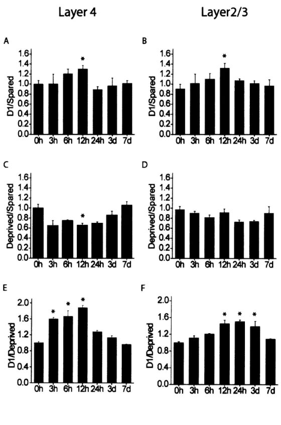
Time Course of Activity-Dependent cpgl5 Expression in Barrel Cortex
Single unit studies have shown that changes in whisker responses can begin after 16h of single whisker experience and continue for weeks thereafter (Barth et al., 2000). If these long-term changes result from alterations in molecular constituents within cortical neurons, one would expect that physiological changes be preceded by changes in gene expression. To investigate the onset and time course of cpgl5 expression, we monitored mRNA levels through all cortical layers of mice after single whisker experience of 6h, 12h, 24h, 3d or 7d. In layer IV, the typical cpgl5 pattern in response to single whisker experience could be seen as soon as 3h after whisker trimming and was clearly visible until 12h after onset of single whisker experience (Figure 2-3, left). In layers II/III, onset of the cpgl5 response was first evident at 6h after whisker trimming, but was apparent up to 3d after onset of single whisker experience (Figure 2-3, right). To quantify these results and to assess whether cpgl5 elevation in Dl and depression in the surrounding barrel field follow a similar time course, we compared the D1/Spared, Deprived/Spared and D1/Deprived whisker ratios at each time point (Figure 2-4). cpgl5 induction in the D1 barrel unit (D1/Spared) peaked after 12h of single whisker experience in layers II/III as well as IV (Figure 2-4A,B) cpgl5 depression in the surrounding barrel field (Deprived/Spared) showed a similar trend, but was significant only in layer IV at 12h (Figure 2-4C,D). Although cpgl5 induction in Dland depression in the deprived barrel units are significant only at 12h (likely due to small sample size at other time points), the
patterns are somewhat different for the two layers, suggesting that with additional sampling it may be possible to discriminate whether cpgl5 induction in the spared Dl barrel unit and depression in the surrounding deprived barrel units differ in their time course between layer IV and the superficial layers. When the net change in cpgl5 expression was quantified by combining the increased cpgl5 mRNA levels in the Dl barrel unit with diminished cpgl5 expression in the adjacent deprived barrel units (D1/Deprived), the difference in the time course of cpgl5 expression was after 3h of single whisker experience, with levels markedly elevated only until 12h (Figure 2-4E). In layers II/III, onset of the cpgl5 response occurred later. A significant increase in the D1/Deprived signal ratio was detectable only after 12h of single whisker experience (Figure 2-4F). However, in contrast to the rapid return to basal expression seen in layer IV, change in cpgl5 levels in the layer II/III region corresponding to the Dl barrel units was maintained for at least 3d after onset of single whisker experience (Figure 2-4F). These laminar differences in the cpgl5 response to single whisker experience are consistent with an initial activity-dependent response in layer IV, the primary thalamic input layer, subsequently progressing to the superficial layers where changes in receptive field properties then persists for days (Glazewski and Fox, 1996; Huang et al.,
1998).
Decreased cpgl5 Induction on a/6 CREB Knockout Mice
The transcription factor CREB is known to be required for experience-dependent receptive field plasticity in response to whisker manipulation (Glazewski et al., 1999). The requirement for CREB is likely due to its regulation of downstream effector genes
that mediate changes in synaptic structure and function. Studies of cpgl5 regulation in cultured cortical neurons indicate that CREB is a mediator of cpgl5 activity-dependent expression in vitro (Fujino et al., 2003). To determine if cpgl5 is a CREB-regulated effector gene that is induced in vivo during receptive field plasticity, we examined cpgl5 expression in barrel cortex of ct/8 CREB knockout mice after 12h of single whisker experience. Since previous studies using the spared whisker paradigm have shown that plasticity in layers II/III of these mutants is relatively unaffected in adolescent animals (1-2 months) but is significantly diminished in adult (>6 months) mice) (Glazewski et al.,
1999), we tested both adolescent and adult knockout a/8 CREB mice.
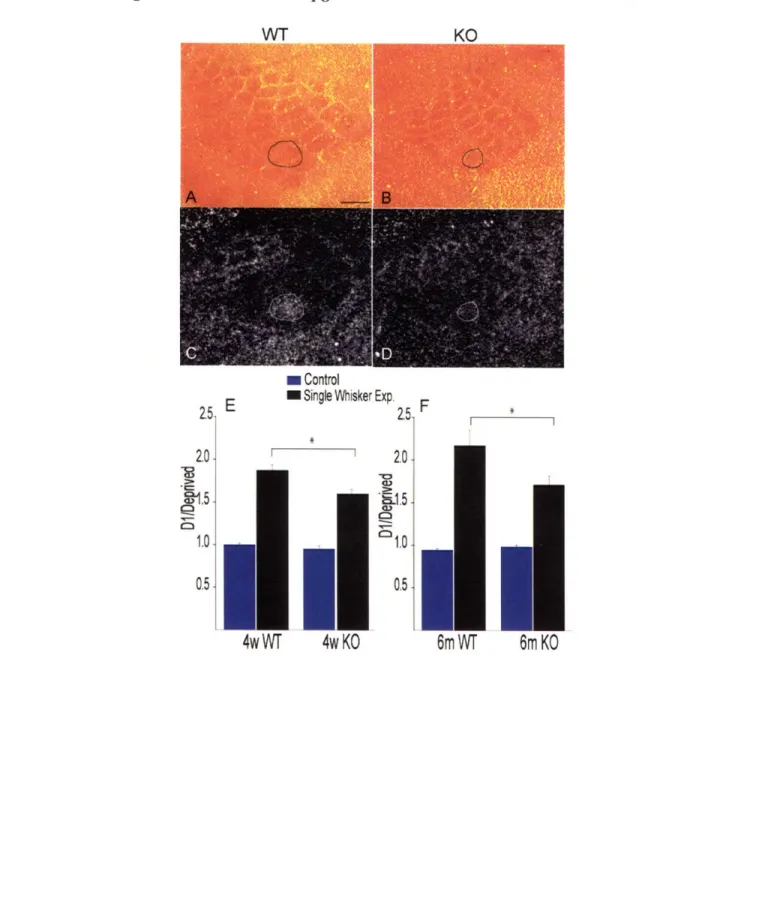
When comparing cytochrome oxidase staining of layer IV sections from cortices of the CREB mutants with those of their wild-type counterparts, it was apparent that the size of the barrel field was smaller in the mutants (Figure 2-5A, B). CREB mutants were previously found to have a thalamus significantly smaller than normal (Pham et al., 2001), suggesting that CREB may have a role in development of sensory brain structures. However, organization of the individual barrels and their relative size and position within the field is normal. The CREB mutants show no overt behavioral abnormalities and the mutation does not seem to cause sensory or motor performance deficits (Glazewski et al.,
1999).
In response to single whisker experience, the qualitative pattern of changes in
cpgl5 expression was similar in the wild-type and CREB mutants; cpgl5 levels in the
spared Dl barrel unit were increased while levels in the surrounding barrel field were decreased (Figure 5C, D). However, the magnitudes of the increase in the Dl barrel unit and the decrease in barrels representing the large deprived whiskers were both less in the
a/8 CREB mutants relative to wild-type mice. To quantify the difference in cpgl5
expression between wild-type and a/8 CREB knockouts, we compared the ratio of cpgl5 signal intensity in the D1 to its level in the surrounding deprived barrel units in control hemispheres or after single whisker experience. We found that cpgl5 levels in control hemispheres of both adolescent 4-week-old mice and 6-month old adults were not significantly different between the wild-type and a/6 CREB knockouts (Figure 5E, F). The change in the D1/Deprived ratio in response to single whisker experience was 16%
smaller in the 4-week-old a/6 CREB knockouts relative to their wild-type control counterparts (Figure 5E, *p < 0.05), and 23% smaller when 6-month-old mice were compared (Figure 5F, *p < 0.01). These results show that lack of a/6 CREB isoforms diminishes activity-dependent changes in cpgl5 expression in response to the spared whisker experience.
Discussion
We measured the experience-dependent expression of cpgl5, a gene encoding a signaling molecule that promotes growth of dendritic and axonal arbors and synaptic maturation in developing neurons, in the adult barrel cortex. We found that the spatial and temporal patterns of cpgl5 expression, and its regulation by CREB, are consistent with a role in mediating adult receptive field plasticity.
Spatial and Temporal Features of Cortical Plasticity
Layer II/III neurons are the primary substrate for receptive field plasticity in the adult barrel cortex (Glazewski and Fox, 1996; Glazewski et al., 1998). After deprivation,
depression of deprived whisker responses, likely due to changes at synapses of layer IV onto layer II/III neurons (Allen et al., 2003; Shepherd et al., 2003), occurs rapidly (Glazewski and Fox, 1996). Potentiation of the response to the spared whisker in deprived barrels happens later. Previous studies in Xenopus and feline visual systems have suggested that cpgl5 expression is presynaptic to the connections undergoing activity-dependent plasticity (Corriveau et al., 1999; Nedivi et al., 2001). Thus, the spatio-temporal patterns of cpgl5 expression we observe are consistent with experience-dependent physiological plasticity in barrel cortex. Early up-regulation (down-regulation) of cpgl5 in layer IV may relate to strengthening (weakening) of layer IV to layer II/III synapses, while its subsequent expression in the superficial layers relates to modification of horizontal connections within these layers. It is interesting to note that cpgl5 expression is regulated in both the barrel and its surrounding septal region since the structures receive input through parallel subcortical pathways (Koralek et al., 1988; Lu and Lin, 1993), and give rise to two distinct intracortical circuits (Kim and Ebner, 1999). The layer IV barrel columns receive input from the ventral posteriormedial nucleus of the thalamus (VPM), and within somatosensory cortex project short distances to superficial layers of the same barrel or immediately neighboring barrels (Koralek et al. 1988; Lu and Lin, 1993; Kim and Ebner, 1999). These observations indicate that modifications in both types of circuits occur, or they may be required for physiological changes in barrel representation.
Only a handful of studies have addressed the immediate effect (on the scale of hours) of sensory experience in the adult on expression of candidate proteins (Barth et al., 2000; Bisler et al., 2002; Filipkowski et al., 2000; Rocamora et al., 1996; Staiger et al., 2002). Here we show that, by altering sensory experience for a few hours, we can produce significant changes in cpg]5 expression within barrel cortex. Transcriptional activation is usually a good indicator of a subsequent increase in protein levels. Indeed following kainic acid seizure in the hippocampus, both the cpgl5 transcript and the CPGl5 protein are dramatically induced (Naeve et al., 1997; Nedivi et al., 1998). Other than transcription factors cpg]5 and BDNF are the only genes shown to be rapidly regulated by activity in a manner spatially and temporally correlated with experience-dependent synaptic modification in adult barrel cortex (Rocamora et al., 1996). Moreover, cpgl5 is the first activity-dependent effector gene demonstrated to be regulated downstream of CREB in the context of barrel cortex plasticity in vivo.
Previous studies have shown that barrel plasticity in a/6 CREB knockout mice is significantly reduced, although not eliminated (Glazewski et al., 1999). Our findings regarding cpgl5 expression in these mice are consistent with these studies, in that activity-dependent cpgl5 induction within the spared whisker barrel unit is diminished, but not completely abolished. In vitro studies of cpg]5 regulation in cultured cortical neurons indicate that CREB is a mediator of cpgl5 activity-dependent expression, but is by no means the only transcriptional activator involved (Fujino et al., 2003). Thus CREB likely works in concert with other transcription factors to modify expression of cpgl5 and other genes that contribute to synaptic plasticity. Another possible explanation for the partial effect of a/6 CREB isoform deletion on both receptive field plasticity and cpgl5
expression in barrel cortex is that in the mutants there is compensation for the loss of the a/8 CREB isoforms by up-regulation of the P3 isoform (Glazewski et al. 1999), or other CREB-like transcription factors (Hummler et al., 1994). These factors may act to partially rescue cortical receptive field plasticity.
Potential Role of cpgl5 in Adult Receptive Field Plasticity
In the developing Xenopus tectum (Nedivi et al., 1998; Cantallops et al. 2000),
cpgl5 functions as an intercellular signaling molecule that promotes process outgrowth
and synaptogenesis. While the role of cpgl5 in adult barrel cortex has not been defined, it would be intriguing to consider that it performs a similar function. Recent in vivo imaging studies have not revealed changes in dendritic arbor morphology of barrel cortex neurons during receptive field plasticity (Trachtenberg et al., 2002). Yet these same studies provide evidence for increased synapse turnover in response to experience-dependent receptive field plasticity and demonstrate that synapse formation and elimination is associated with sprouting and retraction of dendritic spines (Trachtenberg et al., 2002). Expression of cpgl5 in the context of receptive field plasticity could be related to its involvement in small scale synaptic changes that consolidate adaptive cortical responses to peripheral manipulation.
Methods
Animal manipulations and tissue isolation
All animal work was approved by the Massachusetts Institute of Technology and Cold Spring Harbor Laboratory Committees on Animal Care and conforms to NIH
guidelines for the use and care of vertebrate animals. C57BL/6 (+/+) (Charles River, Wilmington, MA) or CREB a, 8 knockout mice (-/-) (Bourtchuladze et al., 1994; Hummler et al., 1994) were used at 4-5 weeks or 6 months of age. For the single whisker experience paradigm, mice were gently restrained by hand and all the large whiskers (Al -A4), Bl-B4, C1-C5, D2-D5, El-E5, a, j3, y, and 6) except D1 on the right side of the muzzle were trimmed close to the face (<1 mm) using microspring scissors. Whiskers on the left muzzle were left intact, and their corresponding hemispheres served as controls. After 12h of sensory deprivation, mice were sacrificed by guillotine decapitation, and brains immediately removed. At 4-5 weeks n = 8 brains per group (+/+ or -/-). At 6 months, n = 6 for +/+ and n = 5 for -/-. For a time course of the cpgl5 response to the single whisker experience, additional 4-5 week old mice were harvested after 3h, 6h, 24h, 3d, or 7d of sensory deprivation (n = 2 brains per time point). For long deprivation periods, whiskers were trimmed every 2d. Cortical hemispheres were rapidly dissected from the freshly removed brains and flattened between two clean microscope slides, rapidly frozen in dry ice and stored at -80'C.
In situ hybridizations
Flattened cortices were tangentially sectioned by cryostat (12um), thaw-mounted on Superfrost/plus microscope slides (VWR Scientific, West Chester, PA), dried, fixed in 4% paraformaldehyde, washed in PBS, dehydrated in ethanol, air-dried, and stored desiccated at -800C. Before hybridization, slides were pretreated (at room temperature,
unless otherwise stated) with 0.2 M HCI (20 min), double distilled water (DDW) (5 min), 2x SSC (30 min at 700C), and DDW (5 min). The next prehybridization treatments, from
pronase (type XIV) (Sigma) to air-drying slides for 1 hr, were conducted as described previously (Hogan et al., 1994.). RNA probes were synthesized with an RNA transcription kit (Stratagene, La Jolla, CA) and 35S-UTP (800 Ci/mmol; Amersham Biosciences, Piscataway, NJ), using linearized cpgl5 cDNA as a template. Hybridizations were done as described previously (Nedivi et al., 1996.). Posthybridization wash conditions were as follows: 3 hr at 500C in 50% formamide and 1 x salt solution (Hogan
et al., 1994.) with 10 mM DTT; 15 min at 370C in TNE (10 mM Tris, pH 7.5, 0.5 M
NaC1, 1 mM EDTA); 30 min at 370C in TNE containing RNase A (20 ptg/ml; Sigma);
30 min at 370C in TNE; and finally overnight at 50'C in 50% formamide and lx salt
solution. Slides were dehydrated with 0.3 M NH4Ac in ethanol, air-dried, and processed for autoradiography as described previously (Hogan, 1994), using autoradiographic emulsion type NTB-2 (Eastman Kodak) diluted 1:1 with 2% glycerol, and exposed for 3-5 d at 40C.
Quantitative Analysis
In situ hybridizations on brains of age-matched wild type and mutant animals
were always carried out in parallel. Brains from the 4-week wild type and mutant animals with 12h of single whisker experience were processed in four different experiments containing two brains from each group. Brains from the 6-month wild type and mutant animals were processed in two experiments, each containing one brain from each time point. Prehybridization treatments hybridizations, posthybridization washes, processing for autoradiography, counterstaining and mounting were done as previously described (Lee and Nedivi, 2002). In all experiments, alternate slides were stained by cytochrome
oxidase to provide a clear anatomical map of the barrel field in layer IV (Wong-Riley, 1979). On the in situ hybridized slides, two to three sections from each brain contained barrel fields sufficiently intact for quantitative analysis. Dark field images of these layer IV sections were imported into Adobe Photoshop 5.0 (Adobe Systems, San Jose, CA) with a Diagnostic Instruments (Sterling Heights, MI) Spot 2 digital camera mounted on a Nikon (Tokyo, Japan) Eclipse E600 using a lx/0.04 Plan ultra-wide objective. Images were saved as gray-scale Tiffs and imported in NIH Image (version 1.62). Mean pixel density measurements were taken from four areas on each slide: the area representing the Dl whisker (barrel and septae), the area representing the other large whiskers (Al-A4, B1-B4, C1-C5, D2-D5, El-E5, a,
13,
7, and 8) (deprived area during single whisker experience), the area corresponding to the anterolateral small vibrissae (spared in single whisker experience), and the background outside of the section. Pixel density was measured on a 0-255 scale, in which 255 is white. To ensure that measurements were unbiased, image files from wild type and mutant animals were coded and measured blind to age and genotype. The background served as a zero labeling negative control and was subtracted from the mean pixel densities of the D1, large barrel (Deprived), and small barrel (Spared) regions to yield the net mean pixel density for each region. The net mean pixel density of the Dl barrel was divided by the net mean density f the large or small barrel regions on the same section. Measurements from 2-3 sections per layer were averaged for each brain. Statistical significance was determined by unpaired Student's t test for all experiments except the time course. The time course data was compared using ANOVA post-hoc analysis with the Bonferroni/Dunn method. In all experiments, n represents number of brains.Figure 2-1. cpgl5 expression in barrel cortex is regulated by single whisker experience
Figure 2-2. Activity dependent cpgl5 expression is localized to layers II/III and IV of barrel cortex.
Figure 2-3. Time course of cpgl5 expression in barrel cortex after spared whisker experience.
Laver 4
Figure 2-4. Quantification of the time course cpgl5 expression after single whisker experience.
Layer
4
Layer2/3
Aa=
1.6- 1.4- 1.2-1.0 0.8 *0-6-0.4'
h37
d 0.2 0 h ýh 6h 1h 24h 3'd7d CL FI8
1.6-1.4
1.2
1.0 0.80.6
0.42
0.2-Oh h 6h 12h24h 3d 7d
Figure 2-5. Induction of cpgl5 is diminished in CREB mutant mice. I A P
m Control
SE m Single Whisker Exp. F
*e 0,
4wWT
4wKO
71, L.· I IFigure Legends
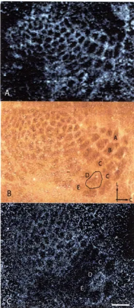
Figure 2-1. cpgl5 expression in barrel cortex is regulated by single whisker experience.
Representative dark-field photomicrographs of in situ hybridizations for cpgl5 mRNA in mice: (A) with normal whisker experience. (C) after 12h of single whisker experience. (B) Cytochrome oxidase staining of a section adjacent to (C) with the region representing the spared Dl whisker outlined. Arrows mark lateral (L) and caudal (C) orientation. (D) A quantitative comparison of sections such as shown in (A) and (C) (n = 8 brains per group, 2-3 sections per brain). In response to single whisker experience, cpgl5 levels are increased in the barrel and septal region (barrel unit) representing the D1 whisker when compared with barrel units of the small (untrimmed in single whisker experience) whiskers in the same section (Spared), and decreased in the barrel units of the large whiskers (trimmed in single whisker experience) (Deprived). The D1/Deprived ratio represents the net change in cpgl5 levels, combining the increase in the D1 barrel unit with the decrease in the surrounding deprived barrel units. *p< 0.05, **p < 0.001. Scale bar, 350pm.
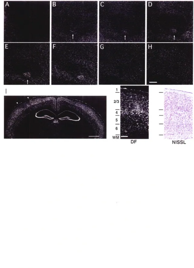
Figure 2-2. Activity-dependent cpgl5 expression is localized to layers II/III and IV of barrel cortex. Representative dark-field photomicrograph of in situ hybridization for
cpgl5 mRNA on serial tangential sections through flattened cortex of a 4-week-old
mouse after 12h of single whisker experience. The sections progress from layer I (A) to layer VI (H). Arrows mark the cortical region representing the spared whisker in layers II through IV (B-F). Scale bar, 0.5mm. (I) cpgl5 mRNA localization is shown in a coronal section through the brain of a mouse treated as above. The contralateral hemisphere to the
spared whisker shows cpgl5 up-regulation in the region representing the D1 whisker, with a concomitant down-regulation in the surrounding barrel field. White arrowheads indicate the region of the barrel field shown at high magnification on the right. Scale bar, Imm. Dark-field view shown side by side with a bright-field view of an adjacent Nissl stained section delineating cortical layers. Scale bar, 0.1mm.
Figure 2-3. Time course of cpgl5 expression in barrel cortex after spared whisker experience. Representative dark-field photomicrographs of cpgl5 expression in layer IV
(left) and layers II/III (right) of the barrel field after different lengths of spared whisker experience. Scale bar, 0.5mm. Arrows mark the spared Dl barrel unit.
Figure 2-4. Quantification of the D1/Spared, Deprived/Spared, and D1/Deprived ratios in layer IV (A, C, E), and II/III (B, D, F) as a fiunction of single whisker experience time (n = 2 brains per time point, 2-3 sections per brain). Compared to Oh controls, *p < 0.0024 (5% significance level). Although cpgl5 induction in Dl (A, B) and depression in the deprived barrels (C, D) are significant only at 12h (likely due to the small sample size at other time points), the net change in cpgl5 expression, combining induction in Dl and depression in the deprived neighboring barrel units (D1/Deprived) shows a significantly different time course in layers IV and II/III.
Figure 2-5. Induction of cpgl5 is diminished in CREB mutant mice. Cytochrome oxidase staining of the barrel field of WT (A) and a/8 CREB knockout mice (B) with the Dl barrel unit outlined. Corresponding in situ hybridizations for cpgl5 mRNA after 12h
single whisker in WT (C) and a/8 CREB knockout mice (D). (E) Quantification of the Di/Deprived ratios show that cpgl5 regulation in response to single whisker experience is significantly reduced in both 4-week-old (n = 8 +/+; n = 8 -/-; *p < 0.05) and 6-month-old animals (n = 6 +/+; n = 5 -/-; *p < 0.01).
Chapter 3
Soluble CPG15 expressed during early development rescues cortical progenitors from apoptosis.
Abstract
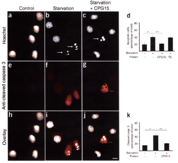
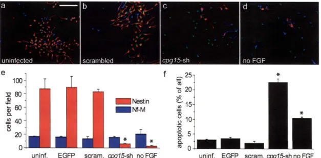
The balance between proliferation and apoptosis is critical for proper development of the nervous system. Yet, little is known about molecules that regulate apoptosis of proliferative neurons. Here we identify a soluble, secreted form of CPGl5 expressed in embryonic rat brain regions undergoing rapid proliferation and apoptosis, and show that it protects cultured cortical neurons from apoptosis by preventing activation of caspase 3. Using a lentivirus-delivered small hairpin RNA, we demonstrate that endogenous CPG15 is essential for the survival of undifferentiated cortical progenitors in vitro and in vivo. We further show that CPGl5 overexpression in vivo expands the progenitor pool by preventing apoptosis, resulting in an enlarged, indented cortical plate and cellular heterotopias within the ventricular zone, similar to the phenotypes of mutant mice with supernumerary forebrain progenitors. CPGl15 expressed during mammalian forebrain morphogenesis may help balance neuronal number by countering apoptosis in specific neuroblasts subpopulations, thus influencing final brain size and shape.
Introduction
During mammalian evolution, the cerebral cortex has greatly expanded through a tremendous increase in the number of cortical neurons. The surface of the cortical plate has extended and become indented and convoluted as a result of neuron addition in columnar radial units (Rakic, 1995). At the onset of cortical neurogenesis, the
proliferative population of founder cells is confined to the ventricular zone of the embryonic cerebral wall (Takahashi et al., 1997). Even modest alterations in the size of this progenitor population during its early exponential growth phase can markedly affect final neuronal numbers (Caviness et al., 1995; Rakic, 1995). Thus, it has been proposed that cellular mechanisms that influence founder cell number may underlie the telencephalic expansion and sculpting that are characteristic of mammalian forebrain development and evolution (Rakic, 1995). Apoptosis within the founder population is one putative mechanism for influencing eventual brain size and shape (Haydar et al., 1999; Kuan et al., 2000).
It has recently been recognized that the role of apoptosis in brain development extends beyond matching of neuronal populations with their appropriate target fields, as specified in the 'neurotrophic hypothesis' (de la Rosa and de Pablo, 2000; Kuan et al., 2000). Caspase 3, a key enzyme in the mammalian apoptotic pathway, is expressed at high levels in the mouse cerebral wall around embryonic day 12 (E12) (Pompeiano et al., 2000), when dying cells are prevalent in proliferative zones of the cerebral cortex(Blaschke et al., 1996; Blaschke et al., 1998; Thomaidou et al., 1997). Consistent with these observations, mutant mice deficient in the pro-apoptotic genes Casp3, Casp9 and Apafl show gross nervous system malformations resulting from improper expansion of specific neural progenitor populations (Hakem et al., 1998; Kuida et al., 1998; Kuida et al., 1996; Yoshida et al., 1998). The excess neurons in some of these mutants are added as extra radial units, expanding the surface of the cortical plate, rather than influencing its thickness. The cortical plate, with increased size, forms convolutions resembling the gyri and sulci of the primate brain. In addition, later generated cells accumulate below the
cortical plate, forming heterotopic cell masses within the ventricles (Kuida et al., 1998; Kuida et al., 1996). Despite the essential role of the core apoptotic pathway in brain morphogenesis, little is known about the signals regulating apoptosis of proliferative neurons. Identification of the molecules involved is vital to understanding the complex morphogenetic processes that shape the mammalian brain.
cpgl5 (also known as Nrnl) was identified in a screen for activity-regulated genes
involved in synaptic plasticity (Hevroni et al., 1998; Nedivi et al., 1993) and encodes a small, highly conserved protein (also termed neuritin-1) (Naeve et al., 1997; Nedivi et al., 1998). In a membrane-bound form attached by a glycosylphosphatidylinositol (GPI) link, CPG15 has been shown to function non-cell autonomously to coordinately regulate growth of apposing dendritic and axonal arbors, and to promote synaptic maturation (Cantallops et al., 2000; Nedivi et al., 1998). As cpgl5 is an activity-regulated gene, late
cpgl5 expression is contemporaneous with critical periods for activity-dependent
plasticity and requires action potential activity. However, cpgl5 is also expressed in an activity-independent manner during early brain development before circuit formation and maturation (Corriveau et al., 1999; Lee and Nedivi, 2002), suggesting that it may have a different role at this stage. We hypothesized that, like the neurotrophic factors, CPG15 has multiple roles during nervous system development. In addition to its previously characterized role as a growth and differentiation factor that affects process outgrowth and synaptic maturation, CPG15 may also function as a survival factor during early brain development. Here we describe the identification of a soluble CPG15 expressed in the embryonic brain that regulates survival of cortical progenitors by preventing caspase-mediated apoptosis.
Results
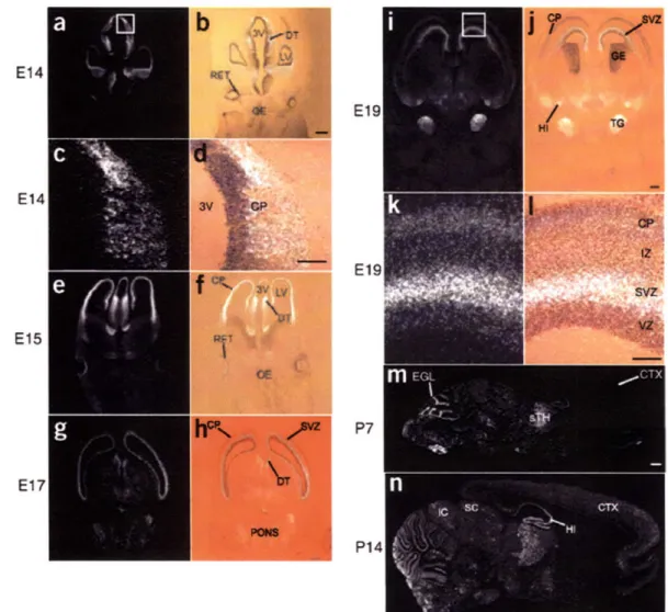
cpgl5 is expressed in embryonic proliferative zones
To examine localization of early, activity-independent cpgl5 expression, we performed in situ hybridizations on sections from embryonic rat brains. At the earliest times tested, embryonic days 14 (E14) and 15 (El15), cpgl5 mRNA is present in the cortical plate, in the ventricular zone of the dorsal thalamus and in retinal ganglion cells (Figure 3-la-f). At E17-E19, cpgl5 is expressed in the telencephalic and dorsal diencephalic subventricular zones (Figure 3-1g-l), is expressed in the hippocampal primordia (Figure 3-li-j), and at postnatal day 7 (P7) appears in the external granular layer of the cerebellum (Figure 3-1m). In all these regions early cpgl5 expression is temporally correlated with expansion of the progenitor pool and apoptotic elimination of superfluous neuroblasts (Blaschke et al., 1998). cpgl5 is not expressed in all proliferative zones and is markedly absent from the olfactory epithelium and ganglionic eminence (Figure 3-la,b,e,f,ij), suggesting that its function may be cell type specific. cpgl5 is also expressed when target-derived trophic support is crucial for protection from apoptosis used to match neuron number with target size. From E19 to P7, cpg]5 mRNA is present in the trigeminal ganglia, sensory thalamus and various brainstem nuclei (Fig. 3-lij,m), at times of afferent ingrowth, target selection and synaptogenesis in these structures. From P0, cpgl5 expression in the cerebral cortex is downregulated to undetectable levels (Lee and Nedivi, 2002), coincident with cessation of apoptosis in this region (Blaschke et al., 1996; Thomaidou et al., 1997). At P14, cpgl5 mRNA re-appears, not in the ventricular or subventricular zones, but in the differentiated cortical layers (Fig. 3-1n), where activity-dependent plasticity is thought to occur postnatally. cpgl5 mRNA patterns