HAL Id: hal-01514635
https://hal.archives-ouvertes.fr/hal-01514635
Submitted on 26 Apr 2017
HAL is a multi-disciplinary open access
archive for the deposit and dissemination of
sci-entific research documents, whether they are
pub-lished or not. The documents may come from
teaching and research institutions in France or
abroad, or from public or private research centers.
L’archive ouverte pluridisciplinaire HAL, est
destinée au dépôt et à la diffusion de documents
scientifiques de niveau recherche, publiés ou non,
émanant des établissements d’enseignement et de
recherche français ou étrangers, des laboratoires
publics ou privés.
On the early diagnosis of Alzheimer’s Disease from
multimodal signals: A survey
Ane Alberdi Aramendi, Asier Aztiria, Adrian Basarab
To cite this version:
Ane Alberdi Aramendi, Asier Aztiria, Adrian Basarab. On the early diagnosis of Alzheimer’s Disease
from multimodal signals: A survey. Artificial Intelligence in Medicine, Elsevier, 2016, 71, pp.1-29.
�10.1016/j.artmed.2016.06.003�. �hal-01514635�
To link to this article : DOI :
10.1016/j.artmed.2016.06.003
URL :
http://dx.doi.org/10.1016/j.artmed.2016.06.003
To cite this version :
Alberdi Aramendi, Ane and Aztiria, Asier and
Basarab, Adrian On the early diagnosis of Alzheimer's Disease from
multimodal signals: A survey. (2016) Artificial Intelligence in
Medicine, vol. 71. pp. 1-29. ISSN 0933-3657
O
pen
A
rchive
T
OULOUSE
A
rchive
O
uverte (
OATAO
)
OATAO is an open access repository that collects the work of Toulouse researchers and
makes it freely available over the web where possible.
This is an author-deposited version published in :
http://oatao.univ-toulouse.fr/
Eprints ID : 17021
Any correspondence concerning this service should be sent to the repository
administrator:
staff-oatao@listes-diff.inp-toulouse.fr
On
the
early
diagnosis
of
Alzheimer’s
Disease
from
multimodal
signals:
A
survey
Ane
Alberdi
a,∗,
Asier
Aztiria
a,
Adrian
Basarab
baMondragonUniversity,ElectronicsandComputingDepartment,GoiruKalea,2,Arrasate20500,Spain
bUniversitédeToulouse,InstitutdeRechercheenInformatiquedeToulouse,CentreNationaldelaRechercheScientifique,UnitéMixtedeRecherche5505,
UniversitéPaulSabatier,118RoutedeNarbonne,31062Toulouse,France
Keywords: Multimodality Behaviour Physiology Alzheimer’sDisease Earlydetection
a
b
s
t
r
a
c
t
Introduction:ThenumberofAlzheimer’sDisease(AD)patientsisincreasingwithincreasedlifeexpectancy and115.4millionpeopleareexpectedtobeaffectedin2050.Unfortunately,ADiscommonlydiagnosed toolate,whenirreversibledamageshavebeencausedinthepatient.
Objective:Anautomatic,continuousandunobtrusiveearlyADdetectionmethodwouldberequiredto improvepatients’lifequalityandavoidbighealthcarecosts.Thus,theobjectiveofthissurveyisto reviewthemultimodalsignalsthatcouldbeusedinthedevelopmentofsuchasystem,emphasizingon theaccuracythattheyhaveshownuptodateforADdetection.Someusefultoolsandspecificissues towardsthisgoalwillalsohavetobereviewed.
Methods:Anextensiveliteraturereviewwasperformedfollowingaspecificsearchstrategy,inclusion criteria,dataextractionandqualityassessmentintheInspec,CompendexandPubMeddatabases. Results:Thisworkreviewstheextensivelistofpsychological,physiological,behaviouralandcognitive measurementsthatcouldbeusedforADdetection.Themostpromisingmeasurementsseemtobe mag-neticresonanceimaging(MRI)forADvscontrol(CTL)discriminationwithan98.95%accuracy,while electroencephalogram(EEG)showsthebestresultsformildcognitiveimpairment(MCI)vsCTL(97.88%) andMCIvsADdistinction(94.05%).AvailablephysiologicalandbehaviouralADdatasetsarelisted,aswell asmedicalimaginganalysisstepsandneuroimagingprocessingtoolboxes.Someissuessuchas“label noise”andmulti-sitedataarediscussed.
Conclusions:ThedevelopmentofanunobtrusiveandtransparentADdetectionsystemshouldbebased onamultimodalsysteminordertotakefulladvantageofallkindsofsymptoms,detecteventhesmallest changesandcombinethem,soastodetectADasearlyaspossible.Suchamultimodalsystemmight prob-ablybebasedonphysiologicalmonitoringofMRIorEEG,aswellasbehaviouralmeasurementslikethe onesproposedalongthearticle.ThementionedADdatasetsandimageprocessingtoolboxesare avail-ablefortheirusetowardsthisgoal.Issueslike“labelnoise”andmulti-siteneuroimagingincompatibilities mayalsohavetobeovercome,butmethodsforthispurposearealreadyavailable.
1. Introduction
Peoples’lifeexpectancyisgrowingcontinuouslyinthe devel-opedcountries,makingthepopulationincreasinglyold.Evenifthis isapositivereality,italsobringsunwantedconsequencessuchas anincreasingnumberofdiseases,includingtheAlzheimer’s Dis-ease(AD).ItisestimatedthatADwilldoubleitsfrequencyinthe next20years[1]andthat115.4millionpeoplewillsufferfromit
∗Correspondingauthor.Tel.:+34647504215.
E-mailaddresses:aalberdiar@mondragon.edu(A.Alberdi), aaztiria@mondragon.edu(A.Aztiria),adrian.basarab@irit.fr(A.Basarab).
in2050[2].Furthermore,whiledeathsattributedtootherhealth problemssuchasheartdiseasehavedecreasedinthelastyears, deathsattributedtoADbetween2000and2010haveincreasedin 68%[3].Nowadays,itdoesnotexista cureforAD[4],neithera 100%reliablediagnosisuntilapost-mortemanalysisisdone.Only somesymptomatictreatmentsthatareeffectiveforlimitedperiods insubgroupsofpatientsareavailable[5].Lifeexpectancyofthe patientsdiagnosedwithADiscurrentlylessthan7years[4].
Even ifit is thought that AD is theresult of a combination ofgenetic,environmentalandlifestylefactors[6,7],theinitiating eventsthatmakesomeonedevelopthistypeofdementiaremain stillunknown[8].ThemosteffectivemethodofcontrollingAD’s progressionis believedtobebased onanearlydiagnosisand a
goodmanagementstrategystartedfromtheverybeginningofthe cognitivedecline[9,10],but, nowadays,thediagnosisismainly accomplishedusingpsychologicalteststhatbecomepositivewhen thediseaseispracticallyirreversible[11].
Thispaperis structuredas follows.In theremainderof this section,ADand itsprogressareexplained(seeSections1.1 and
1.2), andtheneed foranearlydiagnosismethodishighlighted (Section1.3).InSection2themethodologyusedtoconductthe literaturereviewis explained.In Section3,thecurrent stateof cognitive,psychological,physiologicalandbehaviouraldiagnostic methodsandbiomarkersisexposedandinSection4,thereviewed state of the art is critically analysed highlighting the research gaps.InSection5someoftheusefultoolsforthemultimodalAD detectionresearchareconsidered,namely,thepubliclyavailable datasets(Section5.1),thestandardmethodsformedicalimaging analysis(Section5.2)andtheneuroimagingprocessingtoolboxes (Section5.3).InSection6,someofthereal-worlddatasets’issues arediscussed.Finally,inSection7,aconclusionisgivenalongwith cluesforfuturework.
1.1. Definition
ADisaprogressive,degenerativedisorderthatattacksbrain’s nervecells,orneurons,resultinginlossofmemory,thinkingand languageskills,andbehaviouralchanges[12].Itisaneurological disorderthatmostlyaffectspeopleover65yearsoldandwhose incidencerategrowsexponentiallywithage[13].It isthemost commonformofdementia[14]andunlikewhatsomepeoplemay think,ADisnotanormalpartofageing[15].
1.2. Phases
ItisbelievedthatpeopledevelopingADundergothreedifferent stages[16].ThefirstoneisthepreclinicalADstage,wherechanges inthebrain,inthebloodandinthecerebrospinalfluid(CSF)may starthappening,butthepatientdoesnotshowanysymptoms[3]. Therefore,nowadays,thisphasecannotstillbedetected.Infact,it isbelievedthatthisstagecanstart20yearsbeforeanysymptomis evidenced.TheNunStudy,oneofthemostsignificantlongitudinal studiesintheareaofADresearch,hasevenshownevidencesof cor-relationsbetweenyouthlinguisticabilityandlatelifeprogression toAD[17].
Thesecond stageof the diseaseis calledthemild cognitive impairment(MCI).Inthisstage,symptomsrelatedtothethinking abilitymaystarttobenoticeableforthepatientsthemselvesand forthenearestfamilymembers,buttheydonotaffecttheirdaily life[3].NotallthepeoplediagnosedwithMCIdevelopAD,butonly anestimated10–15%ofthemeveryyear[18,19]andthereason whysomepeopledevelopdementiaand othersdonot,remains stillunknown.Whena patientisdiagnosedwithMCI,aspecific diagnosisproceduremuststarttounderstandwhichdiseaseor con-ditionisresponsibleforthedeficit[20].TwodifferenttypesofMCI aredistinguished:amnesicMCI(aMCI)andnon-amnesicMCI[9]. Theformerreferstopatientswhohaveimpairmentinthememory domain,andthelattertopatientswhohaveimpairmentinoneor morenon-memorydomainsofcognition,as,forexample,attention orlanguageprocessing.Itisbelievedthatsubjectssufferingfrom aMCIaremorelikelytodevelopAD[21].
ThefinalstageofthediseaseiscalleddementiaduetoAD,where memory,thinkingandbehaviouralsymptomsarealreadyevident andaffectthepatient’sabilitytofunctionindailylife.
1.3. Theneedforanearlydiagnosismethod
ADsymptomsareoccasionallyrecognizedbythepatients them-selves,butinthevastmajorityofcases,thecaregiversortheclose
familiars and friends are theones who realize thebehavioural andcognitivechangessufferedbytheADpatients.Theseverity ofthesesymptomsisnotalwayseasytonotice.Theproblemisthat ADsymptomsareinmanycasesconfusedwithanormalageing process,andthus,doctorsarenotconsulteduntilbeingtoolate, resultinginalatediagnosis[22].Inthesurveycarriedoutin[23], 64%ofthecaregiversaffirmedthatbeforethediagnosis,they con-sideredthebehaviourchangessufferedfromthepatientsaspart ofthenormalageingprocess.67%ofthemagreethatthismade thediagnosistobedelayed.Furthermore,onceinthehandof spe-cialists,itisnotyeteasytocorrectlydiagnoseAD.Eventhemost experiencedspecialistsfailinabout10–15%ofcasestocorrectly diagnoseAD[24].Infact,thedefinitediagnosecanonlybemade byapost-mortemexaminationofthebrain.Nowadays,apatient suspiciousofsufferingfromADcanbeclinicallydiagnosedwith anaccuracyofabout90%,drawingonmedicalrecords,physical andneurologicalexamination,laboratorytests,neuroimagingand neuropsychologicalevaluation[25].Mostofthesemethodsusedfor ADdiagnosisaretime-consumingandtheyrequireaclinician inter-vention[26],involvingannoyingdisplacementstohospitals,which canbespeciallydifficultwithelderly.Moreover,themonitoring oftheprogression ofthediseaseisvery costly[27]and, there-fore,notwellenoughstudied.Neuroimagingisbeingincreasingly usedbecauseitofferstophysiciansthepossibilitytoanalysethe patients’brainswhiletheyarealive.Nevertheless,whenchanges canbeappreciatedwiththenakedeye,itisnormallytoolate.That meansthatthebrainhasevidentsignsofatrophy,orthattoomany neurofibrillarytangles(NFT)andAmyloidplaquedepositscanbe foundonit.Non-invasive,fast,inexpensiveandreliableAD diag-nosismethodsarestilltobedeveloped[4].
Anearlydiagnosis of thedisorder canbe extremelyhelpful forthepatientsbecausetheycanhaveaccesstotreatmentsthat candelaysomesymptoms,beingmuchmoreeffectiveintheearly stages[4],aswellastoprogramsandsupportservices,whenthe disorderhasnoyetprogressedtoomuch[22].Furthermore,thiscan allowthemtotakepartinthedecisionoftheirfuture,as,for exam-ple,abouttheircareandeverydaylifeoraboutmoneyandlegal concerns.EarlydiagnosiscanalsohelptoimproveADsurvivalrate
[28].
Thereby,foranearlydiagnosisofADitisnecessarytobeableto detectthemostsubtlesymptomsofanytype.Takingintoaccount themultimodalnatureofADsymptomatology,itisclearthatthe mostefficientandreliableearlyADdetectionmethodologycannot onlyrelyonmeasurementsofauniquedomain,i.e.only physiolog-icalorbehaviouralsymptoms,butonthecombinationofseveral modalities,thatcouldallowtodetectallthesubtlechangesofall domainsfromtheverybeginningandtocontrastthemwithother typeofsymptomsforareliablediagnosis.Themultimodalnature ofotherdisorderssuch asstresshasalsobeenanalysed,andan approachforitsearlydetectionproposed[29],demonstratingthe feasibilityandapplicationofthesemethodstomultipledisorders. Nevertheless,subtlechangesarenoteasilynoticeable.People, withouttheaidoftechnology,arenotabletorecognisethesosmall behaviouralshiftsthatADpatientsmayundergo,notsuspecting theproblemuntilbeingtoolate.Technologythatcouldmakethis processeasierishighlyrequiredtospeedupthewholeprocess. PhysiciansmaynotbeabletocorrectlydiagnoseADiftheydonot findthenecessaryphysiologicaltracesforit.Automatedcomputer aideddiagnosis(CAD)techniquesareneededtofacilitate physi-cian’sdiagnosisof complexdiseasesin individualpatients [30]. Thus,technologythatcanhelpintheearlydetectionofADbasedon subtlebehaviouralandphysiologicalchangesmustbedeveloped.
Recently,areviewofnon-invasiveinnovativediagnostictools fortheearlydetectionofADhasbeenpublished[31]. Neverthe-less,thisarticledidnotemphasizeonthemultimodalnatureofthe disorder,neitherintheautomaticassessmentofdementiabased
Fig.1.Searchmethodologyusedfortheliteraturereviewprocess.
onunobtrusively obtainedbehavioural data.Hence, theaim of thisarticleistocompletethepreviousworkbysummingupthe researchcarriedout intheearlyADdetectioninthefourmain modalities,namely,inthephysiological,cognitive,psychological and behaviouraldomains.Itis intendedtoemphasize themost reliableandusefulbiomarkersfounduptodate,andtherefore,to givecluesforthecombinationofmeasurementsandfeaturesthat shouldbeusedforanautomatic,unobtrusiveandreliableearlyAD detectionsystem.
2. Methods
ThefollowingreviewofthestateoftheartconcerningAD detec-tionwasundertakentoaddressthreespecificgoals:
1Toreviewthevarietyoffeatures,thatcancurrentlybeusedin ordertodetectAD,startingfromthemostwidelyaccepted meth-odswhicharealreadyincludedintheclinicaldiagnosisprocess, tothenewemergingways.
2Tocomparetheaccuraciesthatcanbeachievedwitheachsignal ormeasurement,soastohelptodecideamongthemostsuitable signalsforeachsituation.
3Tohighlightthestepsthatshouldbefollowedinordertoachieve anubiquitousearlyADdetectionsystem.
Toattainthesegoals anextensiveliteraturereviewwas per-formed,withthefollowingsearchstrategyandinclusioncriteria. 2.1. Searchstrategy
Publicationswereretrievedbymeansofacomputerizedsearch oftheCompendexandInspecdatabasesviaEngineeringVillage[32]
andofthePubMeddatabase[33]inordertofindrelevantstudies publishedinEnglishfromJanuary2005todate.
Thereviewwascarriedoutinaniterativeway:first,aglobal pointofviewofthecurrentstateinADdetectionwassearched. The search terms applied in the title field in this step were: “Alzheimer’s”OR“dementia”OR“AD”AND“detect*”OR“diagnos*” OR“measure*”AND“survey”OR“review”.Controlledtermswere usedinordertodiscardallthepublicationsrelatedtonon-relevant domains. Afterremoving duplicates, 70 results were achieved. Titles and abstracts of the remaining papers were reviewed, rejectingtheonesthat focusedonaspectsofADotherthanthe assessment.Twentypaperswereconsideredforfurtherreading.
Afteridentifyingthemaindomainsandmodalitiesinvolvedin thecurrentstateofADdetection,amorespecificsearchwascarried outforeachoneofthedomains.Thecombinationofsearchterms usedwerethefollowing:“Alzheimer’s”OR“dementia”OR“AD” AND“detect*”OR“recogn*”OR“identif*”OR“model*”OR“anal*” OR“diagnos*”AND“physiolog*”OR“behavio*”OR“psycholog*” OR“cogniti*”AND“accura*”.Afirstsetof649studyabstractswas retrievedforassessment.Controlledvocabularytermswereused inordertoexcludepublicationsrelatedtonon-relevantresearch areasandduplicateswererejected.Thebibliographiesofall rele-vantarticlesandreviewpaperswerealsohand-searched.Thetitles andabstractsoftheremainingarticleswerereviewedinapplying theinclusioncriteria.Forty-fourpaperswerein-depthread.
Asummaryoftheliteraturereviewmethodologyusedis pre-sentedinFig.1.
2.2. Inclusioncriteria
Allthe selectedpapers were original studiesand journal or conferencearticles,writteninEnglishandpublishedfrom2005 onwards.Forthefirststepofthisliteraturereview,onlythepapers in where objective AD detection systems were reviewed were accepted whilefor the secondstep, the inclusioncriteria were thefollowing:studiesofdiagnosticaccuracyofADusingatleast physiologicalorbehaviouraldataandvalidatedbymeansof cog-nitiveassessmenttestssuchastheMini-MentalStateExamination (MMSE),totalsubjectsinthestudyatleasttenandsufficientdata reportedeitherdirectlyorindirectlytoenabletheaccuracytable construction.
2.3. Dataextractionandqualityassessment
Adataextractionspreadsheetwascreatedforcollectingdata fromthepapers.Eachoneoftheselectedpaperswasfullyreadand assessedbyoneoftheauthors,whereastheresultswereverified byallofthem.Disagreementswereresolvedthroughdiscussion. 3. MeasuringADsymptoms
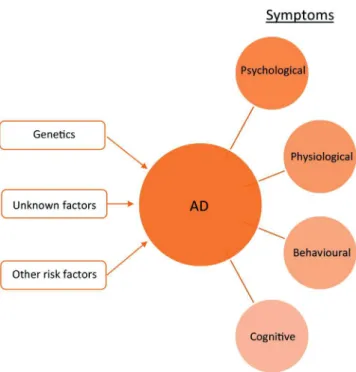
PeoplesufferingfromAD,showsymptomsofseveraltypesand in differentdegrees, depending ontheprogression level ofthe dementia.Thesesymptomscanbedistinguishedintofourmain modalities,whicharephysiological,psychological,cognitiveand behavioural.Thesymptomatologyinthesefourmodalitiescould
ingeneralbeunderstoodasachainprocessthatstartswithsome physiologicalchangesinthepatient,mostlyinthebrain,thatlead tocognitivedifficultieswhichinturnprovokepsychologicaland behaviouralchangesonthepatient.
RegardingthephysiologicalsymptomsofAD,thebest-known changesaretheonesevidencedinthebrain.Ithasbeenfoundthat ADpatients’brainscontainahugenumberofAmyloidplaquesand NFT,whicharealsopresentinmanyhealthyagedsubjects’brains, butinmuchmoremoderateamounts.Amyloidplaquesrefertothe depositsofbeta-amyloid(ˇA)proteinfragments,whichare accu-mulatedbetweentheneuronsandNFTtothedepositsoftauprotein fragmentswhicharepiledupinsidetheneurons[22].The accumu-lationofˇAonthebrainisconsideredanecessarybutnotsufficient conditiontoproducetheclinicalsymptomsofMCIanddementia
[34].Thepresenceoftheseplaquesandtanglesiseventually accom-paniedbythedamageanddeathofneurons[3],andinfact,oneof themostfavourablehypothesisabouttheoriginofADnowadaysis theabnormaldepositionoftheseproteins[35–37].Cerebral hypo-perfusionhasalsobeenfoundtobemoreevidentinADpatients thaninnormaladults,sootherhypothesisblamingthevascular andcardiovascularproblemstobethecauseofthishypoperfusion whichinturncouldtriggerdementiahavebeendeveloped[38]. Corticalandhippocampalatrophiesofthebrainarealsoverywell knownADsymptoms[9].Reductionofthevolumeofthe hippocam-pusisprobablythemostcommonpronouncedchange[26],being asymptomwhichisalreadyevidencedinthemildstageandwhich worsensovertime.Infact,theauthorsof[39]affirmthatatthemild dementiastageofAD,hippocampalvolumeisalreadyreducedby 15–30%andinaMCIthevolumeisreducedby10–15%.Reduced brainactivityand communication betweennervecells hasalso beenfoundtobeanADsymptom,andeyedynamicpattershave alsobeendetectedtochangeinthesepatients.
Cognition-relatedsymptomsareprobablythebestknowninAD. TheclinicalhallmarkandearliestmanifestationofADisepisodic memoryimpairment[40].Notrememberingrecentlylearnt infor-mationisthemostcommonsymptom,whichisdiscerniblefrom theearlystagesof thedisease.Memoryalsostartstofailwhen rememberingimportantdatesorevents.PeopleinearlyADstages may alsohave difficulties solving daily problems, for example, withnumber-relatedtasksasmanagingfinances.Gettingconfused aboutthedates, seasonsand time, aswellas familiarplacesis anothersignofthedisease.Visionmayalsobeaffected,andthe patientsmaynotbeabletoread,tojudgedistancesorto distin-guishcoloursandcontrastsandolfactorydysfunctionhasalsobeen reported[41].Communicationproblemsmayalsoarise:patients maysufferfromdifficultieswhenexpressingthemselves,theymay repeatthingsortheymaystopinthemiddleofaconversation with-outknowinghowtocontinue.Vocabularylossisalsoacommon signofthedisease,aswellasmisplacingand notremembering wheretheyleftthingsandthus,losingthem.Asthedisease pro-gresses, this cognitive symptoms become even worse, and the patientsstarthavingtroublesrecognizingpeoplenearby, includ-ingfamilymembers[22,42].Reducedprevalenceofpaincanalso beanADsymptom[41].
Progressive deterioration of cognition leads to incoherent behaviourandlimitsthepatient’scapacitytoperformhistasksof everydaylife.Therefore,behaviouralsymptomsofADaredirect consequencesofthecognitivechanges.ADpatientsmaytakemuch moretimethanbeforeperformingdailyactivitiesdueto concen-trationdifficulties.Visualproblemsmayleadtomanybehavioural changes,as,forexample,indriving.Duetocommunication diffi-culties,thepatientmaysufferabigchangeofpersonality,anda personwhohasalwaysbeenverysociable,canbeanymore moti-vatedtodealwithpeopleandhavesociallife.Furthermore,they relymoreandmoreonotherpeopleforeverydayactivities,like eating,bathingor dressingbecausetheymayhaveproblemsto
Fig.2.ThemultimodalnatureofAD.
performwellphysically.Theymayalsobeunabletowalkproperly, duetogaitandbalancedysfunction[41],ortositbythemselves andincontinenceandswallowingproblemsmayalsoarise[22,42]. Psychologicalsymptomsincludechangesinmoodand person-ality.ADpatientscanbecomesuspiciousbecausetheymaythink theyhavebeenstolenwhentheylosethings,confusedwhenthey donotrememberthedayitisorhowtheyarrivedwheretheyare, anddepressed,fearfuloranxious,becausetheyrealizethattheydo notrememberbasicthingsandtheydonotknowhowfaritcan arrive[22].Depressionisthemostcommonpsychological symp-tominAD.Nevertheless,itisnotstillclearifdepressionisreallya consequenceofADorariskfactorbyitself[43].Apathy,irritability, agitation,euphoria,disinhibition,delusionsandhallucinationsare alsopartofADsymptomatology[41].
3.1. Cognitionanalysis
ADandMCIlevelscanbeevaluatedbymeansofmanytests, among which some are based on thecognitive abilities of the patients. Someexamplesare theMMSE[44]which isthemost frequentlyusedtestforADdiagnosis,theSevereCognitive Impair-ment Scale [45], the Alzheimer’s Disease Assessment Scale – Cognitive[46]whichfocusesonattention,orientation,language, executivefunctioningandmemoryskills,theNeuropsychological TestBattery[47]whichincludestreatmenteffects’measurements, theBlessedTestwhichassessesmemory,attention,concentration, and theabilitytocomplete Activitiesof DailyLiving(ADL)and theSevereImpairmentBattery[48]whichalternativelyfocuseson measuringtheunaffectedcognitivefunctions[49].
Some other tests are the Neurobehavioral Cognitive Status Examination,theDementiaRatingScale –2andtheCambridge NeuropsychologicalTestAutomatedBattery[50].TheReyAuditory VerbalLearningTestandtheCategoryFluencyTest,whichtestthe abilityofpatientsforrecallingwords,TheTrailMakingTestwhich measuresthefunctionofbrainingeneral,andothercognitivetasks liketheDigitSymbolSubstitutionTestandtheClockDrawingTest canalsohelpinmeasuringcognitivefunctionsofADpatients.
Thistypeofneuropsychologicaltestshavebeenshowntobe effectiveintheassessmentofAD.Nevertheless,theypresentsome
drawbacks. Themost important one is that the assessment by meansofthesetestsislengthyandcomplicated[49].Furthermore, theyarenotsuitableforallthepatientsinallthestagesofthe dementia.Moreover,eveniftheycanmeasurethedementiastatein acertainmoment,itcanbecomplicatedtoearlydetectADbecause theymaynotshowenoughsensitivityorbecauseasinmanycases, itmaybetoolatewhenthetestisperformed.
3.2. Psychologicalevaluation
Asdepressionisoneofthemostfrequentnon-cognitive symp-tomsinAD(totheextentthatapathogeneticrelationbetween depressionandADhasbeensuggested[51]),psychological evalu-ationismainlyfocusedondepressionsymptoms’measurement.
Geriatric Depression Scale (GDS) is an instrument todetect depressionamongoldadults.Depressioncansometimesprovoke similarsymptomstothose of dementia,even areversiblestate called pseudodementia, and therefore, this test allows not to measure thecognitivestateof thepatients butto seewhether depressioncoexistswithADoranotherformofdementiaorto dis-missanykindofdementiaverifyingthatthesymptomsarerelated todepressionwithoutAD[52].
TheMontgomeryand ˚AspergDepressionScale,theCornellScale forDepressioninDementiaandtheNursesObservationScalefor GeriatricPatientsareotherpossibilitiestoassessthedepression levelsinADpatients[51].
Furtherresearchisstillneededinordertoverifyifdepressionis aconsequenceofdementia,or,onthecontraryifitisanotherrisk factor.Onceknowntherelationbetweenbothconcepts,thiskind ofpsychologicalassessmentcouldbeintroducedin aperiodical dementiaprogressiontest.Nevertheless,psychologicalevaluation beingcarriedoutbymeansoftestsandscales,hasthesame draw-backsasthecognitivetests,notbeingsuitableforanautomatic continuousmonitoringsystemfordementia.
3.3. Physiologicalsignals
NowadaysADresearchismainlybasedonphysiological mea-surements, making use of both biological signals and imaging methods.Especiallythelatterarebeingdevelopedandimproved straight off, and this is giving way to AD related physiological changesgettingbetteridentified.Thevolumetricanalysisofthe brain,whichallowstodetectatrophy,hasbeenthemain objec-tiveofimaging,andevenifithasbeendonemanuallyformany years,nowadays,itisevolvingandhasstartedtobedone auto-matically,thankstotechniqueslikethevoxel-basedmorphometry
[53,54], tensor-based morphometry [55,56], object-based mor-phometry[57] and feature-basedmorphometry [58].Currently, “neuroimagingplaysacentralroleintheclinicalresearchof cog-nitive disorders” [59]. Some of theneuroimaging methods are consideredforclinicaluse,namely,PositronEmission Tomogra-phy(PET),ComputedTomography(CT)andstructuralMRIwhile theothersneedstillfurtherresearchinordertobeaccepted.
In this section, thecurrent useand state of the biomedical signalsandimagesthathavebeenconsideredforADresearchis introduced.
3.3.1. CSF
Intherecentyears,someresearcheshavefocusedonidentifying reliableandvalidbiomarkersofADinbiofluids[60].Oneofthese biofluidsistheCSF,whichisaclearfluidthatsurroundsthebrain andspinal cordmainlyforprotection.CSFmustbeobtainedby lumbarpuncture[61].
CSF“istheonlybodyfluidindirectcontactwiththeextracellular spaceofthebrainandthusbiochemicalchangesdueto patholog-icalbrainprocessesaremoreprobabletobereflectedinCSFthan
inotherbodyfluids”[7].Thus,scientistshavemadethe hypoth-esisthattheaccumulationofAˇplaquesonthebraininvolvesa decreaseinCSFAˇ42levelsandthatthiscanbealready appreci-atedintheasymptomaticperiod.CSFtaulevelsarealsoknownto increase,butitisnotclearifthishappensaftertheAˇ accumula-tionstarts[62]orbothprocessesstartindependently[34],thus,the pathophysiologicalprocessoftaumightbeprecedenttotheoneof Aˇ.
Aˇ42 is probably the mosttypical CSF measurement in AD detection,andinthemajorityofthecasesdecreasedvalueshave beenfoundinADpatientscomparedtohealthysubjects[34].Aˇ40 isalsopresentintheliteraturebutnosignificantdifferenceshave beenfoundforADpatients[63],andsometimesratios between bothAˇspecieshavealsobeencomputed,suggestingthatithas potentialforbothfordistinguishingADpatientsfromhealthy sub-jectsandtopredictADinpeoplesufferingfromMCI[60,63–65].CSF totaltau,aswellassomespecifictauepitopes(p-tau231,p-tau181 andp-tau199),havebeenfoundtoincreaseinAD[34,66]andsome researchesalsoaffirmitspredictabilityfromMCItoAD[60,67]. Theratiooftau-epitopestoAˇ42,inagreementwiththe prece-dentresults,havealsobeenfoundtobepredictorsofADinMCI patients [67].OtherchemicalcomponentslikeCSF Isoprostanes which have beenfoundtobeincreasedin ADpatients even in thepreclinicalstage[68]and˛1-antichymotrypsin,Interleukin-6 andvariousmarkersofinflammationwhichhavegivenambiguous results[60],evenifmuchlessfrequently,arealsopresentinthe lit-erature.Recently,ithasbeenconcludedthattheamountofCSFin thehippocampalregionisalsorelatedtoAD[69].Thisisprobably duetothedecreasedsizeofthehippocampusinAD,whichleaves spaceformoreCSF.
“Numerous studies on CSF biomarkers for AD have been publishedduringthelastyears,howeverfrequentlyproviding con-tradictoryand inconclusive results”[34].Furthermore,many of thesebiomarkersarenotuniquetoADdiseasebuttoothertypesof dementia.Inaddition,thistechniqueisveryinvasivebecauseCSF mustbeobtainedbylumbarpuncture,andthus,itisdifficulttouse itasapreventionmethodoftheoverallpopulation.
3.3.2. Bloodtests
Bloodsamplescanbeobtainedinalessintrusiveandlesscostly way [61] andmore frequently thanCSF samples,and thus, AD biomarkersonbloodhavealsobeensearched.Moreprecisely,the bloodcomponentsplasmaandserumhavebeenanalysed,aswell asplatelets[70].
Featuresextractedfrombloodsamplesaresimilartotheones extractedfromCSF.Aˇ42andAˇ40,whichaccordingtothe major-ityoftheresearchesdonotshowsignificantdifferencesbetween ADandhealthysubjects[63,71]orgiveambiguousresults[72,73], Aˇ42/Aˇ40ratiowhichinthestudycarriedoutbyKoyamaetal.
[71],atoddswiththeoneperformedbyHanssonetal.[72],has showndecreasedvaluesinADpatientsand˛1-antichymotrypsin andvariousmarkersofinflammationwhichhavenotprovided evi-denceaboutthepotentialfordistinguishingbetweenADpatients andhealthysubjects[73].
IsoprostanesandInterleukin-6havealsobeenextractedfrom plasma,but theyhave resultedin thesamekindof ambiguous results[60].Inblood platelets,AmyloidPrecursorProtein(APP) forms,beta-secretaseenzyme(BACE)andalpha-secretase(ADAM 10)havealsobeenmeasured.Studieshavereportedthataltered valuesofthisbiologicalparameterscanbefoundinADpatients, evenintheveryearlystagesofthedisease[74,75].
Thus, uptonow, it is notclear ifblood samplescouldhelp indiscriminating ADandhealthypatients, neitheriftheycould serveasapredictor.Thedifferentresultsobtainedcouldbedueto thedifficultytomeasureAˇ42inplasmaortothemethodused for extractingthe peptides,as wellastothedifferences inthe
populationsstudied.Hansonetal.[72]statethatwhetherplasma Aˇ concentrations reflect Aˇ metabolism in the brain is very unclearandothers affirmthereisnorelationatall[76]. Conse-quently,blood-basedbiomarkersofADhavenotbeenstillaccepted duetothe“failuretoreplicatefindings”[61]andtothe ambigu-ousresultsobtainedindifferentstudies.Nevertheless,itwouldbe interestingtoresearchfurtherbecausebloodcanbeeasilyobtained inroutinetests.Recently,ablood-baseddiagnosisprocedurehas beenpatentedbutitsvalidityforclinicaldiagnosisremainstobe seen.
3.3.3. CTscans
CTscanisastructuralimagingmethodthatusesX-raysto cre-atepicturesofcross-sectionsofthebody,achievingwiththesame dosageofradiation,100timesmoreclearimages[77]thanthe reg-ularX-rays.Togetthiskindofimages,specificCT scannersare used.
CThasbeenusedtoobservetheatrophyofmedial temporal regionsyearsago,butitisnoteasytofindrecentstudiesaboutCT asanADdiagnosissource.Vargheseetal.affirmthat[9]CTisnot usedasastandardtechniqueforearlydiagnosisofAD.Thiscould bebecauseothermethodshavedemonstratedtoprovidegreater accuracy,manipulabilityandprecision[60]andbecauseCTisonly capabletoshowlatechangesinAD[9].
EvenifsomestudieshavetriedtoverifyitsutilityinAD diag-nosis[78]due toitssimplicity,availabilityandinexpensiveness comparedtoothermethodssuchasMRI,nowadays,itisonlyused toruleoutotherbrainproblems,liketumoursorhaemorrhages, andit“doesnothaveanyotherroleintheearlydiagnosisofAD”
[9].
3.3.4. PETscans
PETimaging isa molecular imagingtechnique thatprovides three-dimensionalimagesofabrainatthemolecularand cellu-larlevel[79].Itconsistsofinjectingormakinginhaleasubstance, calledradiotracer,thatcontainsapositronemittertothepatients, detectingtheemittedradiationbyascannerandcomputinga dig-italimagethatrepresentsthedistributionoftheradiotracerinthe body[80].Dependingonthechosenradiotracer,differentkindsof PETscanscanbedone.PETScansaredonewithPETscanners,but theuseofcyclotronsforthepreparationoftheradiotracersisalso necessary,elevatingthecostoftheequipment.
InADdiagnosis,manydifferentradiotracershavebeenusedfor fourmainpurposes:Mainlythe11C-PIBtoimagetheaccumulation
oftheˇAplaquesonthebrain,18F-FDGtoimagetheglucose
con-sumptionofthebrain,11C-PMP,11C-MP4A,11C-MP4B,11C-Nicotine
andotherstoimagetheneurotransmittersystemsofthebrainand finally11C-(R)-PK11195toimagetheinflammationinthecentral
nervoussystem(CNS)whichcancauseneuronaldeath[5].The glu-coseconsumptionimagingisbasedontheideathatasbrainmainly usesglucoseforenergyproduction,glucosemetabolismisclosely relatedtoneuronal function,bothatrestand duringfunctional activation[20,81].
CADsystemshavebeendevelopedtotrytoautomatically diag-nose AD and MCI. Su et al. [82] proposeda methodbased on automaticallyselectedROIfeatures,andclassifiedwithasupport vectormachine(SVM)classifierwithalinearkernel,andachieved accuracyratesofupto91.1%indistinguishingADfromcontrols, 79.41%withADandMCIand78.13%withMCIandcontrols.They alsotestedprincipalcomponentanalysis(PCA)andlinear discrimi-nantanalysis(LDA)basedfeatureswithbothlinearandradialbasis function(RBF)kernelSVMsandachievedaccuraciesupto94.6%, 81%and79.7%forthesamecasesasbefore.Dehghan[83]improved theseresultscombiningbothFDGand PiBPETscans,andusing PCAandSVMalgorithmsforfeatureextractionandclassification, theyachieved94.12%ofaccuracydistinguishingADfromhealthy
controlsand82.05% inthecaseofMCIandcontrols.Agroupof investigatorsoftheUniversityofGranadahaspublishedseveral importantworksproposingautomaticPETbasedADdiagnosistools
[84–86],reportinghighaccuraciesofupto98.3%distinguishingAD patientsandhealthycontrols,77.47%separatingCTLsfromboth ADandMCIpatientsand68.79%inclassifyingMCIpatients and controls.
TheadvantageofPETisthatithastheabilitytodisplayverymild symptoms[83].Unfortunately,whiletheoreticallyisnotahighrisk forthepatients,itinvolvesexposuretoradiationandradioactivity, and,therefore,itisamethodthatshouldbetterbeavoided. Fur-thermore,itisanexpensivemethodandisnothighlyavailable, althoughthisfactischanginginrecentyears[87].Thesereasons leadustobelievethatPETimagingisnotthebest-suitedmethod formassivemonitoringofthepopulation.
3.3.5. Singlephotonemissioncomputedtomography(SPECT) SPECTorperfusionSPECTisatypeofradionuclidebrainscan thattrackscerebralbloodflow(CBF)andmeasuresbrainactivity
[88].Itconsistsofinjectingormakingthepatientswallow radioac-tivesubstancesanddistinguishingthebraintissuesbytheradiation emittedbyeachoneofthemduetotheparticularabilityofeach tis-suetoabsorbthiskindofsubstances.ItisasimilarmethodtoPET,as bothconsistonintroducingshort-livedradionuclidesintoan amy-loidbindingmolecule,beingdifferenttheradionuclidesusedfor thetwotechniques:whilePETusesemittingpositrons,SPECTneeds photons[89].ThetwocommonestradiotracersusedforSPECTare
99mTc-hexamethylpropyleneamineoximeand99mTc-ethylcysteine
dimer[90].
SPECThasshowntobeavaluableaidfortheearlydiagnosisof AD[91],becauseitallowstoimagethehypo-perfusionsufferedby ADpatients.AcorrelationbetweentheprogressionofADandthe lossofcorticalCBFinvariousbrainregions[92]hasbeenfoundwith SPECT.Asignificantcorrelationwasalsofoundbetweenthetotal tauandphosphorylatedtauconcentrationsinCSFandperfusionin theleftparietalcortex[93].Nevertheless,itisnotyetclearinwhich brainareasthishypoperfusionismostevidencedandthuswhich onewouldbethemostaccurateoneforADdiagnosis. Temporo-parietalregionhasbeenconsideredpracticalfortheearlydetection ofAD[94],butitssensitivityandspecificityisstillquestioned[91]. Somesuggestthatposteriorcingulategyriandprecuneiregions couldbemoreuseful[95]whilemedialtemporallobe(MTL)and hippocampusregionscannotbeanalysedduetothedepthtowhich theyarelocated[96].
CADsystems have been developed using SPECTimages and machine-learning techniques[84,86,94,97,98]. Lopez et al. [84]
havebeenabletodistinguishADpatientsofAlzheimer’sDisease neuroimaginginitiative(ADNI)database[99]fromCTLswith96.7% accuracy,usingPCAbasedfeaturesofpreselectedslicesof inter-estandanSVMclassifierwithaquadratickernel.Ramirezetal.
[97]usedSPECTimagesof52subjects,andamethodologybased onfirstandsecondorderimageparameterselectionandSVM clas-sification.Featureselectiontechniquesyieldedafeaturevectorof onlytwocoefficients,thatcouldstillprovideahighclassification accuracyof90.38%.Theseresultsstronglysuggestthepotentialof suchasystemtoearlydetectAD.
SPECTshowslowerresolutionandhighervariability[100]than PET,butitsradiotracersarecheaperandeasiertoacquire[101], beingprobablybettersuitedforlongitudinalrepetitivestudies. Fur-thermore,SPECTcanbecarriedoutbymeansofaGammacamera, adevicethatisalreadyavailableinmostofthegreaterhospitals
[102]. SPECThasalsoshown thepotentialtoaiddistinguishing betweenADandotherdementias,namely,frontotemporal demen-tia(FTD),vasculardementia(VD)anddementiawithLewybodies, aswellasbetweenADpatientsandhealthycontrols[90,102]. Nev-ertheless,theheterogeneityoftheresultssuggestthatitshould
becombinedwithothermethods.Weihetal.suggestedintheir review[102]thatSPECTcouldbebetterusedtoruleoutADinstead offordiagnosingit,asitpresentsamuchhigherspecificitythan accuracybothindistinguishingADpatientsfromhealthycontrols andinpredictingprogressionfromMCItoAD.Theresultsreported aboveencourageSPECT-basedADdiagnosisresearch,nonetheless, thiscanbequestionedduetoitsinvasivenatureprovokedbythe useofradiotracers.
3.3.6. StructuralMRI
MRIisanon-invasiveimagingtechniqueforstructural analy-sis.Shortly,itconsistsofapplyingstrongmagneticfieldstothe areathatiswantedtoimagewhilethedifferenttissuesare dis-tinguishedthankstotheirparticularrelaxationresponses,i.e.the radiofrequencysignalemittedbytheprotonsofeachtissue,[87]
whenthemagnetizationstops.ThisisdonewithanMRIscanner. StructuralbrainMRIimaginghasbeenwidelyconsideredfor earlydetectionanddiagnosisofAD[28].Thistechniquecanhelp diagnosingADintwoways:ononehand,itallowstomeasureMTL’s atrophy,whichiscloselyrelatedtocognitionandmemory,with veryhighdefinition[103,104]andontheotherhand,itenables changes ontissue characteristicsdue tovascular damagetobe detected[87].MTL’satrophyisearliestevidencedinthe hippocam-pusandtheentorhinalcortex[28,105–107],followedcloselybythe parahippocampusandtheamygdala[87].
Inthelastyears,thenumberoflongitudinalstudiesbasedon MRIimageshasincreased,thankstodatabasessuchasADNI[99]. Thishasallowedtobetteranalyseandmodeltheprogressionof thedisease [108]and its effects onindividuals’ spatiotemporal brainatrophy patterns[109]. Moreover,ithasbeenpossibleto verifythatthebrainatrophyinADpatientsandinsubjects con-vertingfromMCItoADhappensmuchfasterthaninhealthyadults
[110,111].
Makinguseofimageprocessing techniques,automatic diag-nosissystemshavebeendeveloped,achievingsatisfactoryresults distinguishingAD patients fromCTLs.Recently, Yepes-Calderón etal.[105]havedevelopedarelativelysimpleclassification sys-temtodistinguishbetweenAD,MCIandcontrolpatientswithMRI andtheyachievedclassificationsaccuraciesof98.95%when distin-guishingADfromcontrolpatients.Othershavereportedaccuracies of92%[30],89.22%[112],89%[56],88.9%[106],88.49%[28],87%
[26]and83%[113].Farzanetal.[114]haveachievedcomparable resultsinADdiagnosisfromlongitudinalMRIdata.Theyused per-centageofbrainvolumechangesinformationofaperiodoftwo years,andafterapplyingdiscriminativeanalysis(DA)toselectthe bestsubsetoffeatures,theyachievedaclassificationaccuracyof 91.7%indiscriminatingADpatientsfromCTL.Others[115]have recentlyanalysedwhetheritispossibletopredictADconversion fromMCIpatientsusinglongitudinalMRIdata.Apartfrom affirm-ingthispossibility,theyfoundoutadifferencebetweenmaleand femalepatients:whiletheyachievedanaccuracyof61%inmales, infemalesthisvalueraisedupto84%.
Asdistinguishingcontrolpeoplefromthosewhoaredeveloping MCImightbeparticularlyusefulinearlyADdetection,researches have alsofocusedonthis. Yepes-Calderónet al.[105]achieved 87.3%ofaccuracydistinguishingADandMCIcohortsand90.64% inthecaseofMCIandcontrol.MCIandcontrolsubjectshavealso beendistinguishedinotherstudieswithaccuracyratesof85.4%
[28],84%[56],81.3%[106]and78.22%[26].
Currently,MR’sroleisquiteblurryintheearlydiseasestages
[11].AtrophyofthehippocampuscanbedifferencedclearlyinAD patientscomparedtohealthypeople,but,unfortunately,itmaynot besoobviousattheearlystages,hinderingtheuseofMRIforearly detection.Furthermore,brainatrophyisnotspecifictoADbut char-acteristicofdifferentdiseases[87],orthebraincanevensuffer vol-umechangesduetoreasonsotherthanneuronalloss.Nevertheless,
someresearchersaffirmthepossibilityofpredictingand distin-guishingbetweenthedifferentstagesofADusingautomaticMRI analysisandtheresultsreportedhereinsuggestthatMRIcan con-tributepositivelytoanautomaticADdiagnosissystem,eveninits earlystages.Furthermore,MRIscannersarehighlyavailable nowa-daysandtheyareeasytouse,sofurtherresearchisworth. 3.3.7. FunctionalMRI(fMRI)
FunctionalMRIisanon-invasiveimagingtechniquefor func-tionalanalysisthatallowstodetectsomeoftheabnormalitiesof ADpatientsbrain’sperformance[9].Morespecifically,fMRI con-sistsofmeasuringtheoxygenconcentrationofthedifferentbrain areaswhenthesubjectisdevelopingdifferenttasksorwhenheisat thereststateforevaluatingthedefaultmodenetwork.Theseway, brainareasinvolvedoneachtaskoratthereststatecanbedetected
[116].Thus,abloodoxygenlevel-dependent(BOLD)imagecontrast thatprovidesanindirectmeasureofneuronalactivityisachieved
[59,87].Asintheprecedentcase,anMRIscannerisneededforthis typeofimaging.
“TheuseoffMRIinaging,MCI,andADpopulationsthusfarhas beenlimitedtoarelativelysmallnumberofresearchgroups”[87]. Notwithstanding,fMRIcanhelpdiagnosingADbyobtaining infor-mationabouteachbrainpart’sactivity.IthasbeenfoundthatAD patientshavereducedactivityintheMTL[117],particularlyinthe hippocampus[117–121],butalsointheentorhinalcortex[117], whileanincreasedactivationhasbeenreportedintheprefrontal cortex, probably,due to a compensationmechanism [122,123]. Deactivationinposteromedialcorticalareassuchastheposterior cingulateandthemedialparietalcortexhasalsobeenfoundtobe anomalousinADpatients[124,125].Nevertheless,these anoma-liesaremuchlessevidentinMCIpatients,whichcouldcomplicate theuseoffMRIasanearlydetectioncomponent.Insomecases, conflictingfindingshavebeendoneinhippocampal[118,126]and intheMTL[127,128]activationinMCIpatients.Thesedifferencesin resultsmightbeduetoacompensatoryeffect,wheresomebrain regionsmustactivateinordertocarryouttheworkthatothers cannotdoanymore.Someresearcheshavesuggestedthat some brainpartsfollow aU-curvepatternfor activation[129].Inthe defaultmodenetworkevaluations,a “significantalteration”has beenfoundintheconnectivitybetweenthehippocampusandits surroundingbrainareas[130].DifferencesinBOLDsignalshave alsobeenfoundbetweenADpatientsandotherdementiasufferers
[131–133].
Eveniftheyarefewerthanin thecaseof MRI,some exam-plesofautomaticanalysisoffMRIimagesinADdetectioncanbe foundintheliterature.Khazaeeetal. [134]developed an auto-maticclassificationsystembasedonSVMandfMRIimages,where anaccuracyof97.5%wasachieveddistinguishingADfromhealthy people.Tripolitietal.havecarriedoutseveralworks[135–137]
whereanaccuracyof88%wasachievedinthesametwo-class clas-sification problem. Furthermore,theyalso distinguishedelderly CTLs,patientswithverymildADandthosewithmildADwith80.5% ofaccuracyandintroducingafourthclassofhealthyyoungpeople theyachieved87%ofaccuracy.
ThebiggestadvantagesoffMRIareprobablyitsnoninvasiveand noradioactivenature,allowingitssafeutilization[59]ina repeti-tivemannerandthusfacilitatinglongitudinalstudies.fMRIoffers arelativelyhighspatialandtemporalresolution[9]ofthe activa-tionmapofthebrain,but,unfortunately,itisverysensitivetohead motion.Thiscouldbeaprobleminpeoplethatareinanadvanced stageofcognitiveimpairment,aswellasthefactthattheycanhave difficultiesinperformingthecognitiveactivitiesthatareneededfor thetest[87].Nevertheless,thelattershouldnotbeaproblemfor theearlydiagnosisofthedementiaandfurthermore,theresting statemethodologycanhelpovercomethisobstacle.
3.3.8. Magneticresonancespectroscopicimaging(MRSI)
MRSI, also known as Chemical Shift Imaging, Spectroscopic ImagingorMultivoxelSpectroscopy(orMultivoxelMRS)[138],isa non-invasiveimagingmethodthatcanbeperformedinastandard MRIscanner.UnlikeMRIthatvisualisesanatomyinlivingtissueby onlyusingthesignalofwater,MRSImakesalsouseofMRS tech-nologythatcandetectbiochemistrybyusingsignalsfromorganic molecules,allowinginvivodetectionandmeasureof concentra-tionofsomelowmolecularweightmetabolites[59,138,139].“This techniqueisbasedonthephenomenonofchemicalshiftto dis-tinguishbetweenvariouscerebralmetabolites, whereby theH1 signalsfromthemetabolitesexhibitslightlydifferentresonant fre-quenciesdependentontheirspecificchemicalenvironment”[140]. Asthechemicalshiftofasinglemetaboliteisconstant,itwillalways peakat thesame frequency[141]andthereby, MRSprovidesa spectrain whicheachpeakrepresents ametaboliteorgroupof metabolites.Theareaunderthepeakisrelatedtothe concentra-tionofthemetabolite.Thesemetabolitesincludemyo-Inositol(mI), choline(Cho),N-acetylaspartate(NAA),creatine (Cr),glutamate andglutamine(Glu)[142].
AD patients have shown metabolite abnormalities like decreasedNAAorNAA/Crlevels[143–149],elevatedmI/Crratio
[144,147],increasedordecreasedCho/Crratiolevelsdependingon thestageofthedisease[150]anddecreasedGlulevels[147–149]
inthegraymatter(GM).NAA/mIratiohasbeenfoundtobeuseful fordistinguishingbetweenADpatientsandhealthysubjects.Infact, someaffirm[141,151,152]thatthisisthemostrobustmarkerofthe disease.MRSIcouldalsohelpinthepredictionfromMCIto demen-tia.SomestudieshavereportedlowerNAA/Cr[153–157]andhigher Cho/Cr[158]levelsinseveralbrainregionsinMCIpatientswho developeddementiathanstableMCIsubjects.Nevertheless,some disagreewiththesefindings[159,160]sofurtherresearchisneeded toverifyMRSI’spredictabilityfromMCItoAD.Someresearches havealsoaffirmedthepotentialofMRSItohelpindistinguishing differenttypesofdementiafromAD,suchasFTD[161]or subcor-ticalischemicVD[162,163].
Nevertheless,MRShassomedrawbacks.Theconcentrationof metabolitesinthebodycomparedtowaterconcentrationissmall, resultinginlowSNRimagesandlongacquisitiontimes[139],which inturnmakesthissystemsensitivetomotionartifacts[138]. Fur-thermore,itprovidesalowspatialresolution.Consequently,MRS “islittleusedintheclinicalevaluationofsubjectswithdementia”
[164].Furthermore,forthebestofourknowledge,automaticAD diagnosissystemsbasedonMRSIandmachinelearningtechniques havenotbeenreporteduptodate.
3.3.9. Diffusiontensorimaging(DTI)
DTI is a MRI technique that can provide information about braintissuemicrostructure.Itcanbeobtainednon-invasivelyusing an ordinary MRI scanner. It takes advantage of the Brownian motionphenomenonsufferedbywatermoleculesinhuman tis-sues,whichmakesthemcolliderandomlybetweenthemandwith othermolecules.Inpurewater,moleculesmoveinalldirections isotropically,i.e.withequalprobability,butthecellmembranes and thelargeprotein moleculesof thehumantissues limitthe rateand theorientationof thewatermolecules’ diffusion, ren-deringmovementsan-isotropic[59].Thus,themicrostructureof thehumantissuescanbeinferredfromthewatermolecules’ dif-fusionpatterns[165].Inotherwords,DTIidentifiesindirectly“the microscopicaspectsthatprovidemeasuresreflectingthepatterns in size, orientation and organization of tissue which are sup-posedprecursorstothefinalstageofmacroscopictissueatrophy”
[166,167].
IthasbeenproventhatDTIcanproviderelevantinformation aboutaperson’scognitivestate,beingmean-diffusivity(MD)and fractionalanisotropy(FA)themainmeasuresusedforit[59].FAhas
shownsignificantdifferencesinthecingulum,spleniumofthe cor-puscallosum,uncinatefasciculus,superiorlongitudinalfasciculus andfrontallobesbetweenADpatientsandhealthycontrols,andMD inthehippocampus,spleniumofthecorpuscallosum,parietallobes andtemporallobes.MDhasbeenfoundtoincreasewithcognitive performancedecline,especiallyinthetemporalstructureswhile FAdecreases[168].Thehippocampalarea,theposteriorcingulate andthecorpuscallosumhavealsoshownmoderateearly cogni-tivedysfunctionevidencein DTIimages[166,169],whichcould allowearlydetectionofAD.Forthispurpose,DTIhasshown supe-rioreffectsizescomparedtovolumetricMTLmeasurements[170]. SomefewstudieshavealsoshownabnormalitiesinMDvaluesin healthysubjectsatriskofAD[171,172],whichcouldleadtoavery earlydiagnosiswhencognitivechangeshavenotyetstarted.
Machinelearning methods havebeenappliedtoDTIimages inseveralresearchesbothfor automaticMCIandADdiagnosis. O’Dwyeretal.[19]madeuseofDTIimagesandanSVMalgorithm withanRBFkernelandachieved92.9%accuracyin distinguish-ingMCIfromcontrolhealthysubjects,andaverysimilarresultof 92.785%consideringathreeclassclassificationproblemwithaMCI, non-amnesicMCIandcontrolsubjects.Weeetal.[173]combined bothDTIandfMRIimagesinordertoobtaincomplementary fea-turesrelatedtothewhitematter(WM)andtotheGMrespectively. Thecombinationof thetwotechniquesandtheSVM algorithm withalinearkernelgavesignificantlyhigherclassification accu-raciesdistinguishingMCIfromhealthycontrolsubjectsthanusing eachoneofthetechniquesalone.Specifically,96.3%accuracywas achievedwiththecombinationmethod,comparedto88.89% accu-racyusingDTIaloneand70.37%usingfMRIalone.Dyrbaetal.[174]
createda diagnosismethodologyforADemphasizinginitsreal futureapplicationandtakingintoaccountthevariabilitythatcan befoundinDTIimagestakenwithdifferentMRIscanners.Forthat, theymadeuseofDTItakenfrom9differentscannersandcreated amethodologytodistinguishADfromcontrolsubjects.Thebest classificationaccuracyof83.3%wasachievedfortheMD informa-tionextractedfromtheimages,usinganSVMalgorithmwithRBF kernel.TheirworkshowedthatDTIcanberobustenoughtobe incorporatedtoADdiagnosissystemsifthenecessarytreatments areapplied.
DTIhasshowntobeaverypotentialtoolintheearlydiagnosis ofAD,becauseitcandetectalterationsthatcannotbedetected,for example,byconventionalMRI[175].Itstillpresentssome draw-backs,becausethereisstilluncertaintyaboutthebestchoiceof diffusionparameters and aboutthemethods touse tomanage crossingfibres[59].Nevertheless,theseobstaclesarebeing over-come,sobeforelongDTIcouldbeacceptedasaclinicaldiagnose tool.
3.3.10. TranscranialDoppler(TCD)ultrasonography
TCDultrasonographyisanimagingtechnologythathasbeen usedtoassesscerebralhemodynamics[176],namely,CBF. Tran-quartetal.[177]wereamongthefirstinmeasuringCBFusingthis technology,testingitinrabbits,whileMacéetal.[178]havemore recentlypresentedahighresolutionCBFimagingtechniquebased onthesameprinciple.ThismethodisbasedontheDopplereffect, andisexecutedwithan“ultrasoundprobethatsendshigh-pitched inaudibleandinvisiblesoundwavesintothebody,which“bounce” offofthetissuesinvaryingpatterns”[179].Theparametersofthese patternsallowtocomputethedirectionandspeedoftheblood flow.Theexperienceoftheoperatorandtheindirectparameters likethedepthofthesamplevolume,thepositionofthetransducer andthedirectionoftheflowallowtoassignthereceivedDoppler signaltoaspecificartery[180].
Studiesbasedonthistechnologyhavebeencarriedoutinorder toverifyitspotentialtodetectADandtopredictit.Several find-ingshavebeendone[181]. Silvestriniet al.[182]foundoutan
increasedcarotidintima-mediathickness(IMT),whichisa parame-terofthearterialwall,inADpatientscomparedtohealthysubjects. ThisincreaseinIMTcouldalsoindicateahighershort-termriskof developingADortoconvertfromMCItoAD[183].Ahigherdegree ofcarotidatherosclerosishasbeenfoundtobecorrelatedthesame waywiththediseaseandahigherriskofdevelopingit[184].The total CBFis alsodecreased inAD patients [185,186],aswellas thecerebrovascularreservecapacity(CVRC)and themeanflow velocity(MFV)[176,180,187,188].CVRC“isa parameterof cere-brovascularautoregulationdescribingtheabilityforvasodilation ofcerebralarteriolesinsettingoflowcerebralperfusionpressure”
[180].DecreasedCVRCormiddlecerebralarteryflowvelocitycould alsorevealahigherriskfordevelopingadementia[189].The pul-satilityindex(PI)hasbeenfoundtoincreaseinAD[176].Afew studieshavealsofocusedoncerebralmicro-embolization,andhave concludedthatpeoplesufferingfromdementia,bothADorVD,are moreaffectedbythiseffect[190,191].
Despitethesefindings,fortheverybestofourunderstanding, researchesaimingtocreateanautomaticearlydiagnosissystem forADhavenotyetbeenreported.Nevertheless,advanceshave beendoneinthisareadevelopingimageprocessingalgorithmsto betterfocusonregionsofinterest,i.e.thecarotidarterywall,in ultrasoundimages[192].
Vascularimpairmentcanbedetectedbyseveralimaging meth-odslikePETorSPECT,butultrasonographycanbeanon-invasive andcheaperalternative,thusnoradiationorinjectionsareneeded. Unfortunately,ultrasonographyhasalsosomedrawbacks.Mistakes canbedoneintheidentificationofindividualvesselsandinthe esti-mationofbloodvelocityduetotheanglebetweenthevesseland theultrasonicbeam[180].Nowadays,colour-codedduplex ultra-sonographymightovercomesomeofthesedrawbacks,butitoffers lowerperformanceforlongmonitoring.Furthermore,evenifsome researchessuggestthepossibilityofusingultrasonographyto dis-tinguishbetweenAD’ssymptomsfromotherdementiassuchasthe VaD[193],thisisnotstillpossible[181].Evenworse,giventhatthe relationshipbetweenvasculardegenerationanddementiaisnot clear,itcannotevenbeknownifthistechniquecouldreallyserve asadiagnosismethod.Nowadays,itcanjustserveformonitoring thevascularsystem’sstateforADprevention.
3.3.11. Electroencephalogram(EEG)
EEGsarecalledtotherecordingsoftheelectricfieldofthescalp causedbytheelectricalsignalsexchangedbetweenneurons[4]. Thus,theyreflectthecommunicationactivitybetweennervecells, whichisofgreatimportanceinneurologicaldiseaseslikeAD.
StudieshaveshownthatEEGmayhavethepotentialforanearly ADdetection.Ithasbeenwidelyacceptedthatatleast3typesof changesoccurinADpatients’EEGsignals:theyslowdown(i.e.the poweroflowfrequenciesisfoundtobeincreasedwhilethepower ofthehighfrequenciesisdecreased),theircomplexity,whichis themeasureofthenumberofdifferentpatternsinthesignal[4],is reducedandsynchronyorcorrelationbetweenEEGsignalsofthe differentpartsofthebrainisreduced[4,194].
In therecentyears, EEGhasshown promising resultsin AD and MCIdetection. AnItalian groupof researchershave devel-opedautomaticEEGbaseddiagnosismethods,usinganalgorithm calledIFAST,whichisbasedonArtificialNeuralNetworks(ANN). IFAST consists in synthesizing EEG data by computing spatial featuresofEEGsthatarerepresentedbysomeANNbased connec-tionparameters[195].Usingthesetechniquestheyhaveachieved 93.46% accuracyseparatingMCI fromhealthyelderly[196]and 92.33%distinguishingADpatientsfromMCI[197].Theyhavealso shown thevalidity of the IFAST method to predictconversion fromaMCItoADwithhighaccuracy(85.98%)ina1-year follow-upstudy[195].Recently,theyhaveimprovedtheirmethod[198]
by using a feature extraction technique called MS-ROM and a
combinationofsomeclassificationalgorithms,namely,k-nearest neighbours,naïveBayesandquadraticdiscriminantclassifier.They havereportedverysatisfactoryresults:anaverageof93.48%for ADdetection,97.88%forMCIdetectionand94.05%forADvsMCI discrimination.Trambaiollietal.[199]haveusedSVMtoclassify healthypeopleandprobableADpatients,usingEEGsignals,and 79.9%accuracywasachieved.Individualmodelswerealsotested byanalysingfor eachoneofthesubjectstheratiobetweenthe numberofcorrectlyclassifiedEEGepochsandthetotalnumber ofEEGepochs,resultinginhigheraccuraciesofupto87%.Inthe studycarriedoutbyMcBrideetal.[35],EEGsignalshavebeenused todiscriminatecontrol,MCIandADpatients.Spectraland com-plexityfeatureswereusedforthreeSVM classifiers,that aimed tosolvethreetwo classclassificationproblems:controlvsMCI, controlvsADandMCIvsAD.Amajorityvotingsystemwasused tosolvetheoverallclassificationproblembasedontheresultsof thepreviousclassifiers.91.4%,84.4% and89.9%ofaccuracy was achievedinthefirst,secondandthirdtwoclassclassification prob-lemsrespectively,andanoverallclassificationaccuracyof82.6% ontheclassificationof thethree classes.Recently,anothervery promising methodbased onEEGsynchronization analysishave beenproposedfortheearlydiagnosisofAD[200].
Manyresearchers[1,4]supporttheuseofEEGforalongitudinal monitoringofchanges inthebrain, duetothecheapand non-invasivenatureofthismethodandbecauseoftheeasewithwhich anybodycantakesampleswithouttheneedofgoingtoamedical facilityeachtime.Itisa“simple,relativelyinexpensiveand poten-tiallymobilebrainimagingtechnology”[201]butfurtherresearch isneededforEEGtobeincludedinaclinicalADdiagnosis.
3.3.12. Magnetoencephalogram(MEG)
MEGisanon-invasivemedicalimagingtechnology.MEG iden-tifiesthebrainactivitybymeasuringthemagneticfield created by theelectric current flowing withintheneurons. Thus, mea-surementsfollowasimilarprincipletotheonesobtainedbyEEG becausebothmeasurethesamesourcesofbrainactivity.AMEG scannerisneededforthisimagingpurpose.
MEGfindingsrelatedtoADaresimilartothoseofEEG.Increased delta and theta activity [202–205] in frontal and central areas
[206] and decreased alpha activity in posterior and temporal regions[206]hasbeenreportedinseveralpiecesofresearch,i.e. slowersignals.A generalizedlossof functionalinteractions (i.e. decreasedsynchrony)hasalsobeenfound[202,207,208].People withMCIhave alsobeeninvestigated withMEG, verifyingthat theirsymptomsaresomewherebetweenthoseofADandcontrols
[203,209,210].
MEG hasbeenless studied thanEEG, probably,becausethe resultsobtaineduptodatewiththismethodsuggestthatithasa lowerdiscriminativepotentialthanEEG.Gómezetal.havehighly contributedtotheuseofthesesignalsonADdiagnosis, publish-ing severalresearches based onit. Theyhave analysed several MEG features’ ability to discriminatebetween AD patients and healthycontrols,includingSampleEntropy(SampEn)and Lempel-Ziv complexity(LZC)[211,212],Shannonspectralentropy(SSE), approximate entropy(ApEn), Higuchi’s fractaldimension(HFD)
[213],Maragos and Sun’s fractal dimension (MSFD)and Cross-approximateentropy(Cross-ApEn)[214].Theyhaveconcludedthat MEGreallyhasthepotentialtodiscriminatebetweenthesetwo groups,astheyhaveachievedaccuraciesof70.83%usingthe Cross-ApEn,77.42%withSSE,87.8%inthecaseofHFDand85.37%with SampEnandLZCparametersintroducedtoanANFISclassifier. Nev-ertheless,forthebestofourknowledge,noresearchhasanalysedif theresultsremainsohighwhenanMCIgroupisconsidered,which couldbeinterestingtoanalysethepredictabilitytoADandearly detectionpotentialofMEGsignals.
MEGcanbedonewithoutplacinguncomfortableelectrodesand itislessaffectedbyconductivityissuesrelatedtotheskullandscalp
[215],theydonotrequireareference,theyarelessaffectedby vol-umeconduction,andfurthermore,theycanobtainmoresensitive measurementsofthecorticalactivitythanscalpEEG.The disadvan-tageofMEGistheinterferencethatEarth’smagneticfieldorthe electricaldevicescanintroduce,someasurementsmustbedone inaheavilyshieldedroomwithalltheelectricaldevicesaround switchedoff,whichcomplicatesitsuseaspartofaglobalroutine monitoringsystem.Furthermore,resultsshowthattheaccuracy obtaineduptodatewithMEGdoesn’treachtheoneobtainedby EEGs.
3.3.13. Eyedynamics
Ithasbeenhypothesizedthat“thepatternofAD-specific neu-rodegenerationmayaffectneuralcircuitryoftheeyemovement systemin a uniquemannerthat allows theclinical differentia-tionofADfromothercognitivedisorders”[216].Inordertoverify thishypothesis,eyemovementsofADpatients havebeen com-paredtothoseofhealthysubjectsinmanystudiesandeffectively, ithasbeenproventhatADpatientssufferfromchangesin ocu-lomotorandpupillaryfunctions[217].Moreprecisely,changesin saccades,smoothpursuitfunctionandinthepupillaryresponse havebeenfoundbysomeresearchers.Saccadesare“rapid, conju-gatemovementsoftheeyes,whichservetoorientthehighacuity fovealregionoftheretinaontoaspecificregionofvisualspace”
[218]. Saccadicmovementscan bedistinguishedintothree dif-ferenttypes: prosaccades,which areeyemovementstowardsa target,antisaccades,whicharemovementsintheopposite direc-tionofatarget,andfinally,microsaccadesandsaccadicintrusions, which are minuscule eye movementsthat happen during fixa-tion.Itisthoughtthatsaccadesareofparticularinterestbecause theyareveryrelatedtoattentionandthus,theyarelikelytobe disturbedbycognitiveimpairmentsassociatedwith neurodegen-erativedisorders suchasAD,aswellasbydysfunctions related purelytooculomotorexecution[219].Allthesebehaviourscanbe measuredeasilyandinanon-invasivewayinalaboratory,making thepatientscarryoutspecifictaskslikethereflexiveparadigm,the memory-guidedparadigm[219],thegap/overlaptaskorthe anti-saccadetask[220]whiletheireyesarebeingtrackedbycamerasor infraredsystems[221]andimageprocessingtechniques.Reading taskshavealsobeenused[222].
Some researches have revealed that AD patients show higherlatencythanhealthy subjectswhenstarting prosaccades
[223–226],thatthevelocityoftheseprosaccadesis lower[226]
andthattheaccuracywhenreachingthetargetalsoworsens[220]. Antisaccadeshavealsobeenanalysedinsomeresearches.Ithas beenfoundthatinthiscasetoo,latencyincreasesinADpatients comparedtohealthypeople,andthatthenumberofincorrect sac-cadestowardthetargetincreaseswhilethenumberofcorrections aftertheerrordecreases[220,223].Peltschetal.[227]compared aMCI,mildADandhealthypeople’santisaccadesconcludingthat aMCIandmildADpatientsshowedverysimilarperformanceinthe antisaccadetask.RelatedtoADpatients’microsaccadesand sac-cadicintrusions,ithasbeenfoundthattheyaremuchmoreoblique, frequentandgreaterinamplitude[223]thanhealthypeoples’ones. Hence,theirgaze-fixationismuch moreunstable.Nevertheless, disagreementsexistwiththeseresults.Somehavenotfound dif-ferencesinspeedandaccuracyofADpatients’saccadicmovements
[224],neitheringaptests’results.Smoothpursuitsareslowereye movementsthatservetokeepanobjectfoveatedifitmovesacross ourfieldofvision[218].IthasbeenfoundthatADpatients’smooth pursuitfunctionisaffectedinasimilarwayassaccadicfunctions, i.e.theyshowincreasedlatencyanddecreasedvelocity,velocity gain(pursuitvelocity/targetvelocity)andinitialacceleration.They alsotendtoanticipatetothetargets’movement,resultinginmore
compensatorysaccades.Pupillaryresponseshavealsobeenstudied bysomeresearches,andtheyhavefoundoutthattheseresponses showagreaterlatency,andsmalleramplitude,velocityand accel-erationinADpatientsthatinhealthypeople[228].
Despitethepoweroftheeyedynamics’forADdetection,no manyresearchershavetriedtodevelopanautomaticADdetection systemusingeyemovementfeaturesandmachinelearning tech-niques.Lagunetal.[221]areabouttheonlyonestakingadvantage ofthesebiomarkersforADrecognition.Theyusedthevisualpaired comparisontask,whichconsistsonidentifyingtheeyemovement patternofthepatientswhileintroducingnewvisualstimulus.It hasbeenseenthatcontrolpatientsspend70%ofthetimelooking atthenewstimuluswhereasMCIpatientsspendthesametime lookingattheoldandnewpictures,suggestingthattheyarenot familiarizedwithanyoneofthem[229].Theyusedtheseandother eyedynamics’featuresandanSVMclassifier,achievinganaccuracy of86.9%distinguishingbetweenMCIandcontrolpatients.
The problemwiththese measurementsis that these abnor-malitiesarenotalwayspresentinADpatientsandfurthermore, theyarenotuniquetothem[219].Moreover,itisnotclearifMCI patientsalsoshowsignsoftheseabnormalitiesbecausewhilesome refusethisfact[224],othershavefoundsomeevidences[220,225]. Crutcheret al. [229] have alsofound that someMCI preferred thenewimagesthesameasthecontrolpatients,demonstrating thevariabilitybetweenpatients’patterns.Consequently,further researchis needed toverify if theymight beused both as AD biomarkersandaspredictorsintheearlystages.
3.3.14. Summary
The great effort beingmade in Alzheimer’s research in the lastyearshasallowedtoidentifymanyAD-relatedphysiological biomarkerssothattheADdiagnosisisbecomingmoreandmore accurate.Table1summarizesthephysiologicalbiomarkersofAD thathavebeenfoundintheliterature.
As it has been seen in this section, many types of signal processingtechniquesandimagingmodalities havebeentested soastofindoutthebestsignalsand featuresthatcanbeused todiagnoseADautomatically,oratleasttoassistthespecialistin
Table1
PhysiologicalADbiomarkersoftheliterature. Method Biomarkers
CSF Aˇ42↓,Aˇ40,Aˇ42/Aˇ40↓,totaltau↑,p-tau231↑, p-tau181↑,p-tau199↑,[p-tau231,p-tau181,
p-tau199]/Aˇ42↑,Isoprostanes↑,˛1-antichymotrypsin, Interleukin-6,markersofinflammation,amountofCSFin thehippocampus↑
Blood Aˇ42,Aˇ40,Aˇ42/Aˇ40,˛1-antichymotrypsin,various markersofinflammation,IsprostanesandInterleukin-6, APP,ADAM10,BACE
CT Brainproblemsotherthandementia MRI MTL’satrophy,vasculardamage
fMRI ActivityintheMTL↓,prefrontalcortex↑,capacityof deactivationinPMC↓
MRSI NAA↓,NAA/Cr↓,mI/Cr↑,Cho/Cr↑,Glu↓,NAA/mI↓ TCD CarotidIMT↑,totalCBF↓,CVRC↓,MFV↓,PI↑,cerebral
microenbolization↑ DTI MD↑,FA↓
PET ˇ-Amyloidplaques↑,glucoseconsumption↑,anomaliesin neurotransmittersystems,inflammationintheCNS↑ SPECT CBF↓,CSF↑,perfusion↓
MEG/EEG Deltaandthetaactivity↑,alphaactivity↓,complexity↓, synchrony↓
Eyedyn. Prosaccades’andantisaccades’latency↑,velocity↓and accuracy↓,no.ofincorrectsaccades↑,no.ofcorrections↓, obliquity↑,frequency↑andamplitude↑ofmicrosaccades andsaccadicintrusions,gaze-fixations’stability↓, anomaliesinsmoothpursuitfunction,pupillaryresponses’ latency↑,amplitude↓,velocity↓andacceleration↓
theharddecisionofthediagnosis.Table2showsthesignalsand imagingmodalitiesusedintheliteraturewiththeirrespective fea-tures.Nevertheless,muchlessworkhasbeendonerelatedtothe automaticearlydiagnosisofMCIandAD,whichisthekeyforthe preventionofdementia’sprogression.
Table3showsthebestaccuraciesachievedinthestateofthe artdistinguishingADpatientsfromCTLsusingphysiological sig-nalsandimages,whileTable4isitsequivalentforMCIdiagnosis and Table5 forMCI andAD discrimination.Theresultsforthe threeclassificationproblemsareverypromising.Itisevidentthat
Table2
Physiologicalfeaturesusedintheliterature.
Method References Features Parameters MRI [135–137,113,106,28,56,
26,112,30,105]
GMvoxelvalues Mean,SD
GMvolume Voxellocation,PCAeigenbrains,PLSa-brains,sICAbasis
functions
WMvolume PCAeigenbrains,PLS-brains GM+WMvolume PCAeigenbrains,PLS-brains
Hippocampalvolume Totalvolume,CHF(visualfeatures),CSFvolume Totalbrainvolume PCAeigenbrains,GMvolume/totalvolume Corticalthickness AveragewithinaROI
MBLbfeatures Coordinates
Atrophicvoxels averageJacobianwithinaROI
MTLvolume Grey-leveldata,volumechangeestimation 35ROIs Structuralthickness,contourarea,volume,structural
curvature
fMRI [134–137,173] Headmotion Pathlength(see[230])
Activationpatterns No.ofactivatedvoxels,maxz-score, sizeoftheclusterwherethemax z-scorebelongsto,no.ofsignificant clusters,%ofactivatedregionsthat belongtoaROI,totalactivationofROIs, atlasbasedROIs’clusteringcoefficients offunctionalconnectivitynetworksof severalfrequencysub-bands BOLDresponse Amplitude,undershootandtransittime
CBF Amplitude
Venousvolume Amplitude Vascularsignal Amplitude DeoxyHbsignal Amplitude
Brainnetworkgraph Degree,participationcoefficient,betweennesscentrality,local efficiency,local/globalefficiency
DTI [174,19,173] FA Mean
MD Mean
Axialdiffusion Mean Radialdiffusion Mean Fibreconnectivitynetwork
PET [82–86] Intensityofthewholebrain(VAFc) Eigenbrains(PCA),ICAbasedfeatures,LDAprojections,22
ROIs,averageof48ROIs IntensityofVOId Eigenbrains(PCA)
SPECT [97,86,84] Firstorderhistogram Mean,variance,entropy
Co-occurrencematrix Angularsecondmoment,contrast,inversedifferencemoment, entropy,correlation
SOIs(slicesofinterest) Eigenbrains(PCA) Voxelsofinterest Eigenbrains(PCA) VOIs NMSEefeatures
EEG [196,35,199,197] Delta,theta,alpha,betaandgammabands’spectrum powerdensities,totalspectralpower,specificspectralpower ratios(see[35]),coherencebetweenseveralcombinationsof pairsofelectrodes,peakalphaband,spectralpeaksof biauricularreferences,spectralpeaksofbipolarreferences, medianfrequency,spectralentropy
TemporalsignalsofFp1,Fp2,F7,F3,Fz,F4,F8,T3,C3, Cz,C4,T4,T5,P3,Pz,P4,T6,O1,O2
ConnectionweightsderivedfromIFASTmethodology,activity, mobility,complexity,SampEn,LZC
Firstderivative Totalspectralpower,peakalphabandfrequency,median frequency,spectralentropy,SampEn,LZC
MEG [211–214] Temporalsignals SampEn,LZC,ApEn,MSFD,Higuchi’sfractaldimension(HFD), cross-ApEn
Spectrum SSE
Eyedyn. [221,229] Pupildiameter SD/meanduringtestsandoutsidethem,pupildilatationin tests((meantest−meanfam)/meantest)
Fixations Medianduration,meanre-fixationdepth,totalduration,total no.offixations,totalfixationtime,noveltypreference Saccades Orientation
aPartialLeastSquares. bManifold-basedlearning. c Voxels-as-features. dVoxelsofinterest.
Table3
AccuracyratesreportedintheliteratureforADvsCTLdiscrimination. Reference Signal Accuracy(%) [105] MRI 98.95 [86] PET 98.3 [94] SPECT 98.3 [40] Speech 97.7 [134] fMRI 97.5 [98] SPECT 96.91 [35] EEG 96.9 [84] SPECT 96.7 [82] PET 94.6 [83] PET 94.12 Table4
AccuracyratesreportedintheliteratureforMCIvsCTLdiscrimination. Reference Signal Accuracy(%) [198] EEG 97.88 [35] EEG 96.8 [173] DTI+fMRI 96.3 [196] EEG 93.48 [19] DTI 92.9 [105] MRI 90.64 [221] Eyedyn. 87 [28] MRI 85.4 [56] MRI 84 [83] PET 82.05 Table5
AccuracyratesreportedintheliteratureforMCIvsADdiscrimination. Reference Signal Accuracy(%) [198] EEG 94.05 [19] DTI 92.785 [136] fMRI 93 [197] EEG 92.33 [35] EEG 90.9 [105] MRI 87.3 [135] fMRI+MRI 87 [195] EEG 85.98 [82] PET 81 [115] MRI 83.78a [28] MRI 78.92 aInfemales.
diagnosticratesinreallifeusingthesemethodswouldnotbeas highastheonesthathavebeenachievedinlaboratoryexperiments becauseinreallifethereismuch morevarietyofdiseases, and probably,alsomuchmorevariationintheprogressionof demen-tia.However,theseresultsverifythatautomaticsignalandimage processingmethodshavethepotentialforbothADdiagnosisand earlyADdiagnosis(orMCI)whentheyarecombinedwithother methods.
Nonetheless,mostoftheanalysedneuroimagingmethodsstill lacktohaveestablishedvalidity,sensitivity,specificity,predictive value,repeatabilityandconcordance[231]tohaverealdiagnostic value.Variabilityinparticipantselection,methodological incon-sistency and useofdifferent acquisitionprotocolsbetweenthe differentresearches arethe maincauses forthis problem[59]. Thereby,furtherresearchisrequiredinordertoovercomethese barriersandestablishavalidearlydiagnosismethodbasedon phys-iologicaldata.
3.4. Behaviouralresponses
Althoughbehaviouralchangesarenotlessimportantthan phys-iologicalones,theyhavebeenmuchlessconsideredinADdetection research.Sometestsandscaleshavebeendevelopedinorderto assess patients’ autonomy tocarry out everyday activities, but
muchlesshasbeendonetowardsanautomaticdetectionsystem. Thefollowinglinessumupthebehaviouralassessmentmethods forADpatients.
3.4.1. Generalbehaviourassessmenttests
Behaviouralchanges sufferedby ADpatients mightbe mea-suredbymeansofquestionnairesortests.Thesetoolsmayhelpthe patienthimselfandhisrelativestotakeconscienceoftherealstate ofthedisease.TestsliketheBehaviouralPathologyinADRating Scale(BEHAVE-AD)[232],theBriefPsychiatricRatingScale[233], theBehaviorRatingScaleforDementiaoftheConsortiumto Estab-lishaRegistryforAD[234],NeuropsychiatricInventory(NPI)[50]
andtheDementiaBehaviourDisturbanceScale[235]are question-nairesusedtomeasurethebehaviouralabnormalitiesthattheAD patientscanundergo[49].
Nevertheless,asdo allassessmentsbasedonquestionnaires, theyhavesomedrawbacks,includingthesocommonbelated diag-nosis.
3.4.2. ADLscales
Testswiththemainobjectiveofmeasuringtheprogressofthe dementiasbyanalysingtheabilitiesofthepatientsforcarryingout typicaldailyactivitieswithnormalityhavebeendesigned.These testsofferadditional informationtotheone givenbycognitive tests,becauseeven ifa patienthasachievedquiteencouraging results,hemayhaveproblemsintegratingvisual,motorand cog-nitiveskills,makinghimperformpoorlyinADLs.Often,thereal stateofthedementiacanbebetterassessedandthelevelof sup-portneededcanbemuchbetterunderstoodseeingtheminaction andrecordingthelevelofcognitivesupportrequiredtocomplete acertaintasksuccessfully[236].Hence,ADLscanbeassessedboth bymeansofquestionnairesandbyspecificallydesignedtasks.
Sometestsarebasedonthemostbasicactivities(ADL),like feed-ing,walkingordressing,whileothersmeasuretheabilitiesformore complextasks,calledinstrumentalactivities(IADL)[49].Examples ofIADLarecooking,taskswhichinvolvetheuseofmoney,etc. KatzIndexofADL[237]andtheADCS-ADL19[238]testmeasure theformerkindofactivitieswhiletheADCS-ADL23[238]issuited fortheIADLactivities.Otherquestionnairesandinterviewslikethe DisabilityAssessmentforDementia[239],Interviewfor Deteriora-tioninDailyLivingActivitiesinDementia[240]ortheFunctional AutonomyMeasurementSystem[241]alsoservetomeasurethe behaviourregardingtheADLactivities.
Amongthespecifictasksthatallowtoevaluatetheabilitiesof thepatientinvivo,themostwellknownisprobablythekitchentask assessment[236].Itisafunctionalmeasurethataimstoevaluate theprocessingskillsofinitiation,organization,inclusionofallsteps, sequencing,safetyandjudgement,andcompletionof acooking taskformeasuringthecognitiveaspectsofperformancebymeans ofbehaviour.
3.4.3. Smarthomes
Asmarthomeisaregularhomethathasbeenaugmentedwith varioustypesofsensorsandactuators[242],beingitsmain objec-tivetoovercomethecognitivedisordersofpeopletoenhancetheir autonomy[243].
Sensorized devices and environments allow to capture the actionsoftheresidentswhileactuatorscanserveforautomation orforprovidingcomfort,makingtaskseasierorfinishingthetasks thathavenotbeenaccomplishedbythepatients,forexample,for securityreasons(turnofftheovenafteracertaintime).Prompts orsuggestionscanalsobemadetorecalltothepatientshowto continueaninterruptedtaskandtoprovidethempunctual assis-tancewhenneeded[244].Alltheseactionsshouldbecarriedoutin anon-intrusive[245]andtransparentway,respectingtheprivacy ofthepatients,tomakeiteasierforadultstoacceptthistechnology