HAL Id: tel-02285920
https://tel.archives-ouvertes.fr/tel-02285920
Submitted on 13 Sep 2019HAL is a multi-disciplinary open access archive for the deposit and dissemination of sci-entific research documents, whether they are pub-lished or not. The documents may come from teaching and research institutions in France or abroad, or from public or private research centers.
L’archive ouverte pluridisciplinaire HAL, est destinée au dépôt et à la diffusion de documents scientifiques de niveau recherche, publiés ou non, émanant des établissements d’enseignement et de recherche français ou étrangers, des laboratoires publics ou privés.
Panton–Valentine leucocidin colocalized with retinal
neurons cells and incited early retinal inflammation
through rabbit endophthalmitis and retinal explant
models
Xuanli Liu
To cite this version:
Xuanli Liu. Panton–Valentine leucocidin colocalized with retinal neurons cells and incited early retinal inflammation through rabbit endophthalmitis and retinal explant models. Bacteriology. Université de Strasbourg, 2018. English. �NNT : 2018STRAJ076�. �tel-02285920�
1
UNIVERSITÉ DE STRASBOURG
ÉCOLE DOCTORALE DES SCIENCES DE LA VIE ET DE LA SANTE
Virulence bactérienne précoce: EA 7290
THÈSE
présentée par :Xuanli LIU
soutenue le : 28 Septembre 2018
Pour obtenir le grade de :
Docteur de l’université de Strasbourg
Discipline/ Spécialité: Sciences médicales- médecine
Rôle de la leucocidine de Panton-Valentine
dans l’infection oculaire staphylococcique
Etude des cibles cellulaires et des conséquences
inflammatoires tissulaires rétiniennes sur des modèles
d’endophtalmie in vivo et ex vivo chez le lapin
THÈSE dirigée par :Mr GAUCHER David Professeur, Université de Strasbourg Mr PREVOST Gilles Docteur, Université de Strasbourg RAPPORTEURS :
Mr RENDON Alvaro Docteur émérite, Sorbonne Université
Mr WOLFENSBERGER Thomas Jona Professeur, Université de Lausanne AUTRES MEMBRES DU JURY :
2
Remerciements
Mes plus vifs remerciements vont au Dr Gilles Prévost et au Pr David Gaucher, qui m’ont accueilli dans leur laboratoire à l’Université de Strasbourg, me permettant de soutenir ma thèse en science. Je leur sais grée de l’aide qu’ils m’ont apporté dans l’obtention de la bourse de l’état Chinois.
Merci au Dr Gilles Prévost qui m’a fait confiance, encourageant mes initiatives tout en me guidant pendant 3 années en me prodiguant de précieux conseils utiles pour ma carrière de chercheuse.
Au Pr David Gaucher, qui m’a connu dans le Service d’Ophtalmologie des HUS pendant mon stage dans un cadre du programme d’échange avec l’Université Médicale de Chongqing (en Chine). Particulièrement merci pour tous les conseils sur les aspects relatifs à l’œil du lapin, pour les collaborations extérieures, pour la rédaction des différents écrits, vous m’avez constamment soutenue pour ma thèse.
Au Pr Thomas Jona Wolfensberger, qui m’a fait l’honneur d’accepter de présider mon jury de thèse, et de se déplacer de Lausanne à Strasbourg et d’être mon rapporteur extérieur. Votre compétence dans le domaine des maladies rétiniennes est reconnue.
Au Dr Alvaro Rendon, qui m’a fait honneur d’être également rapporteur externe et venir de l’Institut de la Vision de Paris pour participer à mon jury de thèse. Vos travaux de recherche sur les pathologies de la rétine font de vous un expert dans le domaine.
Au Pr Florence TOTI, qui a accepté d’examiner ma de thèse au nom de l’Université de Strasbourg
et dont l’expertise s’étend entre autres à la physiopathologie vasculaire. Que tous les membres du jury trouvent ici l’expression de ma gratitude.
A M. Daniel Keller, le technicien de notre laboratoire, qui m’a donné beaucoup de conseils pendant les manipulations sur la paillasse. Merci pour ta patience et tes explications qui m’ont permis de mieux m’intégrer pendant mon séjour en France.
A Mme Elodie Collin, pour ta disponibilité et tes aides pour certaines manipulations. Merci à mes collègues de laboratoire :
A Mme le Dr. Pauline Heitz, qui m’a précédé sur le sujet et m’a transmis les résultats de ses travaux et qui m’a initié à la chirurgie sur l’œil du lapin.
A Mlle Gaëlle Zimmermann-Meisse, qui m’a donné des conseils concernant les anticorps et utilisation du microscope.
3
Merci à Emmanuel Jover, Xavier Argemi, Chimène Nanoukon, Viola Mazzoleni, Kevin Prola, Elodie Olivares, Margaux Dreyer, Jimmy Chammas, pour leur amitié, leur aide, leurs encouragements et leur disponibilité.
Merci au « Chinese Scholarship Council », au Gouvernement Chinois, qui m’a attribué la bourse pour mes études et mon séjour en France.
Merci à mes parents, à mon frère en Chine, qui m’ont soutenu pendant ma thèse en France. Grâce à leur soutien et leur compréhension, j’ai pu faire ma thèse sereinement en France.
4
List of abbreviations
BRB, blood-retina barrier
CA-MRSA, community-associated methicillin-resistant S. aureus CGRP, calcitonin gene-related peptide
CHIPS, chemotaxis inhibitory protein of staphylococci Clf A, B, clumping factor A, B;
CoNS, coagulase-negative staphylococci EVS, Endophthalmitis Vitrectomy Study FnBPA, fibronectin-binding protein A. GCL: ganglion cell layer;
GFAP: glial fibrillary acidic protein
HA-MRSA, hospital-acquired methicillin-resistant S. aureus INL: inner nuclear layer;
IOFB, intraocular foreign body LPS, lipopolysaccharides MGE, mobile genetic elements MHC-II, histocompatibility complex-II
MSCRAMMs, microbial surface components recognizing adhesive matrix molecules; MSSA, methicillin-susceptible Staphylococcus aureus
NFL: nerve fiber layer; NLRs, NOD-like receptors NO, nitric oxides
ONL: outer nuclear layer;
PMN, polymorphonuclear leukocytes PVL: Panton-Valentine leukocidin RGCs: retinal ganglion cells RPE: retinal pigment epithelium
SCCmec, staphylococcal chromosomal cassette mec SCIN, staphylococcal complement inhibitor;
TLRs, Toll-like receptors
TSST-1, toxic shock syndrome toxin-1 RPE, retinal pigment epithelium
VEGF, vascular endothelial growth factor
5 Table of Content ...1 Remerciements ...2 1. Introduction ...7 1.1 Staphylococcus aureus...7 1.1.1 Methicillin-resistant S. aureus ...7
1.1.2 HA-MRSA and CA-MRSA ...8
1.1.3 Vancomycin-resistant S. aureus ...9 1.1.4 Horizontal transfer ... 10 1.2 Virulence of S. aureus ... 10 1.2.1 Adhesive factors ... 11 1.2.2 Exoenzymes ... 12 1.2.3 Toxins ... 12 1.3 Leucocytes ... 18 1.4 PVL ... 21
1.4.1 PVL-related clinical diseases ... 21
1.4.2 PVL effects ... 22 1.4.3 PVL treatment ... 24 1.5 Retinal structure ... 25 1.5.1 General structure... 25 1.5.2 Müller cells ... 27 1.5.3 Microglial cells ... 29 1.5.4 Amacrine cells ... 31
1.5.5 Retinal ganglion cells ... 31
1.5.6 Retinal neurotransmitters ... 32
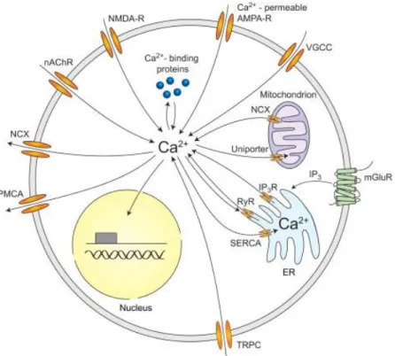
1.5.7 The calcium channels on retinal cells ... 33
1.6 Endophthalmitis ... 34
1.6.1 The classification of endophthalmitis ... 35
1.6.2 Inflammatory changes in retina during endophthalmitis... 36
1.6.3 Treatment: vitrectomy and intravitreal antibiotic ... 37
1.7 Animal model... 38
1.7.1 Intravitreal injection ... 38
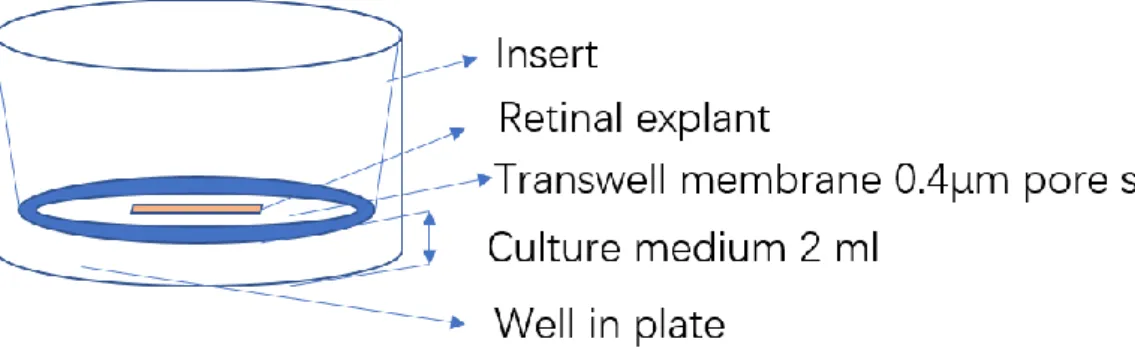
1.7.2 Retinal explant culture... 41
1.8 Objectives ... 44
2. Materials and Methods ... 45
2.1 PVL ... 45
2.1.1 PVL purification ... 45
2.1.2 Evaluation PVL effects by PMNs ... 45
2.1.3 Evaluation of PVL effect in different culture media using cytometry ... 46
2.2 Animal and surgical procedure ... 48
2.2.1 Ethics of the protocol ... 48
2.2.2 Anesthesia, PVL intravitreal injection and euthanasia ... 48
2.2.3 Retinal explant preparation and organotypic culture ... 49
6
2.3.1 Preparation of paraformaldehyde 16% ... 50
2.3.2 Tissue for immunohistochemistry ... 50
2.3.3 Vertical section ... 51
2.3.4 Tissue for whole mount ... 51
2.3.5 Immunohistochemistry for retinal sections ... 51
2.3.6 Immunohistochemistry for retinal whole mounts ... 51
2.3.7 Cell counting ... 51
2.3.8 Statistical analysis ... 52
2.4. Western blotting ... 54
2.4.1 Tissue preparation for western blot ... 54
2.4.2 Extraction protein from retina for western blotting ... 54
2.4.3 Quantify protein using BCA kit ... 54
2.4.4 Migration ... 55
2.4.5 Transfer ... 55
2.4.6 Staining of the membrane ... 55
2.5 Real-time RT-qPCR ... 56
2.5.1 Tissue preparation for RT-qPCR ... 56
2.5.2 RNA extraction ... 56
2.5.3 RNA quantification and integrity ... 56
2.5.4 DNase treatment with DNA-free kit DNase ... 57
2.5.5 Primers design ... 57
2.5.6 RT ... 58
2.5.7 PCR to check cDNA ... 58
2.5.8 Real-time qPCR ... 58
2.5.9 Production specificity verification ... 58
2.5.10 Statistical analysis ... 59
3. Results ... 60
3.1 Article 1: Panton–Valentine Leukocidin Colocalizes with Retinal Ganglion and Amacrine Cells and Activates Glial Reactions and Microglial Apoptosis ... 60
3.2 Article 2: Panton–Valentine Leukocidin Induces Neuronal and Microglial Apoptosis together with Müller and Microglial Cell Activation in a Rabbit Retinal Explant Model ... 80
3.3 Article 3: Bacterial toxins aggravate bacterial endophthalmitis by interacting directly with neurons... 110
4. Discussion ... 130
5. Conclusion ... 133
6. Publications and posters ... 134
7. Bibliography ... 135
7
1. Introduction
1.1 Staphylococcus aureus
Staphylococcus aureus was first discovered in 1871. It was identified as bacteria responsible for purulent infection in 1880. S. aureus is Gram positive, ubiquitous pathogen and commensal to human being. About 20%-25% of the population permanently carry S. aureus and at least 60% carry transiently S. aureus1. It colonizes in wet areas, such as
mucosa from the anterior nostrils to nasopharynx, axilla, wrist and perineum. S. aureus together with Escherichia coli and Pseudomonas aeruginosa are the most common isolated bacteria from hospital environment. It is also the second bacteria responsible for nosocomial infections,after E. coli. The diseases caused by S. aureus vary greatly, from dermal or mucosal infection (folliculitis, boil, impetigo and sinusitis) to visceral organ infections (endophthalmitis, pneumonia, endocarditis and osteomyelitis), and some life-threatening syndromes, such as septicemia and toxic shock syndrome2.
S. aureus is a great burden for health care system, because of its significant morbidity and mortality worldwide. It is hazardous for people that have chronic diseases such as diabetes, eczema, or immune system deficiency (old people, AIDS) and those who are foreign material implant carrier. S. aureus infection can rapidly jeopardize the general health condition and threaten the life, mostly due to its multiple antibiotic resistances and various virulence factors.
S. aureus is frequently found in ocular infections, in which retina can be severely damaged despite prompt and appropriate treatments.
1.1.1 Methicillin-resistant S. aureus
Antibiotic resistance makes S. aureus survive from the antibiotic treatment and spread in hospital and community acquired strains. S. aureus can pass genes of antibiotic resistance and virulence through horizontal transfer among the strains and can adapt rapidly to the new treatment.
β-lactams is the first antibiotic to treat S. aureus infection. The methicillin, one derivative of penicillin of β-lactam antibiotic family, was introduced to clinical usage in 1960. The first methicillin-resistant S. aureus (MRSA) isolate was discovered one year later. Then MRSA prevailed throughout the world as a multi-resistant hospital pathogen. MRSA strains encode a novel specific penicillin-binding protein (PBP2a), which has a low affinity with all β-lactams and makes MRSA resistant to β-lactam antibiotics. This novel PBP2a is encoded by methicillin resistance gene (mecA), which is carried by a variable mobile element, called staphylococcal cassette chromosome mec (SCCmec). SCCmec can be inserted into the chromosome. It is constituted by two complexes, a cassette chromosome recombinase (ccr) gene complex and mec complex. The ccr complex controls the insertion site and integration of SCCmec into the staphylococcal chromosome. The mec complex contains
8
mecA gene and is classified into six different classes, i.e. A, B, C1, C2, D and E. SCCmec could also encode β-lactamases to cleave β-lactams. SCCmec is classified into 9 types according to the classes of ccr and mec complexes3. The type I SCCmec was identified in
1961, type II and III in 1980s, type IV and V in 2000s. The other types of SCCmec were less frequent and identified later. SCCmec varies in size. The type I, II and III are larger than those of type IV and V. The larger SCCmec, such as SCCmec II and III, contains other antibiotic resistances genes.
1.1.2 HA-MRSA and CA-MRSA
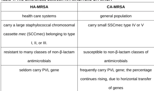
Table 1. The differences between HA-MRSA and CA-MRSA
HA-MRSA CA-MRSA
health care systems general population carry a large staphylococcal chromosomal
cassette mec (SCCmec) belonging to type I, II, or III.
carry small SSCmec type IV or V
resistant to many classes of non-β-lactam antimicrobials
susceptible to non-β-lactam classes of antimicrobials
seldom carry PVL gene frequently carry PVL gene; the percentage continues rising, due to horizontal transfer
of genes
MRSA spreads rapidly and is continuously developing its capacity of resistance to antimicrobial drugs. Considering its ubiquity, virulence and multi-antibiotic resistance, MRSA is a great burden to health care systems. At beginning, MRSA strains were confined to hospitals and other health care systems. However, MRSA infections were reported in healthy populations without exposure to health care system since 1990s. This new emergent MRSA is called as community associated (CA)-MRSA, the previous MRSA as hospital acquired (HA)-MRSA. CA-MRSA is defined as any MRSA infection diagnosed for outpatients or within 48 h of hospitalization without HA-MRSA risk factors.
CA-MRSA strains are different from HA-MRSA strains in terms of genotypic, epidemiological and clinical features4 (Table 1). HA-MRSA strains harbor SCCmec type I,
II, or III and are resistant to many classes of β-lactam and non-β-lactam antibiotics. CA-MRSA strains carry small SCCmec type IV or type V and are susceptible to non-β-lactam antibiotics. The difference in types of SCCmec could well explain the multi-resistance antibiotics of HA-MRSA and the susceptibility of CA-MRSA to non-β-lactam antibiotics5.
The gene encoding PVL can spread from strain to strain by some bacteriophages. About 60 to 100% of CA-MRSA strains carry gene encoding PVL6, 7, while HA-MRSA or
9
methicillin-susceptible staphylococcus aureus (MSSA) are rarely discovered carrying gene encoding PVL. The percentage of CA-MRSA has significantly increased in MRSA infection isolates and may continue to rise in the future. One hypothesis for this increase is due to the horizontal transfer of SCCmec elements and PVL gene to the genomes of MSSA5.
PVL is highly expressed in CA-MRSA strains. The combination of antibiotic resistance and virulent toxin expression might be a great burden in treating S. aureus infection.
1.1.3 Vancomycin-resistant S. aureus
The percentage of MRSA increases and accounts for more than half of S. aureus infections. For severe MRSA infections, such as bacteremia, endocarditis and osteomyelitis, the effective antibiotic treatment is prompt intravenous vancomycin. The adjunctive therapies can also improve the treatment, such as drainage of abscesses or infected lesions, removal of catheters and infected valves8.
Vancomycin is a glycopeptide antibiotic and works by blocking the construction of bacterial cell wall. Vancomycin binds to the precursors of peptidoglycan on the outer surface of bacterial cytoplasmic membrane, preventing the synthesis of peptidoglycan in bacterial cell wall. The mechanism of vancomycin-resistant enterococci is to produce low-affinity precursors of peptidoglycan to eliminate high-affinity precursors. The first resistant clinical isolates were found in Enterococcus in 1988. Since then, vancomycin-resistant enterococci spread rapidly worldwide. In 1997, clinical S. aureus isolates susceptibly resistant to vancomycin was found, called vancomycin-resistant Staphylococcus aureus (VRSA). Then, more evident VRSA strains were found later. Vancomycin resistance genes, such as VanA, VanR, VanH and VanB, are located in plasmid and transferred to MRSA by conjugation to Enterococcus sp9.
Vancomycin is the last therapy for infection caused by multi-resistant strains of staphylococci, streptococci and enterococci. The emergence of resistance to vancomycin means that the bacterial strains can develop resistance to all the antibiotics.
In recent years, alternatives to vancomycin for the treatment of MRSA infection are identified and used. The most effective and wildly used are daptomycin and linezolid. The other alternatives to vancomycin include quinupristin-dalfopristin, tigecycline, trimethoprim-sulfamethoxazole, clindamycin, and tetracyclines. Linezolid is a synthetic oxazolidinone antibiotic, which inhibits the synthesis of bacterial proteins. Linezolid is recommended to treat skin and soft-tissue infections. There was outbreak of linezolid resistance in MRSA strains, which was associated with nosocomial transmission and the extensive usage of linezolid. MRSA strains in patients receiving daptomycin are more likely to develop resistance to daptomycin. Thereby, vancomycin is still the first choice of antibiotic to MSRA infection10.
Other strategies, such as surveillance cultures, more strict usage of antibiotic, and decolonization of S. aureus in bodies, are proposed to control MRSA development in hospital environment.
10
1.1.4 Horizontal transfer
S. aureus could acquire adaptation and evolution very rapidly due to horizontal transfer of mobile genetic elements (MGEs). They could gain new antibiotic resistance and new virulence factors, which facilitate them to escape from immune system and improve their pathogenesis.
MGEs are segments of DNA, which can move around within a genome and be transferred from one stain to another. MGEs encode proteins for antibiotic resistance, virulence and host-adaptation. MGEs include plasmids, bacteriophages and transposons. Plasmids and bacteriophages are the classic MGEs. They are transferred by 3 genetic transfer mechanisms: conjugation (transmission of plasmid), transduction (by the intermediate of viral vector), and/or transformation (that can integrate a DNA fragment into the bacterial genome)11.
SCCmec elements are transferred by bacteriophage, for this reason the prevalence of MRSA increases continuously. VRSA has emerged through the transposons of vancomycin resistance genes; S. aureus pathogenicity islands (SaPIs) carry genes encoding the toxic shock toxin 1 and superantigens. Horizontal transfer of SaPIs relies on the specific “helper” bacteriophages. PVL and staphylococcal enterotoxin A (sea) are found on inserted bacteriophage. PVL gene is frequently found in CA-MRSA, which might be due to selective transfer of PVL-carrying prophages to CA-MRSA12.
1.2 Virulence of S. aureus
S. aureus expresses various virulence factors, including adhesive factors, exoenzymes and toxins (Figure 1). Adhesive factors help S. aureus to adhere to cells and the extracellular matrix, and exoenzymes control colonization and dissemination of S. aureus. The colonization to host tissue is the first stage of microbial infection, which is associated with its structural components and ability to bypass the host defense mechanisms. The dissemination of bacteria can promote the spread of bacteria and enlarge the infection. Whereas, the toxins have a more offensive function, attacking directly the immune system and other cells of the host. For instance, staphylococcal complement inhibitor (SCIN) and chemotaxis inhibitory protein of staphylococci (CHIPS) can modulate neutrophil chemotaxis and phagocytosis. Superantigenes, another type of toxin, can induce activation of lymphocytes, which is responsible for various symptoms, such as toxic shock. Enterotoxins target the intestine cells and are responsible for staphylococcal food poisoning. Hemolysins are able to lyse erythrocytes and lymphocytes. Leukotoxins also lyse other leukocytes. At last, PVL is one of better characterized leukotoxins and is an important virulence factor of S. aureus. This study of its effects on organic tissue and especially retina could help to find new therapeutic solutions to fight S. aureus infection.
11
Figure 1: The virulence of S. aureus
The virulence of S. aureus includes MSCRAMMs, exoenzyme and toxins. MSCRAMMs and exoenzyme have passive functions on S. aureus colonization and dissemination, while toxins target actively immune system or cells of host. CHIPS, chemotaxis inhibitory protein of staphylococci; SCIN, staphylococcal complement inhibitor; Clf A, B, clumping factor A, B; MSCRAMMs, microbial surface components recognizing adhesive matrix molecules; FnBPA, fibronectin-binding protein A.
1.2.1 Adhesive factors
S. aureus adheres to extracellular matrix to form colonization, which is the primary step before opportunistic infection. This adherent ability is relied on microbial surface components recognizing adhesive matrix molecules (MSCRAMMs) on S. aureus surface, including fibronectin-binding protein A and B (FnBPA and FnBPB), a collagen binding protein Cna, a fibrinogen-binding protein and protein A.
FnBPA and FnBPB, expressed by two close genes in most S. aureus strains, attach to
immobilized fibronectin. They are responsible for mediating S. aureus adhering to plasma clots and foreign body surface. The collagen-binding protein Cna adheres to collagen substrates and collagenous tissues. It is expressed by 38%-56% of S. aureus strains and mediates S. aureus binding to cartilage. It is associated with bone and joint infections13.
Clumping factor A (ClfA) and B (Clf B) are the major fibrinogen (Fg) binding proteins of S. aureus. They recognize different parts of fibrinogen. ClfA binds to the carboxyl terminus of the γ chain of fibrinogen, while ClfB binds α and β chains of fibrinogen. They mediate S. aureus clumping in blood plasma during arthritis and endocarditis. Protein A is expressed by 90% of S. aureus stains. Unlikely to other molecules which binds to collagen, fibrinogen or fibronectin, protein A binds to the Fc region of immunoglobulin, which inhibits the opsonization, thus impairing phagocytosis of PMNs13.
12
1.2.2 Exoenzymes
S. aureus secretes many exoenzymes to control its colonization and dissemination. The dissemination of bacteria can promote the spread of the infections. Those enzymes include nuclease, protease, lipase, hyaluronidase and collagenase. The proteases from S. aureus include serine, cysteine and metalloenzymes, and are not sensitive to most human inhibitors of plasma protease14.
Aureolysin is a metalloproteinase of S. aureus. It can transform precursor of V8 protease
into an active form. It inactivates ClfB by cleaving the N-terminal domain of ClfB. ClfB is membrane protein. The activation of ClfB can modify bacterial cell surface proteins, leading to tear off bacteria from colonization and promote the spread of infection. Hyaluronate
lyase cleaves acid residues in hyaluronan, a component of extracellular matrix. This
cleavage can dissociate extracellular matrix and promote bacterial dissemination and migration. Staphylocoagulase binds and activates prothrombin, leading to the cleavage
of fibrinogen to fibrin. It promotes the abscess formation and lethal bacteremia of S. aureus.
V8 protease is a serine protease, which cleaves specifically peptide bonds formed by
aspartate and glutamate residues. It degrades the bacterial cell surface fibronectin-binding protein, facilitating bacterial dissemination. It can also promote the maturation of other virulence factors, such as staphostatins. Lipase can release a large quantity of fatty acids, which can help S. aureus to survive in the fatty human or mammalian skin. Staphopains
A and B are the major secreted cysteine proteases of S. aureus, which may promote the
invasion and metastatic infections of S. aureus. Staphostatin A and B may inhibit Staphopains A and B, respectively, through binding the active sites of these protease14.
1.2.3 Toxins
Bacteria can secret toxic substances, which can directly damage host tissue or disable host immune system to protect bacteria against host defenses. S. aureus could produce a large repertoire of toxins, including CHIPS and SCIN, superantigens, enterotoxins, exfoliative toxin, hemolysins and leukotoxins. S. aureus strains express partially those toxins, presenting various degree of virulence.
1.2.3.1 CHIPS and SCIN
CHIPS and SCIN are toxins simultaneously expressed during the early (exponential) growth stage of S. aureus and can both modulate neutrophil chemotaxis, phagocytosis and cell killing15.
CHIPS is a 121-residue protein excreted by about 60% of S. aureus strains. It is potent inhibitor of chemotaxis of neutrophils and monocytes toward C5a and the N-formylmethionyl-leucyl-phenylalanine (fMLP). Indeed, CHIPS binds directly to the receptors of C5a and fMLP with high affinity and selectivity. Thereby, it prevents C5a and fMLP from binding to their receptors and activating transduction pathways16.
13
SCIN, an 85-residue protein secreted by S. aureus, has an anti-inflammatory action during S. aureus infection. It can inhibit the formation of lytic membrane attack complex (C5b-9 deposition) through disrupting alternative pathway-mediated and classical/lectin opsonization (C3b deposition). Thereby, it reduces phagocytosis following the opsonization and blocks efficiently the functions of the downstream effectors.
1.2.3.2 Superantigens and enterotoxins
Superantigens are proteins that cause non-specific activation of T-cells resulting in polyclonal T cell activation and massive cytokine release. S. aureus produces various superantigens, including the staphylococcal enterotoxins (SEs), the staphylococcal enterotoxin-like (SEls) proteins, and toxic shock syndrome toxin-1 (TSST-1).
Genes encoding superantigens are located on mobile genetic elements, which are carried by about 80% of S. aureus strains. In adaptive immune system, antigen presenting cells present antigens to CD4+ T cells by histocompatibility complex (MHC)-II. TSST-1 can bind to the invariant regions of MHC-II and interact with the β-chains of T cell receptors (Vβ-TCR), which can abnormally make cross-bridge between MHC-II and Vβ2-TCRs of subtype T-cells without specific antigens. This subpopulation of T cells is non-specifically activated and multiplies, resulting in massive release of inflammatory cytokines and toxic shock syndrome. By the same mechanism, superantigens cause other less severe syndromes, neonatal toxic shock syndrome-like exanthematous disease (NTED) and recalcitrant erythematous desquamating disorder (REDD). NTED is found in newborns and REDD is found in patients with AIDS17.
An enterotoxin is an exotoxin released by a microorganism that targets the intestine. Staphylococcal enterotoxins have 20 distinct members, such as SEA, SEB, SED, SEE and SEF. They are responsible for the Staphylococcal food poisoning. Staphylococcal food poisoning, characterized by nausea and vomiting, is usually self-limited and resolves typically within 24-48 h after onset, albeit a few cases may result in a lethal toxin shock-like symptom. Enterotoxins can cause this disease even without the presence of S. aureus18.
1.2.3.3 Exfoliative toxins
S. aureus produces exfoliative toxins (ETs) A, B, D, which cause a blistering of the skin. ETs target desmoglein-1 and act as serine proteases, resulting in mid-epidermal cleavage. ETs can cause dermal diseases ranging from blisters to severe exfoliation, such as bullous impetigo and staphylococcal scalded skin syndrome. About 5% of S. aureus strains carry ETs genes, but only a small proportion cause severe exfoliation. Some authors argued that ETs can act also as superantigens to cause autoimmune dermal disease19.
14
1.2.3.4 Hemolysins
Hemolysins are lipids and proteins that lyse red blood cells by destroying their cell membrane. S. aureus expresses β-hemolysin, δ-hemolysin.
β-Hemolysin, also called beta-toxin, is a sphingomyelinase of S. aureus. It is encoded by a bacteriophage in a small percentage of S. aureus. It lyses erythrocytes and human lymphocytes by cleaving sphingomyelin in cell membranes to scavenge nutrients and escape from immune system. Its structure shows the characteristics of DNase I. However, instead of functioning as DNase, it precipitates extracellular DNA. The precipitated DNA forms non-specifically cross-linking with hemolysin β and other proteins, an insoluble nucleoprotein matrix, which contributes to biofilm formation20.
δ-hemolysin is an alpha-helical and amphipathic 26-amino acid peptide, expressed in 97% of the S. aureus isolates. It is soluble in water and organic solvents. It can perturb the membrane after binding to it, resulting in cell lysis. It can lyse erythrocytes of many species20.
1.2.3.5 Leukotoxins
Leukotoxins kills leukocytes, permitting bacteria to escape from host immune surveillance. Some of them are also hemolytic. Some of them are associated with severe infectious diseases. PVL is one well characterized leukotoxin, which is associated with necrotizing infections. We will review the members of leukotoxins from human isolated S. aureus, to better understand pathogenesis of S. aureus.
1.2.3.5.1 The prevalence of leukotoxins
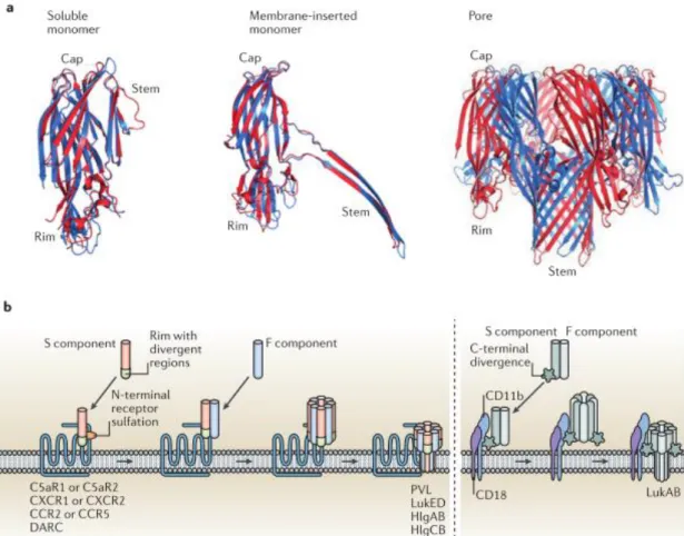
Leukotoxins are composed of two distinct proteins, a class S (31–32 kDa) and a class F component (33–34 kDa). S component binds initially to the cytoplasmic membrane of target cells, then triggering the subsequent F component binding, which organize as alternate octamers, called prepores, then form the pore (Figure 2).
Human S. aureus isolates can produce five leukotoxins: γ-hemolysin consisting of two leukotoxins (HlgA/HlgB and HlgC/HlgB), LukAB, PVL and LukED21. The genes of
HlgA/HlgB, HlgC/HlgB, and LukAB are located in the core genome on chromosome and are presented in almost all human S. aureus isolates. In contrast, PVL and LukED genes are located on mobile gene elements and could be horizontally transferred in a selective way. S. aureus expressing of PVL and LukED is related to some specific infections22. PVL
gene is located in the temperate bacteriophage and is found in 2%-10% of all clinical S. aureus isolates in Europe, about 60% in African or Asian developing countries; PVL gene is positive in 60-100% of CA-MRSA isolates, while it is rare in HA-MRSA strains. LukED gene is located in a stable S. aureus pathogenicity island (a mobile gene element) and is found in 50% -75% clinical S. aureus isolates. S. aureus expressing LukED is associated with bullous impetigo and post-antibiotic diarrhea31.
15
The gene encoding γ-hemolysin (HlgA/ HlgB, HlgA /HlgC) is divided into two parts. The first part is the locus for HlgA, which is about 400 bases upstream of second part which encodes the HlgC and HlgB. In that second party, hlgC and hlgB are separated by a base “T” and are simultaneously co-transcribed. For PVL and LukED, the genes of two components also are separated by one base “T” and are simultaneously co-transcribed. The genes of the two components of LukAB are also co-transcribed together and separated by 21 bases23, 24.
Figure 2: Crystal structure and pore formation of leukotoxins
(a) Crystal structures of leukotoxins: the soluble leukotoxin, leukotoxin in pre-pore formation, the hetero-octameric pore of leukotoxin. (b) The simple illustration of the processes of leukotoxin binding and pore formation. For PVL, LukED, HlgAB and HlgCB, the S component binds first to the receptor, then F component binds consecutively to the complex. Four S and four F components form a hetero-octameric pre-pore and then a pore. For LukA/B, S and F components form a dimer before binding to the receptor and forming the hetero-octameric pre-pore and pore. (Images in part a is from University Medical Center Utrecht, The Netherlands; images in part b is from András N. Spaan et al, 2017)
1.2.3.5.3 The spectra and receptors of leukotoxins
The leukotoxins of S. aureus have different cell-type and species-type targets. HlgA/HlgB could lyse lymphocytes and all the granule cells, while the other leukotoxins target granule
16
cells with different spectra. Also, PVL and HlgC/ HlgB could incite neurons to undergo calcium mobilization and glutamate release25. For a long time, cellular receptor was
hypothesized to play a great role in the effect of leukotoxin. In recent years, the receptors of those leukotoxins are well identified, which can help to explain the spectra of target cells and species specificity of these toxins, and their cytotoxicity22 (Table 2).
γ-Hemolysin
γ-Hemolysin consists of HlgA/HlgB and HlgC/HlgB. 99.5% of human S. aureus isolates express genes encoding γ-Hemolysin, which can aggravate septic arthritis and systemic infection. HlgA/HlgB has a large spectrum of target cells. It can lyse human and rabbit erythrocytes and target most leukocytes: T lymphocytes, granulocytes (neutrophil, basophil, eosinophil) and monocytes and their derivative cells (macrophages and dendritic cells). In contrast to HlgA/HlgB, HlgC/HlgB has a low capacity to lyse lymphocytes and does not lyse erythrocytes. HlgC/HlgB can target human granulocytes, monocytes macrophages and dendritic cells and cell line (HL-69) of the precursor of the neutrophils. To identify the specific receptors for HlgA/HlgB and HlgC/HlgB, human embryonic kidney cells were transfected with a collection of human chemokine receptors and were evaluated for the cytotoxic susceptibility to the two leukotoxins. It was shown that HlgA/HlgB bound to CXCR1, CXCR2 and CCR2, and HlgC/HlgB bound to C5aR and C5L222.
LukE/D
CCR5 is a receptor for LukE/D. Different cell lines (human T cell line, PMN-HL-69 osteosarcoma) are engineered to express CCR5 expression. LukE/D is cytotoxic toward cell lines in presence of CCR5, while the presence of other receptors (CCR1, CCR2, CCR3, CXCR4, CCR8, and CXCR6) does not make cell lines susceptible to LukE/D. The antagonist of CCR5 could effectively suppress LukE/D cytotoxicity. LukE/D can target human, rabbit and murine CCR5-bearing cells, and lyse neutrophils, monocytes and macrophages26.
LukA/B
LukA/B is cytotoxic toward human and rabbit neutrophils, monocytes, macrophages and dendric cells, but not cytotoxic toward murine leukocytes. CD11b, α subunit of the αM/β2 integrin (CD11b/CD18), is known as macrophage-1 antigen or complement receptor 3. CD11b is identified as a host molecule required for LukA/B-mediated cell killing. LukA/B is the most divergent leukotoxin of staphylococcal leukotoxins. The amino acids sequences of LukA and LukB share about 30% and 40% with S component and F component of other leukotoxins, respectively. In contrast, other leukotoxins have 60%-80% similarity of amino acid sequence for S and F components. LukA/B needs to form a stable bicomponent before binding to its receptor, while S and F components of other leukotoxins bind separately and consecutively to the receptors27 (Figure 2).
17
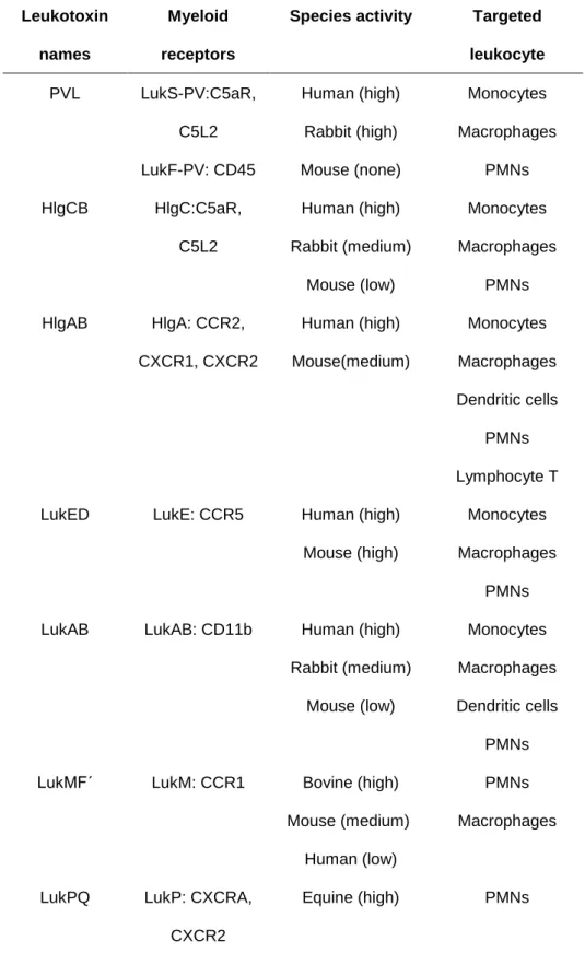
Table 2. The myeloid receptors, species activity and targeted leukocyte of leukotoxins of S. aureus
Leukotoxin names
Myeloid receptors
Species activity Targeted leukocyte PVL LukS-PV:C5aR, C5L2 LukF-PV: CD45 Human (high) Rabbit (high) Mouse (none) Monocytes Macrophages PMNs HlgCB HlgC:C5aR, C5L2 Human (high) Rabbit (medium) Mouse (low) Monocytes Macrophages PMNs HlgAB HlgA: CCR2, CXCR1, CXCR2 Human (high) Mouse(medium) Monocytes Macrophages Dendritic cells PMNs Lymphocyte T LukED LukE: CCR5 Human (high)
Mouse (high)
Monocytes Macrophages
PMNs LukAB LukAB: CD11b Human (high)
Rabbit (medium) Mouse (low) Monocytes Macrophages Dendritic cells PMNs LukMFʹ LukM: CCR1 Bovine (high)
Mouse (medium) Human (low)
PMNs Macrophages
LukPQ LukP: CXCRA, CXCR2
Equine (high) PMNs
Abbreviated symbols: PVL, Panton-Valentine leukocidin; C5aR, C5a receptor; C5L2,
C5a receptor-like 2; PMNs, Polymorphonuclear leukocytes; CCR1, 2, 5, CC-chemokine receptor1, 2, 5; CXCR1, 2, A, CXC chemokine receptor 1, 2, A.
18
LukMFʹ and LukPQ
Leukotoxin MFʹ (LukMFʹ) and leukotoxin PQ (LukPQ) are associated with zoonotic infections and are rarely found in human S. aureus isolates. LukMFʹ is associated to bovine mastitis, not in human infection28. The receptor for LukM is CCR1. LukMFʹ targets actively
bovine and murine neutrophils and macrophages, and human neutrophils to a lesser extent. LukPQ, a newly identified leukotoxin, targets mainly equine neutrophils through equine CXCRA and CXCR229.
PVL
Comparing to other leukotoxins, PVL has the smallest spectrum of target cells. PVL targets macrophages and neutrophils, not lymphocytes. In 2013, it was reported that human C5a receptors, C5aR and C5L2, were receptors for LukS-PV and mediators of PVL-induced cytotoxicity. The antibody against C5aR could effectively block PVL binding to neutrophils and macrophages. PVL has preference for animal species and does not recognize murine C5aR, which is determined by the second extracellular loop of C5aR21. Many previous
studies used mice model to study PVL and might bring out some confusing results30.
Recently, CD45 is identified as one receptor for LukF-PV, which influences the cytotoxicity of PVL towards neutrophils31.
1.3 Leucocytes
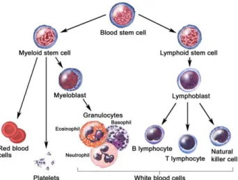
Figure 3: Hematopoietic differentiation tree
In the bone marrow, hematopoietic stem cells differentiate into two types of stem cells: one is myeloid stem cell, leading to the formation of red blood cells, platelets and myeloid cells; another is a lymphoid stem cell, leading to the formation of lymphocytes. (from website national cancer institute https://visualsonline.cancer.gov )
19
All the leukotoxins target and lyse leukocytes, which is the origin of their name. Leukocytes, the body's army of soldiers, are the cells of the immune system. They circulate in the body in blood vessels and lymphatic vessels. They fight the invading germs and prevent the infections. The leukotoxins kill leukocytes and help S. aureus to escape from immune defense and proliferate in human body. It’s necessary to have an overview of leukocytes to understand well the cell targets of leukotoxins.
In the bone marrow, hematopoietic stem cell can differentiate into two types of stem cells: one is myeloid progenitor cell, leading to the formation of monocytes, red blood cells and neutrophils; another is lymphoid stem cell, leading to the formation of lymphocytes (Figure 3). Leukocytes circulate in the blood in a quiescent state with low adhesiveness, they have short life time about 7 to 12 h. They monitor the environment and can migrate into the tissues to defend against the invading microbes. Their main defensive functions are phagocytosis, degranulation and oxidative explosion32.
1.3.1 Polynuclear neutrophils
Polynuclear neutrophils are the major cell type of granulocytes, occupying more than 40-75% of circulating leukocytes. Polynuclear neutrophils are the innate immune cells which react and migrate immediately to the lesion.
Migration
Neutrophils migrate from blood vessels to the tissues, through a process divided into four steps: rolling, activation, adhesion, and transendothelial migration. First, P-selectin ligand (PSGL-1) on neutrophils interacts with P-selectin expressed at the surface of endothelial cells. This interaction is of low-affinity, neutrophils poorly bind to the endothelial cells. Continuously being carried away by the blood flow, they roll on the vessel wall. During this rolling, neutrophils can be activated by the invading pathogens or proinflammatory products. Once being activated, neutrophils could express immediately more adhesive molecules on their surface, such as lymphocyte function-associated antigen 1 (LFA-1) which interacts with ligand intercellular adhesion molecule (ICAM-1) on endothelial cells. This high affinity adhesion allows neutrophils to attach firmly to blood vessels. Then, they transform, sneak through the endothelial cell layer and migrate to lesion33.
Phagocytosis and degranulation
Neutrophils are effective phagocytes. They imprison pathogens inside an endosome, which is internalized into cytoplasm, called phagosome. The granules containing degradation enzymes come to merge with phagosome to degrade the contents.
Neutrophils are granulocytes and have four types of granules with different content. During the maturation of neutrophils, the primary granules are fused with phagosome. They contain myeloperoxidase, elastases, defensins, lysozymes and azurocidine, which digest phagocyted microorganisms. The other granules are formed later than primary granules. The second granules are rich in antimicrobial agents, which are released into extracellular medium. The third granules contain gelatinases, enzymes, and receptors, which are useful
20
for the neutrophils transendothelial migration. The fourth granules are secretory vesicles, which contain many receptors. They are rapidly secreted outside to modify the composition of the cytoplasmic membrane, promoting neutrophil activation and adhesion to the endothelium34.
1.3.2 Eosinophils, basophils and mast cells
Eosinophils are circulating in blood, occupying normally less than 5% of circulating
leukocytes. They produce and store biologically active molecules, including cytotoxic proteins, lipid mediators, chemotactic peptides and cytokines. They are associated with a series of disorders, such as asthma, tropical pulmonary eosinophilia. Basophils occupy normally less than 1% of circulating leukocytes and are associated with fatal asthma, acute and chronic allergy. They abundantly secrete cytokines after being activated to amplify the allergy. Mast cells are not flowing leukocytes, they reside in vascularized tissues. They have many granules, which contain histamine, proteases and cytokines. They can rapidly degranulate after interacting with IgE and release prostaglandins and leukotrienes during allergy35.
1.3.3 Monocytes, macrophages and dendric cells
Monocytes constitute 3-10% of flowing leukocytes. Monocytes express many receptors, such as Toll-like receptors (TLRs), to monitor the environmental changes. In general, they flow in blood for 1-3 days, and then migrate into tissue and become resident macrophages or dendritic cells. In abnormal conditions, monocytes are activated and transform into inflammatory macrophages, which secrete cytokines, myeloperoxidase and superoxide, and help in phagocytosis to eliminate harmful materials. Monocytes and macrophages can eliminate pathogens, cancer cells and cellular debris by phagocytosis.
Dendritic cells are antigen-presenting cells in immune system. They reside in tissue which is in contact with external environment, such as skin and mucosa. Dendritic cells capture and present antigens to T-lymphocytes, which can activate them to undergo immune response. They also can promote B cell activation and differentiation by producing cytokines and other factors36.
1.3.4 Lymphocytes T, B and NK
The flowing lymphocytes constitute 20-40% of the flowing leukocytes in adults and are divided into three types of lymphocytes in the peripheral blood: T, B and NK cells, representing about 80%, 10% and 10% of total lymphocytes, respectively. T lymphocyte is a major cell of the adaptive immune system and can be divided into CD8+ and CD4+ T cells.
CD8+ T cells are cytotoxic T lymphocytes. They bind to target cells and secrete molecules
to destroy these cells. CD4+ T cells bind to antigen fragment presented by CMH II from
21
chemokines and help to develop the clones of B cells which secrete antibodies against the presented antigens. B lymphocytes bind and engulf antigens, which are then presented to T-cells. Helper T cells can aid B cells to produce specific immunoglobulins to antigenic epitopes, called antibodies. Lymphocyte NK is component of innate host defense system. They can lyse virally infected cells and malignant cells37.
1.4 PVL
In the early 1930s, PVL was first discovered by Panton and Valentine. It is a powerful leukotoxin secreted by S. aureus.
1.4.1 PVL-related clinical diseases
PVL is often found in necrotizing S. aureus infections, such as furuncles, acute necrotizing pneumonia and osteomyelitis. Here are a few examples of S. aureus infections potentially aggravated by PVL.
1.4.1.1 PVL pathophysiology in skin infection
The skin abscesses are often related to S. aureus infection. The rate of PVL expression in S. aureus strains isolated from skin infection is much higher than that from systemic infection38. A study included 43 S. aureus isolates from cutaneous infections, among which
12 (28%) strains expressed PVL gene. While only 1 strain among 49 (2%) isolates from systemic S. aureus infection was expressing PVL 39. S. aureus expressing PVL is often
related to primary skin infection, while the S. aureus non-expressing PVL is related to secondary infection after other dermal diseases such as bullous or pruritic diseases39.
The skin lesion generated by PVL is dependent on PVL concentration. PVL concentration ranges from 0.27 mg/L to over 2 mg/L in pus of PVL-positive S. aureus skin abscess. When PVL concentration is superior 1 mg/L, large abscesses (diameter ≥5 cm) are observed38.
Small PVL concentration (30 ng) induces dermal edema and erythema, high PVL concentration (300 ng) provokes widespread infiltrated erythema followed by skin necrosis after intradermal injection in rabbits39. In mice, skin does not react after intradermal
injection PVL, even though the PVL doses reaches up to 3 μg39.
1.4.1.2 PVL pathophysiology in pneumonia
CA-MRSA carrying PVL could cause severe and fatal pneumonia within a short time. PVL-positive S. aureus pneumonia is necrotizing pneumonia characterized by a bloody cough (haemoptysis) and a decrease of circulating leukocytes (leucopenia). Histopathological analysis shows extensive necrotic ulcerations and massive haemorrhagic necrosis in lung tissue40. This pneumonia is preceded by an influenza-like syndrome, then followed by high
fever (above 39°C), tachycardia (>140 beats/min). The patients are healthy children or young adults.
This severity is associated with PVL. Purified PVL is directly instilled into rabbit lung and causes severe inflammation and injury by recruitment and subsequent lysis of PMNs in
22
lung during several hours. Damaged PMNs could release cytotoxic granules and/or reactive oxygen metabolites and significantly increase the level of IL-8 and MCP-1 in lung. These cytokines amplify the recruitment of leukocytes and damage the integrity of alveolus41.
1.4.1.3 PVL aggravates osteomyelitis
S. aureus often induces infection in bone and joint. PVL aggravates osteomyelitis in both clinical and experimental observations. The osteomyelitis or arthritis caused by CA-MRSA carrying PVL have more severe damage and require longer antibiotic course and various surgical procedures. PVL-positive osteomyelitis or arthritis undergo rapid evolution toward multifocal osteomyelitis and/or multiple abscesses despite antibiotic treatment42. In
experimental rabbit model, the bone is infected by PVL-positive CA-MRSA strain and its PVL-negative isogenic derivative. In presence of PVL, rabbit bone is deformed, muscle and joints are involved in infection, which is rarely found in PVL-negative strain osteomyelitis. The anti-PVL antibody significantly reduces the inflammation in osteomyelitis caused by PVL-positive S. aureus43.
1.4.2 PVL effects
1.4.2.1 PVL inducing calcium mobilization
PVL induces an increase of intracellular calcium concentration in PMNs and neurons without membrane damages25, 44. This calcium mobilization is not associated with plasma
membrane Ca2+ channels or pore formation, but with the sarcoendoplasmic reticulum
calcium transport ATPase (SERCA)45,46.
PVL induces an increase of intracellular calcium concentration after 100s, which increases linearly within 10 min. The extracellular calcium influences PVL effects in calcium mobilization. In presence of physiologic calcium concentration, PVL application incites an increase of the intracellular calcium concentration in PMNs. In absence of extracellular calcium, PVL forms pore on PMNs, while the intracellular calcium concentration is not affected47.
PVL induces an increase of intracellular calcium concentration without pore-forming in primary cerebellar granular neurons and dorsal root sensory neurons. This calcium increase is accompanied by glutamate release from primary cerebellar granular neurons25.
1.4.2.2 PVL inducing cell death
PVL can induce necrosis or apoptosis of PMNs, which are dependent on the PVL concentration. At high concentration, PVL induces necrosis by osmotic lysis, which might be due to pore formation on cytoplasmic membrane. At low concentration, PVL induces apoptosis by Bax-independent signaling pathway48. PVL is also related to neutrophil cell
death during neutrophil extracellular traps (NETosis). NETosis is a recently described process of neutrophils trapping and killing of S. aureus, which leads to lytic cell death. PVL has been found to be associated to this process49.
23
Cell apoptosis related to PVL is also demonstrated on keratinocytes: S. aureus strains are engulfed into endosomes by human epidermal keratinocytes (RHEK-1) within 1 h. PVL-positive S. aureus could successfully disrupt endosomes and replicate intracellularly. After 6 h, PVL induces significantly caspase-dependent keratinocyte apoptosis50.
1.4.2.3 PVL inducing release of inflammatory factors
PVL is reported to target human and rabbit monocytes, macrophages and PMNs, but not to lymphocytes, which implies that PVL disturbs directly the innate immune system. PVL can also induce reactions on cerebral and radical ganglion neurons25.
PVL induces different cell reactions in different cell types. PVL incites PMNs to secret granule content in a dose-dependent way. PVL induces proinflammatory factors release such as histamine, leukotriene B4 and IL-8 from neutrophils51, 52. Histamine is a potent
vasodilator. Leukotriene B4 and IL-8 are chemokines which attract leukocytes to inflammatory sites. Those factors promote and facilitate PMNs-tissue infiltration. PVL influences the production of radical superoxide in PMNs: at low concentration, PVL induces PMNs to produce moderate superoxide anion; at high concentration, PVL inhibits radical superoxide formation. The production of free radical superoxide molecules could damage the tissues, while their inhibition could help S. aureus to spread53.
PVL activates monocytes and macrophages to produce NLRP3 inflammasome, a signaling complex, which incites the release of IL-1β and IL-1854. IL-1β and IL-18 can further induce
lung epithelial cells to secret chemokines and recruit leukocytes in PVL-positive MRSA necrotizing pneumonia55.
PVL could incite neurons, resulting in calcium mobilization and glutamate release. In total, PVL effects result in leukocyte recruitment and death, vasodilation, cells invasion and tissue necrosis.
1.4.2.4 PVL retrograde transport
After PVL binding to C5aR, the receptor is phosphorylated and PVL is internalized with the phosphorylated receptor45. Immunolabeling and confocal microscopic techniques were
used to trace the PVL retrograde transport. It showed that PVL accumulated first in lysosomal compartments 10 min after PVL application and reached to Golgi network 3 h after PVL application. Cell apoptosis was observed 3 h and peaked 6 h after PVL application. PVL induced Ca2+ release from the ER within 10 min, unexpectedly not from
lysosome, which indicated that PVL-induced calcium mobilization was not related to the pore formation. The initial process of interaction between PVL and C5aR seems to play great role in PVL-induced calcium mobilization and cytotoxicity, independently from pore formation56.
24
Figure 4: PVL retrograde transport
Step 1. LukS-PV binds to C5aR; Step 2. Recruitment of LukF-PV; Step 3. Internalization of PVL with phosphorylated C5aR; Step 4. Transfer to lysosome (10 min-3 h); Step 5. Transfer to Golgi (after 3h); Apoptosis occurs at 3 h and peaks at 6 h after PVL application.
1.4.3 PVL treatment
Considering the severity of S. aureus infection caused by PVL, PVL should be targeted as a new treatment. Different antibiotic families have very different effects in PVL secretion. Oxacillin enhances PVL release, while clindamycin, linezolid, fusidic acid and rifampicin inhibit PVL expression. The other antibiotics do not have influence on PVL expression. It has been proposed to combine clindamycin, linezolid, fusidic acid and rifampicin with other antibiotics to treat possible PVL-carrying S. aureus infection43. Those antibiotics could
inhibit the production of many staphylococcal exotoxins in vitro. Combination those inhibitory antibiotics with other antibiotics needs be further studied in clinical studies. Humanized heavy chain-only antibodies (HCAbs) against PVL two components (LukS and LukF) prevent PVL binding and pore formation. In a rabbit PVL-induced endophthalmitis in vivo model, HCAbs (anti-LukS-PV, anti-LukF-PV) prevent effectively ocular inflammation and show therapeutic potential57.
Among a series of molecules tested to inhibit PVL, calixarenes show the property of inhibiting PVL. P-sulfonato-calix[n]arenes can abolish S protein binding to membrane. This inhibitory effect is also observed in a rabbit model of PVL-induced endophthalmitis58. All
25
1.5 Retinal structure
1.5.1 General structure
1.5.1.1 The cell layers of retina
The retina is innermost, light-sensitive nervous layer that lines the inner surface of the back
of the eyeball, in which stimulation by light occurs, initiating the sensation of vision.
The light penetrates retina and is absorbed by photoreceptors. The photoreceptors transform the light into electrical message, which is sent to all the succeeding neurons of retina, reaching to central system by the visual pathway (Figure 5). The retina contains several structural layers, from the most inner layer to the most outer layer are as follows: the inner limiting membrane (ILM), which is constituted by a membrane and endfeet of Müller cells. ILM is the interface between retina and vitreous and functions as demi-barrier, which prevents large molecules from penetrating into retina; the nerve fiber layer (NFL), which contains axons of ganglion cells which constitute optical nerve; the ganglion cell
layer (GCL), which contains two kinds of cells, ganglion cells and displaced amacrine cells;
the inner plexiform layer (IPL), which is neuropil containing the axons or synapses of nearby cells and some microglial cells; the inner nuclear layer (INL), which is constituted by cellular soma of bipolar, horizontal, amacrine and Müller cells; the outer plexiform
layer (OPL), which is another neuropil containing process of bipolar cells and horizontal
cells and synaptic pedicles of photoreceptors; the outer nuclear layer (ONL), which contains rod and cone photoreceptors; the outer limiting membrane (OLM), which is adherent junctions between Müller cells and photoreceptors; the pigment epithelium
layer, which constitutes the outer ocular-blood barrier (Figure 6).
1.5.1.2 The retinal vasculature
The arterial intra-retinal branches supply three layers of capillary networks: the radial peripapillary capillaries, and the inner and the outer layers of capillaries (Figure 7). From NFL to OPL, the retinal structure is vascularized by retinal capillaries from central retinal artery. ONL is avascular and gets nutrients from the choriocapillaries which are supplied by choroidal arteria. The fovea is exceptional and avascular, in which cone photoreceptors are maximally concentrated and other retinal layers are absent.
1.5.1.3 The blood-retina barriers
The retina has two blood-retina barriers (BRB), the outer BRB, which is constituted by tight junctions among retinal pigment epithelium and inner BRB which is constituted by tight junctions among intra-retinal vessels. The molecules selectively pass through these BRBs, which is the principal mechanism to maintain retinal homeostasis and function.
26
Figure 5: The structure of eye
(Gaurab Karki, 2018, Human Eye: Anatomy, parts and structure.
http://www.onlinebiologynotes.com/human-eye-anatomy-parts-structure)
Figure 6: The organization of retina
(Helga Kolb, Webvision: Simple Anatomy of the Retina by Helga Kolb. https://webvision.med.utah.edu/book/part-i-foundations/simple-anatomy-of-the-retina/)
27
Figure 7: Simple illustration of ocular vasculature
Left: choroid and retinal vessels consist ocular vasculature. Right: retinal vessels are consisted in three layers: radial peripapillary capillaries (superficial), and the inner and the outer layer of capillaries (intermediate and deep). The choroidal vessels are under RPE and supply blood to the outer portion of the retina.
Abbreviation: GCL: ganglion cell layer; INL: inner nuclear layer; NFL: nerve fiber layer;
ONL: outer nuclear layer; RPE: retinal pigment epithelium. (Image from Jing Chen Laboratory website).
1.5.2 Müller cells
1.5.2.1 Müller physiologic function
Müller cell population is intense and well organized in retina, which provides architectural support to retinal neurons. Their cell bodies are situated in the inner nuclear layer. Their processes extend through the entire retinal thickness, their endfeet forming the inner limiting membrane and their outer processes supporting photoreceptors. They also form endfeet on retinal large retinal blood vessels. Anatomically, Müller cells are tightly related to all retinal cells and blood vessels59.
Müller cells participate in many retinal physiologic functions. They regulate retinal blood vessels and maintain blood-retinal barrier. They provide metabolic and nutritional support to retinal neurons, maintain retinal physiologic homeostasis (water/ion and pH), uptake or recycle neuronal transmitters, provide scaffold for neuronal cells orientation during retinal development, release neuroactive or vasoactive substances59. Müller cells secrete
neuronal trophic molecules such as nerve growth factor to support neuronal survival and neuritogenesis60.
1.5.2.2 Müller gliosis
In some species, Müller cells can regenerate retinal neurons after retinal damage. For example, zebrafish retinal Müller cells produce neural progenitor cells following neuronal damage and death61. While in mammalian, Müller cells undergo gliosis in response to
28
from diverse harmful stimulus. The hallmark of retinal Müller cell gliosis is the shape change resulting from rapid upregulation of glial fibrillary acidic protein (GFAP) following acute retinal injury62. The time-dependent Müller cell gliosis is significantly associated with
retinal functional impairment and alteration of retinal gene expression63. Human Müller
cells may produce IL-6 mRNA and proteins after stimulation of IL-1β or LPS64. Müller gliosis
modifies the expression of glutamine synthetase, reduces the K+-conductance on their
membrane, releases neurotrophic factors and antioxidants. The Müller gliosis can also release detrimental factors such as nitric oxide and VEGF, leading to detrimental glial scar, which could disturb the retinal function59. The increase of intermediate filament expression
in Müller gliosis is correlated with cell stiffness, which could impede the nerve regeneration65. In retinal detachments, Müller cell processes migrate abnormally to the
outer part of retina and undergo mitosis and gliosis, resulting in subretinal glial scar66.
1.5.2.3 The inflammatory reaction of Müller cell
Müller cells express innate immune receptors and are capable to sense both pathogen- and host-derived ligands67. Müller cells express Toll-like receptors (TLRs) 1-10, the major
family of pattern recognition receptors in innate immune response68. Müller cells express
also intracellular NOD-like receptors (NLRs), which are correlated with TLRs and regulate inflammatory reaction69. After detecting these pathogens, Müller cells react intensely and
release proinflammatory cytokines, chemokines and nitric oxide (NO). During S. aureus endophthalmitis, Müller cells are activated and secret proinflammatory cytokines (IL-6, TNF-α, and IL-1β), chemokines (IL-8), and antimicrobial peptide (IL-37)70. In
endotoxin-induced uveitis, Müller cells express NO and TNF-α71. Müller cell-derived VEGF is the key
factor in inducing retinal vascular lesions, vascular leakage and retinal cytokines productions72.
1.5.2.4 Müller cell gliosis results in retinal edema
Müller cells express water transports coupled to potassium channels, which can be influenced during retinal inflammation. Kir4.1, the main potassium channel in retina, and Aquaporin 4 (AQP4), the predominant water channel colocalize on Müller cells. During uveitis, Kir4.1 and AQP4, 5 expressions are significantly decreased on cytoplasm membrane of Müller cells73. Aquaporin 11, expressed exclusively on Müller cells,
decreases significantly during uveitis, which is related to Müller cell swelling and retinal edema74. When the homeostasis of ion and water influx is disturbed by inflammation,
Müller cells undergo swelling and neurons suffer from the abnormal osmotic stress59.
Müller gliosis is related to retinal swelling. In endotoxin (LPS)-induced ocular inflammation, Müller cells hypertrophy and increase the immunoactivity of GFAP. Electrophysiology shows the downregulation of inward K+ currents and depolarization of Müller cell
membrane. The intracellular Müller cell edema can increase extracellular fluid accumulation75. In retinal inflammation, retinal Müller cells are activated and transformed
into gliosis, accompanied by decrease of potassium and water channel protein expression, which results in Müller cell swelling and dysfunction of retinal fluid absorption, leading to retinal edema and degeneration74, 76.
29
1.5.3 Microglial cells
In retina, microglial cells have two origins: blood-cell born cells and mesodermal cells. In rabbit retina, microglial cells are located in nerve layer and inner plexiform layer, with small cell bodies and long ramified processes panning the around areas.
1.5.3.1 Microglial cell is sensitive to the changes of
environment
Microglial cells monitor their environments and can migrate to damaged areas and phagocytize apoptotic cells and debris. In murine newborn, some retinal ganglion cells undergo physiological apoptosis. Microglial cells migrate to ganglion cell layer and transform into amoeboid phagocytic microglia77. In pathological condition when retina is
injured, microglial cells are activated and transform into active amoeboid form to participate in inflammation, removal of cellular debris and glial scar78.
Microglial cells have potential to act as macrophages and dendritic cells during immune responses79. Retinal microglial cells express CD45, CD68 and some macrophage antigens,
suggesting that they are potential macrophages in retina79. Retinal microglial cells share
many characteristics with dendritic antigen presenting cells. They express constitutive MHC class II antigens, HLA-DR, CD45 and nucleotidase80. Microglial cells are very
sensitive to the changes of environment. Time-lapse confocal imaging shows that microglial cell processes are in dynamic movement (extension and retraction) and can rapidly transform their morphology and migrate in response to retinal injury81. In vitrectomy,
microglial cells undergo reversible morphologic transformation without Müller cells activation or obvious neuronal damages82.
1.5.3.2 Retinal microglial cell activation
Retinal microglial cell activation is a common hallmark of various retinal degenerative and inflammatory diseases and the early phenomenon in response to retinal injuries and inflammations83. Retinal microglial cells demonstrate a broad range of morphologic
changes in different activated stages. Early activated changes include enlargement of the soma, retraction and shortening of processes, and increase of the expression of myeloid cell markers. Microglial cells are slightly activated but can easily resume to a resting state. At more activated state, microglial cells are transformed into amoeboid form, a round cellular soma without processes84, which can turn into post-activated state with a reduced
number of processes or undergo apoptosis by overactivation (Figure 8)85. Activated
microglial cells can express proinflammatory cytokines, chemokines, growth factors, neurotrophins, reactive oxygen and nitrogen species. In autoimmune uveoretinitis, microglia migrate to the photoreceptor cell layer where they generate TNF-α and peroxynitrite86. Hypoxia induces microglial cells to express inflammatory factors, TNF-α