HAL Id: hal-03121675
https://hal.archives-ouvertes.fr/hal-03121675
Submitted on 26 Jan 2021HAL is a multi-disciplinary open access archive for the deposit and dissemination of sci-entific research documents, whether they are pub-lished or not. The documents may come from teaching and research institutions in France or abroad, or from public or private research centers.
L’archive ouverte pluridisciplinaire HAL, est destinée au dépôt et à la diffusion de documents scientifiques de niveau recherche, publiés ou non, émanant des établissements d’enseignement et de recherche français ou étrangers, des laboratoires publics ou privés.
To cite this version:
N. Krutyak, V. Nagirnyi, D. Spassky, I. Tupitsyna, A. Dubovik, et al.. Luminescent and structural properties of ZnxMg1-xWO4 mixed crystals. Radiation Measurements, Elsevier, 2016, 90, pp.43-46. �10.1016/j.radmeas.2016.01.007�. �hal-03121675�
ZnxMg1-xWO4 mixed crystals
Article in Radiation Measurements · January 2016 DOI: 10.1016/j.radmeas.2016.01.007 READS 53 6 authors, including: D. Spassky Lomonosov Moscow State University 135 PUBLICATIONS 915 CITATIONS SEE PROFILE Irina A. Tupitsyna National Academy of Sciences of Ukraine 60 PUBLICATIONS 374 CITATIONS SEE PROFILE Available from: D. Spassky Retrieved on: 18 August 2016Luminescent and structural properties of ZnxMg1-xWO4 mixed crystals N. Krutyak, V. Nagirnyi, D. Spassky, I. Tupitsyna, A. Dubovik, A. Belsky PII: S1350-4487(16)30007-5
DOI: 10.1016/j.radmeas.2016.01.007
Reference: RM 5534
To appear in: Radiation Measurements
Received Date: 25 October 2015 Revised Date: 17 December 2015 Accepted Date: 6 January 2016
Please cite this article as: Krutyak, N., Nagirnyi, V., Spassky, D., Tupitsyna, I., Dubovik, A., Belsky, A., Luminescent and structural properties of ZnxMg1-xWO4 mixed crystals, Radiation Measurements (2016), doi: 10.1016/j.radmeas.2016.01.007.
This is a PDF file of an unedited manuscript that has been accepted for publication. As a service to our customers we are providing this early version of the manuscript. The manuscript will undergo copyediting, typesetting, and review of the resulting proof before it is published in its final form. Please note that during the production process errors may be discovered which could affect the content, and all legal disclaimers that apply to the journal pertain.
M
AN
US
CR
IP
T
AC
CE
PT
ED
Luminescent and structural properties of Zn
xMg
1-xWO
4mixed crystals
N. Krutyaka,*, V. Nagirnyib, D. Spasskyb,c, I. Tupitsynad, A. Dubovikd, A. Belskye
aPhysics Department, Moscow State University, Leninskie Gory 1, 119991, Moscow, Russia bInstitute of Physics, University of Tartu, Ravila 14 c, 50411, Tartu, Estonia cSkobeltsyn Institute of Nuclear Physics, Moscow State University, 119991, Moscow, Russia
dInstitute for Scintillation Materials, NAS of Ukraine, 60 Lenin ave., 61001, Kharkiv, Ukraine
eInstitute of Light and Matter, CNRS, University Lyon1, 10 rue Ada Byron, 69622, Villeurbanne CEDEX, France
Abstract
The structural and luminescent properties of perspective scintillating ZnxMg1-xWO4 mixed
crystals were studied. The following characteristics were found to depend linearly on x value:
the energy of several vibrational modes detected by Raman spectroscopy, the bandgap width
deduced from the shift of the excitation spectrum onset of a self-trapped exciton (STE)
emission, the position of thermally stimulated luminescence peaks. It is also shown that the
thermal stability of the STE luminescence decreases gradually when x decreases. These data
indicate that each ZnxMg1-xWO4 mixed crystal is not a mixture of two constituents, but
possesses its original crystalline structure, as well as optical and luminescent properties.
Keywords: ZnxMg1-xWO4 mixed crystals; Raman spectra; self-trapped excitons;
M
AN
US
CR
IP
T
AC
CE
PT
ED
1.IntroductionHigh light output for scintillating materials is one of the main requirements for their
successful application. Recently it has been demonstrated that the scintillation light yield
increases in the mixed crystals relatively to the yield in their constituents. The effect is
observed for the compounds with activator as well as intrinsic luminescence (Gektin et al.,
2014; Spassky et al., 2014) and is presumably due to the decrease of the mean free path of
charge carriers at the stage of thermalization and subsequent migration of thermalized
electrons and holes.
Recently it has been demonstrated that the light output of undoped ZnxMg1-xWO4 mixed
crystals has a maximum value at x = 0.5 at T = 300 K. The luminescence of these crystals is
connected with the emission from excitons, which are self-trapped at WO6 complexes. Using
the data of luminescence spectroscopy and by means of the numerical simulation it has been
shown that the enhancement is connected with the increase of the probability of exciton
formation from separated geminate e-h pairs as far as the distance between thermalized
electrons and holes is minimal at the value of x = 0.5 (Spassky et al., 2014). The constituents
of the MgWO4 and ZnWO4 mixed crystals belong to the same wolframite structural type
(space group P2/c) that allows to expect the ZnxMg1-xWO4 crystals to have the same structure
through the whole set of x values. The abovementioned properties allow considering the
ZnxMg1-xWO4 crystals as a new and promising scintillating material for various applications,
including cryogenic scintillating detectors.
In this work we present new experimental data on ZnxMg1-xWO4 mixed crystals,
M
AN
US
CR
IP
T
AC
CE
PT
ED
studies. Linear dependences of a set of structural and luminescent parameters on x value will
be demonstrated.
2. Experimental details
The set of ZnxMg1-xWO4 (x = 0.3, 0.4, 0.5, 0.6, 0.7, 0.8, 0.9, 0.95, 1) single crystals were
grown by the Czochralski method from platinum crucibles using high-frequency heating.
Growth conditions were the following: gradient ∆Tz ≤ 10–20 deg/cm, pulling speed ν =
1.2-1.4 mm/hr, weight gain dm/dt = 2.0–2.5 g/hr. The MgWO4 single crystal was grown from
melted flux solution by pulling on a rotating seed from a platinum crucible.
The Raman measurements were made using a micro Raman spectroscope Renishaw
inVia at wavelength 514 nm. A deuterium 400 W discharge lamp DDS-400 and a
double-quartz prism monochromator DMR-4 were used for measuring the luminescence excitation
spectra near the fundamental absorption edge (3.4–4.4 eV) at room temperature.
The measurements of radioluminescence spectra were carried out under irradiation by an
X-ray source with a tungsten anode operating at U = 30 keV. The spectra were registered
using Shamrock 500i spectrograph equipped with Newton EMCCD DU970P. TSL glow
curves and spectra were recorded in the temperature region 80–470 K after X-ray irradiation
of samples for 600 s at T = 80 K.
3. Results and discussion 3.1. Raman spectroscopy data
The phase composition of ZnxMg1-xWO4 was controlled by X-ray phase analysis. All
M
AN
US
CR
IP
T
AC
CE
PT
ED
the wolframite structure presents 18 Raman active modes – 8 Ag+10 Bg. In these compounds,
due to the differences in valence and mass between W6+ and A2+ cations, the lattice dynamics
can be understood assuming two kinds of modes (Ruiz-Fuertes et al., 2011). Six of them are
related to the vibrations inside the covalently bounded WO6 octahedra (so-called internal
modes) and other modes originate as motions of the A cations against the WO6 units (external
modes). The internal modes have higher frequencies than the external ones and usually there is
a phonon gap between them.
The Raman spectra of ZnxMg1-xWO4 mixed crystals are presented in Fig. 1. It should be
noted, that some of the modes were not observed due to the uncertain orientation of the
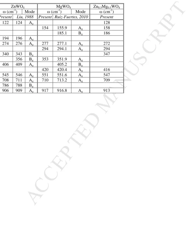
samples or low intensity of these Raman modes. The frequencies of detected Raman bands in
ZnWO4 (x = 1), MgWO4 (x = 0) and Zn0.5Mg0.5WO4 as well as their attribution to the
corresponding modes are listed in Table 1. It can be seen from Table 1 that the observed
Raman-active modes for ZnWO4 and MgWO4 are in good agreement with literature data (Liu
et al., 1988; Fomichev et al., 1994; Ruiz-Fuertez et al., 2011). The frequencies 909 (Ag), 788
(Bg), 708.5 (Ag), 677 (Bg), 409 (Ag) and 341.4 (Ag) cm-1 in ZnWO4 correspond to the
stretching vibrations of W-O atoms in the WO6 group (Liu et al., 1988; Perakis et al., 2000).
The bands with the frequencies 197 (Ag), 165 (Bg), 150 (Bg) and 125 (Ag) cm-1 are attributed
to vibrations involving zinc cations (Fomichev et al., 1994). In the case of MgWO4 the internal
modes are the Ag modes at 420, 552, 713 and 917 cm-1 and the Bg modes at 684 and 809 cm-1
(Ruiz-Fuertes et al., 2011).
A strong Raman mode at ~ 910 cm-1 is typical for wolframites and corresponds to a
M
AN
US
CR
IP
T
AC
CE
PT
ED
variation of the maximum position of this mode from 906 to 917 cm-1 depending on x value in
ZnxMg1-xWO4. The dependence of the mode maximum on x is linear (see Fig. 2, inset). The
same tendency is observed for the Raman mode at ~120 cm-1 connected with external
vibrations involving cations. The observed linear shift is caused by the linear increase of cell
parameters with the increase of x value. The intensity of Raman modes under discussion
decreases with the increase of x value in both cases. The width of the internal vibration mode
increases slightly whereas that of the external vibrations increases drastically reflecting the
gradual substitution of Zn by Mg cation. Presented data indicate that the ZnxMg1-xWO4 mixed
crystals cannot be considered as mixtures of two constituent crystals, but a new unique crystal
is formed at each x value. This statement is supported by the fact, that changes in x values
result rather in a gradual shift of the Raman mode maxima than in the redistribution of the
intensities of Raman modes, characteristic for each of constituent crystals, which could be
expected in the case of phase mixtures.
3.2. Luminescence spectroscopy data
ZnxMg1-xWO4 mixed crystals under X-ray excitation are characterized by a single
emission band at 495 nm, which is connected with the self-trapped exciton (STE) emission.
The position of the STE-related band and its decay characteristics are similar for all studied
crystals. It indicates an insignificant influence of cation electronic states on the formation of
energy bands in the vicinity of the bandgap that is confirmed by the band structure
calculations for ZnWO4 and MgWO4 (Khyzhun et al., 2013; Ruiz-Fuertes et al., 2012).
The crystal bandgap can be estimated from the excitation spectra of the STE
M
AN
US
CR
IP
T
AC
CE
PT
ED
edge. The excitation spectra for all the samples studied in the energy region 3.4-4.4 eV are
presented in Fig. 3. A gradual low-energy shift of the fundamental absorption edge with the
increase of x from 0 to 1 is observed. Actually the bandgap for MgWO4 (Eg = 5.0 eV) is
greater than for ZnWO4 (Eg = 4.5 eV) (Kolobanov et al., 2002). Therefore, this shift indicates
the linear increase of the optical bandgap with the decrease of x value, which is estimated from
the onset of excitation spectra (see. Fig.3 inset).
The temperature dependences of the STE luminescence intensity for ZnxMg1-xWO4 are
shown in Fig. 4. Emission spectra were measured under X-ray radiation in the temperature
region 80–470 K then integrated. A distinct decrease of the emission intensity above ~250 K is
connected with the temperature quenching of the luminescence. It follows from Fig. 4 that
both the stability of the STE emission and the width of the bandgap are increased in the same
sequence of mixed crystals. One can suggest that the exciton bonding energy increases along
with the bandgap width increase in the mixed crystals studied. Different thermal stability of
excitons localized at WO4 complexes has also been observed in a sequence of homologue
scheelites (Laasner et al., 2015).
Temperature dependences measured under X-ray excitation cannot be fitted by the Mott
formula. The deviations appear due to the occurrence of different processes competitive to the
STE emission, e.g. nonradiative relaxation of separated charge carriers on crystal defects,
trapping of charge carriers, crystal structure defect creation under X-ray irradiation. The latter
process is manifested in the degradation of luminescence intensity down to 80% of initial
intensity at T = 88 K in the Zn0.6Mg0.4WO4 sample under continuous X-ray irradiation during
M
AN
US
CR
IP
T
AC
CE
PT
ED
luminescence degradation is not observed. Similar effect has been observed for PbWO4 at low
temperatures and resulted in the degradation of the scintillation light yield (Burachas et al.,
2010). Taking into account that tungstates are considered for the application in cryogenic
scintillating detectors, the poor radiation hardness could become a considerable problem for
their application.
In Fig. 5, the TSL curves of ZnxMg1-xWO4 are presented. Each of the studied samples
demonstrates a set of TSL peaks. At x = 1 (ZnWO4) the intensive peak at 130 K and two weak
peaks at 170 and 230 K are observed. The peak at 230 K is generally ascribed to the emission
related to the Mo impurity contaminating the crystal, while the peak at 170 K is attributed to
the presence of Li as a satellite impurity in the crystal structure (Krutyak et al., 2013). At x = 0
(MgWO4) only one TSL peak at 170 K is observed. It should be noted that the peaks at 170 K
in zinc and magnesium tungstates have different origin in spite of the same position. Actually
throughout the set of mixed crystals the tendency for the linear shift of peak positions to
higher temperatures with the decrease of x is clearly observed. The shift indicates the increase
of trap depths and is correlated to the increase of bandgap width. In the TSL curves, we
indicate by lines two sets of TSL peaks, which demonstrate linear shifts of their positions on x
value.
The most intensive TSL peaks are characteristic for the intermediate values of x –
Zn0.7Mg0.3WO4 and Zn0.6Mg0.2WO4. It may be ascribed to the partial disorder of crystal’s
structure in the mixed crystals that results in the increase of traps concentration.
M
AN
US
CR
IP
T
AC
CE
PT
ED
A gradual shift of Raman bands following the changes of lattice parameters was observed in
Raman spectra of ZnxMg1-xWO4 mixed crystals. The studies of the excitation spectra of the
STE emission revealed the linear modification of the bandgap with x value. The TSL glow
curves demonstrate a high-temperature shift in peak positions with the increase of x value,
thus indicating the increase of trap depth and, consequently, providing additional arguments in
favor of bandgap width increase. The degradation of luminescence under X-ray irradiation
was detected at T = 80 K and ascribed to the creation of crystal structure defects. The data
provided indicate that each ZnxMg1-xWO4 mixed crystal is not a mixture of two constituents,
but possesses its original crystalline structure, as well as optical and luminescent properties.
Acknowledgement
Financial support of grants of the RFASInnovations № RFMEFI61614X0006, RFBR
15-02-07825-а, Mobilitas ESF (MTT83) and Estonian investigation council (IUT02-26) is
gratefully acknowledged.
References
Burachas, S., Ippolitov, M., Manko, V., Nikulin, S., Vasiliev, A., Apanasenko, A., Vasiliev,
A., Uzunian, A., Tamulaitis, G., (2010) Temperature dependence of radiation hardness of
lead tungstate (PWO) scintillation crystals, Rad. Meas., vol. 45, 83-88.
Fomichev, V.V., Kondratov, O.I., (1994) Vibrational spectra of compounds with the
M
AN
US
CR
IP
T
AC
CE
PT
ED
Gektin, A.V., Belsky, A.N., Vasilev, A.N., (2014) Scintillation Efficiency Improvement by
Mixed Crystal Use, IEEE Trans. Nucl. Sc. vol. 61, 262–270.
Khyzhun, O.Y., Bekenev, V.L., Atuchin, V.V., Galashov, E.N., Shlegel, V.N., (2013)
Electronic properties of ZnWO4 based on ab initio FP-LAPW band-structure calculations
and X-ray spectroscopy data, Mater. Chem. Phys. vol. 140, 588–595.
Kolobanov, V.N., Kamenskikh, I.A., Mikhailin, V.V., Shpinkov, I.N., Spassky, D.A.,
Zadneprovsky, B.I., Potkin, L.I., Zimmerer, G., (2002) Optical and luminescent properties
of anisotropictungstate crystals, Nucl. Instr. Meth. Phys. Res. A. vol. 486, 496-503.
Krutyak, N.R., Mikhailin, V.V., Vasil’ev, A.N., Spassky, D.A., Tupitsyna, I.A., Dubovik,
А.M., Galashov, E.N., Shlegel, V.N, Belsky, A.N., (2013) The features of energy transfer
to the emission centers in ZnWO4 and ZnWO4:Mo, J. of Lum., vol. 144, 105–111.
Laasner, R., Nagirnyi, V., Vielhauer, S., Kirm, M., Spassky, D., Sirutkaitis, V., Grigonis, R.,
Vasil’ev, A.N., (2015) Cation influence on exciton localization in homologue scheelites, J.
Phys. Cond. Mat. vol. 27, 385501.
Liu, Y., Wang, H., Chen, G., Zhou, Y.D., Gu, B.Y., Hu, B.Q., (1988) Analysis of Raman
spectra of ZnWO4 single crystals, J. Appl. Phys. vol. 64, 4651–4653.
Perakis, A., Sarantopoulou, E., Raptis, C., (2000) Pressure and temperature dependent Raman
study of ZnWO4, High Pres. Research, vol. 18, 181–187.
Ruiz-Fuertes, J., Errandonea, D., López-Moreno, S., González, J., Gomis, O., Vilaplana, R.,
Manjón, F.J., Muñoz, A., Rodríguez-Hernández, P., Friedrich, A., Tupitsyna, I.A.,
M
AN
US
CR
IP
T
AC
CE
PT
ED
calculations on scintillating MgWO4: Comparison with isomorphic compounds, Phys. Rev.
B, vol. 83, 214112.
Ruiz-Fuertes, J., Lopez-Moreno, S., Lopez-Solano, J., Errandonea, D., Segura, A.,
Lacomba-Perales, R., Munoz, A., Radescu, S., Rodriguez-Hernandez, P., Gospodinov, M.,
Nagornaya, L.L., Tu, C.Y., (2012) Pressure effects on the electronic and optical properties
of AWO4 wolframites (A = Cd, Mg, Mn, and Zn): The distinctive behavior of multiferroic
MnWO4, Phys. Rev. B, vol. 86, 125202.
Spassky, D., Omelkov, S., Mägi, H., Mikhailin, V., Vasil’ev, A., Krutyak, N., Tupitsyna, I.,
Dubovik, A., Yakubovskaya, A., Belsky, A., (2014) Energy transfer in solid solutions
M
AN
US
CR
IP
T
AC
CE
PT
ED
Figure captionsFig. 1 The Raman spectra of ZnxMg1-xWO4 crystals at T = 300 K.
Fig. 2 The modification of Raman bands at ~120 and 910 cm-1 with x value for ZnxMg1-xWO4
crystals. Inset: dependence of the maximum of band at ~910 cm-1 on x.
Fig. 3 Excitation spectra of STE luminescence of ZnxMg1-xWO4 crystals at λex = 500 nm, T =
300 K. Inset: dependence of bandgap width Eg on x value. Eg is estimated from the
interpolation of the onset of spectra to the abscissa axis.
Fig. 4 Temperature dependences of luminescence intensity under X-ray excitation for ZnxMg
1-xWO4 crystals. Inset: intensity degradation after 600 s of irradiation with X–rays at
different temperatures for Zn0.6Mg0.4WO4.
M
AN
US
CR
IP
T
AC
CE
PT
ED
Table 1 Vibrational frequencies of some ZnxMg1-xWO4 compositions with the wolframite
structure
ZnWO4 MgWO4 Zn0.5Mg0.5WO4
ω (cm-1) Mode ω (cm-1) Mode ω (cm-1)
Present Liu, 1988 Present Ruiz-Fuertes, 2010 Present
122 124 Ag 128 154 155.9 Ag 158 185.1 Bg 186 194 196 Ag 274 276 Ag 277 277.1 Ag 272 294 294.1 Ag 294 340 343 Bg 347 356 Bg 353 351.9 Ag 406 409 Ag 405.2 Bg 420 420.4 Ag 416 545 546 Ag 551 551.6 Ag 547 708 711 Ag 710 713.2 Ag 709 786 788 Bg 906 909 Ag 917 916.8 Ag 913