Advance Access publication 24 July 2012
Is the use of the cervical vertebrae maturation method justified
to determine skeletal age? A comparison of radiation dose of
two strategies for skeletal age estimation
Raphael Patcas*
,†, Luca Signorelli*
,†, Timo Peltomäki** and Marc Schätzle*
*Clinic for Orthodontics and Pediatric Dentistry, Center for Dental Medicine, University of Zurich, Zurich,Switzerland and **Dental and Oral Diseases Outpatient Clinic, Department of Eye, Ear and Oral Diseases, Hospital and Department of Otolaryngology, Tampere University, Tampere, Finland
† These authors contributed equally to the accomplishment of the manuscript
Correspondence to: Marc Schätzle, Clinic for Orthodontics and Pediatric Dentistry, Center for Dental Medicine,
University of Zurich, Plattenstrasse 11, Zurich, Switzerland. E-mail: marc.schaetzle@zzm.uzh.ch
SUMMARY The aim of this study was to assess effective doses of a lateral cephalogram radiograph with and
without thyroid shield and compare the differences with the radiation dose of a hand-wrist radiograph. Thermoluminescent dosimeters were placed at 19 different sites in the head and neck of a tissue-equivalent human skull (RANDO phantom). Analogue lateral cephalograms with and without thyroid shield (67 kV, 250 mA, 10 mAs) and hand-wrist radiographs (40 kV, 250 mA, 10 mAs) were obtained. The effective doses were calculated using the 2007 International Commission on Radiological Protection recommendations.
The effective dose for conventional lateral cephalogram without a thyroid shield was 5.03 microsieverts (µSv). By applying a thyroid shield to the RANDO phantom, a remarkable dose reduction of 1.73 µSv could be achieved. The effective dose of a conventional hand-wrist radiograph was calculated to be 0.16 µSv. Adding the effective dose of the hand-wrist radiograph to the effective dose of the lateral cephalogram with thyroid shield resulted in a cumulative effective dose of 3.46 µSv. Without thyroid shield, the effective dose of a lateral cephalogram was approximately 1.5-fold increased than the cumulative effective dose of a hand-wrist radiograph and a lateral cephalogram with thyroid shield.
Thyroid is an organ that is very sensitive to radiation exposure. Its shielding will significantly reduce the effective dose. An additional hand-wrist radiograph, involving no vulnerable tissues, however, causes very little radiation risk. In accordance with the ALARA (As Low As Reasonably Achievable) principle, if an evaluation of skeletal age is indicated, an additional hand-wrist radiograph seems much more justifiable than removing the thyroid shield.
Introduction
In orthodontic treatment planning, there are several rea-sons why the skeletal age of a patient should be evaluated. First, dentofacial orthopaedic appliances are a main treat-ment modality to correct mandibular deficiency by modify-ing mandibular and maxillary growth. The effectiveness of these growth modifications, however, depends on patient’s skeletal maturity stage with the optimal time for growth modification being around the pubertal growth spurt (Hägg and Taranger, 1980a, b; Baccetti et al., 2009). Second, some orthognathic surgical procedures and the insertion of dental implants should not be performed before the cessation of the individual’s growth has been clarified (Kokich, 2004;
Noble et al., 2007; Chen et al., 2010). Some authors, how-ever, have questioned the clinical value of skeletal age as-sessment for craniofacial growth (Moore, 1997).
Numerous approaches to assess skeletal maturation have been suggested: biological indicators such as the rate of body height changes (Bjork, 1963; Hunter, 1966), menarche, or voice changes (Tofani, 1972; Hägg and Taranger, 1980a, b); dental development and eruption
(Hellman, 1923; Lewis and Garn, 1960; Bjork and Helm, 1967); hand-wrist radiographs (Greulich and Pyle, 1959;
Bjork and Helm, 1967; Tofani, 1972; Hägg and Taranger, 1980b); and more recently, cervical vertebrae maturation (CVM) (O'Reilly and Yanniello, 1988; Hassel and Farman, 1995; Baccetti et al., 2002).
For decades, hand-wrist radiographs have been used routinely in orthodontics to determine the peak of growth spurt (Hägg and Taranger, 1980a). However, concern has been voiced over the extra radiation exposure caused by an additional radiograph. Since the introduction of the assess-ment of skeletal age by means of the CVM method on a lateral cephalograph, it seems therefore questionable if an additional hand-wrist radiograph should be taken for the sole purpose of skeletal age evaluation. In accordance to this apprehension, the British Orthodontic Society stated in its guidelines of 2008 that hand-wrist radiographs are no longer indicated to predict the onset of the pubertal growth spurt (Isaacson et al., 2008).
The rationale behind this guideline is obvious. A lateral cephalograph is routinely required for orthodontic diagnosis
and treatment planning. By using the CVM method, no ad-ditional radiograph is needed. Since radiation risks are cumu-lative, it is imperative that strategies for dose reduction, in-cluding the amount of radiographs taken, be considered. And although the amount of radiation utilized in dentistry is typi-cally fairly low (White, 1992), acceptable radiological policies and practices are based on the assumption that some risk does exist, and this risk must be clearly outweighed by benefits, i.e. the quantity and quality of needed diagnostic information.
Another option to reduce radiation exposure is the use of a thyroid shield. This shield, however, would forfeit the pos-sibility to determine the skeletal age with the CVM method and would render a hand-wrist radiograph necessary in cas-es were skeletal maturation has to be asscas-essed.
The aim of this study was therefore to examine the radio-biological risk of a lateral cephalogram with and without a thyroid shield and of a hand-wrist radiograph by means of phantom dosimetry. Based on the obtained average absorbed doses, the effective doses are calculated to estimate the poten-tial risk of the radiographs and to enable a comparison.
Material and methods
Thermoluminescent dosimeter (TLd) chips (3 mm × 1 mm × 1 mm) were used on selected locations in the head and neck region of an adult male tissue-equivalent phantom (RANdO—radiation analog dosimetry system; The Phan-tom Laboratory, Salem, NY, USA) to record the distribution of the absorbed radiation dose. The 19 sites measured in this study are listed in Table 1. These locations reflect critical organs known to be sensitive to radiation. For the assess-ment of the effective dose of hand-wrist radiographs, a TLd was exposed directly in the respective distance to the ray tube. The TLd chips were supplied by the Institute of Ap-plied Radiophysics (IAR.) from the University of Lausanne, Switzerland. The exposed dosimeters were analyzed by the IAR in a blinded fashion. One unexposed dosimeter served as control for environmental radiation.
The radiological examinations were performed on a cus-tom-made X-ray unit (COMeT, 3175 Flamatt, Switzerland) with the following parameters for the lateral cephalogram: 67 kV tube voltage, 250 mA tube current, 0.04 second ex-posure time, and 10 mAs tube current time product and for the hand-wrist radiograph: 40 kV tube voltage, 250 mA tube current, 0.04 second exposure time, and 10 mAs tube cur-rent time product. These parameters correspond to the clini-cal exposure parameters commonly used. For the lateral cephalogram with a thyroid shield, a commercially available thyroid shield (3534-TS, WIROMA, 3145 Niederscherli, Switzerland) with 0.5 mm Pb was placed on the phantom. No intensifying screens were used.
Because of the relatively small amount of radiation re-quired for a single examination, multiple exposures for each radiographic technique were performed to provide reliable measure of radiation in the dosimeters. Ten exposures were made to provide more reliable data and the mean value was used for further calculations. doses from TLds at differ-ent positions within a tissue or organ were averaged to ex-press the average tissue-absorbed dose in micrograys (µGy). These values were used to calculate the equivalent dose HT using the following equation:
HT=
∑
W DR T.The equivalent dose HT for a tissue or organ is defined as the product of the radiation weighting factor WR (WR equals 1 for x-radiation) and the measured absorbed dose DT aver-aged over a particular tissue or organ (Valentin, 2007).
effective dose E has been recommended by the Interna-tional Commission on Radiological Protection (ICRP) as a means of comparing detriment of different exposures to ionizing radiation to an equivalent detriment produced by a full-body dose of radiation. Thus, the risk to the whole body is determined as the summation of the equivalent doses es-tablished for all tissues and organs (Valentin, 2007). The ef-fective dose (EICRP60), expressed in microsieverts (µSv), was calculated using the equation
E=∑W HT T,
where E is the product of the tissue weighting factor WT, which represents the relative contribution of that organ or tissue to the overall risk, and the equivalent dose HT. The weighting factors of the equivalent doses in accordance with the ICRP guidelines of 2007 for the lateral cephalo-gram and for the hand-wrist radiograph are given in Table 2a and 2b, respectively.
Results
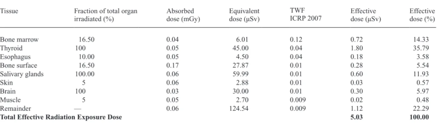
The absorbed doses, equivalent doses, and effective doses are given in Table 3 for lateral cephalogram without a thy-roid shield and in Table 4 for lateral cephalogram with a
Table 1 Locations of thermoluminescent dosimeter (TLd) chips on the RANdO phantom.
Organ Location TLd number Phantom level Brain Anterior/posterior 18, 19 1
Right/left 16, 17 2
Hypophysis 13 3
eyes Right/left lens 14, 15 3
Skull Right/left maxillary sinuses 9, 10 5 Salivary glands Right/left parotid 11, 12 5 Right/left submandibular gland 7, 8 6 Sublingual gland 5 Thyroid Right/left 1,2 9 Spine B2 6 6 Right/left 3, 4 7
Table 3 Radiation exposure: lateral cephalogram without thyroid shield.
Tissue Fraction of total organ
irradiated (%) Absorbed dose (mGy) equivalent dose (µSv)
TWF
ICRP 2007 effective dose (µSv) effective dose (%)
Bone marrow 16.50 0.04 6.01 0.12 0.72 14.33 Thyroid 100 0.05 45.00 0.04 1.80 35.79 esophagus 10.00 0.05 4.50 0.04 0.18 3.58 Bone surface 16.50 0.17 27.87 0.01 0.28 5.54 Salivary glands 100.00 0.06 59.99 0.01 0.60 11.93 Skin 5 0.06 2.88 0.01 0.03 0.57 Brain 100 0.03 30.00 0.01 0.30 5.97 Muscle 5 0.05 2.70 0.009 0.02 0.48 Remainder — 0.06 124.54 0.009 1.12 22.29
Total Effective Radiation Exposure Dose 5.03 100.00
TWF ICRP 2007: Tissue weighting factor according to the International Commission on Radiological Protection.
Table 2b Weighting of the equivalent dose (HT) for hand-wrist radiation exposure.
Tissue ICRP-identified organs
Fraction of total organ irradiated (%)
Corresponding TLd
numbers Fraction irradiated (%)
Weighting Weighting in %
Bone marrow Bone 0.5 — 0.5 0.06 58.71
Bone surface Bone 0.5 — 0.5 0.02 22.70
Skin Skin 1.0 — 1 0.01 9.78
Muscle Muscle 1.0 — 1 0.01 8.81
Remainder — —
Table 2a Weighting of the equivalent dose (HT) for lateral cephalometric radiation exposure.
Tissue ICRP-identified
organ Fraction of total organ irradiated (%) Corresponding TLd numbers Fraction irradiated (%) Weighting
Weighting in %
Bone marrow 16.5 1.98 17.86
Mandibula 1.30 Mean 7, 8
Calvarium 11.80 Mean 16, 17, 18, 19 Cervical spine 3.40 Mean 3, 4, 6
esophagus esophagus 10.00 Mean 1, 2 10.0 4.00 36.08
Thyroid Thyroid 100.00 Mean 1, 2 100.0 0.40 3.61
Bone surface 16.5 0.77 6.91
Mandible 1.30 4.64 Mean 7, 8
Calvarium 11.80 4.64 Mean 16, 17, 18, 19 Cervical spine 3.40 4.64 mean 3, 4, 6
Brain Brain 100.00 Mean 13, 16, 17, 18, 19 100.0 1.00 9.02
Salivary glands 100.0 0.05 0.45
Parotid 33.00 Mean 11, 12
Submandibular 33.00 Mean 7, 8
Sublingual 33.00 5
Skin Skin 5.0 Mean 11, 12, 14, 15 5.0 1.00 9.02
Muscle Muscle 5.0 Mean 1–8, 11–13 5.0 0.05 0.41
Remainder 1.85 16.64
Lymphatic nodes 5.0 Mean 1–8, 11–12 extrathoracic airway 100.00 Mean 1–8, 11–14 Oral mucosa 100.00 Mean 1–8, 11–15
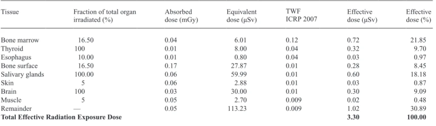
thyroid shield, respectively. Analogously, these doses for the hand-wrist radiograph are listed in Table 5.
It is noteworthy that the thyroid is the most vulnerable organ followed by the salivary glands. The use of a thyroid shield decreased the effective dose of the thyroid considerably (from 1.8 to 0.32 µSv).
The overall effective dose for conventional lateral cepha-lograms without a thyroid shield was 5.03 µSv. By applying a thyroid shield to the RANdO phantom, a remarkable dose reduction could be attained, resulting in an effective dose of 3.30 µSv. This equals a reduction of approximately 34 per cent. The effective dose for a conventional hand-wrist radio-graph was calculated to be 0.16 µSv. Adding the effective dose of the hand-wrist radiograph to the effective dose of the lateral cephalogram with thyroid shield results in a cu-mulative effective dose of 3.46 µSv. This equals a reduction of approximately 31 per cent over the effective dose of a lateral cephalogram without thyroid shield. Hence, without thyroid shield, the effective dose of a lateral cephalogram was approximately 1.5-fold increased than the cumulative
effective dose of a hand-wrist radiograph and a lateral ceph-alogram with thyroid shield.
Discussion
In the last decade, the radiological depiction of the cervi-cal spine area has been the subject of increasing interest in orthodontics. Scientific contributions have shown that di-agnostic data can be obtained from the cervical spine on a lateral cephalogram. In addition to skeletal age evaluation, it is possible to assess the natural head position by using the cervical spine as the reference structure (Kylamarkula and Huggare, 1985). Furthermore, cervical vertebrae anomalies (CVA) such as fusions have been associated with craniofa-cial syndromes, sleep apnea, and dentoskeletal malocclu-sions (Sonnesen, 2010).
However, some of these potential benefits have been severely questioned. Natural head position can be as-sessed clinically without the help of a radiograph. In fact, many perform the lateral cephalogram with the head fixed
Table 5 Radiation exposure: hand-wrist radiograph.
Tissue Fraction of total organ irradiated (%) Absorbed dose (mGy) equivalent dose (µSv) TWF
ICRP 2007 effective dose (µSv)
effective dose (%) Bone marrow 0.50 0.16 0.80 0.12 0.096 58.71 Thyroid 0 — — — — — esophagus 0 — — — — — Bone surface 0.50 0.16 3.71 0.01 0.037 22.70 Salivary glands 0 — — — — — Skin 1 0.16 4.75 0.01 0.016 9.78 Brain 0 — — — — — Muscle 1 0.16 1.60 0.009 0.014 8.81 Remainder 0
Total Effective Radiation Exposure Dose 0.164 100.00
TWF ICRP 2007: Tissue weighting factor according to International Commission on Radiological Protection.
Table 4 Radiation exposure: lateral cephalogram with thyroid shield.
Tissue Fraction of total organ irradiated (%) Absorbed dose (mGy) equivalent dose (µSv) TWF
ICRP 2007 effective dose (µSv)
effective dose (%) Bone marrow 16.50 0.04 6.01 0.12 0.72 21.85 Thyroid 100 0.01 8.00 0.04 0.32 9.70 esophagus 10.00 0.01 0.80 0.04 0.03 0.97 Bone surface 16.50 0.17 27.87 0.01 0.28 8.45 Salivary glands 100.00 0.06 59.99 0.01 0.60 18.18 Skin 5 0.06 2.88 0.01 0.03 0.87 Brain 100 0.03 30.00 0.01 0.30 9.09 Muscle 5 0.05 2.70 0.009 0.02 0.48 Remainder — 0.05 113.23 0.009 1.02 30.89
Total Effective Radiation Exposure Dose 3.30 100.00
parallel to the Frankfort plane and not in natural head posture. Moreover, it has been argued that the oblique facets of the joint render lateral cephalograms entirely inappropriate to evaluate fusions and that Cone Beam CT (CBCT) remains the gold standard for assessing CVA (Koletsis and Halazonetis, 2010; Bebnowski et al., 2012).
All of this reduces the benefit of depicting the cervical area to determine skeletal age. Yet, it has been reported that the exact evaluation of the skeletal age based on the cervical spine is at best very difficult. According to recent studies, the reproducibility of evaluating the skeletal age based on the spine is disappointingly low (Gabriel et al., 2009; Nest-man et al., 2011; Zhao et al., 2012). All of this implies that the benefit of exposing the cervical spine to radiation—and with it the thyroid—is very limited.
In contrast to the CVM method, bone age assessment based on hand-wrist radiography shows good reproducibility (King et al., 1994) and a high reliability (Flores-Mir et
al., 2004). A further strength of a hand-wrist radiograph is that the cumulative results of different methods such as Greulich and Pyle (GP), Tanner and Whitehouse (TW), and Bowden or Fishman can be used to assess the skeletal age (Flores-Mir et al., 2004). For endocrinologists, the concordant result of GP together with TW still remains the gold standard with TW being the method of choice (Gilli, 1996), whereas certain sources of error (such as poor positioning of the hand or inter- and intra-observer reliability) undeniably affect the accuracy of hand-wrist radiography as well (Cox, 1996).
The thyroid is an organ that is highly sensitive to radiation exposure. Our study reveals that a thyroid shield will reduce the effective dose remarkably. In light of the questionable benefits of radiological exposure of this sensitive area, our findings give strong support to the use of a thyroid shield. Alternatively, the beam could be collimated to exclude the thyroid and hence reduce the effective dose. Whatever the personal approach might be to reduce the radiation of the thyroid, should a clinical indication to assess the skeletal age arise, an additional hand-wrist radiograph is to be rec-ommended, even if extra costs of an additional radiography have to be taken into account.
A possible limitation of our study is that different radiographical equipment may generate slightly different results, although a comparison with other studies seems to suggest that the differences are very small (Ludlow et al., 2008). Furthermore, the image acquisition was performed with analogue radiographs and not with digital imaging generating less radiation when a digital system based on a charged-coupled device (CCd) is used. Yet digital image acquisition would reduce all absolute values of the effective doses but will leave the ratios almost unaffected. In fact, absolute values are not very useful for comparative research as they depend on device, protocol, and film sensitivity.
Visser et al. (2001) demonstrated that even analogue cephalometry can generate diverse doses. This is why our
study describes the differences achieved in dose reduction as percentages and as ratios.
Hassel and Farman (1995) have claimed that C3 and also C4 can be visualized even when a thyroid shield is worn. This assertion is based on the fact that in adulthood, the topograph-ical location of the thyroid corresponds to C5–C7. However, it is important to realize that the location of the thyroid is age dependent as the thyroid experiences a caudal movement through puberty (Crelin, 1973). Also, it should be appreci-ated that the thyroid position reportedly highly varies from person to person (Gray et al., 2005). All this implies that the thyroid shield should cover the cervical spine above C5 as well, especially in pre-pubertal children and would hence, if applied correctly, forfeit the CVM method. In post-pubertal subjects, however, the thyroid would most of the time be low-er than C4 and, theoretically, the use of a neck shield would be commendable. In practical setting, it is however very chal-lenging to determine clinically by visual inspection and neck palpation the correct location of C5. A recent retrospective survey also demonstrated that the thyroid shields are used in-consistently and if applied, C3 and C4 were entirely depicted in only 14% of all subjects (Hujoel et al., 2006).
exposure to natural radiation sources is more significant for the world’s population than most exposures to medical radiation sources (UNSCeAR, 2000). The average world-wide exposure to environmental radiation sources of about 2.4 mSv per year would seem to indicate that the described dose reduction achieved with the thyroid shield to be negli-gible. However, it must be stated that the average exposure probably does not pertain to any one individual, since there are wide distributions of exposures from each source and the exposures combine in various ways at each location, de-pending on the specific concentrations of radionuclides in the environment and in the body, the latitude and altitude of the location, and many other factors (UNSCeAR, 2000). And on the other hand, scientific evidence has recently been provided that exposure to routine dental X-rays appears to be associated with an increased risk of intracranial menin-gioma (Claus et al., 2012). Thus, unless proven differently, the task to reduce the ionizing risk of medical radiation will remain on dentists.
It is indeed a perplexing conclusion that an additional X-ray means less radiation exposure (provided the thyroid shield is applied). As mentioned, the British Orthodontic Society considers the hand-wrist radiograph to be obsolete, an opinion echoed by many others (eU, 2004; Turpin, 2008;
Chen et al., 2010; Litsas and Ari-demirkaya, 2010). But our findings strongly corroborate that the conclusions of the British Orthodontic Society ought to be reconsidered and the use of a thyroid shield to be enforced.
In summary, this study demonstrates that, based on the overriding ALARA (As Low As Reasonably Achievable) principle, the assessment of skeletal maturation of cervical vertebrae on a lateral cephalogram is to be questioned and the use of a thyroid shield is strongly to be advocated. If
an evaluation of skeletal age is deemed necessary, an ad-ditional hand-wrist radiograph seems much more justifiable than removing the thyroid shield, which would cause highly vulnerable tissue to be exposed to direct radiation.
References
Baccetti T, Franchi L, Kim L H 2009 effect of timing on the outcomes of 1-phase nonextraction therapy of Class II malocclusion. American Jour-nal of Orthodontics and dentofacial Orthopedics 136: 501–509 Baccetti T, Franchi L, McNamara J A, Jr. 2002 An improved version of
the cervical vertebral maturation (CVM) method for the assessment of mandibular growth. The Angle Orthodontist 72: 316–323
Bebnowski d, Hanggi M P, Markic G, Roos M, Peltomaki T 2012 Cervical vertebrae anomalies in subjects with Class II malocclusion assessed by lateral cephalogram and cone beam computed tomography. european Journal of Orthodontics 34: 226–231.
Bjork A 1963 Variations in the growth pattern of the human mandible: lon-gitudinal radiographic study by the implant method. Journal of dental Research 42 Pt 2: 400–411
Bjork A, Helm S 1967 Prediction of the age of maximum puberal growth in body height. The Angle Orthodontist 37: 134–143
Chen L, Liu J, Xu T, Long X, Lin J 2010 Quantitative skeletal evaluation based on cervical vertebral maturation: a longitudinal study of adoles-cents with normal occlusion. International Journal of Oral and Maxil-lofacial Surgery 39: 653–659
Claus e B, Calvocoressi L, Bondy M L, Schildkraut J M, Wiemels J L, Wrensch M 2012 dental x-rays and risk of meningioma. Cancer doi:10.1002/cncr.26625. [epub ahead of print]
Cox L A 1996 Tanner-Whitehouse method of assessing skeletal maturity: problems and common errors. Hormone Research 45 Suppl 2: 53–55 Crelin e S 1973 Functional anatomy of the newborned. Yale University
Press, New Haven
eU 2004 european guidelines on radiation protection in dental radiologyed. Office for Official Publications of the european Communities, Luxembourg Flores-Mir C, Nebbe B, Major P W 2004 Use of skeletal maturation based
on hand-wrist radiographic analysis as a predictor of facial growth: a systematic review. The Angle Orthodontics 74: 118–124
Gabriel d B, Southard K A, Qian F, Marshall S d, Franciscus R G, Sou-thard T e 2009 Cervical vertebrae maturation method: poor reproduc-ibility. American Journal of Orthodontics and dentofacial Orthopedics 136: 478. e471–e477; discussion 478–480
Gilli G 1996 The assessment of skeletal maturation. Hormone Research 45 Suppl 2: 49–52
Gray H, Standring S, ellis H, Collins P, Wigley C, Berkovitz B K B 2005 Gray's anatomy: the anatomical basis of clinical practice. 39th edn. el-sevier Churchill Livingstone, New York
Greulich W W, Pyle S I 1959 Radiographic atlas of skeletal development of the hand and wrist. Stanford University Press, Stanford
Hägg U, Taranger J 1980a Menarche and voice change as indicators of the pubertal growth spurt. Acta Odontologica Scandinavica 38: 179–186 Hägg U, Taranger J 1980b Skeletal stages of the hand and wrist as indicators
of the pubertal growth spurt. Acta Odontologica Scandinavica 38: 187–200 Hassel B, Farman A G 1995 Skeletal maturation evaluation using cervical
vertebrae. American Journal of Orthodontics and dentofacial Orthope-dics 107: 58–66
Hellman M 1923 The process of dentition and its effects on occlusion. dental Cosmos 65: 1329–1344
Hujoel P et al. 2006 Thyroid shields and neck exposures in cephalometric radiography. BMC Medical Imaging 6: 6
Hunter C J 1966 The correlation of facial growth with body height and skeletal maturation at adolescence. The Angle Orthodontist 36: 44–54 Isaacson K G, Thom A R, Horner K, Whaites e 2008 Orthodontic
radio-graphs—guidelines for the use of radiographs in clinical orthodontics. 3rd edn. British Orthodontic Society, London
King d G et al. 1994 Reproducibility of bone ages when performed by ra-diology registrars: an audit of Tanner and Whitehouse II versus Greulich and Pyle methods. British Journal of Radiology 67: 848–851
Kokich V G 2004 Maxillary lateral incisor implants: planning with the aid of orthodontics. Journal of Oral and Maxillofacial Surgery 62: 48–56 Koletsis d d, Halazonetis d J 2010 Cervical vertebrae anomalies in
ortho-dontic patients: a growth-based superimpositional approach. european Journal of Orthodontics 32: 36–42
Kylamarkula S, Huggare J 1985 Head posture and the morphology of the first cervical vertebra. european Journal of Orthodontics 7: 151–156 Lewis A B, Garn S M 1960 The relationship between tooth formation and
other maturational factors. The Angle Orthodontist 30: 70–77 Litsas G, Ari-demirkaya A 2010 Growth indicators in orthodontic
pa-tients. Part 1: comparison of cervical vertebral maturation and hand-wrist skeletal maturation. european Journal of Paediatric dentistry 11: 171–175
Ludlow J B, davies-Ludlow L e, White S C 2008 Patient risk related to common dental radiographic examinations: the impact of 2007 Inter-national Commission on Radiological Protection recommendations re-garding dose calculation. Journal of the American dental Association 139: 1237–1243
Moore R N 1997 Principles of dentofacial orthopedics. Seminars in Ortho-dontics 3: 212–221
Nestman T S, Marshall S d, Qian F, Holton N, Franciscus R G, Southard T e 2011 Cervical vertebrae maturation method morphologic criteria: poor reproducibility. American Journal of Orthodontics and dentofacial Orthopedics 140: 182–188
Noble J, Karaiskos N, Wiltshire W A 2007 diagnosis and clinical manage-ment of patients with skeletal Class III dysplasia. General dentistry 55: 543–547
O'Reilly M T, Yanniello G J 1988 Mandibular growth changes and matura-tion of cervical vertebrae—a longitudinal cephalometric study. The An-gle Orthodontist 58: 179–184.
Sonnesen L 2010 Associations between the Cervical Vertebral Column and Craniofacial Morphology. International Journal of dentistry 2010: 295728
Tofani M I 1972 Mandibular growth at puberty. American Journal of Or-thodontics 62: 176–195
Turpin d L 2008 British Orthodontic Society revises guidelines for clinical radiography. American Journal of Orthodontics and dentofacial Ortho-pedics 134: 597–598
UNSCeAR 2000 Annex B: exposures from natural radiation sources. In: United Nations Scientific Committee on the effects of Atomic Radia-tion Report to the General Assembly, with scientific annexes, United Nations, Vienna, Austria, pp. 111–112.
Valentin J 2007 Managing patient dose in multi-detector computed to-mography (MdCT). ICRP Publication 102. Annals of the ICRP 37: 1–79, iii
Visser H, Rödig T, Hermann K-P 2001 dose reduction by direct-digital cephalometric radiography. The Angle Orthodontist 71: 159–163 White S C 1992 1992 assessment of radiation risk from dental radiography.
dentomaxillofacial Radiology 21: 118–126
Zhao X G, Lin J, Jiang J H, Wang Q, Ng S H 2012 Validity and reliability of a method for assessment of cervical vertebral maturation. The Angle Orthodontist 82: 229–234