Protein Engineering vol.8 no.l pp.81-89, 1995
Engineered turns of a recombinant antibody improve its in vivo
folding
Achim Knappik1 and Andreas PlUckthun2
Biochemisches Institut, Universitat Zurich, Winterthurerstrasse 190, CH-8057 Zurich, Switzerland
'Present address: Morphosys GmbH, Frankfurter Ring 193a, D-80807 MUnchen, Germany
2To whom correspondence should be addressed
Using recombinant antibodies functionally expressed by secretion to the periplasm in Escherichia coli as a model system, we identified mutations located in turns of the protein which reduce the formation of aggregates during in vivo folding or which influence cell stability during expression. Unexpectedly, the two effects are based on different mutations and could be separated, but both mutations act synergistically in vivo. Neither mutation increases the thermodynamic stability in vitro. However, the in vivo folding mutation correlates with the yield of oxidative folding in vitro, which is limited by the side reaction of aggregation. The in vivo folding data also correlate with the rate and activation entropy of thermally induced aggregation. This analysis shows that it is possible to engineer improved frameworks for semi-synthetic anti-body libraries which may be important in maintaining library diversity. Moreover, limitations in recombinant protein expression can be overcome by single amino acid substitutions.
Key words: E.colifF(ab) and Fv fragments/protein engineering/
protein expression/protein folding
Introduction
Folding in vivo can be the limiting process in the production of many recombinant proteins. It has been shown that aggregation during in vivo and in vitro folding is a specific off-pathway process involving folding intermediates (Jaenicke, 1987; Wetzel, 1994). For a few proteins it has been possible to isolate point mutations which induce or suppress aggregation (Tsai et al, 1991; Jappelli et al, 1992; Mitraki and King, 1992; Chrunyk and Wetzel, 1993; Chrunyk et al., 1993; Dale
et al, 1994), and the solution of this problem may offer the
possibility of improving production of industrially important proteins (Mitraki and King, 1992). We undertook an investi-gation of the molecular basis of this limitation using recombin-ant recombin-antibody fragments secreted into the periplasmic space of
Escherichia coli as a model system. This class of structurally
well characterized p"-barrel proteins offers an almost unlimited database of proteins which have similar sequence and structure but have been found to fold in E.coli in rather different proportions.
The efficient expression of antibody fragments in bacteria is clearly of great technological importance. The routinely achievable amounts of recombinantly expressed antibody will be decisive for the viability of this technology for many medical and technological applications. In this paper we show that there are two principal factors which are limiting the
expression yields. The first can be directly related to folding problems of the antibody fragments in the periplasmic space of E.coli, and the second is a consequence of the stress imposed by the antibody on the bacterial cell secreting it. In its mildest form it leads to periplasmic leakiness and then to lysis with the occasionally observed plasmid loss; in severe cases it leads to plasmid rearrangements and loss of clones from libraries. Since such problems are not universally observed for high-level secretion of proteins in E.coli, it follows that they are a consequence of the antibody sequence and structure. Here we describe experiments that have helped us to analyze and solve some of these problems.
While many expression experiments of antibody fragments in bacteria have now been reported (Pluckthun, 1994), a comparison of functional yields is made essentially impossible by the use of many different vector systems, strains, experi-mental conditions and types of antibody fragment. Moreover, results from high cell density experiments are occasionally directly compared with shake-flask experiments; however, this is not useful for drawing conclusions about the components of the expression system. Here we analyze the expression of different antibody fragments under identical conditions and show that the single most important factor is the primary sequence of the antibody variable domains. We then show that the crucial features within the sequence can be elucidated and that it is possible to engineer an antibody of mediocre expres-sion behavior into one of superior properties without interfering with its binding properties.
The basis for our study was the conclusion derived from previous work that it is the periplasmic folding process itself which limits the whole expression process (Skerra and PlUckthun, 1991; Knappik et al, 1993). This suggestion was based on the observations that (i) substantial amounts of periplasmic, correctly processed, albeit insoluble, protein were formed and (ii) increased overproduction of total protein was easily achieved but led to only very modest increases in soluble processed protein. Only small amounts of soluble precursor could be detected when a strong promoter was used. It is likely that the precursor would be stable enough to observe it if it were present in large amounts, since it has been seen in certain mutants (Glockshuber et al, 1992). The fact that high-level production leads to the formation of insoluble protein is also observed with many other proteins (Shatzman, 1990), and the high achievable amounts of total protein show that the levels, production rates and degradation rates of plasmid, mRNA and protein are not the primary culprits of low yields of functional protein, although they may of course be contributing factors. Furthermore, the observation of processed, insoluble protein suggests that the protein does not have a problem in transport through the membrane, since it has apparently come into contact with the signal peptidase on the periplasmic face of the inner membrane. Taken together, it appears that a side reaction of folding, namely aggregation, limits the yield in vivo. In this paper we show that the
the antibody mutants constructed and examined do correlate directly with the soluble expression yields in vivo.
In previous work we have also investigated the effect of external catalysis on the antibody fragments folding in the periplasm (Knappik et al., 1993). While the antibody fragments absolutely require the E.coli disulfide-forming protein DsbA
in vivo, its overexpression does not increase the yield for any
fragment tested. This suggests that DsbA plays an active role in the folding process but is not limiting under the conditions examined. Similarly, the overexpression of proline-cis-trans-isomerase (PPIase) had only a marginal effect on the yield of one of the fragments. The aggregation processes appear, therefore, to occur before the action of these proteins, or at least to be independent of their action.
Periplasmic folding, and in particular the question of peri-plasmic chaperones, have been reviewed recently (Wtllfing and PlUckthun, 1994a). While indirect evidence for the existence of a periplasmic chaperone was obtained (Wiilfing and PlUckthun, 1994b), the efficiency of this system is still unclear. It appears that the most effective strategy to obtain high-yield folding of periplasmic proteins is to engineer the protein itself. Here we propose that the crucial determinants are loops in the structure, at least in P-barrel proteins.
Materials and methods Materials and strains
For all cloning and expression experiments the E.coli K12 strain JM83 [ara (lac-pro AB) rpsL (strA) thi ((J)80 lacZAM 15), Yanisch-Perron et al., 1985] was used. The plasmid pAK19 (Carter et al., 1992) was obtained from P.Carter (Genentech, South San Francisco, CA).
Construction of plasmids
All in vivo experiments were carried out in the pHJ series of vectors (Knappik et al., 1993; Knappik and PlUckthun, 1994). They were constructed from our previous vectors (Skerra and PlUckthun, 1991; Knappik et al., 1993) and contain unique restriction sites at the 5' and 3' ends of the antibody genes, facilitating subcloning of antibody genes and domain shuffling between the F(ab)—, Fv- and single-chain Fv- vectors (Knappik and PlUckthun, 1994; A.Knappik, unpublished data). Addition-ally, both genes for the heavy and the light chains carry short versions of the FLAG™ tag (Prickett et al., 1989; Knappik and PlUckthun, 1994) attached to the N-terminus of the mature protein to allow detection of the chains in immunoblots, regardless of the particular antibody fragment expressed. The genes of the huMab4D5 antibody (Carter et al., 1992) were inserted in the pHJ vectors by PCR amplifying the coding regions of the variable and the constant domains independently using the vector pAK19 as a template.
The mutant antibody fragments H1-H10 were constructed by site-directed mutagenesis (Kunkel et al., 1987) using the vector pLisc_SE (Knappik et al., 1993) as a single-stranded template and up to six oligonucleotides per reaction. After screening and sequencing, the mutated part of the gene was gel-purified and ligated into the respective vector. Mutant HI 1 was constructed by combining the mutations HI and H3; mutant HI2 was constructed by removing the P40A mutation in mutant H10. Complementarity-determining region (CDR) grafting of the CDRs of McPC603 onto the huMab4D5 framework was carried out by the recursive PCR gene synthesis protocol described by Prodromou and Pearl (1992). All con-structs were verified by DNA sequencing.
Monitoring cell stability during expression
Growth curves and leakiness data were obtained as follows. 20 ml of LB medium containing 100 u.g/ml ampicillin and 25 (ig/ml streptomycin were inoculated with an E.coli JM83 overnight culture harboring the plasmid encoding the respective antibody fragment and incubated at room temperature (24°C) until an OD550 of 0.5 was reached. IPTG was added to a final
concentration of 1 raM and the incubation was continued for several hours. Every 30-60 min after induction the OD550 was
measured. At the same time, 100 |il of the culture were removed and the cells pelleted by centrifugation (4 min at 6000 r.p.m.). In the supernatant, the P-lactamase activity was measured (O'Callaghan et al., 1972) to quantify the degree of cell leakiness. After 3 h of induction, an aliquot of the culture was removed and the cells were lysed (see below). The p-lactamase activity in the cell extract was measured to verify that differences in the P-lactamase activity in the medium were not due to differences in the total amount of p-lactamase. The total amount of P-lactamase was found to deviate by —20% between the clones in repeated experiments.
Cell lysis and immunoblotting
Cell cultures harboring the plasmids encoding the respective antibody Fv fragments were grown and induced (see above). After 3 h of induction, 2-5 ml of the cultures, depending on the OD550, were harvested by centrifugation and lysed using
a modified protocol given by Wetzel et al. (1991). The cell pellets (normalized to the same OD550) were frozen for at least
1 h at -20°C and lysed by the addition of 500 |il lysis buffer (10 mM glycylglycine, 1 mM EDTA, 2 M urea, pH 7.5) to the frozen cells, and incubated on ice until thawed (20 min). 5 (xl hen-egg lysozyme (250 u,g) were added and the suspension was incubated for 10 min at room temperature. After adding 20 ^1 of 100 mM MgCl2 and 10 u.1 of DNase I (1 mg/ml) and
a further incubation of 10 min at room temperature, the lysates were centrifuged (5 min at 14 000 r.p.m.) and the supernatants containing the soluble fractions were withdrawn. The pellets were shaken at 4°C in 535 \x\ lysis buffer until resuspended to give the insoluble fractions. This procedure was chosen because it gave the same distribution of soluble and insoluble antibody fragments as lysing the cells with a French press, which was used for preparative antibody expression. The P~ lactamase activity was measured in all fractions. The fractions were assayed by reducing SDS-PAGE with the samples normalized to the same p-lactamase activity to account for differences in plasmid copy number as well as cell leakiness. After blotting and immunostaining the gel using the anti-FLAG antibody Ml (Prickett et al., 1989) as the first antibody, an Fc-specific anti-mouse antiserum conjugated to horseradish peroxidase as second antibody and a chemoluminescent detec-tion assay as described elsewhere (Ge et al., 1994), the lanes were scanned and quantitated by laser densitometry. All experiments were carried out at least in duplicate.
Thermodynamic stability
Fv fragments were purified using phosphorylcholine-Sepharose affinity chromatography (Skerra and PlUckthun, 1988). The concentration was estimated photometrically using an extinction coefficient calculated according to Gill and von Hippel (1989). Equilibrium denaturation curves were obtained by denaturing 0.1 |iM protein in borate buffered saline/phosphorylcholine (BBS/PC) buffer (200 mM sodium tetraborate, pH 8.0, 160 mM NaCl, containing 5 mM phos-phorylcholine) and increasing amounts of urea (0.0-6.8 M
Engineered turns of a recomblnant antibody
urea in 36 steps) in a total volume of 1.8 ml. After incubating the samples for 15 h at 10°C, the fluorescence spectra were recorded at 10°C from 300 to 400 nm with an excitation wavelength of 280 nm. The emission wavelength of the fluorescence peak shifted from 331.4 to 348.3 nm during denaturation (maximal deviation in repeated measurements 0.3 nm) and was used as the dependent variable. The denatura-tion was found to be reversible (data not shown). The data were fitted according to Pace (1990), assuming a two-state equilibrium system, since the fluorescence of the Fv fragment of McPC603 is dominated largely by VH (>90%), which
contains four Trp and nine Tyr, compared with one Tip and five Tyr in VL (Glockshuber et al., 1992). The 'fraction
unfolded' was calculated from the fit values using the equation:
apparent rate constant using the equation:
F =
was calculated by fitting the data
Fv = [yf + (m{ • [D]) - y]/[(yf + mf • [D]) - (yu [D])],
where yf is the y intercept for the native conformation, ya is
the y intercept for the unfolded conformation, ntf is the slope of the pre-transition baseline, mu is the slope of the
post-transition baseline and [D] is the denaturant concentration. The parameters were obtained by fitting the data according to Pace (1990).
Renaturation kinetics
Purified Fv fragments were denatured for at least 3 h at 10°C in either BBS/PC (200 mM sodium tetraborate, 160 mM NaCl, 5 mM phosphorylcholine, pH 8.0) containing 7 M urea (denaturation under oxidized conditions) or BBS/PC containing 7 M urea and 100 mM dithioerythreitol (denaturation under reducing conditions). The denatured protein was diluted 90-fold in a cuvette containing re90-folding buffer (BBS/PC for refolding of the oxidized protein or BBS/PC containing 5 mM oxidized glutathione for refolding of the reduced protein), leading to a final protein concentration of 0.11 |iM. During refolding, the solution was maintained at 10°C and mixed with a magnetic stirrer. Refolding was monitored for 3 h by scanning the fluorescence every 90 s from 300 to 400 nm (excitation wavelength: 280 nm). The data were analyzed by two methods: (i) the peak-shift from 348.3 to 331.4 nm during refolding was calculated by the estimation of the maximum emission wavelength of every scan. These data indicate the time-course of folding into a 'native-like' structure, where 'native-like' is defined by the property of having an emission maximum of -331.4 nm; and (ii) the fluorescence intensities at 331.4 nm were estimated from the scans. These values represent the yield of the folding process, assuming that a decrease in fluorescence intensity at 331.4 nm during refolding monitors aggregation or loss by adsorption to the cuvette while an increase in fluorescence at that wavelength monitors the conversion into the folded protein.
Thermal denaturation
For measuring the thermal denaturation, 20 |il of purified Fv fragments were diluted into a cuvette containing 1.78 ml of BBS/PC buffer (200 mM sodium tetraborate, 160 mM NaCl, 5 mM phosphorylcholine, pH 8.0), prewarmed to the respective temperature and stirred with a magnetic stirrer. The final protein concentration was carefully adjusted to 1.0 (iM in all experiments and for all mutants. The time-course of formation of aggregates was monitored with a fluorescence spectrometer (Shimadzu, RF-5000) at an excitation/emission wavelength of 400 nm. The kinetics of aggregation were found to be concentration-dependent and followed a pseudo-first-order reaction at the chosen protein concentration of 1.0 (J.M. The
where t is the time, f0 is the apparent starting time-point of
aggregation, and F and F(00] are the emission values at the
time-points t and f(oo), respectively.
Results
Development of the analysis system
We chose a model system consisting of the murine anti-phosphorylcholine antibody McPC6O3 (Satow et al., 1986; Skerra and PlUckthun, 1988) and the humanized version of the anti-HER2 antibody 4D5 (Carter et al., 1992; Eigenbrod
et al., 1993). Despite sharing a high degree of sequence
homology, they seemed to differ to a large extent in expres-sion titers.
An alignment of the sequences of hu4D5 and McPC6O3 Fv, based on a structural definition for the CDRs, showed that 67 of 89 framework residues are identical in VL (75%) and 72 of
92 framework residues are identical in VH (78%). Nevertheless,
the yield of recombinantly expressed and purified McPC6O3 F(ab) was -0.2 mg per liter of bacterial culture and OD (Skerra and PlUckthun, 1991), while the reported yield of the hu4D5 F(ab) fragment was ~50-fold higher (Carter et al., 1992).
Analysis systems were developed to investigate the fate of the antibody proteins in vitro and in vivo. A shortened improved FLAG epitope (Prickett et al., 1989; Knappik and PlUckthun, 1994) on the N-terminus of the heavy and/or light chain, combined with a quantitative luminescence-based Western blot (Ge et al., 1994), were used to quantify antibody protein, in both the soluble and insoluble forms, produced and transported to the periplasm of the bacteria. Correct assembly of the protein was determined independently by antigen affinity chromatography which does not allow either chain to bind separately. The effect on the E.coli growth physiology was measured by determining growth rates and the leakage of periplasmic p-lactamase into the growth medium after induction.
Comparison of hu4D5 and McPC603 antibody expression
The extraordinary expression levels of the hu4D5 F(ab) frag-ment were reported for high cell density ferfrag-mentation (Carter
et al, 1992), while the McPC603 fragments were mostly
studied in shake flasks (Skerra and PlUckthun, 1991; Knappik
et al., 1993). Additionally, the growth media, the expression
vectors and the E.coli strains were different in both systems. Therefore it was not possible to compare the available data directly or to give any statements about the reasons for the differences in expression titers.
For these reasons it was necessary to first investigate the antibody expression in identical E.coli host/vector systems. We PCR-cloned the coding regions of the variable and the constant domains of the hu4D5 F(ab) gene independently, using the vector pAK19 as a template, into the pHJ vector series (leading to Fv-, F(ab)- and scFv- expression vectors) and could then compare the expression behavior of the McPC603- and hu4D5 antibody fragments directly.
We found that in identical E.coli host/vector systems an enormous increase in the amount of folded periplasmic protein of hu4D5 compared with McPC6O3 was found, and this was observed independent of whether Fv, scFv or F(ab) fragments were studied. However, we also observed a marked
physiologi-cal difference: cells expressing McPC603 became leaky and eventually lysed, and major parts of the expressed molecules were found as insoluble aggregates (see below). These unfavor-able effects on folding and growth physiology were absent when the cells expressed the corresponding hu4D5 fragments. We concluded therefore that the primary sequence alone must be responsible for the differences in both expression yield and cell stability during expression.
Point mutations in the McPC603 antibody framework improve its expression yield
We then wished to find out whether these effects could be ascribed to individual amino acid substitutions between the
Table I. Mutations introduced in the McPC603 VH framework
Mutant P40A 148 V S63A AMD 17 IT V72I Q78K A87S 40 48 60 61 68 69 75 82B HI • H2 • H3 • • H4 • H5 • • • H6 • H7 • • • • H8 • • • • H9 • • • • • • H10 • • • • • • • • Hll • • • H12 • • • • • • •
Each line represents a different protein carrying the mutations indicated. The residues are numbered according to the McPC603 VH sequence (Satow et al., 1986). The second line gives the residue numbers according to Kabat et at. (1991) and is provided to simplify comparison of the positions with other antibodies.
two antibodies. After domain switching experiments had shown that both the growth and leakiness phenotype and the folding problems were associated with VH (A.Knappik and
A.PIUckthun, unpublished results), a number of single and multiple mutations (Table I) were constructed and analyzed, first in the form of the Fv fragment. We limited this analysis to framework regions using a structural definition of the CDRs, with a view to defining a generally useful antibody framework. The selection of the mutations was based on a comparison of the framework sequences of the McPC603 and hu4D5 VH
sequences, and any changes introduced during the human-ization of 4D5, which itself appeared to improve folding (Carter et al., 1992).
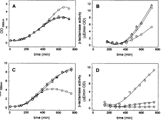
Indeed we found from this analysis that certain point mutations improved the expression behavior of the McPC603 antibody. Surprisingly, the growth/leakiness phenotype and the folding properties could be separated. The substitution P40A (HI mutant; Table 1) led to a slight decrease in leakiness and improved cell growth (with no influence on the amount of insoluble protein), while double mutant H3 (S63A/A64D) eliminated most of the insoluble protein yet had no effect on cell physiology (Figure 1). The H10 mutant, containing all eight mutations tested (Table I), showed a further improved phenotype: the ratio of soluble to insoluble protein was identical to H3, but the leakiness of cells during induction was much more reduced than with the HI mutant. As a control, the Fv fragment carrying all except the P40A mutation (HI2; Table I) was tested and found to lead to a leaky phenotype (data not shown). Therefore we concluded that the improved expression behavior of mutant H10 is due to the combination of mutations HI and H3. We tested this hypothesis by constructing the mutant Fv fragment Hll which carried both the HI and the H3 mutations (Table I). Indeed we found that this combination
200 400 600 time (min) 800 200 400 time (min) 800 800 vrt y icf c tU a CD <u •a a 1 Q
o
c E/m i 10 8 6 4 2 0 D PA
A /-*y
200 400 600 time (min) 800 200 400 time (min) 600 800Fig. 1. Comparison of growth and leakiness phenomena of cells expressing various antibody Fv fragments. (A) Growth curves of cells expressing McPC603
wild-type ( • ) , P40A (O) and S63A/A64D (A) (B) Corresponding leakiness data during inducuon, given as P-lactamase activity per OD in the culture supernatant. The symbols correspond to those in (A). (C) Comparison of growth curves of cells expressing the Fv fragments of McPC603 wild-type ( • ) , the triple mutation P40A/S63A/A64D (V) and the hu4D5 fragment (O). (D) Corresponding P-lactamase activities in the medium. The symbols correspond to those in (C).
Engineered turns of a recomblnant antibody VH Lane. M a b c d a b c d a b c d a d Dilution: 1 2 4 7
B
r
VH L a n e M a b c d a b c d a b c d a d D i l u t i o n : 2 4 10 20Fig. 2. Western blots showing the soluble (A) and insoluble (B) fractions of
cell extracts, prepared as described in Materials and methods, expressing the Fv fragments of the McPC603 wild-type (a), the P40A mutation (b), the S63A/A64D mutation (c) and the triple mutation P40A/S63A/A64D (d). The amounts loaded on the gel were normalized to the same P-lactamase activity and diluted by the factor indicated in the panels. A dilution of 1
corresponds to -100 \l\ of cell culture with an OD5Jo of I. The first lane (M) shows the size marker. Since with the constructs used here the detection of the heavy chain in Western blots is 10-fold more sensitive than the detection of the light chain due to differences in the FLAG epitope (Knappik and PlUckthun, 1994), only the heavy chain is visible in this type of blot (indicated by an arrow). The band visible below the VH chain corresponds to cross-reacting hen-egg lysozyme used to lyse the cells.
Table II. Summary of in vivo properties of the mutated Fv fragments
compared with the McPC603 wild-type Fv fragment P40A S63A/ A64D P40A/S63A/ A64D Soluble (%)' Insoluble (%)'
Increase in soluble/insoluble ratio Functional yield (%)b Cell stability0 Volume yield (%)d 300 100 3-fold 300 + 400 200 10 20-fold 200 ± 0 200 300 5 60-fold 300 1000 "Relative amounts of soluble and insoluble mature VH chain from the Fv fragment after 3 h induction at 24°C as estimated from densitometric scanning of Western blots (see Figure 2). The corresponding values of the wild-type are set to 100%.
'Functional yield (normalized to equal cell densities) given as the relative amount of Fv fragment purified from crude extracts by antigen affinity chromatography after 3 h induction at 24°C, compared with the McPC6O3 wild-type Fv fragment set at 100%.
Stability during expression at 24°C of cells expressing mutated Fv fragments compared with cells expressing the McPC603 wild-type Fv fragment. See Figure 1 for details.
dYield of mutated Fv fragments per liter cell culture, purified from crude extracts by antigen affinity chromatography after 'optimal' induction periods at 24°C is compared with the functional yield obtained with the McPC6O3 wild-type Fv fragment after 3 h of induction at 24°C. The 'optimal' induction period depends on die cell stability during induction and amounts to 5 h for the fragment containing the P40A mutation, 3 h for the fragment containing the S63A/A64D mutation and 12 h for the fragment containing the P40A/S63A/A64D mutation
had a synergistic effect, almost completely eliminating any insoluble protein (Figure 2 and Table II) and approaching the favorable growth behavior of cells expressing hu4D5 (Figure 1).
The relative effect of the mutations on the in vivo folding yields (Table II) was found to be similar in the F(ab) and scFv fragments as in the Fv fragments, suggesting that the diversion from the folding pathway appeared to be governed by the folding of an individual domain. However, the effect of the P40A mutation on the lysis phenotype was much reduced in F(ab) and scFv fragments compared with Fv. We conclude that the lysis phenotype is not proportional to the amount of
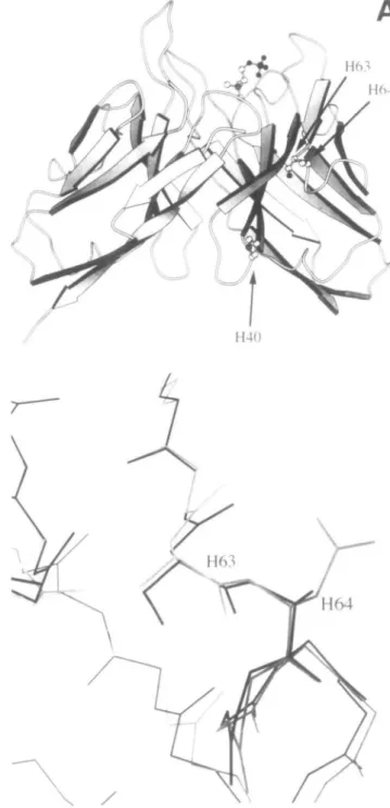
Fig. 3. (A) Location of the mutations in the structure of the
phosphorylcholine binding antibody McPC603 (Satow et al., 1986). (B) Superposition of the heavy chains of McPC603 (black) and hu4D5 (grey) (Eigenbrod et al., 1993).
insoluble protein, and must be caused by the differential interaction of the various proteins with one or more cellular components during transport or folding.
Improved cell stability during induction of the expression of Hll mutant was significant at lower growth temperatures and was lost at 37°C, while the ratio of soluble to insoluble protein was independent of the growth temperature (data not shown). In contrast, cells expressing the hu4D5 Fv fragment remained stable at all temperatures tested. Therefore, additional crucial residues, either in the framework or in the CDRs, must
• o £ c § 1.0 0.6 0 4 0.0 ^ ^ ; / 0 1 2 3 4 5 8 7 urea [M]
Fig. 4. Equilibrium denaturation curves of Fv fragments [McPC603 ( • ) ,
P40A (O), S63A/A64D (A) and P4OA7S63A/A64D (V)]. The fraction unfolded was calculated from the wavelength of the fluorescence maximum (A^j = 280 nm), measured as a function of the denaturant concentration. The calculated values for m (the slope of the curve in the transition region) and the differences in AG(H2O) are given in Table III.
be responsible for the differences in cell stability when the expression was carried out at 37°C. To distinguish between these two possibilities, we also grafted the CDRs of the McPC6O3 Fv antibody on the hu4D5 framework. This chimeric molecule consisted of the hu4D5 framework residues and the McPC603 CDRs. However, we found no additional improve-ments in yield and growth physiology with our assay systems (A.Knappik, H.Bothmann, K.Bauer and A.Plllckthun, manu-script in preparation).
We conclude therefore that the observed difference in stability between cells grown at 37°C and expressing the hu4D5 or the McPC603 Fv fragment containing the Hll mutations must be due to the different CDR sequences of the two antibodies. The mutations we found (Figure 3), however, are sufficient to eliminate the differences between the hu4D5 and the McPC603 framework in yield and cell physiology.
In vitro analysis of the mutant Fv fragments
We next sought to establish if the effects observed in vivo could be related to in vitro properties of the proteins. An
in vitro characterization of the wild-type and mutant Fv
fragments showed that these mutations did not increase the thermodynamic stability of the proteins, as measured by urea-induced fluorescence changes (Figure 4). When measuring the folding rate of the oxidized Fv fragments (containing the intradomain S-S bonds), a kinetic intermediate was detected by fluorescence changes, which was formed faster than precisely measurable by manual mixing and which slowly interconverted
50 100 time (min) 150 200 100 time (min) 150 200 50 100 150 time (min) 200 50 100 time (min) 150 200
Fig. 5. Time-course of refolding of urea-denatured Fv fragments [McPCo03 (D), P40A (O), S63A/A64D (A) and P40A/S63A/A64D (V)] of either oxidized
(A and B) or reduced (C and D) protein, monitored by fluorescence spectroscopy. (A and C) The shift of the emission peak during refolding is plotted against the time. The fully denatured protein shows an emission maximum at 348.3 nm, while the folded protein has an emission maximum at 331.4 nm (see small insert in A). (B and D) The amount of protein reaching a native-like state (as given in A and C) during refolding is plotted using the fluorescence intensity at 331.4 nm. See Materials and methods for details.
Engineered turns of a recombinant antibody
to the native state. The yields and rates of the slow folding step were again identical for all proteins examined (Figure 5A and B). To mimic the in vivo situation more closely, the experiment was repeated for the reduced proteins (Figure 5C and D). This time, the kinetics of the slow step differed dramatically between the proteins: while the McPC603 wild-type and (less dramatically) HI mutant fragment did not fold during the time of measurement, but rather aggregated very rapidly, the H3 and particularly H11 mutant fragments intercon-verted slowly into a native-like structure (Figure 5C). More-over, the yield also followed the in vivo results (Figure 5D). Interestingly, the synergistic effect observed in vivo with the triple mutant fragment could also be observed in vitro. It thus appears that a folding intermediate differs between the mutant proteins in its tendency to aggregate.
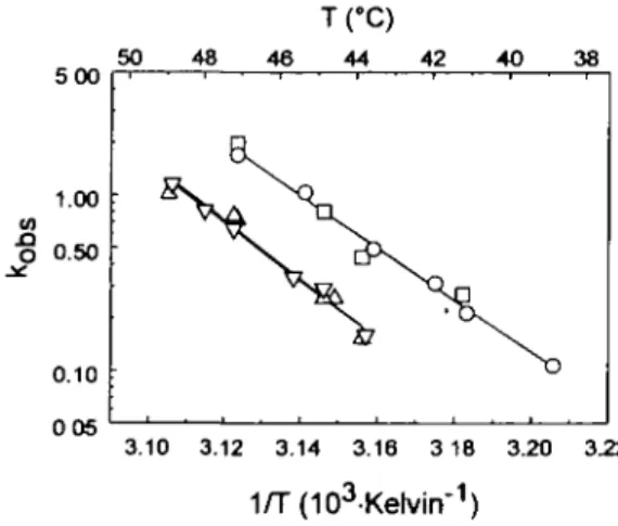
A similar ranking of the mutant fragments could be obtained by measuring by light scattering the rate of thermally induced aggregation, whose time-courses were consistent with a uni-bimolecular process when examined at different concentrations (data not shown) but followed first-order kinetics at 1.0 \iM. Interestingly, since the slopes of the temperature dependence of the aggregation rate were identical for all mutants (Figure 6), the enthalpy of activation is similar for all proteins, while the entropy of activation (the intercept in Figure 6) depends on the nature of the amino acids at position H63 and/or H64, but not on that at H40.
Discussion
What is the mechanistic basis of the observed differences in expression yields? The side chain of AspH64 is exposed in the hu4D5 Fv fragment (Eigenbrod et al, 1993) and other antibodies which carry this amino acid, and the main chain of McPC6O3 and hu4D5 is almost perfectly superimposable in this region (Figure 3). A plausible explanation for the different aggregation behavior of the mutants may thus be a greater ordering of water around AlaH64 in the ground state, compared with AspH64. Thus, less entropy of activation is required to overcome the barrier for forming the crucial transition state with increased hydrophobic surface, which leads to aggrega-tion, since AlaH64 may start at a higher ground state level, characterized by higher ordering of solvent. The folding process in the bacterial periplasm mirrors the in vitro behavior surprisingly well, suggesting that under the experimental
5 00 1.00 0.50 0.10 50 48
fc
46 IfC) 44 42 40N
38 D 3.10 3.12 3.14 3.16 3 18 3.20 3.22 1/T(103-Kelvirr1)Fig. 6. Thermal denaturation of wild-type and mutant antibody Fv fragments [McPC603 ( • ) , P40A (O), S63A/A64D (A) and P40A/S63A/ A64D (V)] measured with light scattering at 400 nm as a function of temperature.
conditions there are no molecular chaperones present in suffi-cient amounts to effisuffi-ciently prevent the aggregation reaction. Actually, this type of mutation seems to free the protein from the need of a chaperone.
While the mechanistic basis of the cell-stabilizing effect of the P40A mutation (HI) is not yet clear, it acts synergistically with the double mutation (H3) in vivo (Table II) and in vitro (Figure 5C and D), particularly if the proteins containing either the double (H3) or triple mutation (HI 1) are compared; thus both mutations may act on crucial steps of the same pathway. A speculative model consistent with the experimental data would state that there are two crucial intermediates during the
in vivo folding pathway. The first is either itself toxic for the
cells or leads to a toxic by-product, possibly by interacting with the transport apparatus of the cells. This effect of the lysis mediating intermediate can be reduced by introducing the P40A mutation in the McPC603 Fv fragment or, much more dramatically, by introducing this mutation in the Fv fragment which already contains the H3 mutations (Figure 1). The second intermediate leads to aggregation of the antibody fragments during in vivo and in vitro folding and can be prevented by the introduction of the H3 mutations.
The folding efficiency of proteins can thus depend on single amino acids which determine the kinetic partitioning between folding and aggregation. In other proteins, where similar mutations have been found (Jappelli et al., 1992; Mitraki and King, 1992; Chrunyk et al., 1993; Deng et al., 1994), they were also found to lie in turns of the protein. We predict that the folding efficiency does not simply depend on a sufficient number of equidistant surface charges in the native structure, but that there are crucial exposed microdomains consisting of turns which, in a folding or unfolding intermediate, become partially detached from the protein and can, if sufficiently hydrophobic, provide an aggregation interface. In vitro studies with, for example, Trp synthase (London et al., 1974), carbonic anhydrase (Cleland and Wang, 1990) and bovine growth hormone (Brems, 1988) are all consistent with the idea that it is an intermediate state, and neither the unfolded nor the native state, which leads to aggregation. Brems (1988) found that by adding helix-forming peptides from the growth hormone sequence to the refolding mixture of this protein, the formation of aggregates could be inhibited, suggesting that they may bind protectively at crucial sites for die aggregation reaction. These data are in excellent agreement with the results observed here and support the hypothesis that specific microdomains of a folding intermediate are susceptible to aggregation.
We suggest that a similar intermediate may be populated in thermal denaturation and in oxidative folding, because the mutations have a similar effect in both directions of folding. Since the NMR signals of the backbone amide proton have now been assigned for the Fv and single-chain Fv fragment
Table III. Summary of stability data of
Figure 4) m (kcal/mol/M) AAG(H2O) (kcal/mol) McPC603 wild-type 1.34 ± 0.08 reference P40A 1.25 ± 0.23 ±
Fv fragments (obtained from
S63A/A64D 0.07 1.52 ± 0.10 0.2 -0.14 ± 0.2 P40A/S63A7 A64D 1.56 ± 0.13 ± 0.10 0.2
The differences of the free energy of folding AAC^HjO) compared with the wild-type Fv fragment and the slope m in the transition region are shown. In this measurement, the protein concentration was 0.1 \\M in all cases.
of McPC603 (Freund et al, 1994), folding studies using NMR may give a very detailed picture of the folding and assembly pathway. There may thus be two solutions to the folding problem. One, as described here, is to inhibit aggregation by designing a charge in a crucial region likely to detach early from the protein or to attach late in the folding. Another may be to stabilize the interaction of this region with the rest of the protein. This strategy might have been operative in the disulfide mutants which stabilize T4 lysozyme against ther-mally induced aggregation, but which do not significantly change the overall AC of the molecule (Wetzel, 1987).
The mutations described here are of a different nature from those described by Hurle et al. (1994). These authors identified destabilizing mutations in VL domains which appear to be
frequent in those light chains found in amyloidosis but very infrequent in normal light chains. The free energy of folding was found to correlate roughly with the tendency to form amyloid-like aggregates. In contrast, the mutations we identi-fied do not change AG within experimental error. Furthermore, the enhanced folding yields observed are not correlated at all with the distribution of these amino acids. The amino acids we replaced were not rare ones. It thus appears that there might be a selection against unstable molecules in the animal, but not necessarily for efficient folding. It rather seems that the eukaryotic organism has other means to overcome these problems.
The antibody folds after transport into the endoplasmic reticulum and it appears to be guided by at least two chaperones, Hsp70 (BiP) and Hsp90 (GRP94) (Melnick et al., 1994). In addition, protein disulfide isomerase (PDI) has been shown to be involved in this process in vivo (Roth and Pierce, 1987). It is not yet known whether calnexin, a membrane-bound protein in the endoplasmic reticulum presumed to be involved in glycoprotein folding (Bergeron et al., 1994), also participates in the folding process of the heavy chain, at least for some subclasses. In contrast, the chaperone content of the periplasmic space is probably far more modest, and there is no evidence for ATP in this compartment (WUlfing and PlUckthun, 1994a,b). Rather than trying to recreate an identical environment as in the endoplasmic reticulum, which may be impossible, we attempted therefore to engineer the protein such that it no longer would be dependent on chaperones.
It is possible that, and it remains to be investigated whether, the susceptibility of the improved variants to disulfide iso-merase and proline-m-fra/w-isoiso-merase overexpression has changed. Depending on the folding conditions, PDI may promote either folding or aggregation in vitro (Lilie et al., 1994; Puig and Gilbert, 1994). While in the eukaryotic system Hsp70 and Hsp90 are probably the main factors in preventing premature aggregation, in the E.coli system it is the protein structure itself which is of primary importance. It can thus not be excluded that even better yields may now become possible, after the first bottleneck of aggregation has been identified and removed, by again increasing other catalysts in the system.
Recent impressive progress in constructing and screening libraries containing large antibody repertoires (Winter et al., 1994) has revealed the problem that limitations in the mainten-ance and stability of the diversity, due to very different effects of distinct antibody sequences on the bacterial cell physiology, might lead to the elimination of clones. This study shows that in the future, semi-synthetic libraries based on engineered frameworks, but carrying the CDR variability, may overcome this problem, facilitating equal representations of clones and
amplification and long-term maintenance of such libraries. Since the aspects of stability, folding and physiology can apparently be separated, such a design may now be possible. Particularly attractive might be the combination of the strategies described here with stabilizing disulfide mutations (Glockshuber et al, 1990).
Moreover, it becomes apparent that mutations influencing the aggregation of proteins during folding are most often located in turns, at least in p"-barrel proteins. An engineering approach as described here, as well as random mutagenesis focused on turn regions, may overcome potential folding problems of other recombinant proteins and, by evaluating different types of folding mutations as described here, may lead to a deeper insight into the mechanisms of protein folding.
Acknowledgements
We thank Konrad Bauer for help in the construction of the loop-grafted antibody genes, Dr Paul Carter (Genentech) for his kind gift of pAK19, and Claus Krebber and Dr Simon Moroney for useful discussions. This work was supported by the Schweizerische Nationalfonds grant 31-37717.93.
References
BergeronJ.M., Brenner.M.B., Thomas.D.Y. and Williams,D.B. (1994) Trends Biochem. Set., 19, 124-128.
Brems.D.N. (1988) Biochemistry, 27, 4541^546. Carter.P. et al (1992) Biotechnology, 10, 163-167.
Chrunyk,B.A. and Wetzel,R. (1993) Protein Engng, 6, 733-738.
Chrunyk,B.A., EvansJ., LillquisU-, Young,P. and Wetzel,R. (1993) J. Biol. Chem., 268, 18053-18061.
ClelendJ.L. and Wang.D.I.C. (1990) Biochemistry, 29, 11072-11078. Dale.G.E., Broger.C, Langen.H., d'Arcy.A. and StUber.D. (1994) Protein
Engng, 7, 933-939.
Deng.S., MacKenzie,C.R., SadowskaJ., MichniewiczJ., Young.N.M, Bundle,D.R. and Narang.S.A. (1994) J. Biol. Chem., 269, 9533-9538. Eigenbrod.C, Randal,M., Presta,L., Carter.P. and Kossiakoff,A.A. (1993)
J. Mol. BioL, 229, 969-995.
Freund.C, Ross.A., PlUckthunA and Holak.T. (1994) Biochemistry, 33, 3296-3303.
Ge,L., Knappik.A., Pack.R, Freund.C. and PlUckthun,A. (1994) In Borrebaeck.C. (ed.), Antibody Engineering. A Practical Approach. Oxford University Press, in press.
Gill.S.C. and von Hippel.P.H. (1989) Anal. Biochem., 182, 319-326. Glockshuberji., Maliajvl, Pfitzinger.I. and PlUckthun.A. (1990) Biochemistry,
29, 1362-1367.
Glockshuber.R., Schmidt,!", and PIUckthunA (1992) Biochemistry, 31, 1270-1279.
Hurle.MR., Helms.L.R., Li,L., Chan.W. and Wetzel.R. (1994) Proc. Natl Acad. Sci. USA, 91, 5446-5450.
Jaenicke.R. (1987) Prog. Biophys. Biol., 49, 117-237
Jappelli,R., Luzzago,A., Tataseo.R, Pemice.I. and Cesareni.G. (1992) J. Mol. BioL, 227, 532-543.
Kabat,E.A., Wu.T.T, Perry,H M., Gottesman,K.S. and Foeller.C. (1991) Sequences of Proteins of Immunological Interest. 5th edition. National Institutes of Health, Bethesda, MD.
Knappik,A and PlUckthun.A. (1994) BioTechniques, 17, 754-761. Knappik,A., Krebber.C. and PlUckthun.A. (1993) Biotechnology, 11, 77-S3. Kunkel.T.A., RobertsJ.D. and Zakour.R.A (1987) Methods Enzvmol., 154,
367-382.
Lilie,H., McLaughlin,S., Freedman.R. and BuchnerJ. (1994) J. Biol. Chem.,
269, 14290-142%.
LondonJ., Skrzynia,C. and Goldberg.M.E. (1974) Eur. J. Biochem., 47, 409-415.
MelnicU., DuU-L. and Argon,Y. (1994) Nature, 370, 373-375. MitrakiA and KingJ. (1992) FEBS Lett., 307, 20-25.
O'Callaghan.C.H., Morris,A., Kirby.S.M. and Shingler.A.H. (1972) Antimicrob. Agents Chemother, 1, 283-288.
Pace.C.N. (1990) Trends Biotechnol, 8, 93-98.
PlUckthun.A. (1994) In Rosenberg.M. and Moore.G.P. (eds), The Pharmacology of Monoclonal Antibodies. Springer Verlag, Berlin, Germany, Vol 113, pp. 269-315.
Prickett,K.S., Amberg.D.C. and Hopp.T.P. (1989) BioTechniques, 7, 580-589 Prodromou.C and Pearl.L.H. (1992) Protein Engng, 5, 827-829.
Engineered tarns of a recombinant antibody
Puig,A. and Gilbert^!. (1994) J. Biol. Chem., 269, 7764-7771. Roth,R.A. and Pierce.S.B. (1987) Biochemistry, 26, 4179-4182.
Satow,Y., Cohen.G.H., Padlan.E.A. and Davies.D.R. (1986)7 Mol. Biol., 190, 593-604.
Shatzman,A.R. (1990) Curr. Opin. Biotechnol., 1, 5-11. SkerraA and PlUckthun.A. (1988) Science, 240, 1038-1041. SkerraA and PlUckthun.A. (1991) Protein Engng, 4, 971-979.
TsaiA-YM-, ltoh,M., Streuli,M., Thai.T. and Saitoh. (1991) /. Biol. Chem.,
266, 10534-10543.
Wetzel.R. (1987) Trends Biochem. Sci., 12, 478-482. Wetzel.R. (1994) Trends Biotechnol., 12, 193-198.
Wetzel,R., Perry.LJ. and Veilleux.C. (1991) Biotechnology, 9, 731-737. Winter.G., Griffiths,A.D., Hawkins.R.E. and Hogenboom.H.R. (1994) Annu.
Rev. Immunol., 12, 433-455.
WUlfing.C. and PlUckthunA (1994a) Mol. Micmbiol., 12, 685-692. WUlfing,C. and PlUckthunA (1994b) / Mol. Biol., 242, 655-669. Yanisch-Perron.C, VieiraJ. and MessingJ. (1985) Gene, 33, 103-119. Received August 23, 1994; accepted October 18, 1994
![Fig. 5. Time-course of refolding of urea-denatured Fv fragments [McPCo03 (D), P40A (O), S63A/A64D (A) and P40A/S63A/A64D (V)] of either oxidized (A and B) or reduced (C and D) protein, monitored by fluorescence spectroscopy](https://thumb-eu.123doks.com/thumbv2/123doknet/14904489.655335/6.925.129.762.570.1039/refolding-denatured-fragments-oxidized-reduced-monitored-fluorescence-spectroscopy.webp)