HAL Id: hal-02163294
https://hal.archives-ouvertes.fr/hal-02163294
Submitted on 24 Jun 2019
HAL is a multi-disciplinary open access
archive for the deposit and dissemination of
sci-entific research documents, whether they are
pub-lished or not. The documents may come from
teaching and research institutions in France or
abroad, or from public or private research centers.
L’archive ouverte pluridisciplinaire HAL, est
destinée au dépôt et à la diffusion de documents
scientifiques de niveau recherche, publiés ou non,
émanant des établissements d’enseignement et de
recherche français ou étrangers, des laboratoires
publics ou privés.
A M3 MUSCARINIC RECEPTOR COUPLED TO
INOSITOL PHOSPHATE FORMATION IN THE RAT
COCHLEA?
Janique Guiramand, Ebrahim Mayat, Sylvain Bartolami, Marc Lenoir,
Jean-Francois Rumigny, Rémy Pujol, Max Récasens
To cite this version:
Janique Guiramand, Ebrahim Mayat, Sylvain Bartolami, Marc Lenoir, Jean-Francois Rumigny, et al..
A M3 MUSCARINIC RECEPTOR COUPLED TO INOSITOL PHOSPHATE FORMATION IN THE
RAT COCHLEA?. Biochemical Pharmacology, Elsevier, 1990, 39, pp.1913 - 1919.
�10.1016/0006-2952(90)90609-o�. �hal-02163294�
Biochemical Pharmacology, Vol. 39, No. 12, pp. 1913-1919, 1990.
Printed in Great Britain. COW-2952/90 Pergamon Press $3.00 + o.cm plc
A M3 MUSCARINIC RECEPTOR COUPLED TO INOSITOL
PHOSPHATE FORMATION IN THE RAT COCHLEA?
JANIQUE GUIRAMAND,* EBRAHIM MAYAT, SYLVAIN BARTOLAMI, MARC LENOIR, JEAN-FRANCOIS RUMIGNY,? R~MY PUJOL and MAX RBCASENS
INSERM-U. 254, Laboratoire de Neurobiologie de l’Audition, Hbpital St Charles, 34059 Montpellier Cedex; and TLaboratoires de Recherche Delalande, 10 rue des Carritres, 92500 Rueil-Malmaison,
France
(Received 26 June 1989; accepted 2 January 1990)
Abstract-Various neuroactive substances, including excitatory and inhibitory amino acids, biogenic amines and neuropeptides, were tested for their ability to stimulate the inositol phosphate (IPs) cascade in the presence of lithium in the rat cochlea. Among them, only the muscarinic agonists (carbachol and oxotremorine M) were able to stimulate the IPs formation in 12-day-old rat cochleas. The carbachol- elicited IPs formation was inhibited by muscarinic antagonists with the following relative order of potency: atropine > 4-DAMP apirenzepine > methoctramine = AF-DX 116. This pharmacological profile suggests that the activation of the M3 muscarinic receptor subtype is responsible for the increase in IPs synthesis in the rat cochlea. However, an interaction with a m5 receptor subtype could not be completely excluded. The unusual link of only one receptor subtype with the phosphoinositide breakdown in the cochlea, as opposed to the usual existence of several receptors coupled to this transduction system in other organs such as the brain, suggest a unique role for muscarinic agonists in the cochlea.
The mammalian cochlea contains two types of hair
cells: namely the inner hair cells (IHCs$) and the outer hair cells (OHCs). The auditory transmission between IHCs and the first auditory neurons (type I afferent neurons) is thought to be assumed by an excitatory amino acid, likely glutamate [l-4]. This transmission may be modulated by the efferent lateral olivocochlear fibers which synapse at the afferent dendrites of the type I auditory neurons. Several neurotransmitters including dopamine, GABA, acetylcholine have been identified in these fibers [5]. The OHCs receive neuronal information via the medial efferent system which is mainly chol- inergic [6]. In addition, the OHCs contact the den- drites of type II neurones, which represent the other afferent system of the cochlea. This afferent system, for which the neurotransmitter remains unknown, represents only 5% of the whole cochlear afferent system.
The OHCs have been postulated to indirectly modulate IHCs activity [7,8] probably due to their motile properties. Motility (shortening and elong- ation) of isolated OHCs has been shown to occur in response to intracellular current administration (fast motility) [9] or in response to neuroactive substances such as potassium or acetylcholine (slow motility) [9, lo]. The molecular mechanisms, which underlie such OHCs motility, may originate from phos- phoinositide metabolism and the subsequent intra- cellular calcium mobilization [ll, 121, which in turn
* To whom correspondence should be addressed. $ Abbreviations: IHC, inner hair cell; OHC, outer hair cell; CDAMF’, 4-diphenylacetoxy-N-methyl pipexidine methiodide; AF-DX 116, ll-((2-((diethylamino)-methyl)- l-piperidinyl)acetyl) - 5,ll - dihydro - 6H - pyrido(2,3 - b)- (1,4)-benzodiazepine-6 one.
controls actin polymerization and the formation of microfilament assemblies [ 131.
Despite the importance of such molecular mech- anisms in the understanding of cochlear functioning, no direct measurement of inositol phosphate for- mation has been performed in this organ. In the present paper, we examine the accumulation elicited by neuroactive substances of these second messenger molecules (inositol mono-, bis- and tris-phosphate) in the rat cochlea.
MATERIALS AND METHODS
Incorporation of[3H]inositol. Wistar rats of age 12 days were killed by decapitation. After dissecting out the bony capsule, whole cochleas were rapidly removed and immediately placed in Krebs-Ringer buffer, pH 7.4. The Krebs-Ringer buffer used con- tained 125 mM NaCl; 3.5 mM KCl; 1.25 mM KH,PO,; 1.2 mM MgSO,; 1.5 mM CaCl*; 10 mM glucose and 25 mM NaHC03. Before use, this buffer was equilibrated to pH7.4 by saturation with a gaseous mixture of 95% 02 and 5% CO* (v/v). Myo-
[2-3H]Inositol incorporation in 75 cochleas was car- ried out at 37”, under gassing of 95% 0,/5% CO*, for 75 min in 5 mL Krebs-Ringer buffer containing 1 mM cytidine and 50 @i of the radioactive inositol (sp. act. 17Ci/mmol, CEA Saclay, France). The labelled cochleas were then washed four times in 5 mL Krebs-Ringer buffer before being individually distributed in plastic tubes containing 500 PL Krebs- Ringer buffer. The tubes were then transferred to a water bath maintained at 37” and continuously gassed with the gaseous mixture of 95% OJS% CO* throughout the subsequent experimental steps.
Agonist stimulation. LiCl (lOpL, final concen- tration 10mM) and 2OpL of an appropriate con- centration of an antagonist (where applicable) were
1914 J. GUIRAMAND et al.
added to each tube. After 15 min, the stimulatory neuroactive substance or buffer (20 pL) was added. Following an additional 20min of incubation, the reaction was stopped by the addition of 5OpL per- chloric acid (PCA 72%) and, by placing the tubes on ice.
Measurement of [3H]inositol phosphates. The labelled IPs were purified and measured according to the method described by Bone et al. [ 141 modified as follows: the cochleas were homogenized using an Ultraturrax homogenizer. The homogenates con- taining the PCA-extracted [3H]IPs were then centri- fuged at 2000g for 20 min. The resulting supernatants were poured off and retained while the pellets were resuspended by homogenization in 1 mL PCA (1%) and recentrifuged at 2000 g for 20 min. The two PCA extracts were then pooled and neu- tralized with a 1.5 M KOH/0.075 M HEPES solution in the presence of universal indicator pH 4-10. The resulting potassium perchlorate salt was then pel- leted by centrifugation (5 min at 2OOOg), resus- pended in 2 mL 0.5 mM EDTA/S mM Na2B40, and washed by centrifugation (5 min at 2000g). The supernatants from the two centrifugations were then pooled. A solution (10mL) containing 0.5 mM EDTA/S mM Na2B407 was then added to the pooled supernatant. The extracts (containing [3H]inositol and [3H]IPs) thus obtained were then applied to Dowex anion-exchange columns (1X8 formate form; height of resin: 4cm; diameter: 0.8cm). Aliquots (1.5 mL) were taken from the flow-through fractions and subjected to liquid scintillation counting. Each column was then washed once with 20 mL water. Glycerophosphoinositides (GPI) were eluted with 20 mL 0.04 M ammonium formate/0.003 M sodium tetraborate. The IPs were eluted with 15 mL 0.8 M ammonium formate/O. 1 M formic acid. For both the GPI and IPs fractions, 4-mL aliquots were taken and radioassayed. Columns were regenerated twice with 10 mL 2 M ammonium formate/O.l M formic acid and twice with 20 mL distilled water prior to re-use. Columns were discarded after five experiments. For liquid scintillation counting, 2.5 mL of Ready Gel scintillation fluid (Beckman Instruments Inc., Ireland) was added to the 1.5-mL aliquots obtained from the flow-through fractions while 6 and 7 mL of Ready Gel were added to the 4-mL aliquots con- taining the eluted GPI and IPs, respectively.
Data calculation. Dose-response curves for agon- ists and antagonists were fitted using non-linear regression with experimental values weighted by the reciprocal of the variance, according to the logistic model described by De Lean et al. [15,16]. The computer programme used was the RS/l release 3.0 program (BBN Software Product Corporation, Cambridge, MA, U.S.A.), run on a VAX 6210 microcomputer.
RESULTS
Time course
Carbachol (carb) enhanced IPi accumulation in the presence of 10 mM Li+ at 37” in the rat cochlea. This increase was exponential during the first 5 min and linear between 5 and 45 min of incubation (Fig. 1 upper panel). Carb did not induce any IPi accumu-
“r
IOmM Li’ - IP, .-. IP, .j/ n -m IP, 100 - IP, 0-e lP2 l -. IP, 100-a
0 5 IO I5 20 25 30 35 40 45 50 Time(min)Fig. 1. Time course of carbachol-induced formation of [‘H]IP,, [3H]IP2 and [3H]IP3 in rat cochleas. Experimental conditions are as described in Materials and Methods except that the cochleas were prelabelled with 500&i [3H]myo-inositol instead of 50 j&i for 75 min, washed five times, and then stimulated for different time periods with 10 mM carbachol in the absence or in the presence of 10 mM LiCl. In addition, the reaction was stopped using 60 PL of a solution containing PCA (72%), EGTA (1 mM) and a nonradioactive mixture of inositol phosphates (2 m9/ mL) in order to inhibit the acidic phosphatases which catalyse the dephosphorylation of the various IPs metab- oiites. The elutions of the various metabolites from the Dowex 1X8 columns were performed as follows: twice with 20mL of water to remove [3H]myo-inositol, once with 15 mL of 0.04 M ammonium formate/0.003 M sodium tetra- borate for GPI, twice with 15mL of 0.15M ammonium formate/O.OOS M sodium tetraborate for IP,, twice with 15 mL of 0.4 M ammonium formate/O.l M formic acid for IPs, twice with 15 mL of 0.8 M ammonium formate/O.l M formic acid for IP3 and once with 15 mL of 2 M ammonium formatejO.1 M formic acid for the other inositol phosphates. Results are means 2 SD of experiments done on at least six different co&leas (one IPs determination is performed for each individual cochlea). The basal IP,, IPs and IP3 accumulation was not significantly affected by the presence
of 10 mM Li+.
lation in the absence of Li+ (Fig. 1 lower panel). Very little radioactivity was found in the IPz and IP3 fractions and this was not affected by Li+. The basal levels of IPi, IPs and IP3 did not change with Li+. The results were expressed as the ratios of dpm contained in the various IP fractions to the dpm of incorporated inositol. Similar patterns were obtained when results were expressed as dpm per mg of pro- tein (data not shown). These preliminary experi- ments prompted us to measure the total IPs (containing about 98% of IPi and IPs) for a 20 min
Muscarinic receptor and inositol phosphate in the rat cochlea 1915 Table 1. Effect of neuroactive substances on the IPs formation in the rat cochlea
Agonist Maximum Concentration Tested (M) Effect on IPs accumulation* Glutamate Quisqualate N-Methyl-D-aspartate Kainate Nicotine Carbachol Oxtremorine M Gamma-amino-butyric acid Glycine Noradrenaline 5-Hydroxytryptamine Dopamine Substance P Neuropeptide Y
Calcitonin gene related peptide Vasoactive intestinal peptide Arginine-vasopressin Cholecystokinin Met-enkephalin Leu-enkephalin Met-enkephalin-Arg6-Phe’ Oxytocin DAGOB DTLETlj DPLPET Potassium A 23187 Monensin Valinomycin K Veratridine loo0 1000 1000 1000 10,ooo 10,ooo loo0 1OCO 1000 luoo loo0 1000 1 1 96 2 13 1 109 + 17 87 f 12 1 81 f 10 1 115 2 29 1 1 1 1 1 60,ooo 5 50 10 100 + 26 118 2 16 101 f 21 124 + 52 79 f 29 617 -c 66$ 727 2 59$ 102 * 23 89 f 8 114 +: 24 135 + 23 126 + 12 113 + 26 86 2 17 100 f 18 109 2 12 99 f 17 82 f 11 72 + 3t 91 f 34 92 2 13 96 & 23 145 * 37 79 + 12 92 f 23 * Results are expressed as percentages of control values. The mean control value is (47 2 13) x lo-’ dpm IPs/dpm [‘Hlinositol incorporated and the mean value of [3H]inositol incorporation is 41,130 & 12,170 dpm per cochlea. Results are expressed as means + SD of at least six individual determinations. The statistical significance of the agonist-stimulated IPs formation versus the control IPs values was calculated by two-tailed Student’s t-test. (t P < 0.01; $ P < 0.001).
§ DAGO: (D-ala’, N methyl-phe4, glyol’) enkephalin. ]I DTLET: (D-thr*, thr6) leu-enkephalin.
1 DPLPE: (D-pen’, L-pen’) enkephalin.
incubation period and to express the results as ratios (dpm IPs/dpm incorporated [3H]myo-inositol) for the following experiments.
Effects of neuroactive substances on the IPs formation in the rat cochlea
A large number of neuroactive substances, some of which are known to be present in the cochlea [S], ions and ionophores were screened for their ability to stimulate IPs formation in the cochlea (see Table 1). With the exception of the muscarinic agonists carbachol and oxotremorine M, none of the sub- stances tested (nicotine included) had any significant stimulatory effect.
Dose-response curve
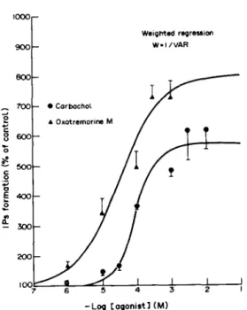
Carbachol and oxotremorine M induced IPs accumulation in a dose-dependent manner (Fig. 2). From the dose-response relationships, the ECs,, values were calculated, using a curve fitting pro- gramme indicated in Materials and Methods. They
were 28 +- 20 PM and 87 k 19 PM for oxotremorine
M and carbachol, respectively. A pre-analysis according to the logistic model proposed by Black et
al. [17] shows that for the two agonists the slopes
are approximately equal to 1. The E,,,, values for carbachol and for oxotremorine M are significantly different. These values expressed as percentages of the basal values are 570 f 60% and 799 + 93%, respectively.
Effect of cholinergic antagonists on carbachol-elicited IPs accumulation
A broad range of choline@ receptor antagonists were assessed for their ability to block the increased accumulation evoked by carbachol. The nicotinic antagonists D-tubocurarine and cu-bungarotoxin had no effect. On the contrary, muscarinic antagonists inhibited all the IPs formation induced by 10e3 M carbachol (Fig. 3). None of these choline@ antag- onists affected the basal IPs accumulation, except for a high concentration of methoctramine. At
1916 J. GUIRAMAND er al.
100 I I I
7 6 5 4 3 2
-Log Cogonistl (Ml
Fig. 2. Concentration-response relationships of carbachol- and oxotremorine M-induced IPs formation in the rat coch- lea. Cochleas were labelled for 75 min with 50 &i [‘H)myo- inositol, and then washed four times to remove the excess of radioactive inositol. After preincubation with 10mM LiCl at 37” for 15 min, increasing concentrations of car- bachol or oxotremorine h4 were added and the cochleas were further incubated for 20min. The accumulation of (3H]IPs was measured by scintillation counting following Dowex 1X8 column chromatography. Results which are means f SE of experiments done on at least five different cochleas, show stimulation as a percentage of the basal 1,Ps formation. Basal IPs formation was 54,180 k 4913 dpm/mg
protein (mean ? SE).
10e3 M, methoctramine alone stimulates IPs accumu- lation (about 400% of basal value). The same level of stimulation is also obtained in the co-presence of 10m3 M carbachol. The relative order of potency of the muscarinic antagonists were atropine > 4-DAMP S- pirenzepine > methoctramine = AF-DX 116. The IC, values which were calculated from the inhibition curves and were 0.007 & 0.007, 0.030 + 0.008, 2.0 2 0.8, 33 2 7 and 63 2 42pM for atropine, 4- DAMP, pirenzepine, AF-DX 116 and methoc- tramine respectively. For each antagonist, pseudo- Hill numbers were determined (Table 2). In all cases they are not significantly different from 1. However, for methoctramine, this number is relatively low.
DISCUSSION
Our results indicate that among the neuroactive substances tested (Table l), some of which are known to occur in the cochlea [5], only choline@ agonists are able to stimulate the accumulation of IPs in the cochlea. Although nicotinic receptors seem to occur in the cochlea, (nicotine potentiates the submaximal effects of the stimulation of the crossed olivocochlear bundle [18]) their activation did not lead to IPs stimulation. Conversely, cholinergic
50 40 34 20 IO o- . o- O- O- 0- -Log [antagonisll (Ml
Fig. 3. Elects of muscarinic antagonists on carbachol- induced IPs formation in the cochlea. Experimental con- ditions are as described in Materials and Methods. Chol- inergic antagonists (IO-’ M-10e3 M) were tested for their ability lo inhibit IPs formation induced by 1 mM carbachol.
Results which are means 2 SE of experiments done on at least six different cochleas, show stimulation as a per- centage of the basal IPs formation. Antagonists pe.r se (atropine, 4-DAMP, methoctramine, pirenzepine and AF- DX 116) did not affect the basal IPs formation at the concentrations tested (except for high concentrations of methoctramine greater than 1O-4 M). D-Tubocurarine and a-bungarotoxin were without effect both on the basal and
carbachol-stimulated IPs accumulation.
Table 2. Calculated ICE and pseudo-Hill values for chol- inergic antagonists for inhibition of 1 mM carbachol-
induced IPs formation in the rat cochlea Antagonist ‘%I (W) nH Atropine 0.007 c 0.007 0.83 2 0.47 4-DAMP 0.03 2 0.008 0.94 + 0.15 Pirenzepine 2.0 2 0.8 0.90 c 0.17 AF-DX 116 33 + 7 0.91 t- 0.15 Methoctramine 63 ? 42 0.79 2 0.49 These values were calculated from the results presented in Fig. 3 as described in Materials and Methods. They are expressed as means -t SE of at least six individual determinations.
agonists of the muscarinic receptor subtype (car- bachol and oxotremorine M) increase the metab- olism of IPs with a high apparent affinity. The fact that the E,,, values calculated for these two agonists are significantly different may indicate that carbachol is a partial agonist. The carbachol-stimulated IPs formation is not blocked by nicotinic antagonists such as D-tubocurarine and cu-bungarotoxin while it is inhibited by muscarinic antagonists (atropine, pirenzepine, 4-DAMP, methoctramine and AF-DX 116). This clearly demonstrates that a muscarinic
Muscarinic receptor and inositol phosphate in the rat cochlea 1917
receptor is responsible for the IPs formation in the cochlea. Muscarinic receptors have recently been divided into five subtypes,* named ml, m2, m3, m4 and m5 on the basis of molecular cloning experiments and the expression of this cloned muscarinic receptor in various cells [19-211. The ml and m2 receptor subtypes have also been cloned from porcine brain and heart [22-241. In order to determine which muscarinic receptor is implicated in the cholinergic- induced IPs formation in the cochlea, we have measured the inhibition, by various muscarinic antagonist of the IPs accumulation, induced by 1 mM carbachol. Although it would be better to determine
Ki values from Schild plot analyses, we have only
measured the lcso value for each antagonist in this paper (Table 2). The reasons for this are technical limitations such as the time taken for dissection, the time-restricted period for cochlear viability and the very small quantities of biological tissue present per cochlea. However, since the same concentration of carbachol was used for studying the antagonist inhi- bition, ICKY values thus determined will provide an approximate rank order of potency of the antag- onists. Subsequently, the subtype of receptor involved can be determined. The most widely used classification of muscarinic receptor subtypes was based on the relative affinities of the antagonists, pirenzepine and AF-DX 116 in particular [25-271. M1 receptors express a high apparent affinity toward pirenzepine and an intermediate affinity toward AF- DX 116 [26,28], whereas Mz receptors “cardiac type” (MZalpha) possess a low apparent affinity for pirenzepine and a high affinity for AF-DX 116 [26- 28]. M3 receptors, previously known as the M2 “glandular type” (MZbeta) present a low apparent affinity toward both pirenzepine and AF-DX 116 and a high affinity toward 4-DAMP [28,29]. Methoc- tramine was found to be as effective and selective as AF-DX 116 for the Mz receptor [30,31]. No specific antagonists, yet tested, allowed the distinction of m4 or m.5 from the rest of the muscarinic receptor subtypes [32]. By using a large variety of antagonists, it is possible to approximate the identification of the specific subtype of muscarinic receptor involved as demonstrated by a recent displacement-binding study carried out in CHO-Kl cells, in which cloned muscarinic receptors were expressed [32]. However, the pharmacological profile determined in this study cannot be easily applied to muscarinic receptor sub- type identification in other experimental models. Indeed, artificial receptor gene expression does not necessarily simulate exactly the expression occurring
in vivo. Another indirect manner of characterization of receptor subtypes is the study of the biochemical
* The nomenclature of the muscarinic receptor subtypes
used in this paper is that recommended recently in the supplement of Trends in Pharmacological Sciences, December 1989, p. VII by the nomenclature committee of the fourth symposium on muscarinic subtypes. According to this nomenclature, the pharmacologically characterized receptor subtypes are known as M,, Mz and M,, while those characterized by molecular cloning techniques are named ml, m2, m3, m4 and m5. It is likely that the ml sequence corresponds to that of the M, receptor, m2 to the Mz receptor and m3 to the M3 receptor.
responses triggered by their activation. Muscarinic receptors are coupled to a variety of second mess- enger systems involving adenylate cyclase and phos- phatidylinositol metabolism as well as to ion channels [20, 33-411. In fact, it was reported that M1 (ml), M3 (m3) and m5 receptors are coupled with the stimulation of phosphoinositides hydrolysis [20,26,29,35-37,39,40] whereas m2 and m4 are mainly linked to the inhibition of adenylate cyclase [36,38].
Our pharmacological results indicate that the muscarinic receptor subtypes involved in the IPs formation in the rat cochlea is probably a M3 recep- tor. In fact, pirenzepine has a low apparent affinity (IQ,, = 2 x 10m6 M) in inhibiting IPs synthesis which rules out the possibility of an interaction with the M1 receptor. A similar low apparent affinity of piren- zepine was recently reported in receptor-binding studies to cochlear membranes using [3H]-l-qui- nuclidinylbenzylate [42]. We found a relatively low efficacy of AF-DX 116 or methoctramine for inhi- biting the carbachol-induced IPs accumulation in the rat cochlea (ICKY = 33 and 63 fi, respectively). This excludes the involvement of a M2 receptor subtype in this IPs response since these two antagonists present a high affinity for the M2 receptor subtype [26-301. The antagonist 4-DAMP inhibits the carb- achol-induced IPs response with a high apparent affinity, thus suggesting an action via the M3 receptor [29]. However, the potency of 4-DAMP has not been tested for its inhibitory action neither on the binding of muscarinic ligands to m4 or m5 receptor subtypes nor on the biochemical responses associated with the activation of these two latter receptor subtypes. Nevertheless, m4 receptors are rather shown to be coupled to adenylate cyclase inhibition than to IPs formation [36], suggesting that the response observed here, is probably not mediated by a m4 muscarinic receptor. On the other hand, the expression of m5 receptors has not yet been observed in any tissue or cell line [32]. The paucity of phar- macological data concerning this receptor subtype, does not allow us to rule out the possibility of the existence of a m5 muscarinic receptor linked to inosi- to1 phosphate formation in the cochlea. If this is so, this will be the first indication of the natural expression of the m5 receptor gene in an organ. In fact, the antagonists used present about the same rank order of potencies in the binding studies on both the m3 and m5 receptors expressed in CHO- Kl cells: atropine ti methoctramine > pirenzepine % AF-DX 116 [32]. In our experiments, we found the following order of inhibitory action on carbachol- stimulated IPs formation: atropine * pirenzepine % AF-DX 116 = methoctramine. The apparent dis- crepancy concerning the inhibitory effect of methoc- tramine between the two sets of results may originate from the fact that methoctramine at concentrations greater than 10m4 M becomes an agonist in the coch- lea. This finding, which has also been reported in the rat cerebral cortex [43], may explain why the Hill number for methoctramine is not equal to 1. A low Hill number for methoctramine has also been obtained in [3H]-N-methylscopolamine displacement experiments on rat submaxillary gland membranes, thought to possess only M3 receptors [32]. Taken
1918 J. GUIRAMAND et al.
together our results seem to indicate that cholinergic- induced IPs formation in the cochlea is probably mediated by the activation of a Ms muscarinic recep- tor.
Our results also show that neither glutamate nor quisqualate stimulate IPs formation in the rat cochlea. Nevertheless several reports have shown that an excitatory amino acid is probably involved in the transmission between the IHCs and the primary auditory neurons [44] by activating a quisqualate receptor [1,45]. If so, the quisqualate receptor involved did not correspond to that linked to the phosphoinositide metabolism [46]. These facts reinforce the conclusion of our previous work [46] which indicate that two subtypes of quisqualate receptors do indeed exist in the central nervous system, one linked to ion channels, the other one to phosphoinositides metabolism.
In conclusion, our data strongly suggest that among the known neuroactive substances found in the rat cochlea only muscarinic agonists mediate the stimulation of IPs turnover likely via a Ms receptor subtype. Acetylcholine is thought to be the main neurotransmitter between the medial efferent system and the OHCs [6], and inositol triphosphate causes the contraction of permeabilized OHCs in vitro [ll, 121. Thus, the activation of this second mess- enger pathway in the cochlea by acetylcholine via a M3 receptor may play a key role in the triggering or the control of slow OHCs motility, although the accurate cellular location of this second messenger remains to be elucidated.
Acknowledgements--We thank Dr C. Melchiorre (Uni- versity of Bologna, Italy) for kindly providing us with the methoctramine and the Laboratoires Boehringer Ingelheim, Reims, France for the gift of AF-DX 116. We are also grateful to Dr M. Eybalin for helpful discussion, M. Gallego for technical assistance and A. Bara for typing this manuscript. The work was supported by grants from M.R.E.S., C.N.A.M.T.S.-INSERM, IPSEN, Air Liquide and the Institut H. Beaufour (Paris).
REFERENCES 1. 2. 3. 4. 5. 6. I. 8.
Jenison GL and Bobbin RP, Quisqualate excites spiral ganglion neurons of the guinea pig. Hearing Res 20: 261-265, 1985.
Bobbin RP, Glutamate and aspartate mimic the affer- ent transmitter in the cochlea. Exp Brain Res 34: 389 393, 1979.
Evbalin M and Puiol R, A radioautoaraphic study of the [3H]-L-glutam&e and [‘HI-L-glut~mine uptake in the euinea-Die cochlea. Neuroscience 9: 863-871.1983.
KlinYke R aAd”Oertel W, Amino acids-putative affer- ent transmitter in the cochlea? Exp Brain Res 30: 14% 148, 1977.
Eybalin M and Pujol R, Cochlear neuroactive substances. Arch Oto-Rhino-Laryngol 246: 228-234, 1989.
--- -.
Eybahn M and Pujol R, Choline acetyltransferase (ChAT) immunoelectron microsconv distinguishes at ieast three types of efferent synap& in theorgan of Corti. Exp Brain Res 65: 261-270, 1987.
Mountain DC, Changes of endolymphatic potential and crossed olivocochlear bundle stimulation alter cochlear mechanics. Science 210: 71-72, 1980.
Siegel JH and Kim DO, Efferent control of cochlear mechanics? Olivocochlear bundle stimulation affects
cochlear biomechanical nonlinearity. Hearing Res 6: 171-182, 1982.
9. Brownell WE, Bader CR, Bertrand D and de Ribau- Pierre Y, Evoked mechanical responses of isolated cochlear outer hair cells. Science 227: 194-196, 1985. 10. Slepecky N, Ulfendahl M and Flock A, Shortening and elongation of isolated outer hair cells in response to application of potassium gluconate, acetylcholine and cationixed ferritin. Hearing Res 34: 119-126, 1988.
11. Schacht J and Zenner H, The phosphoinositide cascade in isolated outer hair cells: possible role as second messenger for motile responses. Hearing Res 22: 94, 1986.
12. Schacht J and Zenner H, Evidence that phos- phoinositides mediate motility in cochlear outer hair cells. Hearing Res 31: 155-160, 1987.
13. Lassing I and Lindberg U, Evidence that the phos- phatidylinositol cvcle is linked to cell motilitv. Exe Cell
kes 174: l-15,1988. , ‘ 14. Bone EA, Fretten P, Palmer S, Kirk CJ and Michell
RH, Rapid accumulation of inositol phosphates in iso- lated rat superior cervical sympathetic ganglia exposed to V,-vasopressin and muscarinic cholinergic stimuli.
Biochem J 221: 803-811, 1984.
15. De Lean A, Munson PJ and Rodbard D, Simultaneous analysis of families of sigmoidal curves. Am J Physiol 235: E97-E102, 1978.
16. De Lean A, Hancock AA and Lefkowitz RJ, Validation and statistical analysis of a computer modeling method for quantitative analysis of radioligand binding data for mixtures of pharmacological receptor subtypes. Mol Pharmacol21: 5-16, 1982.
17. Black JW, Leff P, Shanklev NP and Wood J, An
operational model of pharmacological agonismi the effect of E/[Al curve shane on aeonist dissociation constant es&naiion. Br .l Piarmacol-84: 561-571,1985.
18. Klinke R, Neurotransmission in the inner ear. Hearing Res 22: 235-244, 1986.
19. Bonner TI, Buckley NJ, Young AC and Brann MR, Identification of a family of muscarinic acetylcholine receptor genes. Science 237: 527-532, 1987.
20. Bonner TI, Young AC, Brann MR and Buckley NJ, Cloning and expression of the human and rat m5 musca- rinic acetylcholine receptor genes. Neuron 1: 403-410, 1988.
21. Peralta EG, Ashkenazi A, Winslow JW, Smith DH, Ramachandran J and Capon DJ, Distinct primary struc- ture, ligand-binding properties and tissue-specific expression of four human muscarinic acetylcholine receptors. EMBO J 6: 3923-3929, 1987.
22. Braun T, Schofield PR, Shivers BD, Pritchett DB and Seeburg PH, A novel subtype of muscarinic receptor identified by homology screening. Biochem Biophys Res Commun 149: 125-132, 1987.
23. Kubo T, Fukada K, Mikami A, Maeda A, Takahashi H, Mishima M, Haga T, Haga K, Ichiyama A, Kangawa K, Kojima M, Matsuo H, Hirose T and Numa S, Cloning, sequencing and expression of complementary DNA encoding the muscarinic acetylcholine receptor. Nature 323: 411-416, 1986.
24. Peralta EG, Winslow JW, Peterson GL, Smith DH, Ashkenazi A, Ramachandran J, Schimerlik MI and Capon DJ, Primary structure and biochemical prop- erties of an M2 muscarinic receptor. Science 236: 600- 605, 1987.
25. Hammer R, Berrie CP, Birdsall NJM, Burgen ASV and Hulme EC, Pirenzepine distinguishes between dif- ferent subclasses of muscarinic receptors. Nature 283: 90-92, 1980.
26. Ladinsky H, Giraldo E, Monferini E, Schiavi GB, Vigano MA, De Conti L, Micheletti Rand Hammer R, Muscarinic receptor heterogeneity in smooth muscle:
Muscarinic receptor and inositol phosphate in the rat cochlea 1919
27.
28.
29.
30.
binding and functional studies with AF-DX 116. Trends 37. Lai J, Mei L, Roeske WR, Chung F-Z, Yamamura
Pharmacol Sci Suppl February: 44-48, 1988. HI and Venter JC, The cloned murine M, muscarinic Barnes PJ, Minette P and Maclagan J, Muscarinic receptor is associated with the hydrolysis of phos- receptor subtypes in airways. Trends Pharmacol Sci 9: phatidylinositols in transfected murine B82 cells. Life
412-416, 1988. Sci 42: 2489-2502, 1988.
Micheletti R, Montagna E and Giachetti A, AF-DX 116, a cardioselective muscarinic antagonist. J Phar- macol EXD Ther 241: 628-634. 1987.
Doods HN, Mathy M-J, Davidesko D, van Charldorp KJ, de Jonge A and van Zwieten PA, Selectivity of muscarinic antagonists in radioligand and in vivo
experiments for the putative M,, M2 and MS receptors.
J hharmacol Exp Ther 242: 257-262, 19871
Melchiorre C. Aneeli P, Lambrecht G. Mutschler E, Picchio MT and -Wess’ J, Antimuscarinic action of methoctramine, a new cardioselective M-2 muscarinic receptor antagonist, alone and in combination with atropine and gallamine. Eur J PharmacoZl44: 117-124, 1987.
38. Ashkenazi A, Winlow JW, Peralta EG, Peterson GL, Schimerlik MI, Capon DJ and Ramachandran J, A M2 muscarinic receptor subtype coupled to both adenylyl cyclase and phosphoinositide turnover. Science 238: 672-675, 1987.
39. Marty A, Control of ionic currents and fluid secretion by muscarinic agonists in exocrine glands. Trends Neurosci 10: 373-377, 1987.
40. Fukuda K, Higashida H, Kubo T, Maeda A, Akiba I, Buko H, Mishina M and Numa S, Selective coupling with K+-currents of muscarinic acetylcholine receptor subtypes in NG 108-15 cells. Nature335: 355-358,198s. 41. Jones SVP, Barker JL, Bonner TI, Buckley NJ and
Brann MR, Electrophysiological characterization of cloned Ml muscarinic receptors expressed in A9 L cells. Proc Nat1 Acad Sci USA 85: 4056-4060, 1988. 42. Van Megen YJB, Klaassen ABM, Rodrigues de
Miranda JF and Kuijpers W, Choline@ muscarinic receptors in rat cochlea. Brain Res 474: 185-188, 1988. 43. Lee NH, Forray C and El-Fakahany EE, Methoc- tramine, a cardioselective muscarinic antagonist, stimulates phosphoinositide hydrolysis in rat cerebral cortex. Eur J Pharmacol167: 295-298, 1989. 44. Jenison GL, Winbery S and Bobbin RP, Comparative
actions of quisqualate and N-methyl-D-aspartate, excit- atory amino acid agonists, on guinea-pig cochlear potentials. Comp Biochem Physiol84C: 38s389,1986. 45. Littman T, Bobbin RP, Fallon M and Puel J-L, The
quinoxalinediones DNQX, CNQX and two related congeners suppress hair cell-to-auditory nerve trans- mission. Hearing Res 40: 45-54, 1989.
46. Recasens M, Guiramand J, Nourigat A, Sassetti I and Devilhers G, A new quisqualate receptor subtype (SAA,) responsible for the glutamate-induced inositol phosphate formation in rat brain synaptoneurosomes.
Neurochem Int 13: 463-467, 1988.
31. Melchiorre C, Minarini A, Angeli P, Giardina D, Gulini U and Quaglia W, Polymethylene tetramines as muscarinic receptor probes. Trends Pharmacol Sci
Suppl December: 55-59, 1989.
32. Buckley NJ, Bonner TI, Buckley CM and Brann MR, Antagonist binding properties of five cloned muscarinic receptors expressed in CHO-Kl cells. Mel Pharmacol 35: 469-476, 1989.
33. Neer EJ and Clapham DE, Role of G protein subunits in transmembrane signalling. Nature 333: 129-134, 1988.
34. Berridge MJ and Irvine RF, Inositol trisphosphate, a novel second messenger in cellular signal transduction.
Nature 312: 315-321, 1984.
35. Lazareno S, Kendall DA and Nahorski SR, Pirenzepine indicates heterogenicity of muscarinic receptors linked to cerebral inositol phospholipid metabolism. Neuro- vharmacolonv 24: 593-595. 1985.
36. Peralta EGY Ashkenazi ‘A, Winslow JW, Rama- chandran J and Capon DJ, Differential regulation of PI hydrolysis and adenylyl cyclase by muscarinic receptor subtypes. Nature 334: 434-437, 1988.
![Fig. 1. Time course of carbachol-induced formation of [‘H]IP,, [3H]IP2 and [3H]IP3 in rat cochleas](https://thumb-eu.123doks.com/thumbv2/123doknet/14696944.563471/3.756.397.679.95.495/fig-time-course-carbachol-induced-formation-ip-cochleas.webp)