DEVELOPMENT OF A POTENTIAL BIOARTIFICAL LIVER: SELECTIVE ADHESION OF HEPATOCYTES
by
Sangeeta N. Bhatia B. Sc., Biomedical Engineering
Brown University, 1990
Submitted to the Department of Mechanical Engineering in Partial Fulfillment of the Requirements for the Degree of
Master of Science in Mechanical Engineering
at the
Massachusetts Institute of Technology
ARCHIVE,
" 'A INjSMS I f~qIOLOGYAUG 10
1993
L2t41AIES June 1993 Sangeeta N. Bhatia 1990 All rights reservedThe author hereby grants to MIT permission to reproduce and to distribute copies of this thesis document in whole or in part.
Signature of Author
S
Certified byS
Certified by_Signature redacted
ignature redacted
ignature redacted
Signature redacted
Accepted by__Department of Mechanical Engineering May 7, 1993 Mehmet Toner Thesis Supervisor Martin L. Yarmush Thesis Supervisor 'k
DEVELOPMENT OF A POTENTIAL BIOARTIFICIAL LIVER:
SELECTIVE ADHESION OF HEPATOCYTES
by
Sangeeta Bhatia
Submitted to Department of Mechanical Engineering on May 7, 1993 in partial fulfillment of the requirements for the degree of Master of Science in Mechanical Engineering
ABSTRACT
Liver disease is the cause of death for 30,000 Americans every year. For those who suffer from acute liver failure, transplantation is rarely a viable option. Bioartificial liver devices address this problem by combining live cells, performing an array of liver-specific functions, with extracorporeal machinery. Most conventional
approaches to bioartificial liver support suffer from two problems: (1) unstable culture configurations and (2) mass transport limitations. Micropatterning technology can hope to address both of these issues by sandwiching hepatocytes aligned in rows, to preserve phenotypic function. Rows of cells would alternate with hepatocyte-free areas. creating channels for fluid flow and an efficient transport system. To achieve this goal, hepatocytes must first be selectively adhered to a single, solid substrate.
The experimental portion of this study focused on obtaining reproducible, selective adhesion of
hepatocytes on a glass substrate with large regions of adhesive and non-adhesive coatings . The 'banded', circular surfaces of 5 cm diameter were entirely non-adhesive except for a band of adhesive coating approximately 2 cm
wide. Surface coatings of entirely adhesive or non-adhesive regions were first characterized for their hepatocyte-surface interaction after exposure to various aqueous solutions (bovine serum albumin, poly-L-lysine, and collagen type I). The adhesive surface has hydrophilic characteristics allowing adsorption of collagen molecules from an aqueous solution, and subsequent hepatocyte adhesion, whereas the non-adhesive surface has hydrophobic properties and remains hepatocyte-free. A reproducible processing technique for obtaining patterns of hepatocytes was developed and optimized. This was achieved by spin-coating an aqueous collagen type I solution [0.1 mg/mi] on a 'banded' surface at 500 rpm for 25 seconds. Finally, the morphology and long4erm function of the
hepatocytes in this configuration was assessed by overlaying the patterned hepatocytes with a top layer of collagen gel to mimic sandwich culture. The hepatocytes in the patterned configuration with a gel overlay were found to function as well as stable, differentiated sandwich cultures. The creation of micropatterns utilizing this technology can now be attempted.
The actual creation of a micropatterned flow chamber is dependent on some critical design criteria which can be explored through mathematical modeling. The oxygen distribution and viscous pressure drop were modeled along a typical microchannel. Furthermore, by limiting these parameters to in vivo values, an optimal channel length of 0.7 cm and a flow rate of 2.2 x 10- ml/s were obtained. These values are reasonable in terms of
practical implementation.
Therefore, the creation of micropatterned flow device can now be attempted utilizing spin-coating for selective adhesion of hepatocytes to micropatterns and the results of the mathematical model to approximate design parameters.
Thesis supervisors:
Mehmet Toner, Ph.D.
Assistant Professor of Surgery (Bioengineering)
Massachusetts General Hospital and Harvard Medical School Lecturer, Department of Mechanical Engineering
Massachusetts Institute of Technology Martin L. Yarmush, M.D., Ph. D.
Professor of Chemical Engineering and Biochemical Engineering Rutgers University
ACKNOWLEDGEMENTS
I would like to thank Dr. Mehmet Toner. He has been an inspiration to me in his ability to combine engineering with cell biology. I am grateful for all the time he spent with me, patiently coaching me in research, presentations, technical writing, and how to survive through qualifying exams. I am also grateful to Drs. Martin Yarmush and Ron Tompkins for their direction and support. Their expertise was invaluable.
Many thanks to Dr. Jim Brown for the time, effort, and expense he devoted to this project. In addition, I thank him for his patience with a novice in the world of surface chemistry.
I have enjoyed working with all the various members of our Surgical Research team. I would like to thank Frangois Berthiaume, Inne Borel Rinkes, John Bischof, Brent Foy, Joe Horowitz, Howard Matthews, Avi Rotem, Peter Stefanovich, and Bill Thorpe for the many informative conversations as well as Yogcsh Pancholi and Kristin Hendricks for their technical assistance and friendship.
Financial support came from the Department of Defense and Shriners's Bums Institute for Crippled Children.
Emotional support came from everywhere. I thank all of the Beantown crew, including Theresia Gouw who has recently migrated to the west coast, and the latest Canadian addition, Jagesh Shah. Finally, I thank my family for helping me keep things in perspective and for their love.
TABLE OF CONTENTS
ABSTRACT ... 2 ACKNOW LEDGEM EN TS...3 NOM ENCLATURE ... 5 CHAPTER I -INTRODUCTION 1.1 Background... 7 1.2 Hepatocyte Culture...91.3 Conventional Approaches to Bioartificial Extracorporeal Liver Assist Devices ... 11
1.4 M icropatterning... 18
1.5 Scope of This Study... 21
CHAPTER II - SELECTIVE ADHESION OF HEPATOCYTES 2. 1 Introduction ... 24
2.2 M aterials and M ethods... 26
2.3 Results...36
2.4 Discussion...53
CHAPTER III- MODEL OF OXYGEN DISTRIBUTION AND VISCOUS PRESSURE DROP IN MICROCHANNELS 3.1 Introduction... 65
3.2 M odel for Oxygen Distribution... 66
3.3 M odel for Viscous Pressure Drop... 70
3.4 Results...72
3.5 Discussion...75
CHAPTER IV- CONCLUSIONS AND OUTLOOK 4.1 Conclusions... 83
4.2 Outlook...84
REFERENCES ... 86
7-NOMENCLATURE
A
cross-sectional area of channel [cm2]C oxygen concentration [nmol / ml] C1 inlet oxygen concentration [nmol / ml]
C dimensionless concentration
D
diflusivity of oxygen in liquid [ cm 2/s] Dh hydraulic diameter [cm]h
channel height [cm]k
solubility of oxygen in liquid [nmol/ ml/ mm Hg]Km
P0 at which half-maximal oxygen uptake rate occurs [mm Hg]L
half channel width [cm]LC
channel length in axial direction [cm] p cell density [cells/cm2]P
mechanical pressure [mm Hg]P02
partial pressure of oxygen [mm Hg]Q
volumetric flow rate [cm3/s]R
constant oxygen uptake rate [nmol/ ml / cm]Re
Reynolds Number = Dh urm VSC
Schmidt Number = -D pt viscosity [ g/cm/s] U velocity [cm / s] U dimensionless velocity UM mean velocity [cm / s]V oxygen uptake rate [nmol / s / 106 cells]
Vm maximal oxygen uptake rate [nmol / s / 106 cells] W channel width [cm]
X axial coordinate [cm]
X dimensionless axial coordinate
y
radial coordinate [cm]CHAPTER I
INTRODUCTION
1.1 Background
The liver has a variety of metabolic and anabolic functions: detoxification, aiding
digestion, regulating blood clotting, protein synthesis, and regenerating its own damaged tissue. Failure of the liver is the eighth leading cause of disease-related death in the United States and is responsible for the deaths of 25,000 Americans annually. Those who suffer from acute liver failure confront an 80% mortality rate whereas those suffering from chronic liver failure face the third major cause of death between the ages of 25 and 59. The significant forms of liver disease include viral hepatitis, cirrhosis, gallstones, alcohol-related disorders, cancer of the liver, and more than 100 different types of liver disorders in children (Yarmush et al., 1992B).
Liver transplantation is currently the most effective treatment for liver failure. 7 out of 10 recipients survive the first year; however, organ scarcity is a major limitation. In
1991, approximately 3000 liver transplants were performed and it is estimated that 375 prospective recipients died while waiting for a transplant. Alternative treatments have been attempted utilizing a variety of biological, non-biological, and hybrid approaches. Hemodialysis, hemoperfusion, immobilized enzyme systems, and ion exchange resins have been employed by a number of investigators with limited beneficial results (Yarmush et al., 1992B). This mainly because non-biological approaches generally focus on only a few of the many liver functions. In contrast, biological approaches such as xenograft cross circulation, exchange transfusion, plasma exchange, and liver tissue hemoperfusion
approaches have been proposed. The potential solutions can be broadly classified as hepatocyte-based (i) transplantable and (ii) extracorporeal devices. A variety of
approaches to hepatocyte transplantation have been developed. Cells have been directly injected in the liver or spleen (Matas et al., 1976, and Mito et al., 1978),
microencapsulated (Dixit et al., 1990), or implanted on some support structure. These substrates include microcarriers (Demetriou et al., 1986B), and biodegradable polymers (Vacanti et al., 1988). Although these transplantable devices have had some success in animal models, the clinical applicability still faces the problems of immunological host response and transport limitations due to inadequate perfusion. Cyclosporin has eased the immunological problems at the expense of lifetime immunosuppression and a large
financial burden. Even so, transplantation is unlikely to be a viable solution for acute liver failure patients. The livers of these patients are undergoing a regenerative process where the pace of regeneration cannot compete successfully with that of the injury. If the patients were sustained during this regenerative period, their own livers would fully regenerate thereby eliminating the necessity for any lifelong treatment. Therefore, the most viable alternative for these acute liver failure patients is temporary liver support using an hepatocyte-based extracorporeal device to sustain a full range of liver functions. In addition, this device would be useful as a bridge to transplantation for potential transplant recipients. Furthermore, 25 % of liver transplant recipients undergo post-surgical
complications and require a second surgical procedure; these patients are also candidates for an extracorporeal device.
Extracorporeal devices confront a variety of obstacles which are discussed below in more detail. In particular, the hybrid device is most dependent on its cellular
components and their performance. Therefore, stable, differentiated cell cultures are critical to the success of an extracorporeal bioartificial device.
1.2 Hepatocyte Culture
The development of an extracorporeal device requires a stable culture system.
Hepatocytes, the most abundant cells in the liver, are responsible for a large portion of the liver-specific functions. They are, however, notoriously difficult to maintain in vitro. When cultured in monolayer cultures they dedifferentiate rapidly and lose adult liver phenotype within one week of isolation. The hepatocytes detach from the underlying substrate and die. To address this problem, investigators have developed a number of methods to sustain differentiated function of primary hepatocytes in vitro. One approach utilizes a matrix known as Matrigel, derived from Engelbroth-Holm-Swarm tumors, as an underlying substrate. This culture configuration induces the formation of spherical
aggregates which maintain long-term liver functions (Bissell et al., 1987); however the introduction of tumor-derived compounds in a clinical setting is unacceptable. In addition, Matrigel is prohibitively expensive. Other nonadherent substrates also seem to produce multicellular spheroids with high liver-specific activity (Koide et al., 1990). The clinical applicability of the latter culture configuration is severely limited because the formation of these spheroids is completely reversed by the addition of serum to the culture. Other investigators have attempted to utilize hormonally-defined medium (Dich et al., 1988) and medium supplemented with high concentrations of dimethyl sulfoxide (Isom et al.,
1985). Both of these approaches are severely limited in their applicability to
extracorporeal devices by the potential toxic effects of systemic patient exposure to the components of the media. Yet another approach has included co-culture with a liver-derived epithelial cell line (Gugen-Guillouzo et al., 1983). Like Matrigel, it is unlikely to be practically implemented because of its tumorigenic origins.
Recently, in our laboratory, sandwich culture of a monolayer of hepatocytes between two layers of collagen gel, mimicking the in vivo environment of hepatocytes,
Protein Secretion
Sandwich
s Single Gel
Bile Salt Secretion
ADULT HEPATOCYTES
IN COLLAGEN SANDWICH
Urea Secretion
0
2
4
6
WEEKS IN
CULTURE
Figure 1.1. Hepatocytes cultured in collagen gel sandwich configuration. Schematic of typical protein secretion, bile salt secretion, and urea secretion stability as compared to cells cultured on single, underlying layers of collagen gel.
have a belt of apical (bile canilucular) surface that surrounds each cell and divides two basolateral (sinusoidal) surfaces, each of which is in contact with extracellular matrix (Yarmush et al., 1992A). The sandwich system introduces the normal cellular polarity in
vitro and produces maintained differentiated function of the cells. Thus, the sandwich
system provides a defined, non-tumoriginic, non-toxic, and relatively simple approach to the maintaining stable hepatocyte cultures for use in an extracorporeal bioartificial device. This culture configuration combined with an efficient mass exchanger could then provide the basis for the development of an extracorporeal bioartificial liver.
1.3 Conventional Approaches to Bioartificial
Extracorporeal Liver Assist Devices
Not only does the development of a bioartificial device require a stable culture system, but efficient mass transport is also essential to its performance. In fact, the design of most bioartificial extracorporeal devices is focused on optimal configurations for exchange of nutrients and proteins. Because of the large metabolic requirements of the liver,
specifically its oxygen uptake, and the low solubility of oxygen in liquids, mass transport limitations in bioartificial extracorporeal liver devices are especially critical. A variety of approaches have been taken to maximizing mass transport between hepatocytes, the functional unit of the liver, and the perfused liquid, including microcarriers, hollow fibers, monolayer cultures, and cell suspensions. Microcarriers are spherical support structures with dimensions on the order of 200 im. Approximately 100 hepatocytes are attached to each microcarrier (Foy et al., 1993). Microcarriers also provide a large surface area in a relatively small volume and can be configured either in packed arrays or fluid beds. Packed arrays are hollow column shells filled with microcarriers, whereas fluid beds are created by fluidizing a bed of microcarriers with fluid flow from below. Hollow fibers, on
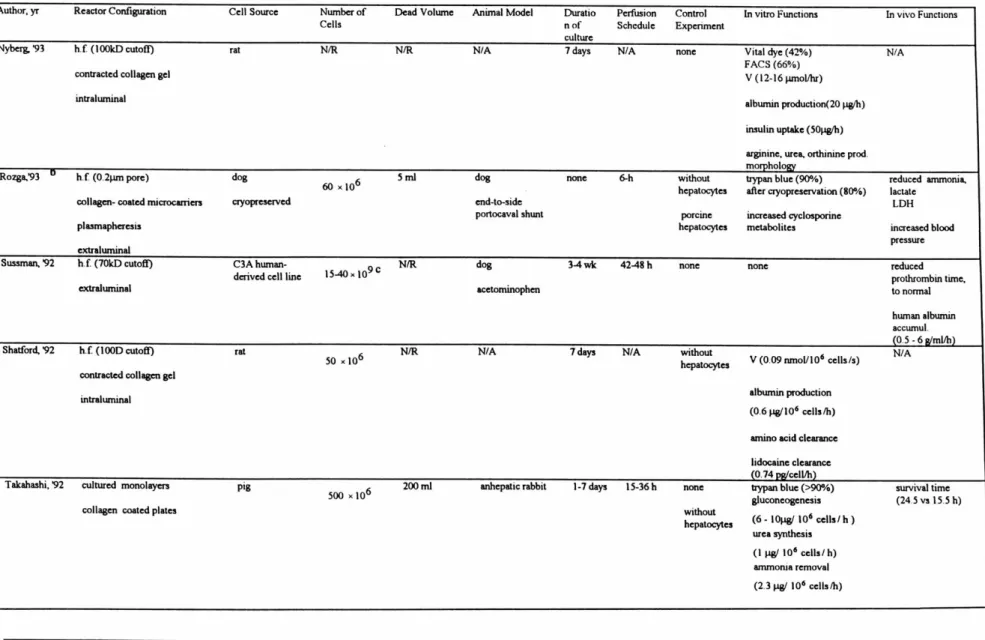
Table 1.1 A. In Vitro and Animal Studies Utilizing Extracorporeal Bioartificial Liversa
Author, yr Reactor Configuration Cell Source Number of Dead Volume Animal Model Duratio Perfusion Control In vitro Functions In vivo Functions
Cells n of Schedule Experiment
culture
Nyberg. 93 h.f. (100kD cutoff) rat N/R N/R N/A 7 days N/A none Vital dye (42%) N/A
FACS (66%)
contracted collagen gel
V (12-16 pmol/hr) intraluminal
albumin production(20 pg/h)
insulin uptake (50pg/h)
arginine, urea, orthinine prod. morphology
Rozga,'93 h.f (0.2pm pore) dog 6x 6 5 mil dog none 6-h without trypan blue (90/0) reduced ammonia,
hepatocytes after cryopreservation (SO%) lactate
collagen- coated mcrocamers cryopreserved end-to-side LDH
portocaval shunt porcine increased cyclosporine
plasmapheresis hepatocytes metabolites increased blood
pressure extraluminal
Sussman,'92 hf. (70kD cutoff) C3A human- 9 C N/R dog 34 wk 4248 h none none reduced
derived cell line 15-40 x 10 prothrombin time,
extraluminal acetominophen
to normal human albumin accumul. (0.5 - 6 g/ml/b)
Shatford, '92 h.f (IOOD cutoff) rat N/R N/A 7 days N/A without N/A
hepatocytes V (0.09 nmol/106 cells/a)
contracted collagen gel
albumin production (0.6 pg/'10 cells /h) amino acid clearance lidocaine clearance (0.74 pg/cell/h)
Takahashi, 92 cultured monolayers pig 6 200 ml anhepatic rabbit 1-7 days 15-36 h none trypan blue (>90%) survival time
500 X 10 gluconeogenesis
(24.5 vs 15.5 h)
collagen coated plates without
hepatocytes (6 - 1p/ 10' cells/ h) urea synthesis (I pg/ 106 cells / h) ammonia removal (2.3 pg/ 10' cells /h)
a h.f. = hollow fiber, N/R = not reported; N/A = not applicable, V= oxygen uptake rate
bDmetnou, 1986A
Shnyra, '91 microcarrier rat 6 1.6 ml rat 1-5 days 3-h without trypan blue (80%) survival Shnyra. 90 fibronectin-coated 50 -10 hepatocytes (60% vs 5%)
D-galactosamine albumin production
packed column (4-5 pg/06 cells/h) decreased GOT,
GPT, bilirubin pre-oxygenated with
perfluorodecalin bilirubin
Yanagi, '89 rotating disks rat, rabbit 9 80 ml cat none 4-h without trypan blue (20-50%) ammonium
1.7 10 hepatocytes removal
cells embedded in algmate portojugular ammonium removal hydrogel with DMSO shunt, ligation of
hepatic artery urea synthesis no plasmapheresis
Amaout, '90 h.f (0.2 pm pore) rat 3 6 7 ml Gunn rat none 3-4 h without trypan blue (30-85%) conjugation of 30-40 x 10 hepatocytes 5 pM bilurubin
collagen-coated microcarriers cryopreserved
extraluminal
Uchino,'88 cultured monolaycrs dog 9 f not reported anhepatic dog 1-14 24-65 h no glyconeogenesis survival
6 xl0 days treatment (110 ng/ug DNA/min) (55 vs 27.8h)
collagen-coated borosilicate
without urea synthesis albumin decline
multiplated hepatocytes (3.6 ng/ug DNA/min) reduced
plasmapheresis albumin production ammonia normal
(0.12 or 1.2 pg/106 cells /h) bilirubin maintained at ammonia removal
amino acids not elevated
decreased bleeding Demetriou, microcarriers (packed column) rat 10 ml N/A none N/A N/A trypan blue (55-60%) N/A
'86 160 x 106
collagen coated cryopreserved increased ratio of conjugated/ unconjugated bilirubin
albumin production
Jauregui, '84 h.f (50 kD cutoff) rat not reported N/A N/A 1-18 N/A N/A LDH leakage N/A
days
collagen-coated glucose uptake
extraluminal total protein (30% drop)
Kasai,'84 h.2f (40 kD cutoff) dog 9 not reported dog none I-h on, none trypan blue (85-60%) ammonia removal.
I -h off 0.083 pgt
hepatocyte suspensions (viable cells) D-galactosamine 6-h total ammonia removal 106 cells /h
plasmapheresis BUN production
(2. 5pg/106 cells/h)
glucose
glucose production maintenance (36.8 pig/g 106 cells /h)
Hager, '83 h.f (50 kD cutoff) mice 6 N/R N/A 1-58 N/A N/A protein synthesis N/A
10 10 days (0.23 mg/ml/day)
extraluminal
urea accumulation deamination of cytidine
h.f ; 100 kD cutoff rat 6 N/R N/A 1-6 days N/A none V (0.09 - 0.03 nmol/ N/A
Shatford, '91 38 x 1061'cll s
contracted collagen gel
Albumin production
(0. 28 -0.45 pg / 106 cells /h)
lidocaine clearance
(I pg/cll/h)
Hager, '78 h.f (50 kD cutoff) neonatal mice N/R N/R N/A 6-8 wks N/A none diazepam metabolism N/A
extralummnal uridine (65.9 pM/h)
urea
(increased 13% in 15 days)
Olumide, '77 dialysis against hepatocyte pig N/R N/R anhepatic pig none 2- 4h without trypan blue (95-100/) survival suspension (5-10 kD cutoff) hepatocytes (34.8 vs 35 h)
pyruvate metabolism:
(decreased 70% in I h) lightened coma
V ( 0.25-0.01 m/g of glucose liver/min) maintenance BUN decrease (2.4 mg % in 36 h) bilirubin conjugation Wolf, '75 hf. (30-50 kD cutoff) hepatoma 6 N/R Gunn rat 1-43 N/R none conjugated bilirubin bilirubin
Reuben- 10 - 15 x10 days total bilirubin conjugation
extralumial H4-II-E
glucosen consumption
LDH, GOT, GTP
accumulation
g approximately 40 g
Table 1. 1B. Clinical Findings from the Utilization of Extracorporeal Bioartificial Livers
Author, year Reactor Cell Source Number of Cells Dead Volume Duration of Treatment Control Findings
Configuration culture Schedule Experiment
Neuzil, '93 h.f (0.2pm pore) pig 9 N/R none 6-h none increased mental status after 2 h; began to
collagen-coated I X10 deteriorate 12 h post support
microcamers cryopreserved
extraluminal patient's liver function and mental status
improved over 3 weeks and patient received orthotopic liver transplantation after 6 mo. ammoia (120 to 32 pAM/L)
increased clotting factors; oozing recurred 15 h post support
twofold increase in most amino acids Porcine hepatocyte viability post support(900/)
Sussman, '92 h.f C3A-human N/R 3-4 wk 144-h none increased mental status in I h
derived cell line 15-40 -09
new cartridge patient died 132 h post support every 5-36 h
decreased bilirubin (20 mg/dL) decreased alkaline phophatase
increased c-fetoprotein demonstrates recovery of native liver function
Margulis, '89 hemoperfusion porcine N/R 20 ml none I-h per cartridge, 67 patients (30 in 59 patients (20 in coma, 39 in precoma)
through a cell 6-h total coma, 37 in survival (63% vs 41%)
suspension with precoma) treated
actived charcoal with commonly coma lightened
used curative
measures free bilhrubin decreased 45.5% ammonium decreased 50.3% EEG increased in normalcy
Matsumara'87 hemodialysis rabbit 325 ml none 5.2-h on, heat-deactivated mentation, appetite retured
against cell cryopreserved 10 x 103 2 days off, control
suspension 4.5-h on patient discharged
plasmapheresis decreased bilirubin (25 to 16.8 mg/dL)
morphology nonpolygonal
in vitro (urea synthesis, lactate to glucose conversion )
extraluminal compartment and transport takes place through the membrane of each fiber (Berthiaume et al., 1993). In addition, some investigators have coated the fibers to promote hepatocyte attachment or pre-attached cells to microcarriers which were
subsequently inserted in a hollow fiber device. Table 1.1 displays the range of bioartificial extracorporeal devices that have been reported. Table 1. 1A summarizes in vitro and animal studies whereas Table 1. LB contains clinical data.
Two classes of limitations become apparent in the examination of Table 1.1 A: transport issues and problems with culture stability. Hollow fiber devices, are prone to transport limitations because of the relatively large distance between the perfused fluid and the peripheral cells. In addition, the interfiber distance is difficult to control, creating variations in transport distance, thereby limiting the efficiency of extraluminal seeded devices. Studies by Nyberg et al. (1993) and Wolf et al. (1975) have included data
indicating necrotic cell masses. Microcarriers, in contrast, suffer from transport limitations in their scale-up. The two studies which utilized packed columns have a small cell number of 50- 160 x106 (Shnyra et al., 1991 and Demetriou et al., 1986B). The scale-up of these
devices will be hampered by transport limitations as the column height increases. All nutrients being provided at the entrance must traverse the entire device. If one increases the flow rate to ameliorate this problem, packed bed configurations generate large shear stresses associated with a greater fluid velocity through small pathways. Furthermore, the amount by which the column diameter can be increased to reduce the fluid velocity, has a practical upper limit in relation to the column height. In fact, the aspect ratio
(height/diameter) is limited to 1:1 by the practicality of evenly distributing the liquid over the entire column cross section (Berthiaume et al., 1993). Other bioreactor
configurations, such as vertical disks rotating in a bath of fluid, face limitations from the low hepatocyte viability and the dissolution of the embedding gel (Yanagi et al., 1989). Finally, monolayer culture configurations necessitate a large dead volume which is clinically impractical (Takahashi et al., 1992).
Table 1. lA also elucidates a number of problems with the stability of hepatocyte cultures in these devices. All investigators who measure cell viability in primary
hepatocyte systems found cultures were stable for less than 7 days. Hepatocyte suspensions, in fact, fair even worse with a cell viability on the order of hours. Furthermore, some investigators utilized freshly thawed, cryopreserved, primary hepatocytes even though other studies indicate that cryopreserved hepatocytes to not regain stable, differentiated function for several days while the cells recover from freezing stresses (Borel Rinkes et al., 1992). Finally, the device which included a C3A human-derived cell line is difficult to assess (Sussman et al., 1992). This device may potentially have problems due to the utilization of transformed cells, cell growth, or maintenance of liver-specific functions.
The aforementioned transport and culture stability limitations have hindered the progress of any large scale clinical trials. A few studies have been reported as seen in Table 1. 1B. The primary effect of the extracorporeal bioartificial devices was an increase in mental status in the treated patients with a drop in serum ammonia and bilirubin concentrations. The treatments are difficult to assess because of the general lack of any controlled studies. One study did compare hemoperfusion through a cell suspension with active charcoal in addition to conventional acute liver failure treatment as compared to acute liver failure treatment alone (Margulis et al., 1989). They found a 63% survival rate as compared to a 41% survival rate in the controlled group. On closer inspection of the data, it should be noted that only 33% of the hemoperfused group were initially comatose whereas 45% of the conventionally treated group were already comatose, therefore, the margin of increased survival with hemoperfusion through a cell suspension may be effectively decreased.
-A
1.4 Micropatterning
Micropatterning techniques rely on creating surfaces with selective adhesiveness on which cells can be organized into microstructures. This technology may also be used in
designing a bioartificial liver system which mimics the sandwich culture configuration (Figure 1.1) as well as the efficient transport properties of the liver. A liver lobule, the functional unit of the liver is shown schematically in Figure 1.2. Cells are aligned in rows and stacked vertically in plate-like structures. The blood flows along the sinusoids, on both sides of the hepatocytes, where it comes in contact with the cells and mass exchange takes place. Figure 1.3 shows a schematic of a micropatterning approach to mimicking the liver geometry in hopes of preserving differentiated cell function and facilitating mass transfer between the perfused fluid and the hepatocytes. This figure depicts hepatocytes aligned in rows with dimensions on the order of 50-200 pm. These micropatterned hepatocytes are sandwiched between two layers of collagen and perfused on both sides by fluid flow through the microchannels. Rows of hepatocytes, in the sandwich
configuration, alternate with channels of fluid flow allowing for efficient mass transfer and preservation of differentiated function. The sites to which the hepatocytes adhere may be a biocompatible, adhesive surface (AS), whereas the hepatocyte-free sites for fluid flow may be created using some non-adhering surface (NAS). Creation of such a
micropatterned device in the sandwich configuration will requires selective adhesion of hepatocytes on a single underlying substrate.
Although there have been no attempts to use selective adhesion to create a liver support device, investigators have been interested in the ordered arrays of cells in vitro for a variety of reasons: tumor invasion, wound healing, embryogenesis, mechanisms of cell locomotion and orientation, synapse formation and the creation of bioelectric circuits in culture. Weiss (1945), seeking to find support for his theory of contact guidance, first
Hepotic Lobule Central Vein - Sinusoid Bile Conoliculus --&. Ductide - -tl venule Hepatocyte-
-Figure 1.2. Schematic of the liver lobule. Blood flows inward from the portal triad (i.e. portal venule, hepatic arteriole, and bile ductule) along the sinusoid and to the central vein (Yarmush et al., 1992B).
Non-Adhering Biocompatible Surface
flo W in Hepatocyte
Figure 1.3. Schematic of a hypothetical micropatterned device. Hepatocytes are
sandwiched and aligned along a collagen-coated adhesive surface in alternating rows. The intermediate rows are composed of a non-adhesive, biocompatible surface and are
plated a suspension of cells on a glass substrate ruled with fine parallel grooves. Later, Carter (1965) coined the term haptotaxis 1 to describe passive cell movement directed by the relative strength of its peripheral adhesions. Since then many studies have reported different types of cellular patterning (see Table 1.2). Most recently, with the advent of photolithography, a number of groups have published results indicating selective adhesion of cells can be obtained on the order of 20 to 150 pm. Britland et al. (1992B) describe patterning of Baby Hamster Kidney (BHK) cells whereas Stenger et al. (1992)
demonstrate the selective adhesion of porcine aortic endothelial cells and fetal rat hippocampal neurons directly to a photochemically modified substrate. In contrast, Matsuda et al. (1992) achieved selective adhesion of rat-derived PC-12 cells and neuroblastoma C6 glial cells to a collagen treated substrate. None of these studies examined the toxic effects of the surfaces or the function of the adhered cells over time. Furthermore, none of these studies utilized selective adhesion for potential use in a bioreactor configuration.
1.5 Scope of This Study
Our overall goal is to create the micropatterned device shown in Figure 1.3. for use in bioartificial liver development. The critical experimental parameter in obtaining this goal is the creation of ordered arrays of hepatocytes. Thus, the specific aim of this study was to obtain selective adhesion of hepatocytes to a single substrate with the ultimate goal of creating a micropattern. The critical design parameters for the construction of the device can be approximated by mathematical modeling of the oxygen distribution and viscous pressure drop along the channels.
The experimental portion of the study utilized collagen as an adhesive molecule. As an abundant component of the extracellular matrix, it is known to promote
Table 1.2. Studies Utilizing Cellular Patterninga
Investigator, Application Material Pattern Resolution Cells
year [pm] bioelectnc circuits range Matsuda, '92 Britland, '92A,B Baier, '92 Corey, '91 Matsuda. '90 Hammarback '88 Klebe. '88 Kleinfeld. '88 Ireland, '87 Dow, '87 Gundersen. '87 Dunn, '86 Brunette, '86 Hammarback, '86 Gundersen. '85 Hammarback. '85 Tumer. '83 Dunn, '82 Albrecht-Buehler, '79 Cooper , '76 Letoumeau. '75 Harns, '73 Rovensky, '71 Carter, '67 Carter, '65 Weiss. '45
indium tin oxide, glass
organosilanes
neurocircuits biosensors
retinal nerve cell guidance in development
synapse formation
neurocircuits
contact guidance and density deposition of fibronectin cytoarchitecture and elect. activity of nerv. tissue
limiting spreading and intercellular contact alignment,
electron microlithography
biological substrata
migration and orientation
dentistry
guidepost theory
growth cone age and guidance neurite outgrowth fibronectin
contact guidance immune response, cancer, development synapses in culture embryogenesis locomotion locomotion / orientation cytokinesis cancer contact inhib contact guidance Aizawa, '92 Stenger,'92
copolymer with collagen depostition of bioactive peptides
glyco-protein suspensions,glass polylysine, glass
copolymer with phenylazide laminin, fibronectin collagen fibronectin, agar silane derivatives-quartz
poly (HEMA)- Palladium
glass IV collagen, laminin, fibronectin quartz silicone agarose-albunin, laminin adsorbed NGF on collagen laminin
fibronectin, urea, glass
cell. acetate-glass gold, albumin, glass
silicon monoxide
collagen, polystyrene, Palladium
cell. acet- Palladium, glass
PVC, nickel cellulose acetate- Palladium cellulose acetate-Palladium glass. mica lines lines outlined polygons outlined hexagons lines lines lines lines lines letters squares grids circles step grooves spirals circles lines lines squares lines circles lines fibrils of fibronectin lines lines radial lines squares lines squares, lines circles squares lines lines
line. D = diameter of circle or width of square. T= spatial frequency
not reported W,1=100 W= 1-20 T = 200 not reported W=1-2 T= 1-2 W=90 W=3-10 T= 80-160 W=20-200 W= 7-10 W=25-100 W=50-100 t= 50-100 D= 22.5- 80 range >0.3 D= 300 W=1-9 T =3-32 D= 5-92 W=36-162 D=100 W=40-50 300 W=1 W=0.7-8 T =5-10 W=10-50 W=17 W=40 D=80 W=30 D=50-85 W=33 D=5-65 W= 100-200 100-400 not reported not reported neuron like PC-12 porcine endothelial rat hippocampal neuron like PC- 12 baby hamster kidney
embryonic chick retinal nerve cells hippocampal neurons
bovine endothelial range
fibroblasts mouse spinal and rat cerebellar fibroblasts
baby hamster kidney, chick myocytes
dorsal root ganglia
chick heart fibroblasts
human gingivial fibroblasts dorsal root ganglia
dorsal root ganglia dorsal root ganglia range
chick heart fibroblasts
3T3 fibroblasts mouse clonal neuroblastoma chick embryo range fibroblasts mouse fibroblasts mouse fibroblasts
rat Schwann cells
differentiated function of hepatocytes. Surface coatings with adhesive and non-adhesive properties after exposure to an aqueous collagen solution were characterized for their hepatocyte-surface interaction. The adhesive surface (AS) has hydrophilic characteristics allowing adsorption of collagen molecules from an aqueous solution, and subsequent hepatocyte adhesion, whereas the non-adhesive surface (NAS) has hydrophobic properties and remains hepatocyte-free. Furthermore, the combination of AS and NAS results in preliminary patterns on a large grid with dimensions on the order of centimeters. A reproducible processing technique for obtaining these patterns was developed and optimized. The selective adhesion was shown to correlate with preferential protein deposition on the AS. Finally, the morphology and long-term function of hepatocytes was assessed by overlaying the patterned hepatocytes with a top layer of collagen gel to mimic sandwich culture. They were found to be normal as compared to stable, differentiated sandwich cultures.
The modeling portion of this work provided optimal channel lengths and
associated flow rates for a typical microchannel in the hypothetical micropatterned device. The creation of a micropatterned flow chamber can now be attempted: first, by the
application of the selective adhesion process to micropatterns on a single substrate, and then by the design and construction of a sandwich-type device.
~IF
CHAPTER II
SELECTIVE ADHESION OF HEPATOCYTES
2.1 Introduction
The experimental portion of this study was directed towards obtaining reproducible, selective adhesion of hepatocytes to a solid substrate. This approach will ultimately be applied to the design and construction of a microchannel device, as described previously. The microchannel device will potentially have application as an extracorporeal bioartificial liver.
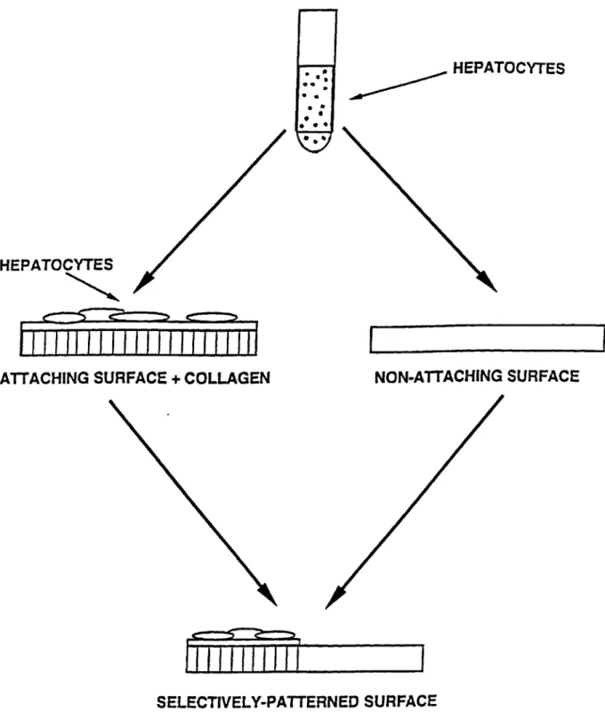
The theory behind achieving the selective adhesion of hepatocytes is based upon the process depicted in Figure 2.1. Polymer coated glass substrates are exposed to a collagen solution. The polymer coatings have differential wettability. The wettable, hydrophilic coating has a small contact angle whereas the non-wettable, hydrophobic coating has a much larger contact angle. Exposing these coatings to a water-based collagen type I solution causes wetting and subsequent deposition of collagen molecules on the hydrophilic surface whereas the hydrophobic surface remains bare. Collagen, known to be an adhesive molecule with corresponding integrin receptors on the cell surface, causes hepatocytes seeded on the two individual surfaces to preferentially adhere to the collagen-coated surface.
Combining the hydrophobic and hydrophilic coatings onto a patterned grid on the same glass substrate would create the base of a microchannel device. The patterned substrate can be manufactured by standard photolithographical techniques. The surface is then treated with a collagen solution and seeded with hepatocytes. Subsequent agitation of the hepatocyte-substrate complex causes removal of any loosely bound hepatocytes from the hydrophobic coating while hepatocytes remain adhered to the collagen-coated,
HEPATOCYTES
I I
ATTACHING SURFACE + COLLAGEN
HEPATOCYTES
NON-ATTACHING SURFACE
1111111111
SELECTIVELY-PATTERNED SURFACE
hydrophilic polymer regions. Finally, the patterned base of the microchannel device is obtained with hepatocytes aligned in regular, alternating rows.
This study focused on the characterization and optimization of selective adhesion of hepatocytes on a large-scale pattern of the two coatings prior to applying the
technology to smaller 'micropatterns'. Specifically, attachment efficiency, spreading and toxicity were investigated on individual surfaces. A reproducible process was developed and optimized for obtaining selective adhesion on a large grid substrate. Finally,
differentiated, sustained function of hepatocytes in the selectively adhered configuration was demonstrated.
2.2
Materials and Methods
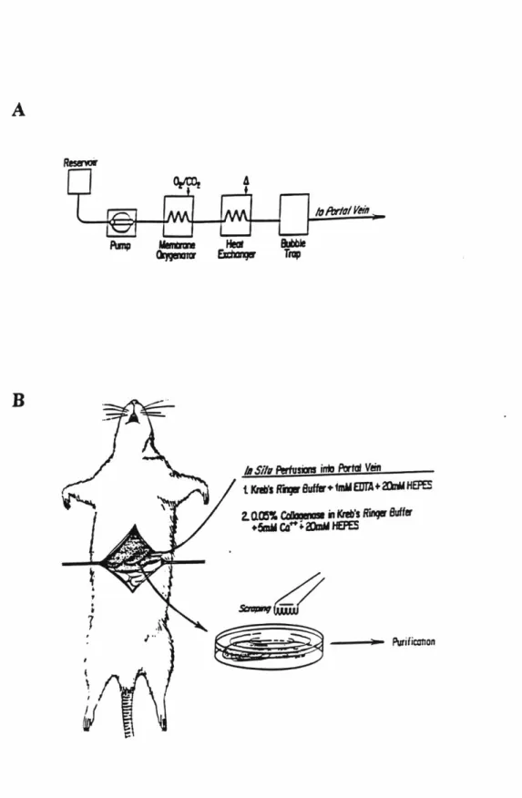
2.2.1 Preparation of Hepatocytes
Hepatocytes were isolated from 2-3 month-old female Lewis rats (Charles River, MA), weighing 180-220 g, by a modified procedure of Seglen (1976) and as described in detail elsewhere (Dunn et al, 1991). Figure 2.2 depicts a schematic diagram of the procedure used for isolating rat hepatocytes. Animals were anesthetized in a chamber containing saturated ether. The liver, weighing 7-8 g, was first perfused through the portal vein in situ with 400 mL of perfusion buffer with 1mM ethylenediaminetetraacetic acid (EDTA) at 45 mL/min. Perfusion buffer is 154 mM sodium chloride, 5.6 mM potassium, 5.5 mM glucose, 25 mM sodium bicarbonate, and 20 mM N-(2-hydroxyethyl)piperazine-N'-2-ethanesulfonic acid (Hepes), pH 7.4 (see Figure 2.2A).I The perfusate was equilibrated with 5 L/min 95% 02 and 5% CO2 through 5 m of silicone tubing (inner diameter 0.058
in., outer diameter 0.077 in.) and was maintained by a 100-mm heat exchanger (reflux
IUnless specified, chemicals were purchased from Sigma (St. Louis, MO), Aldrich (Milwaukee, WI), EM Science (Gibbstown, NJ), JT Baker (Phillipsburg, NJ), and Mallinckrodt (Paris, KY).
A
-mff
wG nOurm ExWg Tap
B
A, Sio Pefusions ii d Putr Vein
i Krjis riuri U KiEnM+ RMw mmE
a.m% C eaU in KE'S RIr er
+5ws C+mm E
s wV .zw
Purificanon
condenser 283000, Kontes, Vineland, NJ) at 37 *C before entering the liver. The liver was subsequently perfused with 200 mL of 0.05% type IV collagenase (Sigma) in
perfusion buffer with 5 mM calcium chloride at the same flow rate. During this time, the liver swelled to about twice the original size. The swollen liver was dissected away from ligaments and the diaphragm and was transferred to a 100-mm dish with 20 mL of ice-cold perfusion buffer (see Figure 2.2B). The liver capsule was teased apart, and the resulting cell suspension was filtered through two nylon meshes with grid sizes 250 and 62 Am (Small Parts, Miami, FL). The cell pellet was collected by centrifugation at 50g for 5 min.
Cells were further purified by a modified procedure of Kreamer et al. (1986). The cell pellet was resuspended to 50 mL, and 12.5 mL of cell suspension was added to 10.8 mL of Percoll (Pharmacia, Piscataway, NJ) and 1.2 mL of lOx HBSS. Hanks' balanced salt solution (IX HBSS) is 138 mM sodium chloride, 5.4 mM potassium chloride, 0.33 mM sodium phosphate, 0.33 mM potassium phosphate, 0.8 mM magnesium sulfate, and
5.6 mM glucose, pH 7.4. The mixture was centrifuged at 500g for 5 min, and the cell
pellet was washed twice with Dulbecco's modified Eagle's medium (DMEM) with 4.5 g/L glucose (Gibco, Grand Island, NY). Routinely, 200-300 million cells were isolated with viability between 92% and 99% as judged by trypan blue exclusion. Nonparenchymal cells, as judged by their size (less than 1 Opm in diameter) and morphology (nonpolygonal or stellate), were less than 1%.
2.2.2 Preparation of Rat Tail Collagen
Type I collagen was prepared from Lewis rat tail tendons by a modified procedure of Elsdale and Bard (1972). Four tendons were dissected from each rat tail and stirred in 200 mL of 3% (v/v) acetic acid overnight at 4*C. The solution was filtered through four layers of cheesecloth and centrifuged at 12000g for 2 h. The supernatant was precipitated with 40 mL of 30% (w/v) sodium chloride, and the pellet was collected by centrifugation at 4000g for 30 min. The pellet was dissolved in 50 mL of 0.6% (v/v) acetic acid, and the
solution was dialyzed against 500 mL of ImM hydrochloric acid five times. For
sterilization, 0.15 mL of chloroform was added to the solution. The solution was stirred for 2 days loosely capped to allow evaporation of chloroform. A 5-mL aliquot was lyophilized and weighed to determine the yield of collagen. Generally 100 mg was isolated per rat tail. This preparation yields type I collagen molecules mostly in its native, not cross-linked, triple-helical form (Elsdale and Bard, 1972).
2.2.3 Hepatocyte Culture
Hepatocyte culture was performed on a variety of collagenous and non-collagenous substrates. Standard single gel refers to a gel consisting of 9 parts collagen solution at 1.11 mg/ml, and 1 part I OX DMEM, pH 7.4, chilled on ice, mixed just prior to use. Collagen forms a gel at physiological pH and ionic strength at room temperature, but the rate of gelation is accelerated at higher temperature. Standard sandwich culture utilizes an overlay of the same collagen gel solution on top of the hepatocytes.
Some collagen gels were formed without the use of DMEM. Gels were formed by utilizing a salt solution consisting of six times the inorganic salt concentration found in DMEM. This salt solution had the following composition: 5.5 mM sodium phosphate, 10.8 mM calcium chloride, 4.9 mM magnesium sulfate, 32.3 mM potassium chloride, 0.264 M sodium bicarbonate, and 0.657 M sodium chloride brought to pH 7.4. Dilute collagen gels of 0.5 mg/ml collagen (1/2 as concentrated as standard collagen gel) were formed by mixing 5 parts of 1.11 mg/mI collagen gel solution, 3.3 parts of distilled deionized water, and 1.67 parts of the salt solution. Collagen gels of higher
concentrations (i.e., greater than standard gel concentration) were prepared by lyophilizing 40 mL of 1.11 mg/mL collagen gel solution in a FTS Systems lyophilizer (Model FD-3-55A-MP, Stone Ridge, NY). The collagen was weighed and dissolved in 10 ML of imM
solution was then used in conjunction with 1.67 parts salt solution and distilled deionized water to obtain intermediate collagen gel concentrations.
Collagen gels were applied by distributing 1 mL of the appropriate collagen gel solution evenly over a 60-mm tissue culture dish (Falcon, Lincoln park, NJ) and incubated at 37*C at least 1 h before use. Two million viable cells were seeded in 2 mL of medium, consisting of DMEM supplemented with 10% (v/v) fetal bovine serum (Hazleton, Lenexa, KS), 0.5 unit/mL insulin (Squibb, Princeton, NJ), 7 ng/mL glucagon (Lilly, Indianapolis, IN), 20 ng/mL epidermal growth factor (Collaborative Research, Bedford, MA), 7.5 p. g/mL hydrocortisone (Upjohn, Kalamazoo, MI), 200 units/mL penicillin (Hazleton), and 200 pg/mL streptomycin (Hazleton). This constituted the underlying, single gel, system. For the sandwich systems, an additional 1 mL of collagen gel solution was distributed over the cells after 1 day of culture at 37 *C and 10% CO2. Culture medium was first removed
and care was taken to ensure that the second layer of collagen gel was evenly spread over the entire dish. Thirty minutes of incubation at 37*C was allowed for gelation and
attachment of the second gel layer before the medium was replaced. Culture medium was changed daily. The collected media samples were stored at 4*C prior to analyses.
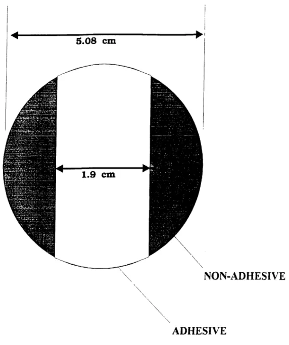
2.2.4 Surface Specifications
Disks of glass (5.08 cm diameter) with various coatings were obtained from Cytonix (Beltsville, MD). Adhesive surfaces (AS) were formed by spin-coating with urethane epoxy and curing in ultraviolet light. Non-adhesive surfaces (NAS) were formed by overcoating with a solution of a perfluorinated polymer. 'Banded' patterns were created by masking the urethane epoxy coated surface with a single strip of adhesive tape of 1.9 cm width. Disks were then overcoated with a perfluorinated polymer followed by removal of the adhesive tape. A banded surface is depicted in Figure 2.3.
5.08 cm
1.9 cm p.
NON-ADHESIVE
ADHESIVE
Figure 2.3. A banded surface with adhesive (AS) and non-adhesive (NAS) regimes.
2.2.5 Processing of Surfaces
All surfaces were washed for 2 min in 3 mL of distilled, deionized water by shaking at 400 RPM on a shaker (Model R-2, New Brunswick Scientific, Edison, NJ).
For patterning, dilute (non-gelling) collagen solutions (0.01% (w/v)) were prepared by mixing 1 part of 1.11 mg/ml collagen solution with 9 parts of distilled, deionized water. 0.01% (w/v) bovine serum albumin (Sigma, Lot Number 118F005) solution and 0.01% (w/v) PL (Sigma) were prepared in distilled, deionized water.
Surfaces were coated with various solutions, described in section 2.2.3, by
immersion into 300 mL of the appropriate solution at a 900 angle and a rate of Imm/s by a modified Harvard Syringe pump (Cambridge, MA) at 40C. Surfaces were then placed in 60-mm tissue culture dishes. In addition, some surfaces were spin-coated at 4*C by utilizing the following procedure. Surfaces were clamped to the center of a modified centrifuge rotor (Dynac, Cat # 0101, Parsippany, NJ). The technical drawing of the machined accessory is depicted in Figure 2.4. A fixed volume of solution (2 ml) was pipetted onto the center of the surface over 5 seconds. Solution was allowed to spread for 5 s prior to spinning. Spinning was done at 500 RPM (centrifuge setting of 18) for 20s. The brake was applied for a duration of approximately 5 s until rotation had ceased. Surfaces were then unclamped, and placed in a 60-mm tissue culture dish (Falcon).
Following processing, all surfaces were incubated at 37 *C and 10% CO2 for 30
min. Dilute collagen coatings evaporated during this time whereas gelation occurred in the salt-based collagen solutions. Surfaces that were analyzed for differentiated, long-term function were pre-treated by ethylene oxide gas sterilization.
MATERIAL: STAINLESS STEEL DIMENSIONS: IN MM BASE PIECE: 53
V,
0
TOP VIEW 53 38 3 ...~j... 110i).-
4
AA CROSS SECTION CORNER PIECE: i-ss TOP VIEW Figure 2.4. Technical drawing of machined coating.BB CROSS SECTION
accessory to modify a centrifuge for
-A4
2.2.6 Attachment Assay and Morphological Measurements
As described above, two million freshly isolated, viable cells were seeded in 2 mL of medium on the appropriate pre-treated surface or single layer of collagen gel in a 60-mm tissue culture dish (Falcon). Cells were spread evenly and incubated for 45 minutes at 37*C and 10% CO2. The following washing procedure was repeated twice. Unattachedcells and medium were then aspirated and 3 mL of medium was added to each dish. Dishes were agitated at 200 RPM on a shaker (New Brunswick Scientific) for 4 minutes. After 2 minutes, the orientation of the dishes was changed by rotating each dish 90 *and shaking was resumed for the remaining 2 minutes. Finally, media was aspirated and 2 mL of media was added to each dish. Cells were recorded at 50X original magnification using a video system consisting of an Olympus microscope (CK2, Japan), camera
(Hamamatsu C-2400, Japan), monitor (Sony PVM1343MD, Japan), and VCR (Panasonic, AG-6750, Japan) as described in detail elsewhere (Rotem et al., 1992). The number of remaining cells in 10 random fields were counted from the recorded images using an image analysis system (Argus 10, Hamamatsu) and compared to the number of remaining cells on a single layer of collagen gel which had been similarly treated. Morphological measurements were made by recording the cells at 200X magnification after 24 h of incubation at 37 'C and 10% CO2. The projected surface area (PSA) of the cells was
analyzed from the recorded images using an image analysis system (Argus 10) which had been previously calibrated using a hemocytometer grating.
2.2.7 Biochemical Analysis
Collected media samples were analyzed for rat albumin content by enzyme-linked immunosorbent assays (ELISA). Chromatographically purified albumin was purchased from Cappel (Cochranville, PA). The 96-well plates (NUNC-Immuno Plate, Maxisorp, Newbury Park, CA) were coated with 100 jiL of rat albumin in 25 mM carbonate buffer, pH 9.6, overnight at 4 C. The wells were washed four times with PBS plus 0.5% (v/v)
Tween 20 (PBS-Tween). Fifty microliters of sample was mixed with an equal volume of antibody (800ng/mL in PBS-Tween) before it was transferred to the wells. After
overnight incubation at 4 'C, the wells were washed four times with PBS-Tween and were developed with 100 pL of 25 mM citrate and 50mM phosphate, pH 5, containing
0.4mg/mL o-phenylenediamine by the conjugated peroxidase. The absorbance was measured at 490 nm with the a Dynatech (Chantilly, VA) MR600 microplate reader. Positive controls included known concentrations of purified rat albumin added to the culture medium and negative controls included the culture medium and PBS-Tween. Concentrations of standards were calibrated by their absorbances at 280 nm, by using 0.6 as an extinction coefficient for 1 mg/mL solutions of albumin. Concentrations of samples were determined from a standard curve generated for each ELISA plate. Absolute rates of secretion were calculated from the concentration by multiplying the total volume of the medium and dividing by the elapsed time. Results were given in micrograms per hour per 2x 10⁶ cells .
2.2.8. Atomic Force Microscopy
Two banded surfaces with regions of AS and NAS were analyzed by Imaging Services (Santa Barbara, CA). Both disks were washed by agitation and 500 rpm for 1 minute in distilled, deionized water. One disk was imaged without any further treatment whereas the other sample was spin-coated with 2 ml of 0.1 mg/ml collagen solution at 500 rpm for 25 seconds.
2.2.9 Statistics and Data Analysis
Generally, two duplicate wells were averaged for each ELISA sample. Experiments were repeated two to three times. The absolute secretion rate of the control culture on the
missing data points. Attachment data was quantified by dividing number of cells attached by the number of cells attached to a single layer of collagen gel (control) and expressed as a percentage. Each data point represents the average of 10-20 fields. Error bars indicate standard deviation of the mean. Morphological measurements were quantified by
analyzing the projected surface area for 5 random cells in 10 random fields, per condition. Error bars indicate standard deviation of the mean.
2.3 Results
Hepatocyte Culture
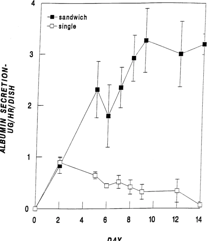
Albumin secretion is typically utilized as a marker for differentiated hepatocyte function because it involves many complex cellular functions such as transcription, translation, intracellular packaging and transport, and secretion. Figure 2.5 shows the albumin secretion of cells cultured in two different collagen gel configurations, namely; a single layer of collagen gel and sandwich culture. In the sandwich configuration, albumin secretion shows a marked increase followed by a plateau at day 8 at approximately 3 pg/h/2 x 106 cells. In contrast, single gel culture shows a maximum at day 2 at 0.89 + 0.1 pg/h/2 x 106 cells followed by a steady decline in albumin secretion and differentiated function. In the original study by Dunn et al (1991), similar trends were reported for albumin and other proteins. These results indicate the importance of utilizing a sandwich configuration to create a stable, differentiated culture system.
The aforementioned experiments were done using a collagen gel concentration of 1 mg/ml. In order to test the effect of varying the collagen concentration in the extracellular matrix on the differentiated function of hepatocytes, a series of experiments were
performed where the collagen gel concentration was varied between 3 and 0.5 mg/ml. Figure 2.6 shows the albumin secretion of hepatocytes sandwiched between two gels of varying concentrations. Altering the collagen concentrations within the examined range
-~1
4
-+-sandwichsingle
3
00
LUj ~CL"
2
0
2
4
6
8
10
12
14
DAY
Figure 2.5. Albumin secretion of hepatocytes cultured in sandwich and single gel configurations.
seemed to have no effect on albumin secretion. All cultures showed an increase in albumin secretion to a maximal level around day 8. Lower collagen gel concentrations were not investigated because a stable gel does not form below a concentration of 0.4 mg/ml.
Surface Characterization
Adhesive (AS) and non-adhesive surfaces (NAS) were characterized before and after surface coating utilizing atomic force microscopy (AFM). The methodology for surface coating with collagen is described in the previous section. Figure 2.7 shows
3-dimensionally rendered AFM images of untreated and treated (by spin-coating with a collagen solution), AS and NAS, on a banded surface. The term 'banded surface' refers to circular 5.08 cm diameter surfaces with a 1.9 cm strip of AS in the center flanked by NAS as depicted in Figure 2.3. As seen in Figure 2.7, spin-coating a banded surface resulted in selective adsorption of collagen to the AS with no observable change in the NAS. Table 2.1 summarizes the roughens of the four surfaces.
Table 2.1. Comparison of surface roughness for various sites on a banded surface.a
UNTREATED [nm] TREATED [nm]
ADHESIVE 0.190 (0.240) 0.350 (0.382)
NON-ADHESIVE 0.303 (0.459) 0.304 (0.382)
Clearly, the roughness of the AS increases after collagen treatment whereas the surface of the NAS is unchanged. Figure 2.8 shows the interface of the two coating regions after
1.5
1.0
.5
co
Lu
C,)0.0
0
COLLAGEN GEL
COLLAGEN GEL CONCENTRATION +u1mg/ml
-+-2 mg/ml
-+--3
mg/ml
---
0.8
mg/ml
--
0.5
mg/ml
-7-2
4
6
8
10
12
14
DAY
Figure 2.6. Normalized albumin secretion of hepatocytes cultured in sandwiches of varying collagen concentration. All samples were normalized to their own control
UNTREATED
ADHESIVE
C
NON-ADHESIVE
Figure 2.7. Atomic Force Microscopy image of (going clockwise from upper left) A. adhesive surface (AS) untreated, B. AS treated by spin-coating with aqueous collagen [0. 1 mg/ml], C. non-adhesive surface (NAS) treated D. NAS untreated by spin-coating with aqueous collagen [0.1 mg/ml].
Figure 2.8. Atomic Force Microscopy image of interface between adhesive (on the left) and non-adhesive regions of a banded surface after treatment by spin-coating with aqueous collagen [0.1 mg/mI]. The adhesive region is covered with a matte coating of collagen whereas the non-adhesive region exhibits the beginning of striations resulting from drying during spin-coating.
collagen treatment. A matte layer of protein is visible on the AS region. In contrast, the NAS portion of the image displays striations of 10-100 nm in height. In addition, other images taken at lower magnification show that the NAS-associated striations begin approximately 5 pm before the NAS interface begins, possibly indicating a resolution limitation to this processing technique.
Hepatocyte-Surface Interactions
The hepatocyte-surface interaction was assessed separately for both AS and NAS. Surfaces were evaluated for the level of hepatocyte attachment, spreading, and differentiated function. Attachment to various processed surfaces was expressed as a percent of the cells that attached to a similarly processed standard collagen gel. PSA was used to quantify the hepatocyte-substrate interaction at later time points following the early attachment phase. Analysis of the preservation of differentiated function was done by seeding cells on a pre-treated AS with a collagen gel overlay to mimic the sandwich culture described in the previous section. This sandwich culture was compared to the conventional sandwich culture by measuring albumin secretion. Finally, preservation of differentiated function in the presence of the NAS was evaluated by culturing cells in the sandwich configuration with media that had been incubated with NAS.
2 x 10⁶ cells were seeded on AS and NAS treated as described previously by immersion into a 300 ml bath of 0.01% (w/v) collagen solution, 0.01% (w/v) BSA, 0.01% (w/v) PL solution, or distilled, deionized water. The NAS should tend to discourage wetting by a water-based solution because of its hydrophobic properties. Conversely, the AS is wettable and thereby should promote adhesion of the water-based protein solution. Figure 2.9 compares adhesion of hepatocytes to a variety of pre-treated surfaces after 45 minutes. Collagen-treated AS showed the largest levels of attachment at 82 15 % of control. AS treated with a PL solution and water alone also displayed small
U UJ cc) LUI Lu
1.0
0.9
0.8
0.7
0.6
0.5
0.4
0.3
0.2
0.1
0.0
'U 0 in.l in.l 0T
'U 0 .ini .ini 0_F
"C IK. C; 41n III @1 -.1 ~I. ..1 0 0. 'U @5 a 0. @2Figure 2.9. Attachment of hepatocytes to various treated substrates as percentage of hepatocytes attached to collagen gel after 45 minutes. A signifies adhesive surface and