HAL Id: hal-01838588
https://hal.umontpellier.fr/hal-01838588
Submitted on 17 Mar 2021
HAL is a multi-disciplinary open access
archive for the deposit and dissemination of
sci-entific research documents, whether they are
pub-lished or not. The documents may come from
teaching and research institutions in France or
abroad, or from public or private research centers.
L’archive ouverte pluridisciplinaire HAL, est
destinée au dépôt et à la diffusion de documents
scientifiques de niveau recherche, publiés ou non,
émanant des établissements d’enseignement et de
recherche français ou étrangers, des laboratoires
publics ou privés.
The Complement System and Antibody-Mediated
Transplant Rejection
Erik Stites, Moglie Le Quintrec, Joshua Thurman
To cite this version:
Erik Stites, Moglie Le Quintrec, Joshua Thurman. The Complement System and Antibody-Mediated
Transplant Rejection. Journal of Immunology, Publisher : Baltimore : Williams & Wilkins, c1950-.
Latest Publisher : Bethesda, MD : American Association of Immunologists, 2015, 195 (12), pp.5525
-5531. �10.4049/jimmunol.1501686�. �hal-01838588�
The Complement System and Antibody Mediated Transplant
Rejection
Erik Stites
*, Moglie Le Quintrec
†, and Joshua M. Thurman
**
Department of Medicine, University of Colorado School of Medicine, Denver, CO.
†
Department of Nephrology, Nephrology and renal Transplantation, Lapeyronnie hospital,
Montpellier, France
Abstract
Complement activation is an important cause of tissue injury in patients with antibody mediated
rejection (AMR) of transplanted organs. Complement activation triggers a strong inflammatory
response, and also generates tissue-bound and soluble fragments that are clinically useful markers
of inflammation. The detection of complement proteins deposited within transplanted tissues has
become an indispensible biomarker of AMR, and several assays have recently been developed to
measure complement activation by antibodies reactive to specific donor human leukocyte antigens
(HLA) expressed within the transplant. Complement inhibitors have entered clinical use and have
shown efficacy for the treatment of AMR. New methods of detecting complement activation
within transplanted organs will improve our ability to diagnose and monitor AMR, and will also
help guide the use of complement inhibitory drugs.
Transplantation is the best treatment for end stage disease of the kidneys, heart, liver, or
lungs. Acute rejection events are associated with worse graft outcomes, and AMR imparts a
poorer prognosis compared to cellular rejection (1-3). AMR is now the leading cause of late
graft failure in kidney transplant recipients (4-7). Complement activation within allografts is
caused by ischemia at the time of transplantation, and also by immunoglobulin bound to
antigens within the allograft during acute and chronic AMR (8).
Antibody mediated rejection
AMR is a clinical and histopathologic diagnosis based on detection of allograft dysfunction
with evidence of endothelial inflammation, and it is mediated by circulating antibodies
directed against donor antigens in the allograft (9). Allelic variation in the genes for ABO
blood group antigens and HLA cause these proteins to be recognized as foreign by the
recipient’s immune system (10). Recipients of organs that are mismatched at these loci can
develop antibodies (referred to as donor specific antibodies, or DSA) that bind these
antigens on the surface of endothelial cells in the microvasculature and activate the classical
pathway of complement (Figure 1A). Although protocols for desensitizing patients to the
ABO antigens have shown some promise, ABO-incompatible transplantation is not widely
HHS Public Access
Author manuscript
J Immunol. Author manuscript; available in PMC 2016 December 15.
Published in final edited form as:
J Immunol. 2015 December 15; 195(12): 5525–5531. doi:10.4049/jimmunol.1501686.
Author Manuscript
Author Manuscript
Author Manuscript
performed; thus, class I and class II HLA antigens are the most common targets of DSA.
Preformed DSA can exist prior to transplant due to exposure to the antigens though
pregnancy, blood transfusions, or previous transplants. De novo DSA can develop after
transplantation. Recipients can also generate a humoral response to non-HLA antigens (11,
12).
Terasaki and Patel first observed the high incidence of immediate graft failure due to
widespread capillary thrombosis and necrosis in sensitized recipients over 40 years ago (13).
Terasaki assessed preformed DSA by mixing recipient sera with donor lymphocytes. This
method became known as the complement-dependent cytotoxicity (CDC) crossmatch and
was the gold standard for assessing donor/recipient compatibility for decades (14). The rapid
occurrence of rejection and poor outcomes of transplantation across a positive CDC
crossmatch highlighted the importance of preformed antibodies and complement activation
in hyperacute rejection and graft loss.
Prior to the early 1990s, acute rejection events were widely thought to be primarily due to
T-cell mediated immunity (15). However, several studies later showed that acute rejection in
renal transplant recipients who developed DSA after transplant was clinically and
pathologically distinct from rejection events in patients without DSA. Rejection events with
DSA were associated with swollen and detached endothelial cells, glomerulitis
(inflammation of glomerular capillaries), small-vessel vasculitis, neutrophil infiltration, and
vascular occlusion. Acute rejection with DSA was also more severe and resulted in worse
outcomes. Interestingly, deposited immunoglobulin was rarely seen in the allografts (16,
17). A similar pattern of endothelial inflammation is also seen in cardiac and lung
transplants with DSA and antibody-mediated injury (18, 19).
The link between complement and rejection was solidified when Feucht demonstrated the
complement split product C4d in transplant biopsies, implicating the classical complement
pathway in acute and chronic rejection (20, 21). This landmark observation revealed that
C4d is a durable biomarker of AMR. These and subsequent, corroborating studies led to the
development of consensus diagnostic criteria in renal allografts for acute AMR at the 2001
Banff Conference on Allograft Pathology (22), and for chronic, active AMR at the 2007
Banff conference (9). These criteria included detection of DSA in serum and C4d in tissues
as part of the diagnosis of renal AMR, and similar criteria have been proposed for
monitoring cardiac AMR (23).
Classical pathway activation by immune-complexes
Classical pathway activation is initiated when plasma C1q binds to the Fc segments of IgM
and IgG (24). The relative ability of human immunoglobulin to activate the classical
pathway is: IgM > IgG3 > IgG1 > IgG2 >> IgG4 (25). Although the C1q binding sites on
free IgG are ordinarily exposed, the relative affinity of C1q for a single IgG is low (the Kd is
approximately 100 μM) (26, 27). C1q is hexameric, however, and when IgG is aggregated
on a target surface C1q can bind multiple IgGs, greatly increasing its affinity (~10 nM) (26).
The affinity of C1q for IgG is even greater if the Fc regions of IgG are packed in a hexamer
conformation, leading to effective classical pathway activation (27). Thus, antibody isotype,
Author Manuscript
Author Manuscript
Author Manuscript
antigen density, and conformation of aggregated IgG all affect C1q binding affinity and
classical pathway activation.
Complement regulatory proteins also affect the degree of complement activation (Figure
1A). C4 binding protein (C4bp) is a soluble regulatory protein that inactivates C4b and the
classical pathway convertase (28). Membrane cofactor protein (MCP, or CD46) and decay
accelerating factor (DAF, or CD55) control activity of the classical and alternative pathway
convertases, and are expressed on endothelial cells. Antibody mediated complement
activation on endothelial cells may upregulate the expression of MCP and CD59, increasing
the resistance of the cells to complement-mediated injury (29). Complement receptor 1
(CR1, or CD35) is another membrane-bound regulator with both classical and alternative
pathway regulatory activity. CR1 is not expressed on endothelial cells constitutively but has
been shown to be inducible in vitro (30). The alternative pathway amplifies complement
activation, even when activation is initially triggered by the classical pathway, and this
process is limited by a soluble protein called Factor H and also by a cell surface regulator
called CRIg (Figure 1A) (31). Efficient complement regulation limits downstream
complement activation (32, 33), and it is possible to have abundant C4 fragment deposition
on a surface in the absence of detectable C3 (34).
The extent of complement activation by immune-complexes is determined by multiple
factors, including the isotype of the antibody, the abundance of the target antigen and
density of immunoglobulin, and the local concentration of complement regulatory proteins.
Prior exposure to the target antigens and treatment with immunosuppressive drugs affects
antibody generation. In vitro studies have shown that expression of HLA antigens and
complement regulator proteins on endothelial cells is altered by inflammatory stimuli (35).
Thus, antibody-mediated allograft injury is not simply a function of the presence of DSA in
serum, but is influenced by numerous local and systemic factors.
Mechanisms of complement-mediated endothelial injury
Complement activation produces several biologically active fragments (Figure 1B). C3b is
covalently fixed to surfaces and acts as an opsonin. C5b-9 (the membrane attack complex or
MAC) forms pores through the outer membrane of cells and can cause cellular activation,
signaling, cell lysis, and possibly proliferation (36). MAC on the surface of endothelial cells
rapidly induces activation of the non-canonical pathway of NF-κB, which then induces the
expression of several pro-inflammatory proteins including IL-6, E-Selectin, and VCAM-1
(37). Although ion flux through the MAC can activate endothelial cells, clathrin mediated
endocytosis of MAC from the cell surface was also shown to activate NF-κB via
stabilization of NF-κB-inducing kinase on the surface of early endosomes (38).
C3a and C5a signal through G-protein coupled receptors expressed on a wide variety of
cells, including hematopoietic cells, endothelial cells, and epithelial cells (39). These protein
fragments, referred to as anaphylatoxins, are potent inducers of inflammation. Signaling
through the C5a receptor on endothelial cells causes the release of heparan sulfate from the
cell surface (40) and it amplifies production of inflammatory chemokines when the cells are
Author Manuscript
Author Manuscript
Author Manuscript
also exposed to other cytokines (41). C5a also directly triggers the release of pre-formed von
Willebrand Factor from endothelial cells (42).
Although the complement system is often regarded as a downstream effector system for
antibody-mediated injury, complement activation also promotes the adaptive immune
response. Several studies have shown that complement activation increases T cell
alloreactivity against transplanted tissues (43, 44). Furthermore, antibody-induced MAC on
endothelial cells enhances the response of alloreactive T cells and increases their production
of IFN-γ (37). IFN-γ, in turn, increases the expression of HLA I and II by the endothelial
cells, leading to further antibody-mediated complement activation on the cell surface.
Complement receptor-2 (CR2) is expressed on B cells, and the ligation of CR2 by C3d
amplifies the B cell response to target antigens (45). Complement activation within the
ischemic kidney was found to amplify the humoral immune response to foreign antigens,
possibly via this mechanism (46). Thus, the complement system potentially influences AMR
at many different steps. It may promote the development of DSA, it mediates the
downstream pathogenic effects of DSA, and it can increase the expression of HLA on
endothelial cells.
Complement activation on endothelial cells is not always pathogenic, and it may be
protective under some circumstances. Transient depletion of DSA in recipients of ABO
incompatible allografts leads to long-term graft survival in some patients. Yet DSA titers
rebound in these subjects and C4d can be detected within the allografts. This phenomenon is
termed “accommodation”, and it may require complement activation to occur (47). In a
study of sensitized primates, depletion of C3 with Yunnan-cobra venom factor (Y-CVF) for
two weeks prior to kidney transplant prevented accelerated AMR and resulted in prolonged
graft survival (29). The animals had persistent DSA and C3 levels returned to normal after
Y-CVF was discontinued, yet none of the animals subsequently developed AMR. The
acquired resistance to complement mediated injury may involve increased expression of
complement regulatory proteins and upregulated mechanisms for removing sC5b-9 from the
plasma membrane (29, 48).
C4d deposition within allografts
A unique feature of the complement system is that circulating C3 and C4 proteins are
cleaved and covalently fixed to nearby surfaces during activation, providing a durable
marker of inflammation. The detection of C4d deposits in capillaries is a marker of acute
and chronic AMR in transplanted kidneys (21, 49) and hearts (50) (Figure 2A). C4d is a
more sensitive indicator of AMR than IgG deposition, probably because it is more abundant
and it is covalently fixed to the cell surface. Isolated C4d deposition in kidney and heart
allografts is not specific for AMR, however. In biopsies performed on patients who did not
have evidence of renal allograft injury (“protocol biopsies”), only about half of the
C4d-positive patients developed AMR (51). As mentioned above, C4d deposition in ABO
incompatible allografts can actually be associated with accommodation and improved
outcomes (52). Thus, detection of C4d without histologic or functional evidence of allograft
injury is not sufficient to predict AMR or to guide treatment. The significance of C4d
Author Manuscript
Author Manuscript
Author Manuscript
staining in lung and liver allografts is controversial (53-55). C3 and C4 are produced in the
liver, and hepatocytes may be intrinsically resistant to complement-mediated injury (56).
The convertases deposit multiple C3b molecules on target surfaces, and the abundance of
C3b can be many fold higher than that of C4b (57). C3d deposits can be seen in an identical
pattern to C4d in some, but not all, allografts (58, 59). Cleavage of C3 occurs downstream of
C4 in the classical pathway. Consequently, C3d is not seen in the absence of C4d in AMR
(58, 60). On the other hand, the detection C4d in the absence of C3d may represent effective
complement regulation (Figure 2B). Thus, C3d may be more specific than C4d as a marker
of complete complement activation and AMR (58), although this requires further validation.
Although detection of C4d has long considered essential for the diagnosis of AMR, there is
emerging evidence that DSA cause injury to allografts in the absence of C4d deposits.
Ultrastructural changes and endothelial-specific gene expression profiles characteristic of
AMR are observed in some C4d-negative biopsies (61, 62). Some reports now suggest that
up to 20% of acute cases and up to 60% of cases of chronic AMR may be C4d-negative (9,
63). The absence of C4d in these biopsies may be due to technical reasons, but DSA may
also cause injury through complement-independent mechanisms, such as signaling through
FcR receptors on natural killer (NK) cells or other cell types (64-66). Based on these
observations, a category of C4d-negative AMR was included in the most recent revision of
the Banff criteria for the diagnosis of AMR (9). Diagnosis of C4d-negative AMR requires
DSA, histologic evidence of injury, and detection of microvascular injury by histology or
gene expression profile. Patients with C4d-negative AMR are at risk for long-term graft
failure (62) and treatment of these patients improves outcomes, confirming the clinical
importance this subgroup (67).
In Vitro Assays for Donor Specific Antibodies
The original CDC assay provides a functional readout of complement activation by DSA
(i.e. lymphocyte lysis) (13). A positive crossmatch with this assay is highly predictive of
acute rejection and is a contraindication to transplantation. The assay is not sensitive and is
cumbersome to perform, however, and several other assays for DSA have subsequently been
developed [the technical aspects of these assays have recently been reviewed (68)]. In 1983,
flow cytometric crossmatching using recipient serum and donor lymphocytes was
introduced as an alternative method (69), and flow cytometry has largely replaced the CDC
crossmatch. In addition, methods to detect DSA reactive to specific donor antigens were
developed in the mid-1990s (70). In these assays, recipient serum is mixed with beads
coated with either HLA class I or class II antigens, and IgG bound to specific antigens is
detected by flow cytometry (71, 72).
Hyperacute rejection has become rare since the adoption of crossmatching using a
combination of flow cytometry crossmatch and the solid phase single antigen bead assay.
Unfortunately these assays do not predict or detect AMR as well as originally hoped. Most
studies agree that pre-transplant DSA is associated with worse graft outcomes and a higher
incidence of acute and chronic AMR in kidney, heart, lung, liver, and pancreas
transplantation (73-77). In a study by Amico et al, for example, pre-transplant DSA was
Author Manuscript
Author Manuscript
Author Manuscript
associated with an increased incidence of AMR (55% vs 6%) and a reduced 5-year
death-censored graft survival compared with patients without detectable DSA. However, 45% of
the patients with detectable DSA did not develop AMR and did not have reduced 5-year
graft survival (78, 79). The poor specificity of the standard DSA assays for predicting AMR
may be due to the fact that they do not capture information about antibody isotype or its
ability to activate complement.
DSA assays that measure complement binding/activation
To improve the ability of solid-phase assays to predict allograft injury, the single antigen
bead assay has been modified to measure complement activation by DSA against specific
antigens. Assays have been developed that measure C1q or C3d binding by the DSA, or that
measure C4d deposition onto the bead surface (80-83). The C4d-deposition DSA assay
correlates with C4d deposition in renal allografts (84), and it predicts worse allograft
survival in cardiac (85) and renal transplants (86). In a recent study of 1016 renal transplant
patients, those with C1q-binding DSA had worse graft survival than those who did not: 18%
of the patients developed T cell mediated rejection and 48% of patients developed AMR
(87). However, detection of C1q-binding DSA has not correlated with AMR or allograft
failure in all studies (88). Patients with C3d-binding DSA were found to have worse renal
allograft survival than those who do not, even in patients with low overall DSA titers (81).
The evaluation of C3d-binding DSA was performed at the time of biopsy and was not
evaluated over time, so the significance of these findings is limited to acute AMR.
A recent study reported that detection of IgG3 DSA in the single antigen bead assay is
associated with a higher risk of allograft loss than other DSA isotypes (89). This is probably
due to the effects of isotype on the ability of immunoglobulin to activate complement.
Complement activation is also a function of DSA titer, however, since bound IgG must
achieve a certain degree of density to bind C1q. Indeed, positivity in the C1q-binding assay
is more strongly correlated with the overall level of DSA than it is with the isotype (90, 91).
Thus, further study will be needed to determine whether the complement binding assays
yield clinically useful information beyond that provided by the standard single antigen bead
assay and antibody titer, and whether the results of these assays will improve patient care.
Complement-targeted therapies
Currently, the treatment of AMR is based upon strategies to remove pre-formed antibodies
(plasmapheresis), or to prevent production of new antibodies (IVIg, rituximab, bortezomib).
Unfortunately, the treatment of acute and chronic AMR is often unsuccessful. Inhibition of
the complement cascade is an attractive therapeutic option, particularly in patients with
acute disease and complement-activating DSA detected by the solid-phase assays (81, 87).
Eculizumab is a monoclonal antibody to C5 that blocks C5 cleavage and activation. The
FDA has approved eculizumab for treatment of paroxysmal nocturnal hemoglobinuria and
atypical hemolytic uremic syndrome. The first case report of eculizumab used for AMR
involved a highly sensitized kidney transplant recipient who developed severe AMR within
days of transplantation (92). The patient was resistant to standard antibody-depleting
therapies but recovered renal function after treatment with two doses of eculizumab.
Author Manuscript
Author Manuscript
Author Manuscript
Additional cases have been reported of successful treatment of acute AMR in highly
sensitized recipients of kidney (93, 94), kidney/pancreas (95), heart (96), lung (97), and
intestine (98) transplants. In the largest published trial to date, prophylactic treatment of
sensitized renal transplant patients with eculizumab in addition to plasmapheresis reduced
the incidence of acute AMR at one year compared to historical controls (7.7% versus
42.2%), but it did not significantly reduce the incidence of chronic AMR at two years (99,
100). Of patients with high DSA levels post-transplant, positive C4d staining on biopsy was
common in both eculizumab treated and control patients (100% and 91%, respectively),
consistent with the downstream position of C5 in the complement cascade (Figure 1B). In
patients with high DSA levels and positive C4d staining, however, only 15% of patients in
the treatment group had inflammation consistent with AMR compared to 100% of the
historical controls.
Further analysis of the two patients in the eculizumab treatment group that developed AMR
revealed an association of IgM DSA with eculizumab failure (101). Burbach et al reported
two other cases of eculizumab failure, one in prevention of acute AMR and one in treatment
of acute AMR (102). Both cases met Banff criteria for acute AMR but were C4d-negative
on biopsy. These cases may represent a subset of patients who develop injury through
complement-independent mechanisms. It is also unknown how long treatment with
eculizumab should continue, and it is possible that prolonged treatment will impede the
development of accommodation. These are particularly important issues given the expense
of the drug.
The early successes of eculizumab highlight the potential of complement modulating
therapies for preventing and treating AMR, while its failures underscore the heterogeneity of
acute and chronic AMR. Clinical trials of eculizumab in transplant patients are ongoing and
should provide further insights into the optimal approach to complement inhibition in AMR
(NCT01895127, NCT02113891, and NCT02013037). Several novel complement inhibitors
are in development, some of which are being investigated as possible therapies for AMR
(103). C1 blockade prevented acute AMR of kidney allografts in allosensitized baboons
(104), for example, and a recent phase I/II trial of C1 inhibitor infusions showed promise in
preventing acute AMR in highly sensitized renal transplant patients (105). Inhibition at the
level of C3 may also be an effective approach given the importance of C3 fragments in the
innate and adaptive immune responses (29). An agent that prevents C3 activation within the
transplanted organs is currently being evaluated in a phase II study (106).
Conclusions
AMR is an important cause of acute and chronic allograft injury. Treatment is not always
effective, and chronic AMR is now the most common cause of long-term allograft failure.
The degree of tissue injury caused by the humoral immune system is a function of the titer,
affinity, and isotype of the DSA, as well as the expression of antigens and complement
regulatory proteins within the target tissue. These elements are influenced by tissue injury,
illness, and by immunosuppressive medications (or non-compliance). Consequently, the
overall tendency of DSA to cause injury varies over time, and the diagnosis of AMR
Author Manuscript
Author Manuscript
Author Manuscript
requires the detection of DSA in serum and microvascular injury and/or C4d deposition
within the allograft.
In recent years there have been important advances in our understanding of the role of
complement in AMR. One innovation has been the development of assays to examine the
complement-activating potential of DSA. These assays provide further evidence of the
importance of the complement cascade in the pathogenesis of AMR. Eculizumab has shown
efficacy for the prevention and treatment of acute AMR, and new therapeutic complement
inhibitors are in development. Additional clinical experience with these drugs will provide
important information about the benefits and limitations of complement inhibition in this
disease. For example, there may be a subset of AMR patients in whom antibody-mediated
injury of the allograft does not involve complement activation. Furthermore, C3a and/or C3b
may contribute to allograft injury, and complement inhibitors that block the complement
cascade at the level of C3 may have advantages over agents that block the cascade at the
level of C5.
One of the primary remaining challenges in caring for transplant recipients is detecting and
monitoring AMR. An ideal biomarker of AMR would specifically indicate active
complement activation and tissue injury, not just future risk of injury. Furthermore,
biomarkers that indicate ongoing complement activation could be used to stratify patients to
treatment with complement inhibitory drugs. Molecular imaging methods have been
developed to non-invasively detect deposition of complement fragments within tissues
(107), and these techniques may be useful for monitoring AMR. These diagnostic tools and
other future discoveries in the field of complement biology will undoubtedly have an
important impact on prevention and treatment of AMR.
Acknowledgments
1This work was supported in part by National Institutes of Health Grant R01 DK076690 (JMT).
References
1. Everly MJ, Arend LJ, Brailey P, Susskind B, Govil A, Rike A, Roy-Chaudhury P, Mogilishetty G, Alloway RR, Tevar A, Woodle ES. Reducing de novo donor-specific antibody levels during acute rejection diminishes renal allograft loss. Am J Transplant. 2009; 9:1063–1071. E. J. [PubMed: 19344434]
2. El Ters M, Keddis MT, Rodrigo E, Chopra B, Dean PG, Stegall MD, Cosio FG. Kidney allograft survival after acute rejection, the value of follow-up biopsies. Am J Transplant. 2013; 13:2334– 2341. G. J. [PubMed: 23865852]
3. Nair N, Ball T, Uber PA, Mehra MR. Current and future challenges in therapy for antibody-mediated rejection. The Journal of heart and lung transplantation : the official publication of the International Society for Heart Transplantation. 2011; 30:612–617.
4. Einecke G, Reeve J, Mengel M, Campbell PM, Hidalgo LG, Kaplan B, Halloran PF. Antibody-mediated microcirculation injury is the major cause of late kidney transplant failure. Am J Transplant. 2009; 9:2520–2531. S. B. [PubMed: 19843030]
5. Sellarés J, Mengel M, Sis B, Hidalgo LG, Matas AJ, Kaplan B, Halloran PF. Inflammation lesions in kidney transplant biopsies: association with survival is due to the underlying diseases. Am J Transplant. 2011; 11:489–499. d. F. D. [PubMed: 21342447]
6. El-Zoghby ZM, Lager DJ, Kremers WK, Amer H, Gloor JM, Cosio FG. Identifying specific causes of kidney allograft loss. Am J Transplant. 2009; 9:527–535. S. M. [PubMed: 19191769]
Author Manuscript
Author Manuscript
Author Manuscript
7. Everly MJ, Rebellato LM, Haisch CE, Ozawa M, Parker K, Briley KP, Catrou PG, Bolin P, Kendrick WT, Kendrick SA, Harland RC, Terasaki PI. Incidence and impact of de novo donor-specific alloantibody in primary renal allografts. Transplantation. 2013; 95:410–417. [PubMed: 23380861]
8. Cravedi P, Heeger PS. Complement as a multifaceted modulator of kidney transplant injury. The Journal of clinical investigation. 2014; 124:2348–2354. [PubMed: 24892709]
9. Haas M, Sis B, Racusen LC, Solez K, Glotz D, Colvin RB, Castro MC, David DS, David-Neto E, Bagnasco SM, Cendales LC, Cornell LD, Demetris AJ, Drachenberg CB, Farver CF, Farris AB 3rd, Gibson IW, Kraus E, Liapis H, Loupy A, Nickeleit V, Randhawa P, Rodriguez ER, Rush D, Smith RN, Tan CD, Wallace WD, Mengel M. Banff 2013 meeting report: inclusion of c4d-negative antibody-mediated rejection and antibody-associated arterial lesions. American journal of transplantation : official journal of the American Society of Transplantation and the American Society of Transplant Surgeons. 2014; 14:272–283.
10. Colvin RB. Antibody-mediated renal allograft rejection: diagnosis and pathogenesis. J Am Soc Nephrol. 2007; 18:1046–1056. [PubMed: 17360947]
11. Zou Y, Süsal C, Döhler B, Opelz G. Antibodies against MICA antigens and kidney-transplant rejection. N Engl J Med. 2007; 357:1293–1300. S. P. [PubMed: 17898098]
12. Jackson AM, Sigdel TK, Delville M, Hsieh SC, Dai H, Bagnasco S, Montgomery RA, Sarwal MM. Endothelial cell antibodies associated with novel targets and increased rejection. J Am Soc Nephrol. 2015; 26:1161–1171. [PubMed: 25381426]
13. Patel R, Terasaki PI. Significance of the positive crossmatch test in kidney transplantation. N Engl J Med. 1969; 280:735–739. [PubMed: 4886455]
14. Gebel HM, Bray RA. Technical aspects of HLA antibody testing. Curr Opin Organ Transplant. 2013; 18:455–462. L. R. [PubMed: 23838651]
15. Terasaki P. Humoral Theory of Transplantation. Am J Transplant. 2003; 3:665–673. [PubMed: 12780557]
16. Halloran PF, Solez K, Srinivasa NS. The significance of the anti-class I response. II. Clinical and pathologic features of renal transplants with anti-class I-like antibody. Transplantation. 1992; 53:550–555. S. J. [PubMed: 1549846]
17. Trpkov K, Pazderka F, Cockfield S, Solez K, Halloran PF. Pathologic features of acute renal allograft rejection associated with donor-specific antibody, Analysis using the Banff grading schema. Transplantation. 1996; 61:1586–1592. C. P. [PubMed: 8669102]
18. Kobashigawa J, Ensminger SM, Reichenspurner H, Angelini A, Berry G, Burke M, Czer L, Hiemann N, Kfoury AG, Mancini D, Mohacsi P, Patel J, Pereira N, Platt JL, Reed EF, Reinsmoen N, Rodriguez ER, Rose ML, Russell SD, Starling R, Suciu-Foca N, Tallaj J, Taylor DO, Van Bakel A, West L, Zeevi A, Zuckermann A. Report from a consensus conference on antibody-mediated rejection in heart transplantation. J Heart Lung Transplant. 2011; 30:252–269. C.-L. M. [PubMed: 21300295]
19. Wallace WD, Farver CF. Update on pathology of antibody-mediated rejection in the lung allograft. Curr Opin Organ Transplant. 2014; 19:303–308. W. S. [PubMed: 24759186]
20. Feucht HE, Felber E, Gokel MJ, Hillebrand G, Nattermann U, Brockmeyer C, Held E, Riethmuller G, Land W, Albert E. Vascular deposition of complement-split products in kidney allografts with cell-mediated rejection. Clinical and experimental immunology. 1991; 86:464–470. [PubMed: 1747954]
21. Feucht HE, Schneeberger H, Hillebrand G, Burkhardt K, Weiss M, Riethmuller G, Land W, Albert E. Capillary deposition of C4d complement fragment and early renal graft loss. Kidney Int. 1993; 43:1333–1338. [PubMed: 8315947]
22. Racusen LC, Solez K, Mihatsch MJ, Halloran PF, Campbell PM, Cecka MJ, Cosyns JP, Demetris AJ, Fishbein MC, Fogo A, Furness P, Gibson IW, Glotz D, Hayry P, Hunsickern L, Kashgarian M, Kerman R, Magil AJ, Montgomery R, Morozumi K, Nickeleit V, Randhawa P, Regele H, Seron D, Seshan S, Sund S, Trpkov K. Antibody-mediated rejection criteria - an addition to the Banff 97 classification of renal allograft rejection. Am J Transplant. 2003; 3:708–714. C. R. [PubMed: 12780562]
Author Manuscript
Author Manuscript
Author Manuscript
23. Colvin MM, Cook JL, Chang P, Francis G, Hsu DT, Kiernan MS, Kobashigawa JA, Lindenfeld J, Masri SC, Miller D, O'Connell J, Rodriguez ER, Rosengard B, Self S, White-Williams C, Zeevi A, F. American Heart Association Heart; C. Transplantation Committee of the Council on Clinical; F. American Heart Association Heart; P. Transplantation Committee of the Council on
Cardiopulmonary Critical Care, Resuscitation; F. American Heart Association Heart; Y. Transplantation Committee of the Council on Cardiovascular Disease; F. American Heart Association Heart; C. o. C. Transplantation Committee of the Council on Clinical Cardiology; Stroke N, F. American Heart Association Heart; R. Transplantation Committee of the Council on Cardiovascular, Intervention; F. American Heart Association Heart; S. Transplantation Committee of the Council on Cardiovascular, and Anesthesia. Antibody-mediated rejection in cardiac transplantation: emerging knowledge in diagnosis and management: a scientific statement from the American Heart Association. Circulation. 2015; 131:1608–1639. [PubMed: 25838326]
24. Duncan AR, Winter G. The binding site for C1q on IgG. Nature. 1988; 332:738–740. [PubMed: 3258649]
25. Bindon CI, Hale G, Bruggemann M, Waldmann H. Human monoclonal IgG isotypes differ in complement activating function at the level of C4 as well as C1q. The Journal of experimental medicine. 1988; 168:127–142. [PubMed: 3260935]
26. Hughes-Jones NC, Gardner B. Reaction between the isolated globular sub-units of the complement component C1q and IgG-complexes. Molecular immunology. 1979; 16:697–701. [PubMed: 119162]
27. Diebolder CA, Beurskens FJ, de Jong RN, Koning RI, Strumane K, Lindorfer MA, Voorhorst M, Ugurlar D, Rosati S, Heck AJ, van de Winkel JG, Wilson IA, Koster AJ, Taylor RP, Saphire EO, Burton DR, Schuurman J, Gros P, Parren PW. Complement is activated by IgG hexamers assembled at the cell surface. Science. 2014; 343:1260–1263. [PubMed: 24626930]
28. Blom AM, Villoutreix BO, Dahlback B. Complement inhibitor C4b-binding protein-friend or foe in the innate immune system? Molecular immunology. 2004; 40:1333–1346. [PubMed: 15072852] 29. Chen Song S, Zhong S, Xiang Y, Li JH, Guo H, Wang WY, Xiong YL, Li XC, Chen Shi S, Chen
XP, Chen G. Complement inhibition enables renal allograft accommodation and long-term engraftment in presensitized nonhuman primates. American journal of transplantation : official journal of the American Society of Transplantation and the American Society of Transplant Surgeons. 2011; 11:2057–2066.
30. Collard CD, Bukusoglu C, Agah A, Colgan SP, Reenstra WR, Morgan BP, Stahl GL. Hypoxia-induced expression of complement receptor type 1 (CR1, CD35) in human vascular endothelial cells. The American journal of physiology. 1999; 276:C450–458. [PubMed: 9950773] 31. Ollert MW, David K, Bredehorst R, Vogel CW. Classical complement pathway activation on
nucleated cells. Role of factor H in the control of deposited C3b. J Immunol. 1995; 155:4955– 4962. [PubMed: 7594501]
32. Gershov D, Kim S, Brot N, Elkon KB. C-Reactive protein binds to apoptotic cells, protects the cells from assembly of the terminal complement components, and sustains an antiinflammatory innate immune response: implications for systemic autoimmunity. The Journal of experimental medicine. 2000; 192:1353–1364. [PubMed: 11067883]
33. Trouw LA, Bengtsson AA, Gelderman KA, Dahlback B, Sturfelt G, Blom AM. C4b-binding protein and factor H compensate for the loss of membrane-bound complement inhibitors to protect apoptotic cells against excessive complement attack. The Journal of biological chemistry. 2007; 282:28540–28548. [PubMed: 17699521]
34. Renner B, Strassheim D, Amura CR, Kulik L, Ljubanovic D, Glogowska MJ, Takahashi K, Carroll MC, Holers VM, Thurman JM. B cell subsets contribute to renal injury and renal protection after ischemia/reperfusion. J Immunol. 2010; 185:4393–4400. [PubMed: 20810984]
35. Iwasaki K, Miwa Y, Ogawa H, Yazaki S, Iwamoto M, Furusawa T, Onishi A, Kuzuya T, Haneda M, Watarai Y, Uchida K, Kobayashi T. Comparative study on signal transduction in endothelial cells after anti-a/b and human leukocyte antigen antibody reaction: implication of accommodation. Transplantation. 2012; 93:390–397. [PubMed: 22222784]
36. Cole DS, Morgan BP. Beyond lysis: how complement influences cell fate. Clin Sci (Lond). 2003; 104:455–466. [PubMed: 12580763]
Author Manuscript
Author Manuscript
Author Manuscript
37. Jane-Wit D, Manes TD, Yi T, Qin L, Clark P, Kirkiles-Smith NC, Abrahimi P, Devalliere J, Moeckel G, Kulkarni S, Tellides G, Pober JS. Alloantibody and complement promote T cell-mediated cardiac allograft vasculopathy through noncanonical nuclear factor-kappaB signaling in endothelial cells. Circulation. 2013; 128:2504–2516. [PubMed: 24045046]
38. Jane-Wit D, Surovtseva YV, Qin L, Li G, Liu R, Clark P, Manes TD, Wang C, Kashgarian M, Kirkiles-Smith NC, Tellides G, Pober JS. Complement membrane attack complexes activate noncanonical NF-kappaB by forming an Akt+NIK+ signalosome on Rab5+ endosomes. Proceedings of the National Academy of Sciences of the United States of America. 2015; 112:9686–9691. [PubMed: 26195760]
39. Klos A, J. Tenner A, Johswich KO, Ager RR, Reis ES, Kohl J. The role of the anaphylatoxins in health and disease. Molecular immunology. 2009; 46:2753–2766. [PubMed: 19477527] 40. Platt JL, Dalmasso AP, Lindman BJ, Ihrcke NS, Bach FH. The role of C5a and antibody in the
release of heparan sulfate from endothelial cells. European journal of immunology. 1991; 21:2887–2890. [PubMed: 1936126]
41. Laudes IJ, Chu JC, Huber-Lang M, Guo RF, Riedemann NC, Sarma JV, Mahdi F, Murphy HS, Speyer C, Lu KT, Lambris JD, Zetoune FS, Ward PA. Expression and function of C5a receptor in mouse microvascular endothelial cells. J Immunol. 2002; 169:5962–5970. [PubMed: 12421982] 42. Yamakuchi M, Kirkiles-Smith NC, Ferlito M, Cameron SJ, Bao C, Fox-Talbot K, Wasowska BA,
Baldwin WM 3rd, Pober JS, Lowenstein CJ. Antibody to human leukocyte antigen triggers endothelial exocytosis. Proceedings of the National Academy of Sciences of the United States of America. 2007; 104:1301–1306. [PubMed: 17229850]
43. Vieyra M, Leisman S, Raedler H, Kwan WH, Yang M, Strainic MG, Medof ME, Heeger PS. Complement regulates CD4 T-cell help to CD8 T cells required for murine allograft rejection. The American journal of pathology. 2011; 179:766–774. [PubMed: 21704012]
44. Gueler F, Rong S, Gwinner W, Mengel M, Brocker V, Schon S, Greten TF, Hawlisch H, Polakowski T, Schnatbaum K, Menne J, Haller H, Shushakova N. Complement 5a receptor inhibition improves renal allograft survival. J Am Soc Nephrol. 2008; 19:2302–2312. [PubMed: 18753257]
45. Dempsey PW, Allison ME, Akkaraju S, Goodnow CC, Fearon DT. C3d of complement as a molecular adjuvant: bridging innate and acquired immunity. Science. 1996; 271:348–350. [PubMed: 8553069]
46. Fuquay R, Renner B, Kulik L, McCullough JW, Amura C, Strassheim D, Pelanda R, Torres R, Thurman JM. Renal ischemia-reperfusion injury amplifies the humoral immune response. J Am Soc Nephrol. 2013; 24:1063–1072. [PubMed: 23641055]
47. Park WD, Grande JP, Ninova D, Nath KA, Platt JL, Gloor JM, Stegall MD. Accommodation in ABO-incompatible kidney allografts, a novel mechanism of self-protection against antibody-mediated injury. American journal of transplantation : official journal of the American Society of Transplantation and the American Society of Transplant Surgeons. 2003; 3:952–960.
48. Shibata T, Cosio FG, Birmingham DJ. Complement activation induces the expression of decay-accelerating factor on human mesangial cells. J Immunol. 1991; 147:3901–3908. [PubMed: 1719094]
49. Collins AB, Schneeberger EE, Pascual MA, Saidman SL, Williams WW, Tolkoff-Rubin N, Cosimi AB, Colvin RB. Complement activation in acute humoral renal allograft rejection: diagnostic significance of C4d deposits in peritubular capillaries. J Am Soc Nephrol. 1999; 10:2208–2214. [PubMed: 10505698]
50. Crespo-Leiro MG, Veiga-Barreiro A, Domenech N, Paniagua MJ, Pinon P, Gonzalez-Cuesta M, Vazquez-Martul E, Ramirez C, Cuenca JJ, Castro-Beiras A. Humoral heart rejection (severe allograft dysfunction with no signs of cellular rejection or ischemia): incidence, management, and the value of C4d for diagnosis. American journal of transplantation : official journal of the American Society of Transplantation and the American Society of Transplant Surgeons. 2005; 5:2560–2564.
51. Sund S, Hovig T, Reisaeter AV, Scott H, Bentdal O, Mollnes TE. Complement activation in early protocol kidney graft biopsies after living-donor transplantation. Transplantation. 2003; 75:1204– 1213. [PubMed: 12717204]
Author Manuscript
Author Manuscript
Author Manuscript
52. Haas M, Segev DL, Racusen LC, Bagnasco SM, Locke JE, Warren DS, Simpkins CE, Lepley D, King KE, Kraus ES, Montgomery RA. C4d deposition without rejection correlates with reduced early scarring in ABO-incompatible renal allografts. J Am Soc Nephrol. 2009; 20:197–204. [PubMed: 18776120]
53. Glanville AR. Antibody-mediated rejection in lung transplantation: myth or reality? The Journal of heart and lung transplantation : the official publication of the International Society for Heart Transplantation. 2010; 29:395–400.
54. Magro CM, Pope Harman A, Klinger D, Orosz C, Adams P, Waldman J, Knight D, Kelsey M, Ross P Jr. Use of C4d as a diagnostic adjunct in lung allograft biopsies. American journal of transplantation : official journal of the American Society of Transplantation and the American Society of Transplant Surgeons. 2003; 3:1143–1154.
55. Neil DA, Hubscher SG. Current views on rejection pathology in liver transplantation. Transplant international : official journal of the European Society for Organ Transplantation. 2010; 23:971– 983. [PubMed: 20723179]
56. Koch CA, Kanazawa A, Nishitai R, Knudsen BE, Ogata K, Plummer TB, Butters K, Platt JL. Intrinsic resistance of hepatocytes to complement-mediated injury. J Immunol. 2005; 174:7302– 7309. [PubMed: 15905577]
57. Ollert MW, Kadlec JV, David K, Petrella EC, Bredehorst R, Vogel CW. Antibody-mediated complement activation on nucleated cells. A quantitative analysis of the individual reaction steps. J Immunol. 1994; 153:2213–2221. [PubMed: 8051421]
58. Haas M, Rahman MH, Racusen LC, Kraus ES, Bagnasco SM, Segev DL, Simpkins CE, Warren DS, King KE, Zachary AA, Montgomery RA. C4d and C3d staining in biopsies of ABO- and HLA-incompatible renal allografts: correlation with histologic findings. American journal of transplantation : official journal of the American Society of Transplantation and the American Society of Transplant Surgeons. 2006; 6:1829–1840.
59. Tan CD, Sokos GG, Pidwell DJ, Smedira NG, Gonzalez-Stawinski GV, Taylor DO, Starling RC, Rodriguez ER. Correlation of donor-specific antibodies, complement and its regulators with graft dysfunction in cardiac antibody-mediated rejection. American journal of transplantation : official journal of the American Society of Transplantation and the American Society of Transplant Surgeons. 2009; 9:2075–2084.
60. Rodriguez ER, Skojec DV, Tan CD, Zachary AA, Kasper EK, Conte JV, Baldwin WM 3rd. Antibody-mediated rejection in human cardiac allografts: evaluation of immunoglobulins and complement activation products C4d and C3d as markers. American journal of transplantation : official journal of the American Society of Transplantation and the American Society of Transplant Surgeons. 2005; 5:2778–2785.
61. Sis B, Halloran PF. Endothelial transcripts uncover a previously unknown phenotype: C4d-negative antibody-mediated rejection. Current opinion in organ transplantation. 2010; 15:42–48. [PubMed: 20009933]
62. Loupy A, Hill GS, Suberbielle C, Charron D, Anglicheau D, Zuber J, Timsit MO, Duong JP, Bruneval P, Vernerey D, Empana JP, Jouven X, Nochy D, Legendre CH. Significance of C4d Banff scores in early protocol biopsies of kidney transplant recipients with preformed donor-specific antibodies (DSA). American journal of transplantation : official journal of the American Society of Transplantation and the American Society of Transplant Surgeons. 2011; 11:56–65. 63. Sis B, Campbell PM, Mueller T, Hunter C, Cockfield SM, Cruz J, Meng C, Wishart D, Solez K,
Halloran PF. Transplant glomerulopathy, late antibody-mediated rejection and the ABCD tetrad in kidney allograft biopsies for cause. American journal of transplantation : official journal of the American Society of Transplantation and the American Society of Transplant Surgeons. 2007; 7:1743–1752.
64. Zhang X, Reed EF. Effect of antibodies on endothelium. American journal of transplantation : official journal of the American Society of Transplantation and the American Society of Transplant Surgeons. 2009; 9:2459–2465.
65. Lee CY, Lotfi-Emran S, Erdinc M, Murata K, Velidedeoglu E, Fox-Talbot K, Liu J, Garyu J, Baldwin WM 3rd, Wasowska BA. The involvement of FcR mechanisms in antibody-mediated rejection. Transplantation. 2007; 84:1324–1334. [PubMed: 18049118]
Author Manuscript
Author Manuscript
Author Manuscript
66. Hirohashi T, Chase CM, Della Pelle P, Sebastian D, Alessandrini A, Madsen JC, Russell PS, Colvin RB. A novel pathway of chronic allograft rejection mediated by NK cells and alloantibody. American journal of transplantation : official journal of the American Society of Transplantation and the American Society of Transplant Surgeons. 2012; 12:313–321.
67. Haas M, Mirocha J. Early ultrastructural changes in renal allografts: correlation with antibody-mediated rejection and transplant glomerulopathy. American journal of transplantation : official journal of the American Society of Transplantation and the American Society of Transplant Surgeons. 2011; 11:2123–2131.
68. Konvalinka A, Tinckam K. Utility of HLA Antibody Testing in Kidney Transplantation. J Am Soc Nephrol. 2015; 26:1489–1502. [PubMed: 25804279]
69. Horsburgh T, Robson AJ. The application of flow cytometry to histocompatibility testing. Transplant Immunology. 2000; 8:3–15. M. S. [PubMed: 10834606]
70. Pei R, Shih NJ, Chen M, Terasaki PI. Single human leukocyte antigen flow cytometry beads for accurate identification of human leukocyte antigen antibody specificities. Transplantation. 2003; 75:43–49. L. J. [PubMed: 12544869]
71. Sumitran-Karuppan S. The use of magnetic beads coated with soluble HLA class I or class II proteins in antibody screening and for specificity determinations of donor-reactive antibodies. Transplantation. 1996; 61:1539–1545. M. E. [PubMed: 8633385]
72. Pei R, Tarsitani C, Rojo S, Chen T, Takemura S, Liu A, Lee J. Simultaneous HLA Class I and Class II antibodies screening with flow cytometry. Hum Immunol. 1998; 59:313–322. W. G. [PubMed: 9619770]
73. Otten HG, Borst HP, Hené RJ, van Zuilen AD. Pretransplant donor-specific HLA class-I and -II antibodies are associated with an increased risk for kidney graft failure. Am J Transplant. 2012; 12:1618–1623. V. M. [PubMed: 22404993]
74. Dunn TB, Gillingham K, Maurer D, Ozturk OG, Pruett TL, Bray RA, Gebel HM, Matas AJ. Revisiting traditional risk factors for rejection and graft loss after kidney transplantation. Am J Transplant. 2011; 11:2132–2143. N. H. [PubMed: 21812918]
75. Amico P, Hönger G, Gürke L, Mihatsch MJ, Steiger J, Hopfer H, Schaub S. Risk stratification by the virtual crossmatch: a prospective study in 233 renal transplantations. Transpl Int. 2011; 24:560–569. H.-M. P. [PubMed: 21332582]
76. Lefaucheur C, Hill GS, Andrade J, Nochy D, Antoine C, Gautreau C, Charron D, Glotz D, Suberbielle-Boissel C. Preexisting donor-specific HLA antibodies predict outcome in kidney transplantation. J Am Soc Nephrol. 2010; 21:1398–1406. L. A. [PubMed: 20634297] 77. Tait BD, Gebel HM, Nickerson PW, Zachary AA, Claas FH, Reed EF, Bray RA, Campbell P,
Chapman JR, Coates PT, Colvin RB, Cozzi E, Doxiadis II, Fuggle SV, Gill J, Glotz D, Lachmann N, Mohanakumar T, Suciu-Foca N, Sumitran-Holgersson S, Tanabe K, Taylor CJ, Tyan DB, Webster A, Zeevi A, Opelz G. Consensus guidelines on the testing and clinical management issues associated with HLA and non-HLA antibodies in transplantation. Transplantation. 2013; 95:19– 47. S. C. [PubMed: 23238534]
78. Amico P, Mayr M, Steiger J, Hopfer H, Schaub S. Clinical relevance of pretransplant donor-specific HLA antibodies detected by single-antigen flow-beads. Transplantation. 2009; 87:1681– 1688. H. G. [PubMed: 19502960]
79. Süsal C, Mahmoud K, Döhler B, Scherer S, Ruhenstroth A, Tran TH, Heinold A, Opelz G. No association of kidney graft loss with human leukocyte antigen antibodies detected exclusively by sensitive Luminex single-antigen testing: a Collaborative Transplant Study report. Transplantation. 2011; 91:883–887. O. J. [PubMed: 21325993]
80. Chen G, Sequeira F, Tyan DB. Novel C1q assay reveals a clinically relevant subset of human leukocyte antigen antibodies independent of immunoglobulin G strength on single antigen beads. Human immunology. 2011; 72:849–858. [PubMed: 21791230]
81. Sicard A, Ducreux S, Rabeyrin M, Couzi L, McGregor B, Badet L, Scoazec JY, Bachelet T, Lepreux S, Visentin J, Merville P, Fremeaux-Bacchi V, Morelon E, Taupin JL, Dubois V, Thaunat O. Detection of C3d-binding donor-specific anti-HLA antibodies at diagnosis of humoral rejection predicts renal graft loss. J Am Soc Nephrol. 2015; 26:457–467. [PubMed: 25125383]
Author Manuscript
Author Manuscript
Author Manuscript
82. Lachmann N, Todorova K, Schulze H, Schonemann C. Systematic comparison of four cell- and Luminex-based methods for assessment of complement-activating HLA antibodies.
Transplantation. 2013; 95:694–700. [PubMed: 23354296]
83. Wahrmann M, Exner M, Haidbauer B, Schillinger M, Regele H, Kormoczi G, Bohmig GA. [C4d]FlowPRA screening--a specific assay for selective detection of complement-activating anti-HLA alloantibodies. Human immunology. 2005; 66:526–534. [PubMed: 15935890]
84. Bartel G, Wahrmann M, Exner M, Regele H, Huttary N, Schillinger M, Kormoczi GF, Horl WH, Bohmig GA. In vitro detection of C4d-fixing HLA alloantibodies: associations with capillary C4d deposition in kidney allografts. American journal of transplantation : official journal of the American Society of Transplantation and the American Society of Transplant Surgeons. 2008; 8:41–49.
85. Rose ML, Smith JD. Clinical relevance of complement-fixing antibodies in cardiac transplantation. Human immunology. 2009; 70:605–609. [PubMed: 19375471]
86. Wahrmann M, Bartel G, Exner M, Regele H, Kormoczi GF, Fischer GF, Bohmig GA. Clinical relevance of preformed C4d-fixing and non-C4d-fixing HLA single antigen reactivity in renal allograft recipients. Transplant international : official journal of the European Society for Organ Transplantation. 2009; 22:982–989. [PubMed: 19619171]
87. Loupy A, Lefaucheur C, Vernerey D, Prugger C, Duong van Huyen JP, Mooney N, Suberbielle C, Fremeaux-Bacchi V, Mejean A, Desgrandchamps F, Anglicheau D, Nochy D, Charron D, Empana JP, Delahousse M, Legendre C, Glotz D, Hill GS, Zeevi A, Jouven X. Complement-binding anti-HLA antibodies and kidney-allograft survival. N Engl J Med. 2013; 369:1215–1226. [PubMed: 24066742]
88. Crespo M, Torio A, Mas V, Redondo D, Perez-Saez MJ, Mir M, Faura A, Guerra R, Montes-Ares O, Checa MD, Pascual J. Clinical relevance of pretransplant anti-HLA donor-specific antibodies: does C1q-fixation matter? Transplant immunology. 2013; 29:28–33. [PubMed: 23907088] 89. Lefaucheur C, Viglietti D, Bentlejewski C, Duong van Huyen JP, Vernerey D, Aubert O, Verine J,
Jouven X, Legendre C, Glotz D, Loupy A, Zeevi A. IgG Donor-Specific Anti-Human HLA Antibody Subclasses and Kidney Allograft Antibody-Mediated Injury. J Am Soc Nephrol. 2015 90. Schaub S, Honger G, Koller MT, Liwski R, Amico P. Determinants of C1q binding in the single
antigen bead assay. Transplantation. 2014; 98:387–393. [PubMed: 24918611]
91. Yell M, L. Muth B, Kaufman DB, Djamali A, Ellis TM. C1q Binding Activity of De Novo Donor-specific HLA Antibodies in Renal Transplant Recipients With and Without Antibody-mediated Rejection. Transplantation. 2015; 99:1151–1155. [PubMed: 25839705]
92. Locke JE, Magro CM, Singer AL, Segev DL, Haas M, Hillel AT, King KE, Kraus E, Lees LM, Melancon JK, Stewart ZA, Warren DS, Zachary AA, Montgomery RA. The use of antibody to complement protein C5 for salvage treatment of severe antibody-mediated rejection. American journal of transplantation : official journal of the American Society of Transplantation and the American Society of Transplant Surgeons. 2009; 9:231–235.
93. Kocak B, Demiralp E, Yelken B, Karatas C, Gorcin S, Gorgulu N, Uzunalan M, Turkmen A, Kalayoglu M. Eculizumab for salvage treatment of refractory antibody-mediated rejection in kidney transplant patients: case reports. Transplant Proc. 2013; 45:1022–1025. A. E. [PubMed: 23622614]
94. Orandi BJ, Dagher NN, Bagnasco SM, Garonzik-Wang JM, Van Arendonk KJ, Gupta N, Lonze BE, Alachkar N, Kraus ES, Desai NM, Locke JE, Racusen LC, Segev DL, Montgomery RA. Eculizumab and splenectomy as salvage therapy for severe antibody-mediated rejection after HLA-incompatible kidney transplantation. Transplantation. 2014; 98:857–863. Z. A. [PubMed: 25121475]
95. Biglarnia AR, Nilsson T, von Zur-Mühlen B, Wagner M, Berne C, Wanders A, Magnusson A, Tufveson G. Prompt reversal of a severe complement activation by eculizumab in a patient undergoing intentional ABO-incompatible pancreas and kidney transplantation. Transpl Int. 2011; 24:e61–66. N. B. [PubMed: 21696455]
96. Irving CA, Carter V, Wallis JP, Hasan A, Griselli M, Kirk R. ABO-incompatible cardiac transplantation in pediatric patients with high isohemagglutinin titers. J Heart Lung Transplant. 2015; 34:1095–1102. G. A. [PubMed: 26116516]
Author Manuscript
Author Manuscript
Author Manuscript
97. Dawson KL, Seethamraju H. Treatment of hyperacute antibody-mediated lung allograft rejection with eculizumab. J Heart Lung Transplant. 2012; 31:1325–1326. P. A. [PubMed: 23063321] 98. Fan J, Tekin A, Nishida S, Selvaggi G, Amador A, Jebrock J, Weppler D, Levi D, Vianna R, Ruiz
P, Tzakis A. Eculizumab Salvage Therapy for Antibody-Mediated Rejection in a Desensitization-Resistant Intestinal Re-Transplant Patient. Am J Transplant. 2015; 15:1995–2000. T. P. [PubMed: 25649227]
99. Stegall MD, Raghavaiah S, Cornell LD, Burns J, Dean PG, Cosio FG, Gandhi MJ, Kremers W, Gloor JM. Terminal complement inhibition decreases antibody-mediated rejection in sensitized renal transplant recipients. Am J Transplant. 2011; 11:2504–2513. D. T.
100. Cornell LD, Gandhi MJ, Kremers WK, Stegall MD. Positive crossmatch kidney transplant recipients treated with eculizumab: outcomes beyond 1 year. Am J Transplant. 2015; 15:1293– 1302. S. C. [PubMed: 25731800]
101. Bentall A, Sequeira F, Everly MJ, Gandhi MJ, Cornell LD, Li H, Henderson NA, Raghavaiah S, Winters JL, Dean PG, Stegall MD. Antibody-mediated rejection despite inhibition of terminal complement. Transpl Int. 2014; 27:1235–1243. T. D. [PubMed: 24990476]
102. Burbach M, Brochériou I, Ridel C, Mesnard L, Dahan K, Rondeau E, Hertig A. Report of the inefficacy of eculizumab in two cases of severe antibody-mediated rejection of renal grafts. Transplantation. 2014; 98:1056–1059. S. C. [PubMed: 24839895]
103. Ricklin D, Lambris JD. Progress and Trends in Complement Therapeutics. Advances in experimental medicine and biology. 2013; 735:1–22.
104. Tillou X, Poirier N, Le Bas-Bernardet S, Hervouet J, Minault D, Renaudin K, Vistoli F, Karam G, Daha M, Soulillou JP, Blancho G. Recombinant human C1-inhibitor prevents acute antibody-mediated rejection in alloimmunized baboons. Kidney Int. 2010; 78:152–159. [PubMed: 20336054]
105. Vo AA, Choi J, Cisneros K, Toyoda M, Kahwaji J, Peng A, Villicana R, Puliyanda D, Reinsmoen N, Haas M, Jordan SC. A phase I/II placebo-controlled trial of C1-inhibitor for prevention of antibody-mediated rejection in HLA sensitized patients. Transplantation. 2015; 99:299–308. Z. A. [PubMed: 25606785]
106. Pratt JR, Jones ME, Dong J, Zhou W, Chowdhury P, Smith RA, Sacks SH. Nontransgenic hyperexpression of a complement regulator in donor kidney modulates transplant ischemia/ reperfusion damage, acute rejection, and chronic nephropathy. The American journal of pathology. 2003; 163:1457–1465. [PubMed: 14507653]
107. Serkova NJ, Renner B, Larsen BA, Stoldt CR, Hasebroock KM, Bradshaw-Pierce EL, Holers VM, Thurman JM. Renal inflammation: targeted iron oxide nanoparticles for molecular MR imaging in mice. Radiology. 2010; 255:517–526. [PubMed: 20332377]
Author Manuscript
Author Manuscript
Author Manuscript
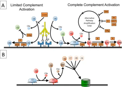
Figure 1. Complement activation in antibody mediated rejection
A) The classical pathway of complement is activated when C1q binds to IgG clustered on
endothelial HLA. Each C1q deposits multiple C4 molecules on target surfaces. Efficient
complement regulation stops the reaction before C3 molecules are deposited. In contrast,
complete complement activation results in covalent fixation of multiple C3 fragments to the
target tissue. This process is augmented by the alternative pathway amplification loop,
although several regulatory proteins control the alternative pathway. Complement regulatory
proteins metabolize the C3b to iC3b and then to C3dg. B) Complete complement activation
generates several biologically active products, including C3a, C3b, C5a, and C5b-9.
Abbreviations: C4bp, C4 binding protein; CR1, complement receptor 1; fI, Factor I; fH,
Factor H; MCP, membrane cofactor protein; DAF, decay accelerating factor.
Author Manuscript
Author Manuscript
Author Manuscript
Figure 2. C4 and C3 deposition in tissues by the classical pathway of complement