RETINAL DISORDERS
Intravitreal ranibizumab (Lucentis®) in the treatment
of retinal angiomatous proliferation (RAP)
Lazaros Konstantinidis&Evangelia Mameletzi&
Irmela Mantel&Jean-Antoine Pournaras&
Leonidas Zografos&Aude Ambresin
Received: 17 January 2009 / Revised: 12 March 2009 / Accepted: 6 April 2009 / Published online: 29 April 2009
# Springer-Verlag 2009 Abstract
Background Retinal angiomatous proliferation (RAP) is a distinct variant of neovascular age-related macular degen-eration (AMD). The aim of this study is to evaluate the functional and anatomic outcome after intravitreal ranibi-zumab (Lucentis) treatment in patients with RAP.
Methods Prospective study of consecutive patients with newly diagnosed or recurrent RAP treated with intravitreal ranibizumab at the Jules Gonin Eye Hospital between March 2006 and December 2007. Baseline and monthly follow-up visits included best-corrected visual acuity (BCVA), fundus exam and optical coherence tomography. Fluorescein and indocyanine green angiography were performed at baseline and repeated at least every 3 months. Results Thirty-one eyes of 31 patients were treated with 0.5 mg of intravitreal ranibizumab for RAP between March 2006 and December 2007. The mean age of the patients was 82.6 years (SD:4.9). The mean number of intravitreal injections administered for each patient was 5 (SD: 2.4, range 3 to 12). The mean follow up was 13.4 months (SD: 3, range 10 to 22). The baseline mean logMAR BCVA was 0.72 (SD: 0.45) (decimal equivalent of 0.2). The mean logMAR BCVA was improved significantly (P<0.0001) at the last follow-up to 0.45, SD: 0.3 (decimal equivalent 0.35). The visual acuity (VA) improved by a mean of 2.7 lines (SD 2.5). Mean baseline central macular thickness (CMT) was 376μm, and decreased significantly to a mean of 224μm (P<0.001) at the last follow-up. Mean reduction of CMT was 152μm (SD: 58). An average of 81.5% of the
total visual improvement and 85% of the total CMT reduc-tion occurred during the first post-operative month after one intravitreal injection of ranibizumab. During follow-up, an RPE tear occurred in one eye (3.2%) of the study group. No injection complications or systemic drug-related side-effects were noted during the follow-up period.
Conclusions Intravitreal ranibizumab injections appeared to be an effective and safe treatment for RAP, resulting in visual gain and reduction in macular thickness. Further long-term studies to evaluate the efficacy of intravitreal ranibizumab in RAP are warranted.
Keywords Age-related macular degeneration . Retinal angiomatous proliferation . Ranibizumab
Introduction
Retinal angiomatous proliferation (RAP) is a distinct variant of neovascular age-related macular degeneration (AMD) in which, principally, new vessels proliferate within the neurosensory retina itself (CNV) [1–3]. The typical clinical presentation of RAP appears to be unique compared with more common forms of neovascular AMD, represent-ing 12% to 15% of newly diagnosed patients with neo-vascular AMD [3,4].
RAP lesions have been identified as a poor prognostic factor, and most of the older established treatments did not seem to alter the natural history of these eyes significantly [1], in contrast to typical exudative AMD [5]. Various treatment modalities have been used in the treatment of RAP, including direct laser photocoagulation, transpupillary thermotherapy, surgical removal, and photodynamic thera-py (PDT) with verteporfin (Visudyne; Novartis Pharma AG, Basel, Switzerland) with or without concomitant intra-L. Konstantinidis
:
E. Mameletzi:
I. Mantel:
J.-A. Pournaras:
L. Zografos
:
A. Ambresin (*)Hôpital Ophtalmique Jules Gonin, University of Lausanne, 15 Av. de France,
1004 Lausanne, Switzerland e-mail: [email protected]
vitreal triamcinolone [5–22]. However, often their results were disappointing, leading to limited success or to poor func-tional results, while in some cases concerns were aroused with regard to risks of severe complications [5,16,17].
It has been reported that RAP lesions demonstrate a predisposition to vascular endothelial growth factor (VEGF) expression [23], which consists of diffusible cytokines that promotes angiogenesis and has been implicated as an important factor promoting neovascularization [24,25].
Recently, some studies have reported promising results with off-label use of the intravitreal anti-VEGF drug beva-cizumab (AvastinTM, Genentech, South San Francisco, CA, USA) for the treatment of RAP [26–30], while in 2007 a study of four patients who presented with RAP reported very favorable results with use of intravitreal ranibizumab (Lu-centis®, Genentech, South San Francisco CA, USA) [31].
Ranibizumab is a specific, affinity-mature fragment of a recombinant, humanized IgG1 monoclonal antibody that neutralizes all active forms of VEGF-A. It has been found to be effective in the treatment of exudative age-related macular degeneration (AMD), demonstrating encouraging signs of biologic activity, with acceptable safety [32].
The present study evaluated the safety and efficacy of intravitreal ranibizumab (Lucentis®) in the treatment of RAP.
Patients and methods Patient selection
This was a retrospective study of 31 consecutive patients with RAP treated with intravitreal injection of ranibizumab in the Jules Gonin University Eye Hospital between March 2006 and December 2007. Institutional approval was ob-tained for this study.
Inclusion criteria were presence of RAP (stage II and III) on the basis of clinical and fluorescein angiographic findings according to Yannuzzi et al. [3]; a minimum of 10 months of follow-up, and best-corrected visual acuity (BCVA) of 2.0 (logMAR) or better.
We excluded from this study patients that had previously received laser photocoagulation or submacular surgery as treatment for RAP. Previous PDT treatment was not an exclusion criterion, on condition that it had been adminis-tered at least 6 months before Lucentis® treatment.
Furthermore, we excluded patients who presented any significant ocular disease (other than RAP) that could compromise vision in the studied eye.
Examination
Patient age, gender, affected eye and any previous treatment administered were recorded.
The patients were followed every month. Both initial ophthalmic examination and each follow-up included evaluation of best-corrected distant and near VA using an EDTRS chart and a nearpoint Snellen acuity card at 40 cm respectively; a fundus exam with dilated pupils; fundus photography; and an evaluation of retinal architecture and measurement of foveal thickness using the OCT Stratus (Carl Zeiss Meditec, Inc.). Digital fluorescein angiography (FA) and indocyanine green (ICG) angiography were performed at baseline and at least every 3 months. Any ocular or systemic adverse events also were recorded.
Main indication for retreatment was the presence of any amount of intraretinal or subretinal fluid shown on OCT. Persistent activity from RAP lesions shown on FA or ICG angiograms, such as late leakage on FA or focal areas of intense hyperfluorescence (hot spots) on ICG, were also criteria for retreatment.
An informed consent was obtained from all patients after a thorough discussion before each injection.
Surgical procedure
All injections were performed using the same technique. Ranibizumab 0.5 mg (0.05 ml) was administered by intra-vitreal injection under sterile conditions in the operating theatre. Before injection, tetracaine 0.5% was applied topi-cally. Povidone iodine was applied to eyelid margins, eyelashes, and conjunctival surface, and a lid speculum was placed. An additional drop of povidone iodine was applied to the site of injection. Using a 30-gauge needle, 0.05 ml ranibizumab was injected 3.5–4.0 mm posterior to the corneal limbus into the vitreous cavity. The injection site was compressed with a cotton swab to avoid reflux when the needle was removed. Postoperatively, a topical antibiotic (ofloxacin) was administered three times daily for 7 days. Statistical analysis
The statistical analysis focused on the evaluation of the change in BCVA and the foveal thickness measured by OCT before treatment and at the final follow-up, which was at least 1 month distally to the last ranibizumab treatment. Additionally, the absence or persistence of RAP activity on FA, ICG angiography at the final follow-up was evaluated. BCVA values were converted to logarithm of the minimum angle of resolution (logMAR) for statistical analysis. To determine the equivalent mean decimal, logMAR values were converted back to decimal notation [33].
Serial changes in BCVA and central focal thickness (CFT) were compared using the two-tailed t-test. A p≤0.05 was considered to be statistically significant.
These parameters were analyzed using SPSS statistical software (version 16, SPSS Inc., Chicago, IL, USA).
Results
The study included 31 eyes of 31 patients. There were nine male (29%) and 22 female (71%) patients. The mean age of the patients was 82.6 years (SD: 4.9, range 73–92). Mean time of follow-up was 13.4 months (SD: 3, range 10– 22 months). Eight eyes (26%) had previously received PDT (mean number of PDT: 2.1, SD: 0.8, range 1–3). The mean time between last PDT and first treatment with ranibizumab for these eight patients was 9.6 months (SD: 3.7, range 6– 16 months). The mean number of intravitreal injections administered for each patient was five (SD: 2.4, range 3–12). At the time of treatment, 20 eyes (65%) had a stage II lesion without PED, six (19%) had a stage II lesion with PED, and five (16%) had a stage III lesion.
A summary of clinical data is shown in Table1. Visual acuity
The mean initial VA was 0.2 (decimal equivalent). Mean logMAR initial VA was 0.72 with a range from 2.0 to 0.4
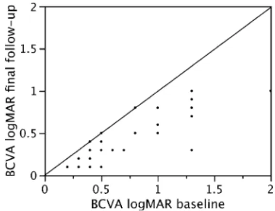
(SD: 0.45). A statistically significant improvement to a mean VA of 0.35 decimal equivalent (logMAR 0.45, range: 1.0–0.1, SD: 0.3) was demonstrated at the final follow-up (p<0.0001, two-tailed t-test). The VA improved by a mean of 2.7 (SD: 2.5) lines. Fourteen patients (45%) demonstrat-ed a gain of 3 or more lines. Twenty-eight patients (90%) showed improvement of VA, while VA remained un-changed in three patients (10%). No patient had worse BCVA at the last follow-up than at baseline (Fig. 1). On average, 81.5% of the total visual improvement occurred during the first post-operative month after one intravitreal injection of ranibizumab (logMAR BCVA at month 1: 0.5, range 1.0–0.1, SD: 0.32) (Fig.2).
Optical coherence tomography and fluorescein angiography results
Mean CMT measured with OCT was 376μm (range 525– 216μm, SD: 67) at baseline and decreased significantly at the final follow-up to 224μm (range 152–358 μm, SD: 51) (p<0.0001, two-tailed t-test). Average CMT reduction was
Fig. 1 Scattergram showing the baseline logMAR visual acuity and at the final follow-up after a mean of 13.4 months. Note that all patients improved their visual acuity apart from three, who maintained the pre-operative level
Fig. 2 Scattergram showing the change of mean logMAR best corrected visual acuity (BCVA) from baseline over time
Characteristics Value P-value
Patients Total 31 (100%)
Males 9 (29%) Females 22 (71%)
Mean age 82.6 years (SD: 4.9, range: 73–92) Mean follow-up 13.4 months (SD: 3, range: 10–22) Mean best-corrected visual acuity Baseline 0.72 (logMAR) (SD: 0.45, range: 2.0–0.4)
Last follow-up 0.2 (decimal equivalent) <0.0001 0.45 (logMAR)
SD: 0.3, range: 0.1–1)
Mean lines gain 0.35 (decimal equivalent) 2.7 (SD 2.5)
Mean central macular thickness Baseline 376 μm (SD: 67, range: 216–525) <0.0001 Last follow-up 224 μm (SD: 51, range 152–358)
Mean reduction 157 μm (SD: 57, range 45–247) Table 1 Summary of clinical
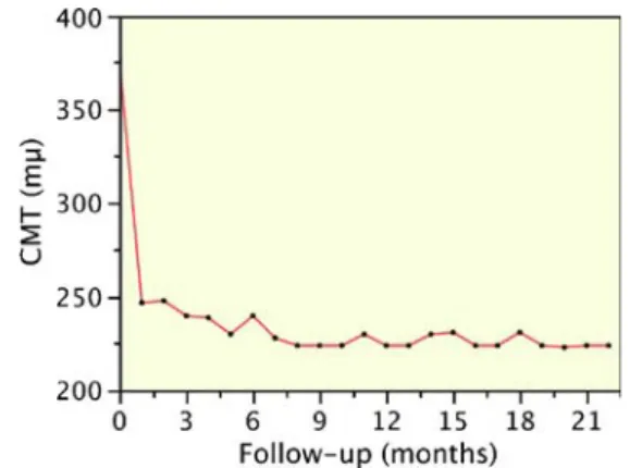
152μm (range: 45–247 μm, SD: 58) (Fig.3). On average, 85% of the total CMT reduction occurred during the first post-operative month after one intravitreal injection of rani-bizumab (average CMT reduction at month 1 was 129μm 0.5, range: 45–221 μm, SD: 54) (Fig.4).
A reduction of both intraretinal and subretinal fluid was observed in all cases.
Four (36%) of the 11 eyes that presented PED at pre-sentation demonstrated a total flattening of the PED after treatment.
All 31 eyes exhibited diminution or absence of RAP activity on FA, ICG after treatment, while 20 eyes (64.5%) showed total absence of signs of angiographic RAP activity on the final follow-up (Fig. 5). At the final follow-up we identified five patients (16%) who had not shown any signs of recurrence for a period of more than 6 months (mean: 6.8 months, SD: 0.8, range: 6–8) after a mean of 4.4 (SD:1.1, range: 3–6) injections. Four of them had a stage II lesion without PED, and one had a stage II lesion with PED at baseline.
RAP stage in this study appeared to have no influence in a statistically significant way on VA improvement or changes in mean OCT CMT.
Complications
During follow-up, RPE tear occurred in one eye (3.2%) of the study group which presented a stage II RAP associated with PED. No injection complications or systemic drug-related side-effects were noted during the follow-up period.
Discussion
Treatment of RAP is probably the most challenging among the different forms of neovascular AMD, while various
treatment modalities have been associated with unfavorable or contradictory results [28]. Extensive, stringent, multi-center, randomized clinical trials such as the MARINA trial have shown that ranibizumab provides significant VA benefit to patients with all angiographic subtypes of CNV related to AMD (i.e., minimally or predominantly classic, or occult with no classic component), with a low incidence of serious ocular and non-ocular adverse events and an ac-ceptable rate of non-serious adverse events [32,34]. How-ever, these studies did not specifically investigate the role of Lucentis in the treatment of RAP, although the MARINA trial did not exclude RAP lesions.
Fung et al. in the PrONTO study [35], an open-label, prospective, single-center, nonrandomized clinical study reported good anatomical and functional results by OCT-guided treatment of patients with AMD with intravitreal ranibizumab. In this study, the authors reported that 25% of the 40 studied patients had RAP lesions, and required a higher mean number of ranibizumab treatments compared with patients with non-RAP lesions. However, the anatom-ical and functional outcomes of patients with RAP lesions were not analyzed independently, and consequently the ef-ficacy of ranibizumab in the treatment of RAP was not further elucidated.
Lai et al. [31] in a small case series study of four patients with RAP lesions demonstrated a mean BCVA improve-ment of 3.0 lines at 3 months, and in addition resolution of subretinal fluid on OCT examination following ranibizu-mab injections. Freund et al. [1] reported rapid resolution of the intraretinal edema, hemorrhage, and neovascular lesion of three patients with RAP lesions treated with a single injection of intravitreal ranibizumab.
However, several studies have reported promising short-term results for the treatment of RAP with off-label use of the intravitreal anti-VEGF drug bevacizumab; bevacizumab is approved by the FDA for the treatment of colorectal cancer. Ghazi et al. [28] reported visual improvement by at least 2 lines in 61.5% of the patients treated with beva-Fig. 3 Scattergram showing the pre-operative and post-operative
central macular thickness (CMT) inμm at the final follow-up after a mean of 13.4 months. Note that all patients reduced their CMT during the post-operative period
Fig. 4 Scattergram showing the change of mean central macular thickness (CMT) from baseline over time
cizumab for RAP lesions. Joeres et al. [30], in their study of 16 eyes with RAP, reported a mean BCVA improvement of 1.7 ± 2 lines 4 weeks after a bevacizumab injection, with decrease of leakage angiographically in the majority (75%) of the eyes.
Meyerle et al. [27] reported similar outcomes after beva-cizumab treatment of RAP, with 94% of 17 treated eyes having stable or improved vision 3 months after treatment. On the other hand, in a recent study [36] that inves-tigated the efficacy of different treatment modalities on RAP lesions, 13 eyes were treated with ranibizumab. In this study, the authors report stabilization but not statistically
significant improvement of visual acuity. It is difficult to explain why the results of this study are so different com-pared with the other studies, including ours, which used anti-VEGF treatment.
One important difference, nevertheless, between this and our study concerns the baseline patient characteristics. In the above-mentioned study, 61.5% of patients presented RAP with PED while 65% of our patients had RAP without PED.
It is additionally worth mentioning that in both the previous-mentioned and also the present study, FA and ICG was performed once every 3 months. So OCT solely was Fig. 5 Selected case: photo
(top row), fluorescein angiogra-phy (second row), ICG angiog-raphy (third row) and OCT (bottom row) of an eye before (left column) and after (right column) ranibizumab treatment for RAP, showing disappearance of hot spot (arrow) on ICG and significant improvement of reti-nal architecture on OCT. BCVA improved from 0.3 (decimal equivalent) at the baseline to 0.8 (decimal equivalent) at the final follow-up 14 months later and after five injections of ranibizumab
used in many instances in order to detect RAP activity and dictate treatment decisions. One should consider, however, that OCT (especially in cases of PED) cannot accurately demonstrate RAP activity, as OCT changes of the PED itself cannot always be quantified. Consequently, the study by Rouvas et al. that included a higher percentage of patients with PED may be biased by a higher percentage of possible cases of undetected RAP activity, leading to patients missing treatment.
In our study, a mean VA improvement of 2.7 lines (SD 2.5) was demonstrated after a mean follow-up of 13.4 months. Furthermore, an average reduction of 152μm (SD: 58) of foveal thickness was documented.
Our results are also relatively similar to those of the MARINA trial of ranibizumab, suggesting that RAP, while classically considered as a distinctive subset of exudative AMD, may respond to ranibizumab treatment in a manner similar to that for the other types of exudative AMD.
It has been hypothesized that RAP lesions might respond more rapidly to intravitreal anti-VEGF therapies [1]. This is in accordance with our results, considering that 81.5% of the total visual improvement and 85% of the total CMT reduction occurred during the first post-operative month, after one intravitreal injection of ranibizumab.
Of our patients, 64.5% showed total absence of signs of angiographic RAP activity on the final follow-up, while 16% of all cases had not shown any signs of recurrence for a period of more than 6 months. However, we believe that ranibizumab treatment may not lead to complete healing of the RAP lesions, but rather may effectively control RAP activity with repeated injections over time.
One potential risk to be considered, as has also been reported by others [37], is the formation of retinal pigment epithelium (RPE) tears, especially in the presence of pigment epithelium detachment (PED). One case of RPE tear (3.2%) was observed in the treated eyes during follow-up. This prevalence seems to be lower, however, than other subtypes of exudative AMD, considering that during the same study period we observed RPE tears in 12.3% of 55 patients presenting a PED associated with occult CNV, and in 5.4% of 74 eyes presenting a predominantly classic CNV, after treatment with ranibizumab (unpublished data). In conclusion, in this small series of eyes with limited follow-up, intravitreal ranibizumab seems to be a safe and effective treatment for RAP, resulting in functional and anatomic improvements. The limitations of this study include the small number of eyes, the relatively short follow-up, and the absence of a control group.
In view of the encouraging results from this study, we consider that randomized, long-term clinical trials are warranted to determine more accurately the potential clinical benefit of intravitreal ranibizumab and its safety.
References
1. Freund KB, Ho IV, Barbazetto IA, Koizumi H, Laud K, Ferrara D, Matsumoto Y, Sorenson JA, Yannuzzi L (2008) Type 3 neo-vascularization: The expanded spectrum of retinal angiomatous proliferation. Retina 28:201–211, doi:10.1097/IAE.0b013e318 1669504
2. Yannuzzi LA, Freund KB, Takahashi BS (2008) Review of retinal angiomatous proliferation or type 3 neovascularization. Retina 28:375–384, doi:10.1097/IAE.0b013e3181619c55
3. Yannuzzi LA, Negrao S, Iida T, Carvalho C, Rodriguez-Coleman H, Slakter J, Freund KB, Sorenson J, Orlock D, Borodoker N (2001) Retinal angiomatous proliferation in age-related macular degeneration. Retina 21:416–434, doi:10.1097/00006982-20011 0000-00003
4. Gross NE, Aizman A, Brucker A, Klancnik JM Jr, Yannuzzi LA (2005) Nature and risk of neovascularization in the fellow eye of patients with unilateral retinal angiomatous proliferation. Retina 25:713–718, doi:10.1097/00006982-200509000-00005
5. Bottoni F, Massacesi A, Cigada M, Viola F, Musicco I, Staurenghi G (2005) Treatment of retinal angiomatous proliferation in age-related macular degeneration: A series of 104 cases of retinal an-giomatous proliferation. Arch Ophthalmol 123:1644–1650, doi:10. 1001/archopht.123.12.1644
6. van de Moere A, Kak R, Sandhu SS, Talks SJ (2007) Anatomical and visual outcome of retinal angiomatous proliferation treated with photodynamic therapy and intravitreal triamcinolone. Am J Ophthalmol 143:701–704, doi:10.1016/j.ajo.2006.10.045
7. Shiragami C, Iida T, Nagayama D, Baba T, Shiraga F (2007) Recurrence after surgical ablation for retinal angiomatous prolif-eration. Retina 27:198–203, doi:10.1097/01.iae.0000224938. 61915.f0
8. Rutishauser-Arnold Y, Tholen AM (2007) Periocular sub-tenon triamcinolone acetonide injections for the treatment of retinal angiomatous proliferation (rap) and occult choroidal neovascular-isation. Klin Monatsbl Augenheilkd 224:269–273, doi: 10.1055/s-2006-927396
9. Roth DB, Scott IU, Gloth JM, Green SN, Yarian DL, Wheatley M (2007) Micropulsed laser photocoagulation and intravitreal triam-cinolone acetonide injection for the treatment of retinal angioma-tous proliferation. Retina 27:1201–1204, doi:10.1097/IAE.0b01 3e3180ed45a6
10. Reche-Frutos J, Calvo-Gonzalez C, Donate-Lopez J, Garcia-Feijoo J, Saenz-Frances F, Fernandez-Perez C, Garcia-Sanchez J (2007) Retinal angiomatous proliferation reactivation 6 months after high-dose intravitreal acetonide triamcinolone and photody-namic therapy. Eur J Ophthalmol 17:979–982
11. Nicolo M, Ghiglione D, Lai S, Calabria G (2006) Retinal angiomatous proliferation treated by intravitreal triamcinolone and photodynamic therapy with verteporfin. Graefes Arch Clin Exp Ophthalmol 244:1336–1338, doi:10.1007/s00417-006-03 06-6
12. Nakata M, Yuzawa M, Kawamura A, Shimada H (2006) Combining surgical ablation of retinal inflow and outflow vessels with photodynamic therapy for retinal angiomatous proliferation. Am J Ophthalmol 141:968–970, doi:10.1016/j.ajo.2005.12.029
13. Mantel I, Ambresin A, Zografos L (2006) Retinal angiomatous proliferation treated with a combination of intravitreal triamcino-lone acetonide and photodynamic therapy with verteporfin. Eur J Ophthalmol 16:705–710
14. Krieglstein TR, Kampik A, Ulbig M (2006) Intravitreal triamcin-olone and laser photocoagulation for retinal angiomatous prolif-eration. Br J Ophthalmol 90:1357–1360, doi:10.1136/bjo.2006. 092536
15. Klais CM, Eandi CM, Ober MD, Sorenson JA, Sadeghi SN, Freund KB, Spaide RF, Slakter JS, Yannuzzi LA (2006) Anecortave acetate treatment for retinal angiomatous proliferation: A pilot study. Retina 26:773–779, doi:10.1097/01.iae.0000244261. 52901.67
16. Freund KB, Klais CM, Eandi CM, Ober MD, Goldberg DE, Sorenson JA, Yannuzzi LA (2006) Sequenced combined intra-vitreal triamcinolone and indocyanine green angiography-guided photodynamic therapy for retinal angiomatous proliferation. Arch Ophthalmol 124:487–492, doi:10.1001/archopht.124.4.487
17. Bottoni F, Romano M, Massacesi A, Bergamini F (2006) Remodeling of the vascular channels in retinal angiomatous proliferations treated with intravitreal triamcinolone acetonide and photodynamic therapy. Graefes Arch Clin Exp Ophthalmol 244:1528–1533, doi:10.1007/s00417-006-0311-9
18. Boscia F, Parodi MB, Furino C, Reibaldi M, Sborgia C (2006) Photodynamic therapy with verteporfin for retinal angiomatous proliferation. Graefes Arch Clin Exp Ophthalmol 244:1224–1232. doi:10.1007/s00417-005-0205-2
19. Shimada H, Mori R, Arai K, Kawamura A, Yuzawa M (2005) Surgical excision of neovascularization in retinal angiomatous proliferation. Graefes Arch Clin Exp Ophthalmol 243:519–524, doi:10.1007/s00417-004-1073-x
20. Boscia F, Furino C, Prascina F, Delle Noci N, Sborgia L, Sborgia C (2005) Combined surgical ablation and intravitreal triamcino-lone acetonide for retinal angiomatous proliferation. Eur J Ophthalmol 15:513–516
21. Borrillo JL, Sivalingam A, Martidis A, Federman JL (2003) Surgical ablation of retinal angiomatous proliferation. Arch Ophthalmol 121:558–561, doi:10.1001/archopht.121.4.558
22. Kuroiwa S, Arai J, Gaun S, Iida T, Yoshimura N (2003) Rapidly progressive scar formation after transpupillary thermotherapy in retinal angiomatous proliferation. Retina 23:417–420, doi:10. 1097/00006982-200306000-00027
23. Shimada H, Kawamura A, Mori R, Yuzawa M (2007) Clinico-pathological findings of retinal angiomatous proliferation. Graefes Arch Clin Exp Ophthalmol 245:295–300, doi: 10.1007/s00417-006-0367-6
24. Rakic JM, Lambert V, Devy L, Luttun A, Carmeliet P, Claes C, Nguyen L, Foidart JM, Noel A, Munaut C (2003) Placental growth factor, a member of the vegf family, contributes to the development of choroidal neovascularization. Invest Ophthalmol Vis Sci 44:3186–3193, doi:10.1167/iovs.02-1092
25. Otani A, Takagi H, Oh H, Koyama S, Ogura Y, Matumura M, Honda Y (2002) Vascular endothelial growth factor family and receptor expression in human choroidal neovascular membranes. Microvasc Res 64:162–169, doi:10.1006/mvre.2002.2407
26. Costagliola C, Romano MR, dell’Omo R, Cipollone U, Polisena P (2007) Intravitreal bevacizumab for the treatment of retinal
angiomatous proliferation. Am J Ophthalmol 144:449–451, doi:10. 1016/j.ajo.2007.05.025
27. Meyerle CB, Freund KB, Iturralde D, Spaide RF, Sorenson JA, Slakter JS, Klancnik JM Jr, Fisher YL, Cooney MJ, Yannuzzi LA (2007) Intravitreal bevacizumab (avastin) for retinal angiomatous proliferation. Retina 27:451–457, doi:10.1097/IAE.0b013e31 8030ea80
28. Ghazi NG, Knape RM, Kirk TQ, Tiedeman JS, Conway BP (2008) Intravitreal bevacizumab (avastin) treatment of retinal angiomatous proliferation. Retina 28:689–695, doi:10.1097/IAE. 0b013e318162d982
29. Bashshur ZF, Haddad ZA, Schakal A, Jaafar RF, Saab M, Noureddin BN (2008) Intravitreal bevacizumab for treatment of neovascular age-related macular degeneration: A one-year pro-spective study. Am J Ophthalmol 145:249–256, doi:10.1016/j. ajo.2007.09.031
30. Joeres S, Heussen FM, Treziak T, Bopp S, Joussen AM (2007) Bevacizumab (avastin) treatment in patients with retinal angio-matous proliferation. Graefes Arch Clin Exp Ophthalmol 245: 1597–1602, doi:10.1007/s00417-007-0580-y
31. Lai TY, Chan WM, Liu DT, Lam DS (2007) Ranibizumab for retinal angiomatous proliferation in neovascular age-related macular degeneration. Graefes Arch Clin Exp Ophthalmol 245: 1877–1880, doi:10.1007/s00417-007-0679-1
32. Rosenfeld PJ, Brown DM, Heier JS, Boyer DS, Kaiser PK, Chung CY, Kim RY (2006) Ranibizumab for neovascular age-related macular degeneration. N Engl J Med 355:1419–1431, doi:10.1056/ NEJMoa054481
33. Holladay JT (1997) Proper method for calculating average visual acuity. J Refract Surg 13:388–391
34. Regillo CD, Brown DM, Abraham P, Yue H, Ianchulev T, Schneider S, Shams N (2008) Randomized, double-masked, sham-controlled trial of ranibizumab for neovascular age-related macular degenera-tion: Pier study year 1. Am J Ophthalmol 145:239–248 e235 35. Fung AE, Lalwani GA, Rosenfeld PJ, Dubovy SR, Michels S,
Feuer WJ, Puliafito CA, Davis JL, Flynn HW Jr, Esquiabro M (2007) An optical coherence tomography-guided, variable dosing regimen with intravitreal ranibizumab (lucentis) for neovascular age-related macular degeneration. Am J Ophthalmol 143:566– 583, doi:10.1016/j.ajo.2007.01.028
36. Rouvas AA, Papakostas TD, Vavvas D, Vergados I, Moschos MM, Kotsolis A, Ladas ID (2009) Intravitreal ranibizumab, intravitreal ranibizumab with PDT, and intravitreal triamcinolone with PDT for the treatment of retinal angiomatous proliferation: a prospective study. Retina 29:536–544
37. Forooghian F, Cukras C, Chew EY (2008) Retinal angiomatous proliferation complicated by pigment epithelial tear following intravitreal bevacizumab treatment. Can J Ophthalmol 43:246– 248, doi:10.3129/I08-017