ION CHANNELS, RECEPTORS AND TRANSPORTERS
P2Y
2
receptor activation inhibits the expression
of the sodium-chloride cotransporter NCC in distal convoluted
tubule cells
P. Gailly&M. Szutkowska&E. Olinger&H. Debaix& F. Seghers&S. Janas&V. Vallon&O. Devuyst
Received: 11 October 2013 / Revised: 28 December 2013 / Accepted: 29 December 2013 / Published online: 25 January 2014 # Springer-Verlag Berlin Heidelberg 2014
Abstract Luminal nucleotide stimulation is known to reduce Na+ transport in the distal nephron. Previous studies suggest that this mechanism may involve the thiazide-sensitive Na+ -Cl−cotransporter (NCC), which plays an essential role in NaCl reabsorption in the cells lining the distal convoluted tubule (DCT). Here we show that stimulation of mouse DCT (mDCT) cells with ATP or UTP promoted Ca2+transients and decreased the expression of NCC at both mRNA and protein levels. Specific siRNA-mediated silencing of P2Y2receptors almost completely abolished ATP/UTP-induced Ca2+ transients and significantly reduced ATP/UTP-induced decrease of NCC ex-pression. To test whether local variations in the intracellular Ca2+concentration ([Ca2+]i) may control NCC transcription, we overexpressed the Ca2+-binding protein parvalbumin selec-tively in the cytosol or in the nucleus of mDCT cells. The
decrease in NCC mRNA upon nucleotide stimulation was abolished in cells overexpressing cytosolic PV but not in cells overexpressing either a nuclear-targeted PV or a mutated PV unable to bind Ca2+. Using a firefly luciferase reporter gene strategy, we observed that the activity of NCC promoter region from−1 to −2,200 bp was not regulated by changes in [Ca2+]i. In contrast, high cytosolic calcium level induced instability of NCC mRNA. We conclude that in mDCT cells: (1) P2Y2 receptor is essential for the intracellular Ca2+signaling induced by ATP/UTP stimulation; (2) P2Y2-mediated increase of cyto-plasmic Ca2+concentration down-regulates the expression of NCC; (3) the decrease of NCC expression occurs, at least in part, via destabilization of its mRNA.
Keywords Distal convoluted tubule . mDCT cells . P2 receptor signaling . Cytosolic calcium level . Posttranscriptional modifications
Abbreviations
ATP/UTP Adenosine-5′-triphosphate/ uridine-5′-triphosphate
[Ca2+]e Extracellular Ca2+concentration [Ca2+]i Intracellular Ca2+concentration
DCT Distal convoluted tubule
mDCT Mouse DCT
EGTA-AM Ethylene glycol tetra (acetoxymethyl ester) FITC Fluorescein isothiocyanate GAPDH Glyceraldehyde 3-phosphate
dehydrogenase NCC Na+-Cl−cotransporter
nCaRE Negative calcium response element
PLC Phospholipase C
PV Parvalbumin
TRPM6 Transient receptor potential cation channel, subfamily M, member 6 P. Gailly and M. Szutkowska contributed equally to this work.
P. Gailly (*)
:
F. Seghers Laboratory of Cell Physiology,Université catholique de Louvain (UCL) Medical School, Avenue Hippocrate, B1.55.12, 1200 Brussels, Belgium e-mail: philippe.gailly@uclouvain.be
M. Szutkowska
:
E. Olinger:
H. Debaix:
S. Janas:
O. Devuyst Division of Nephrology, Université catholique de Louvain (UCL) Medical School, Brussels, BelgiumM. Szutkowska
:
E. Olinger:
H. Debaix:
S. Janas:
O. Devuyst (*) Institute of Physiology and Mechanisms of Inherited Kidney Disorders Group, University of Zurich,Winterthurerstrasse, 190, 8057 Zürich, Switzerland e-mail: olivier.devuyst@uzh.ch
V. Vallon
Department of Medicine, Division of Nephrology/Hypertension, University of California San Diego, San Diego, CA, USA V. Vallon
Introduction
The strict control of NaCl excretion by the kidney is essential for maintaining body fluid homeostasis as well as blood pressure. The distal convoluted tubule (DCT) reabsorbs 5 % to 10 % of the filtered Na+, via the thiazide-sensitive Na+-Cl− cotransporter (NCC), located on the apical membrane. NCC is phosphorylated and activated by WNK via specific SPAK isoforms that are expressed in the DCT [9]. The functional importance of NCC is illustrated by Gitelman syndrome, an autosomal recessive tubulopathy characterized by salt wasting and secondary aldosteronism responsible for hypokalemia and metabolic alkalosis, and by hypomagnesemia and hypocalciuria [13]. Gitelman syndrome is most often due to invalidating mutations in the SLC12A3 gene that codes for NCC [46].
Recently, we provided in vitro and in vivo evidence that NCC expression was modulated by the presence of parvalbumin (PV), a cytosolic "EF-hand" protein able to bind divalent cations and specifically expressed in early DCT (DCT1) [4]. The fact that DCT segment as well as immortal-ized mouse DCT cell line (mDCT) that expresses NCC and other typical markers were sensitive to nucleotide stimulation, known to induce Ca2+signaling, led us to hypothesize that NCC expression might be controlled by ATP/UTP-induced Ca2+transients [4,8].
Response to ATP and UTP involves nucleotide P2 recep-tors. Seven ionotropic P2X (P2X1-7) and five metabotropic P2Y (P2Y1, -2,-4, -6 and -11) receptors have been identified in the kidney, each with a specific agonist response profile (for review, see refs. [24,44,45]). Several P2Y receptor subtypes are coupled to G-protein subtype Gαq, which activates PLC-β and promotes mobilization of Ca2+from intracellular stores. One of them, the P2Y2receptor, which is activated by both ATP and UTP, has previously been linked to electrolyte and water transport processes in the kidney (for review, see ref. [45]). Indeed, in the intact mouse collecting duct, luminal ATP/UTP stimulation via P2Y2-like receptors inhibits electro-genic Na+ transport and decreases K+ secretion, thus inhibiting transport processes for salt and water absorption in this nephron segment [23,24,26]. Making use of P2Y2 receptor knockout mice, it was subsequently shown that this receptor contributes to blood pressure regulation, and renal fluid and NaCl reabsorption by inhibitory effects on the ex-pression of the Na-2Cl-K cotransporter NKCC2 and the water channel aquaporin-2 [34]. These studies further showed that local P2Y2receptor tone in the aldosterone-sensitive distal nephron exerts paracrine down-regulation of epithelial sodium channel (ENaC) activity by lowering channel open probability [31]. This mechanism explains the inhibition of ENaC activity following an increase in dietary NaCl intake and contributes to blood pressure regulation [32]. The precise role of P2Y recep-tors in the DCT is however not yet characterized.
In the present study, we aimed at identifying the P2 recep-tors involved in Ca2+signaling in DCT and how the nucleotide stimulus could regulate the NCC expression in the mDCT cells. We show that the P2Y2receptor is present in DCT cells and that it is the main functional receptor, essential for the intracellular Ca2+ signaling induced by extracellular ATP/ UTP in mDCT cells. P2Y2 stimulation induces cytosolic Ca2+transients, regulating the NCC expression by decreasing the stability of its mRNA.
Methods Cell culture
The immortalized mDCT cell line was kindly provided by Prof. P.A. Friedman (University of Pittsburgh School of Med-icine, Pittsburgh, PA, USA). mDCT cells have been previously characterized as a model for thiazide-sensitive Na+ and Ca2+ transport occurring in the DCT and they express sizeable en-dogenous NCC as well as other markers of the DCT1 [4,12]. Cells were grown in DMEM/Ham’s F12 medium (Lonza, Verviers, Belgium) supplemented with 5 % (v/v) fetal bovine serum (Lonza), 2 mM ultraglutamine (Lonza) and a mixture of penicillin/streptomycin (Invitrogen, Carlsbad, CA, USA) in a humidified atmosphere of 5 % (v/v) CO2at 37 °C. Studies were performed in 24-well plates between passages 25 and 35. Drug treatments
During nucleotide stimulation experiments, confluent cells were treated with either ATP (Boehringer Mannheim, Roche Applied Science) or UTP (Sigma-Aldrich) at different con-centrations (1, 5, 10, 50 or 100μM). For repetitive stimula-tions protocol, cells were stimulated with ATP or UTP 10μM for 10 min, then rinsed (to avoid receptor down-regulation) and re-stimulated every hour, for 6 h. To suppress phospholi-pase C (PLC) activity, cells were treated with 10μM of the PLC inhibitor U73122 (Tocris, Bioscience), 10 min before Ca2+measurements. In mRNA stability experiments, the tran-scriptional inhibitor 6-dichloro-1-b-ribofuranosylbenzimi-dazole (DRB; Sigma) was used at 75 μM throughout the duration of the experiment.
Transient transfections
Subconfluent cultures (approximately 80 %) were transfected using Lipofectamine™ 2000 (Invitrogen) with pCMV-GFP (Mock), pCMV-PV-cyto-GFP (PV-cyto) or pCMV-PV-nuc-GFP (PV-nuc) plasmids (Addgene, Cambridge, MA, USA).
Site-directed mutagenesis was carried out to generate the mutant PV plasmid pCMV-PV-cyto-CDEF-GFP coding for PV in which both functional Ca2+-binding sites were
inactivated by substituting a glutamate for a valine residue at position 12 of each Ca2+-binding loop. The mutant construct was generated by using pCMV-PV-cyto-GFP plasmid as a template and by using the QuickChangeR Lightning Site-Directed Mutagenesis Kit (Stratagene, Agilent) following the manufacturer’s protocol. Mutant plasmid was generated by polymerase chain reaction using the following synthetic oli-gonucleotides containing mismatches in codon 62 of the CD loop and in codon 101 of the EF loop were used: CD62: 5′-TTCATTGAGGAGGATGTGCTGGGGTCCATTCTG-3′, and EF101, 5′- GCAAGATTGGGGTTGAAGTGTTCTCC ACTCTGGTGGCC-3′ (mutated nucleotides are underlined). We checked the identity of the mutant plasmid by sequencing. RNA Interference
To knock down the endogenous P2ry2 expression, a pool of three different double-strand siRNAs (15nM, SilencerRSelect Pre-designed siRNA synthesized by Ambion) was introduced into mDCT cells using Lipofectamine™ RNAiMAX (Invitrogen). Cells were cultured on plate wells containing the transfection complexes. Seventy-two hours after transfection, RNA was extracted with RNAqueousR-Micro kit and subjected to real-time polymerase chain reaction (PCR). Transfection efficiency was assessed using BLOCK-iT™ Alexa FluorR Red Fluorescent Oligo (Invitrogen). Fluorescent siRNAs were used to follow transfection in fluorescence microscopy. Extraction of RNA, quantitative RT-PCR
mDCT mRNAs were extracted with RNAqueousR-Micro kit (Ambion, Invitrogen) and reversed-transcribed using iScript™ cDNA Synthesis Kit (Bio-Rad). Gene-specific PCR primers were designed using Primer3 [36] (see Table1). Total RNA samples were stored at -80 °C. Real-time RT-PCR was performed using 1μg cDNA, 10 μl of SybrGreen Mix (Bio-Rad) and 100 nM of each primer in a total reaction volume of 20μl.
PCR conditions were 95 °C for 3 min followed by 40 cycles of 15 s at 95 °C, 30 s at 60 °C. The PCR products were sequenced with the BigDye terminator kit (Perkin-Elmer Applied Biosystems). The multiScreen SEQ384Filter Plate (Millipore, Billerica, MA, USA) and Sephadex G-50 DNA Grade Fine (Amersham Biosciences, Piscataway, NJ, USA) dye terminator removal were used to purify sequences reac-tions before analysis on an ABI3100 capillary sequencer (Perkin-Elmer Applied Biosystems).
The efficiency of each set of primers was determined by dilution curves (Table1).
Each cDNA was amplified in duplicate and cycle threshold values (Ct) were averaged for each duplicate. The average Ct value for GAPDH was subtracted from the average Ctvalue for the gene of interest. ThisΔCtvalue, determined in specific
experimental conditions, was then subtracted from the ΔCt value determined in control conditions to obtain a ΔΔCt value. As amplification efficiencies of the genes of interest and GAPDH were comparable, the amount of mRNA, nor-malized to GAPDH, was given by the relation 2−ΔΔCt[7,25]. Semiquantitative RT-PCR
We used RT-PCR to assess the presence of P2X and P2Y receptors in microdissected nephron segments. PCR condi-tions used were: 94 °C for 3 min followed by 35 cycles of 30 s at 95 °C, 30 s at 60 °C and 1 min at 72 °C with FastStart Taq polymerase (Roche, Vilvoorde, Belgium). The PCR products were separated on a 2 % agarose gel.
[Ca2+]imeasurements
mDCT cells were cultured on coverslips until they reached 70 % confluence. Cell cultures were incubated with 1 μM Fura2-AM for 60 min at room temperature, prior to the measurement of fluorescence in individual cells. Coverslips were rinsed with Krebs medium containing (in mM): 135 NaCl, 5.9 KCl, 1.8 CaCl, 1.2 MgCl2, 11.6 Hepes and 10 glucose, (pH 7.3) for 30 min and mounted in a thermostatized (20 °C) chamber. The chamber was continuously superfused (1 ml/min, or 4 ml/min for quick exchanges of solutions).
Cytosolic concentration of free Ca2+was measured at room temperature by radiometric measurements of fluorescence intensity monitored at 510 nm [11]. Fura-2 loaded cells were excited alternatively at 340 and 380 nm and fluorescence emission was monitored at 510 nm using a Deltascan spectro-fluorimeter (Photon Technology International) coupled to an inverted microscope (Nikon Diaphot, oil immersion objective 40× NA 1.3). Fluorescence intensity was recorded over the entire surface of the single cells. [Ca2+]iwas calculated from the ratio of the fluorescence intensities excited at the two wavelengths, using a standard intracellular calibration proce-dure performed after cell permeabilization with 5 μM ionomycin. In Ca2+-free solution, CaCl2 was omitted and 0.2 mM EGTA (Molecular Probes, Invitrogen) was added. In the experiments designed to investigate the role of PV in [Ca2+]iresponses, [Ca2+]imeasurements were performed only on cells expressing PV-GFP plasmids (cells selected by GFP fluorescence).
Antibodies
The rabbit anti-P2Y2 (Abcam: ab 46537), rabbit anti-NCC [40], goat anti-PV (sc-7448; Santa Cruz Biotechnology, Inc., Santa Cruz, CA, USA) and mouse anti β-actin (Sigma-Aldrich) primary antibodies were used during experiments. To visualize, secondary fluorescent antibody: Alexa Fluor (647 anti-rat, 633 anti-rabbit, 633 anti-sheep, 488 anti-goat
and 488 anti-rabbit) were used for immunofluorescent assays (Molecular Probes).
Immunoblotting
Total membrane -and cytosolic proteins extracts were pre-pared from mDCT cells after homogenization with a Tissue Tearor Homogenizer. The lysis buffer contained 250 mM sucrose, 20 mM imidazole pH 7.2, 1 mM EDTA and a protease inhibitors mix Complete (Roche). The sample (total extract) was centrifuged at 1,000×g for 15 min at 4 °C for nucleus elimination. The supernatant was then centrifuged at 38,500 rpm for 120 min at 4 °C. The pellet (membrane extract) was suspended in 300μl of lysis buffer. The super-natant contains the cytosolic fraction. Protein concentrations were determined with the bicinchoninic acid assay using BSA as standard. Membrane proteins were solubilized 1/4 in sam-ple buffer (50 mM Tris–HCl, pH 6.8, 7.5 % SDS, 30 %
glycerol, 0.004 % bromophenol blue) containing 6 % of DTT and heated at 95 °C for 5 min.
SDS-PAGE was performed under reduced conditions. For Western blotting, 40μg of protein was loaded onto a SDS-polyacrylamide gel and separated by electrophoresis. After blotting on nitrocellulose, the membranes were incubated overnight at 4 °C with primary antibodies, washed and incu-bated for 1 h at room temperature with peroxidase-labeled antibodies (Dako). Secondary antibodies conjugated to horseradish-peroxidase were detected with ECL reagent (Amersham Biosciences). The molecular weight of proteins was estimated by running the Precision Plus Protein™ All Blue standard (Bio-Rad).
Immunostaining
Cell cultures were grown on permeable filter supports until confluence. Twenty-four hours before cell fixation, standard Table 1 Primers used in real-time
RT-PCR analyses and expression of P2 nucleotide receptors in mDCT cells
Gene (protein) Forward and reverse primers Amplimer length (bp) Efficiency Expression in mDCT cells (Ct) Slc12a3 (NCC) 5′-CCTCCATCACCAACTCACCT-3′ 5′-AGGAGGAAGAGGACGACTC-3′ 151 0.98 Pvalb (PV) 5′-GACGCCATTCTTCTGGAAAT-3′ 5′-ATACCCC CACTGCCCTAAAA-3′ 136 0.99 Trpm6 (TRPM6) 5′-TCTTCCTTCGAGAGCCATCA-3′ 5′-TCCACCAGGATTGGAGTCAC-3′ 156 1.01 β- actin 5′-CCTGAACCCCAAAGCTAACA-3′ 5′-CGTCACCAGAGTCCATGACA-3′ 146 0.98
P2ry1 (P2Y1) 5′-ACCCTACCAGCCCTCATCTT-3′ 5′-CTGTACCTGTGTGCGCTGAT-3′
146 1.01 26.4±0.15
P2ry2 (P2Y2) 5′-CGTGCTCTACTTCGTCACCA-3′ 5′-GAAAAGGGCACAGCAAAAAG-3′
135 0.97 27.5±0.06
P2ry4 (P2Y4) 5′-CACATCACCCGCACAATTTA-3′ 5′-G TCCCCCGTGAACAGATAGA-3′
150 1.02 32.3±0.3
P2ry6 (P2Y6) 5′-CGCTTTGTACGCTTCCTCTT-3′ 5′-TCCACACACTACCCAAGCAG-3′ 150 1.09 27.5±0.4 P2rx1 (P2X1) 5′-ACTGGGAGTGTGACCTGGA-3′ 5′-AGAGGTGACGACGGTTTGTC-3′ 150 0.99 33.9±0.51 P2rx2 (P2X2) 5′-CCATGTCGGAACACAAAGTG-3′ 5′-GGCAGGTAGAGCTGTGAAC-3′ 153 0.95 32.9±0.14 P2rx3 (P2X3) 5′-GACACCGTGGAGATGCCTAT-3′ 5′-AT GGAAGCGGCACTTCTTTA-3′ 147 0.97 34.1±0.36 P2rx4 (P2X4) 5′-CCTCGACACTCGGGACTTA-3′ 5′-GCCTTTCCAAACACGATGAT-3′ 147 0.99 26.8±0.13 P2rx5 (P2X5) 5′-ACTTCCCTGCAGAGTGCTGT-3′ 5′-GGAGTCACGATCAGGTTGGT-3′ 155 1.03 30.2±0.37 P2rx6 (P2X6) 5′-CCCAGAGCATCCTTCTGTTC-3′ 5′-CACCAGCTCCAGATCTCACA-3′ 150 0.98 35.2±0.34 P2rx7 (P2X7) 5′-AAGCTGTACCAGCGGAAAGA-3′ 5′-CCTGCAAAGGGAAGGTGTAG-3′ 152 0.97 33.5±0.17 Gapdh 5′-TGCACCACCAACTGCTTAGC-3′ 5′-GGATGCAGGGATGATGTTCT-3′ 176 0.99
culture medium was replaced by DMEM/F12 supplemented with REGM™ Single Quots (Lonza). Cells were fixed 10 min using 4 % formaldehyde, washed three times with DPBS and permeabilized using 0.2 % Triton X-100. Cells were then preincubated for 1 h with DPBS containing 3 % blocking serum, 1 h with primary antibodies at room temperature, 30 min with the secondary fluorescent antibodies and washed with DPBS before mounting with DAPI-ProLongR Gold (Invitrogen). Sections were viewed with a Zeiss LSM 410 confocal microscope or Leica CLSM confocal microscope. Images were processed (overlays) using Adobe Photoshop. mRNA half-life time determination
mDCT cells were cultured up to 80 % confluence and treated with 75μM DRB for different time intervals. NCC, β-actin and TRPM6 expressions were measured using RT-qPCR. mRNA half-life time was estimated by exponential regression analysis [35].
Plasmid construction for reporter assay
The plasmid pGL3-basic (Promega) was used to examine the promoter activity of the 5′-flanking region in the mouse Slc12a3 gene. The 5′-flanking region of Slc12a3 was generat-ed by PCR and ligatgenerat-ed into KpnI and XhoI sites of pGL3-basic vector. Sense primer: 5′-TtggtaccGTGCCATCCTTCCTCA TTCC-3′ (nucleotide −1502) containing an engineered KpnI restriction site was derived from the Slc12a3 gene sequence, and the antisense primer: 5′-TtctcgagTATGGCTCTGGGTA TCAAAGG-3′ was corresponding to nucleotides −1 to −21 and containing an engineered XhoI restriction site. Construct (pGL3-1500/Slc12a3) was confirmed by sequence analysis. The pGL3-1000/Slc12a3 was generated with Sense primer: TtggtaccGATGATTCAGGGAAACACTGG-3′ (nucleotide −1015) and the antisense primer was the same than for pGL3-1500. The pGL3-2200/Slc12a3 was constructed by PCR amplification using an engineered KpnI restriction site in the Sense primer: 5′-TtggtaccAGAGTCCCACCA-3′ corresponding to nucleotide−2246 and the antisense prim-er: 5′-AagcatgcTACTTGGCTATCAA (nucleotide −1274). The Slc12a3 gene sequence contained a restriction site for SphI in position−1280. The PCR fragment was then ligat-ed into the pGL3-1500/Slc12a3 plasmid digestligat-ed with KpnI and SphI restriction enzymes to generate pGL3-2200/Slc12a3 plasmid.
Plasmids were sequenced with the BigDye terminator kit (Perkin Elmer Applied Biosystems). The multiScreen SEQ384 Filter Plate (Millipore) and Sephadex G-50 DNA Grade Fine (Amersham Biosciences) dye terminator re-moval were used to purify sequences reactions before analysis on an ABI3100 capillary sequencer (Perkin-Elmer Applied Biosystems).
Luciferase assay
mDCT were transiently transfected with 500 ng firefly lucif-erase reporter plasmid and 10 ng Renilla luciflucif-erase vector using Lipofectamine 2000 (Invitrogen).
Forty-eight hours after transfection, luciferase activity was measured with Dual-Luciferase Reporter Assay System (Promega), using a GloMax™ 96 luminometer (Promega) with a 10-s integration time for each luciferase reaction. Firefly luciferase activity was corrected for transfection effi-ciency by using Renilla luciferase measurements. The corrected activity (Firefly luciferase divided by Renilla lucif-erase activity) was compared to the promoterless pGL3-basic corrected activity, used as a negative control (results expressed as percentages).
Extracts from each transfection were assayed in duplicate for at least three independent transfection experiments. Statistical analysis
The results are presented as means±SEM. One-way analysis of variance was used to investigate statistical differences among the studied groups. Individual groups were compared by an unpaired Student’s ttest. A pvalue of <0.05 was taken as significant.
Results
Stimulation of mDCT cells by ATP and UTP decreases NCC mRNA level
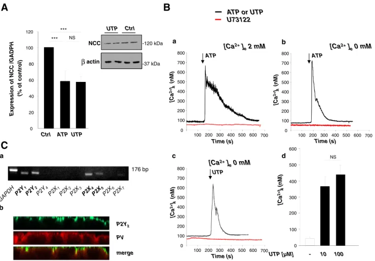
Based on our previous observation that Ca2+signaling regu-lates the expression of NCC in mDCT cells [4], we first investigated whether the nucleotides ATP and UTP, known to trigger changes in intracellular free calcium concentration ([Ca2+]i), regulate the mRNA expression of NCC. mDCT cells were treated with ATP or UTP 10μM for 10 min every hour for 6 h. Both treatments produced a significant decrease in NCC mRNA level compared to control (59±12 % and 58± 7 %, respectively, n=8; Fig.1a). We also observed that the effect on NCC expression obtained at this concentration (10μM ATP/UTP) was maximal (data not shown). Accord-ingly, repetitive stimulation with 10μM UTP for 10 min every hour for 6 h decreased the amount of NCC protein expressed after 24 h by ~40 % (Fig.1ainsert).
ATP and UTP increase [Ca2+]iin mDCT cells
The involvement of P2 receptors was further characterized in mDCT cells by analyzing [Ca2+]i transients after ATP and UTP stimulation. mDCT cells were loaded with the calcium indicator fura-2 acetoxymethyl ester (Fura-2 AM) to measure
[Ca2+]i. In the presence of Ca2+in the extracellular medium, the response was constituted of a fast peak of [Ca2+]ifollowed by a sustained plateau (Fig.1b(a)). The response was largely inhibited by suramine (300μM) suggesting the involvement of a P2 receptor (data not shown). In the absence of external Ca2+, ATP was still able to induce the fast initial peak of [Ca2+]i, but the long-lasting plateau phase was lost, suggesting that ATP stimulation triggers both the release of Ca2+from internal stores and the entry of Ca2+from the external milieu (Fig.1b(b)). This also suggested that the purinergic receptor involved in the first phase is metabotropic, belonging to the P2Y family. UTP elicited a similar increase in [Ca2+]i as observed for ATP and exerted a maximal effect at 10μM (Fig.1b(c–d)). We further characterized the presence of func-tional P2Y receptors coupled to Gq protein by testing the
effect of the PLC antagonist, U73122 (10 nM), on [Ca2+]i response elicited by ATP/UTP. U73122 completely inhibited the increase in [Ca2+]i, both in the presence and in the absence of external Ca2+(Fig.1b(a–c)) confirming that the receptor involved is metabotropic and coupled to PLC, and suggesting that the entry of Ca2+is not due to P2X receptor stimulation but is subsequent to stores depletion. Taken together, these observations indicate that mDCT cells express functional P2Y receptor subtypes sensitive to ATP and UTP.
mDCT cells express P2Y2 receptor in cell membrane Among P2Yand P2X receptors, P2Y1, P2Y2, P2Y4, P2Y6and P2X1–7subtypes have been previously identified in kidney [1, 24,37,42,43]. Semiquantitative (Fig.1c(a)) and quantitative
PV P2Y2 merge
A
U73122 ATP or UTP Time (s) 0 100 200 300 400 500 600 700 800 100 200 300 400 500 600 700 [Ca 2+ ]I (nM ) ATP [Ca2+] e2 mM a c b dB
C
176 bp a b 0 100 200 300 400 500 600 700 800 100 200 300 400 500 700 [Ca 2+ ]I (nM ) 600 Time (s) UTP [Ca2+] e0 mM Time (s) 0 100 200 300 400 500 600 700 800 100 200 300 400 500 700 [Ca 2+ ]I (nM ) 600 ATP Expression of NCC /GADPH (% of control) NS *** *** Ctrl ATP UTP 0 20 40 60 80 100 120 [Ca 2+ ]I (nM ) UTP [µM] - 10 100 NS 0 100 200 300 400 500 600 [Ca2+] e0 mM NCC actin UTP Ctrl -37 kDa -120 kDa βFig. 1 Nucleotide stimulation of mDCT cells activates metabotropic P2 receptors and decreases NCC expression. a Repetitive stimulation with 10μM ATP/UTP induced a 2-fold decrease of NCC mRNA expression (RT-qPCR) (n=8). ***p<0.0001 and a 40 % reduction in NCC protein expression (n=2 independent experiments, withβ-actin as control). b Stimulation of mDCT cells with ATP/UTP induced Ca2+ transients. Representative recording of mDCT cells stimulated by ATP in the pres-ence (2 mM [Ca2+]e; a) and ATP or UTP in the absence (0 mM [Ca2+]e; b and c) of extracellular calcium. ATP or UTP induces the release of [Ca2+]i from internal stores (dark lines). Cells preincubated for 10 min with
10μM PLC inhibitor (U73122) lacked nucleotide-induced [Ca2+]i tran-sients (a–c; red lines). Similar results were obtained for n=3–11 in each condition. Stimulation with 10 and 100μM UTP (d) induced a similar amplitude of [Ca2+]itransient (n=4–5). The time points of drug stimula-tion are indicated by arrows. c P2 receptors expression in mDCT cells. (a) RT-PCR analyses showed that P2Y1, P2Y2, P2X4 and P2X5are highly expressed in mDCT cells (see also Table1). (b) Immunofluorescence staining (confocal images in XZ plane) is compatible with a P2Y2receptor (green) expression in the apical membrane and a PV expression (red) in the cytosol
(Table1) RT-PCR experiments showed that P2Y1, P2Y2and P2Y6 as well as P2X4 and P2X5 were by far the most expressed subtypes in mDCT cells.
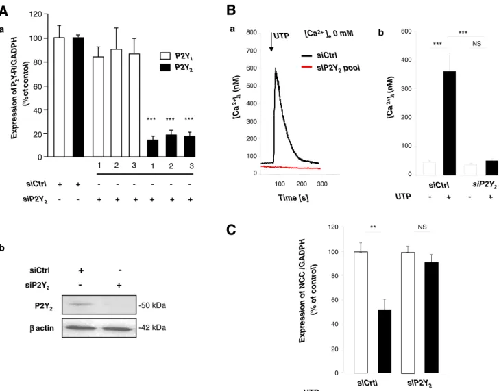
Immunofluorescent microscopy was used to study subcel-lular localization of the P2Y2receptor subtype in mDCT cells (see the reason below). As expected, the P2Y2receptor was found in the membrane compartment, whereas PV, a specific marker of DCT1, was found in the cytosol (Fig.1c(b)). P2Y2receptor silencing suppresses nucleotide-induced increases in [Ca2+]iand suppression of NCC expression The similar and high potency of both ATP and UTP in the above studies pointed to a possible role of the P2Y2 and/or P2Y4 receptor, which in rodents are both and similarly activated by both nucleotides, whereas P2Y1 and P2Y6 subtypes are not responsive to UTP and ATP, respectively. To identify the P2Y receptor involved, and considering the high expression of P2Y2 versus P2Y4 receptors in mDCT cells, we repressed the expression of the P2Y2receptor. mDCT cells were transfected with nonspecific siRNA (siCtrl) or treated independently with three specific siRNAs targeted against P2Y2receptor (siP2Y2). Treatment with siP2Y2resulted in a significant reduction of about 80 % in P2Y2mRNA levels, with no effect on the P2Y1mRNA level (Fig.2a(a)). Accordingly, 72 h after transfection with a pool of the three siP2Y2, Western blot analysis revealed a strong reduction in the P2Y2 receptor protein expressed (Fig.2a(b)).
We then observed that in mDCT cells transfected with this pool of siP2Y2, the Ca2+ response to 10 μM UTP was completely abolished, emphasizing the essential involvement of P2Y2 receptors in nucleotide-induced release of [Ca2+]i (Fig.2b).
We previously reported that the ATP-induced decrease in NCC expression was dependent on intact [Ca2+]itransients [4]. We therefore used the same siRNA strategy to determine the role of P2Y2receptor signaling in the nucleotide-induced regulation of NCC expression. Compared to baseline condi-tions, P2Y2receptor knock-down prevented the decrease in NCC expression induced by nucleotide activation (10μM UTP) (Fig.2c).
Altogether, these observations indicate that P2Y2 re-ceptors mediate the [Ca2+]i transients and the negative regulation of NCC expression induced by extracellular nucleotides.
Selective [Ca2+]ibuffering prevents the nucleotide-induced decrease of NCC mRNA expression
We next investigated the relative role of localized [Ca2+]i transients (nuclear vs. cytoplasmic) in nucleotide-induced regulation of NCC expression. Toward this aim, we
overexpressed PV specifically in the cytosol or in the nucleus. mDCT cells were transfected with plasmids coding for rat PV targeted to the cytosol (PV-cyto) and rat PV targeted to the nucleus (PV-nuc), respectively [33]. We used, as controls, a plasmid coding GFP alone (Mock) and a plasmid coding for a mutated form of PV in which both calcium binding sites were rendered nonfunctional (PV-cyto-CDEF). These proteins were built as GFP-fusion proteins and their proper targeting con-firmed by fluorescent detection (Fig.3a).
At rest, cells transfected with PV-cyto, PV-cyto-CDEF or PV-nuc did not show any significant difference in [Ca2+]iin comparison to Mock transfected cells. However, UTP-induced cytosolic [Ca2+]itransients were largely reduced in PV-cyto transfected cells compared to control Mock transfected cells (274±42 vs. 755±71 nM). As expected, overexpression of PV in the nucleus and overexpression of the mutated PV unable to bind Ca2+did not affect the UTP-induced [Ca2+]iresponse (824±43 and 826±62 nM, respec-tively) (Fig.3b).
Having validated the experimental model, we investigated the effect of local changes in [Ca2+] on the regulation of NCC expression. UTP stimulation of Mock as well as PV-cyto-CDEF and PV-nuc transfected cells induced a significant decrease in NCC expression. In contrast, buffering of cyto-plasmic Ca2+ by overexpression of PV-cyto completely inhibited the effect (Fig.3c), suggesting that an increase of cytosolic but not of nuclear [Ca2+] inhibits NCC expression. This also suggests that if Ca2+ exerts its effects on NCC expression by acting on transcription, it is not a direct effect of Ca2+on the promoter but an indirect effect passing through a cytosolic factor.
Nucleotide stimulation does not influence NCC gene transcription
To evaluate whether nucleotide stimulation could interfere with NCC transcription, we measured the activity of NCC gene promoter using a firefly luciferase reporter gene. As it has been reported that maximal activity of the promoter re-quires a sequence of 1,019 bp for humans and of 2.1 kb for rat [42], mDCT cells were transfected with three Slc12a3 promoter-luciferase gene constructs containing a sequence of the promoter of 1, 1,5 and 2,2 kb, respectively (pGL3-1000/ SLC12a3, pGL3-1500/Slc12a3 and pGL3-2200/Slc12a3, re-spectively). Red firefly luciferase activity was corrected for transfection efficiency by measuring the activity of the simul-taneously transfected green Renilla luciferase. Firefly lucifer-ase activities measured in cells transfected with pGL3-1000/ Slc12a3, pGL3-1500/Slc12a3 and pGL3-2200/Slc12a3 were significantly higher than in cells transfected with the control vector (pGL3-basic), suggesting that the constructs were tran-scriptionally active. Treatment of the cells with 10μM UTP for 10 min every hour for 6 h did not reduce the expression of
luciferase, suggesting that the activity of the NCC promoter region from−1 to −2,200 bp is not regulated by nucleotide stimulation (Fig. 4). These results argued against important regulatory sequences for nucleotide-induced and Ca2+ -medi-ated inhibition of NCC gene transcription in the area of the NCC promoter.
Nucleotide stimulation decreases NCC mRNA stability Since UTP-induced decrease of NCC expression seemed in-dependent of transcription, we checked whether UTP might
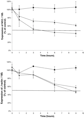
interfere with mRNA stability. To this aim, we blocked transcription with 75 μM of DRB and measured the progressive decay of mRNA amount by RT-qPCR. Analysis showed that NCC mRNA half-life time signif-icantly decreased upon UTP stimulation, from 11.2 to 5.9 h (Fig. 5). In comparison, nucleotide stimulation did not affect the relative half-life time of other genes such as β-actin or the magnesium transporter TRPM6 (10 and 10.3 h, respectively). We conclude that UTP stim-ulation specifically decreases NCC mRNA stability in mDCT cells.
A
C
B
a UTP siCtrl siP2Y2pool 0 200 400 600 [Ca 2+] I (nM ) 100 200 300 Time [s] 800 [Ca2+] e0 mM 100 300 500 700 *** NS 0 100 200 300 400 *** 600 [Ca 2+ ]I (nM ) siCtrl UTP - + - + b a Expression of NCC /GADPH (% of control) 0 20 40 60 80 100 NS siCrtl UTP - + - + ** 120 siP2Y2 ββactin P2Y2 siP2Y2 - + siCtrl + --50 kDa -42 kDa b 0 20 40 60 80 100 120 1 2 3 1 2 3 siCtrl + + siP2Y2 + + + + + + *** *** *** P2Y2 P2Y1 Exp ression of P2 Y R/GA D P H (% of conto l)Fig. 2 Effects of P2Y2receptor knock-down on intracellular Ca2+release and NCC mRNA expression in mDCT. a (a) P2Y receptor mRNA expression in mDCT cells after transfection with three different siP2Y2. P2Y1(white columns) and P2Y2receptors (black columns) mRNAs were quantified by RT-qPCR, related to GAPDH expression and expressed in proportion to control situation (siCtrl). Cells treated with siP2Y2RNA showed a significant decrease in the expression of P2Y2, whereas P2Y1 stayed unchanged. ***p<0.0001 versus siCtrl (n=4). Efficiency of trans-fection was around 90 %, verified with BLOCK-iT™ Alexa FluorRRed Fluorescent Oligo (Invitrogen). (b) Immunoblot analysis of P2Y2protein expression of cells transfected with siP2Y2versus siCtrl. b Specific P2Y2
receptor knock-down inhibits intracellular Ca2+release in mDCT. (a) Representative recording of changes in [Ca2+]imeasured with the fluo-rescent indicator Fura2-AM. Stimulation of mDCT cells with 10μM UTP induced a release of Ca2+in the absence of [Ca2+]eafter siCtrl treatment (dark line) but not after transfection with siP2Y2(red line). The time point of drug stimulation is indicated by arrow. (b) Quantification (n=6 inde-pendent measurements; ***p<0.0001). c Stimulation with 10μM UTP for 10 min every hour for 6 h induced a 2-fold decrease of NCC expression (RT-qPCR), an effect that was blocked after P2Y2silencing (n=8, **p<0.001)
Discussion
We previously reported that ATP stimulation of mDCT cells significantly reduced NCC expression [4]. In the present study, we investigated how ATP-induced Ca2+signaling reg-ulates NCC expression and elucidated the involved P2 recep-tor subtype. We show that P2Y2receptor plays an essential role in this response, which is induced by both ATP and UTP. We further show that the ATP/UTP-induced increase in cyto-plasmic Ca2+concentration down-regulates the expression of NCC, at least in part, by decreasing its mRNA stability.
The DCT segment is not readily accessible by regular microdissection of the mouse kidney. Therefore, most recep-tor studies of the distal tubule have been performed thus far on cell lines. Nucleotide receptors have been studied on Xenopus laevis A6, canine MDCK and rabbit DC1 cells [2,5, 47].
Here, we used mDCT cells, an established cell model that expresses the thiazide-sensitive cotransporter NCC typical for the DCT [12] and which has recently been used to study the regulation of NCC and the expression of other DCT1 markers such as PV [4].
Cytosolic ATP concentrations exceed 5 mM in most cell types [14, 20], whereas the pericellular concentrations re-quired for P2Y2receptor stimulation (EC50 values for ATP) range between 0.085 and 0.23 μM in humans and 0.7 and 1.8 μM in mice. Similar concentrations are necessary for activation of this receptor by UTP [39]. Lazarowski and colleagues detected UTP in nanomolar concentrations in the medium bathing a variety of cells including platelets and leukocytes, primary airway epithelial cells, rat astrocytes and several cell lines cultures [21]. This suggests that constitutive release of UTP may provide a mechanism of regulation of the PV GFP 3xnuc Myc
PV-nuc cyt PV GFP Myc
PV-cyt
cyt PV(CDEF) GFP Myc PV-cyt-CDEF
B
a 0 100 200 300 400 500 600 700 800 900 100 200 300 400 500 Time (s) [Ca2+] e0 mM UTP cytosolic [Ca 2+ ]i (nM ) 0 200 400 600 800 1000 cytosolic [Ca 2+ ]i (nM ) UTP + + + + *** [Ca2+]e0 mM Mock bC
120 UTP - + - + - + - + Expression of NCC /GADPH (% of relati ve control) 0 20 40 60 80 100 Mock *** NS ** **A
Fig. 3 Cytoplasmic overexpression of PV modulates [Ca2+]itransients and NCC expression induced by nucleotide stimulation. a Subcellular localization of targeted PV-GFP fusion proteins. Schematic representation of GFP expression vectors and fluorescent detection (green) of PV-GFP fusion proteins targeted to the cytoplasm (PV-cyt and PV-cyt-CDEF) or to the nucleus (PV-nuc). Nuclei stained with DAPI dye (blue). b Overexpression of PV-cyt but not of PV-cyt-CDEF or PV-nuc decreases [Ca2+]iresponse to 10μM UTP. (a) Representative recording of [Ca2+]i
transients of cells transfected with PV-cyt (black line), PV-cyt-CDEF (red line) or PV-nuc (green line). (b) Quantification (mean±SEM, n=19–40; ***p<0.001). c Overexpression of PV-cyt inhibits UTP-induced decrease of NCC expression. mDCT cells were transfected with Mock and PV-cyt, PV-cyt-CDEF and PV-nuc constructs and treated after 72 h with 10μM UTP for 10 min every hour for 6 h. NCC expression was measured by RT-qPCR (mean±SEM, n=6–25; **p<0.001, ***p<0.0001)
basal activity of uridine nucleotide sensitive receptors. Inter-estingly, high dietary NaCl intake is paralleled by increased urinary levels of UTP and ATP. Such change in NaCl intake,
reflected by modifications in aldosterone concentration, may change P2Y2-receptor activation, in turn affecting ENaC open probability and therefore NaCl reabsorption [32].
+1 -2200bp Slc12a3 Luciferase 5’ 3’ -1500bp Slc12a3 Luciferase 5’ 3’ +1 -1000bp Slc12a3 Luciferase 5’ 3’ +1 Luciferase /Renilla-Basic 5 10 15 20 25 30 35 40 UTP + -+ +
B
A
Fig. 4 Nucleotide stimulation does not affect NCC gene transcription. a Luciferase reporter constructs containing 1-, 1.5- and 2.2-kb fragments of promoter of the mouse Slc12a3 gene coding for NCC. b Luciferase activity was assessed after 24 h by Dual-Luciferase Assay in cells that were stimulated with 10μM UTP for 10 min every hour for 6 h. Firefly luciferase activity corrected for transfection efficiency by relating to Renilla luciferase measurements and related to the corrected activity of the promoterless pGL3-basic activity (mean±SEM, n=4 independent measurements) Ctrl DRB DRB + UTP 0% 20% 40% 60% 80% 100% 120% 0 1 2 3 4 5 6 7 8 9 10 Expression of NCC / 18S (% of control) Time (hours) 0% 20% 40% 60% 80% 100% 120% 0 1 2 3 4 5 6 7 8 9 10 Time (hours) 0% 20% 40% 60% 80% 100% 120% 0 1 2 3 4 5 6 7 8 9 10 Time (hours) Expression of -actin / 18S (% of control) Expression of TRPM6 / 18S (% of control)
Fig. 5 Nucleotide stimulation decreases NCC mRNA half-life time in mDCT cells. mDCT cells were incubated with 75μM DRB for 0, 1, 3, 6 and 9 h and stimulated with 10μM UTP for 10 min every hour for 6 h.
NCC,β-actin and TRPM6 mRNAs were quantified by RT-qPCR (mean ±SEM, n=4–11 independent experiments)
Extracellular nucleotides can activate two families of re-ceptors: (1) the ionotropic P2X receptors that open in response to the binding of extracellular ATP as their principal ligand [6]; and (2) the metabotropic P2Y receptors that are function-ally coupled to G proteins and that are activated by ATP (P2Y1 and P2Y2receptors) or UTP (P2Y2, P2Y4and P2Y6receptors) [22, 41, 45]. By RT-qPCR, we identified the presence of several subtypes of P2Y and P2X receptors among which P2Y1, P2Y2, P2Y6, P2X4and P2X5 were the most highly expressed. ATP and UTP induced an increase in [Ca2+]ieven in the absence of extracellular calcium, suggesting the in-volvement of metabotropic receptors. Accordingly, the PLC antagonist U73122 inhibited the ATP/UTP-elicited calcium peak in mDCT cells. Among the P2Y metabotropic receptors, the similar and high potency of ATP and UTP suggested the possible involvement of P2Y2 in mDCT cells, which was highly expressed in these cells. siRNA-mediated knock-down of P2Y2receptor protein confirmed that this isoform was responsible of both ATP/UTP-induced [Ca2+]itransients and ATP/UTP-induced inhibition of NCC expression. P2Y2 r e c e p t o r a n d N C C ( t h e l a t t e r n o t s h o w n ) w e r e immunodetected in the apical membrane of the mDCT cells cultivated on filter whereas PV, a protein specifically expressed in mouse DCT1, was found in the cytosol. P2Y2 has also been described in both apical and basolateral mem-branes of principal cells of the inner medullary collecting duct (IMCD) [18,43,45].
Our understanding of the distinct roles of cytosolic and nuclear Ca2+transients in gene expression is still limited. The generation of PV expression constructs targeted to the cytosol or to the nucleus allowed to investigate the relative contribu-tion of cytoplasmic and nuclear Ca2+transients to the regula-tion of MAPK-mediated gene expression in response to stim-ulation with EGF [33]. We used the same strategy and found that cytosolic and not nuclear Ca2+transients played a decisive role in the regulation of NCC expression. This observation was confirmed by the fact that the expression of neither the mutated PV unable to bind Ca2+nor the nuclear-targeted PV was able to inhibit UTP-induced modulation of NCC expres-sion. Taken together with the specific buffering properties of PV [30], these data suggest that the calcium signal peak is indeed the determinant factor for NCC regulation.
Gene expression may be regulated at the level of RNA transcription, splicing, polyadenylation, capping, trafficking, stability, translation, or at the level of protein processing and stability [28]. Genes down-regulated by Ca2+transients have been identified in Arabidopsis, but also in mammals [16,17]. For example, it was shown that Ca2+transients inhibit expres-sion of the protooncogene c-myb in an erythropoietin-responsive murine erythroleukemia cell line [38]. Similarly, calcium inhibits renin gene expression by transcriptional and posttranscriptional mechanisms [10]. It inhibits renin tran-scription by inducing translocation of trantran-scription factor
Ref-1 to the nucleus, where it binds to a negative calcium response element (nCaRE) of the renin promoter/enhancer. Besides its indirect action on transcription, Ca2+also induces a destabilization of renin mRNA, a process involving dynamin-1 protein [dynamin-19].
In the present study, we showed that NCC mRNA levels decreased after UTP stimulation, suggesting that UTP stimu-lation inhibits NCC transcription or makes NCC mRNA less stable [35,37]. We therefore studied the role of UTP-induced Ca2+transients in both these processes.
Maximal promoter activity of NCC was observed in mDCT cells using a human promoter containing 1,019 bp of the 5′ flanking region of SLC12A3. MacKenzie and col-leagues [27] observed a significant repressor effect from po-sition–1019 to –1885. Luciferase reporter gene analysis using the rat promoter sequence of Slc12a3 showed a maximal activity with a promoter containing 2,039 bp and demonstrat-ed that the most important region was locatdemonstrat-ed between posi-tion–580 and –141 [42]. In silico analysis of NCC sequence did not reveal any nCaRE element in the promoter region. We nevertheless studied the possible effect of [Ca2+]itransients on NCC gene transcription by using a firefly luciferase reporter gene using the rat promoter sequence up to 2.2 kb upward as the start codon. Based on these observations in mDCT cells, it seems that nucleotide stimulation, shown to increase intracel-lular Ca2+concentrations, has no direct or indirect influence on NCC gene transcription. However, we cannot exclude a possible involvement of a region upstream this 2.2-kb sequence.
We next turned to mRNA stability investigation. Important-ly, we found that repeated stimulations with 10μM UTP (for 10 min every hour for 6 h), eliciting repeated pulses of high intracellular calcium concentrations, reduced NCC mRNA half-life by 50 %. Messenger RNA stability is determined by specific sequences (cis-acting elements) in the 3′-UTR of RNA such as AUUUA, U(U/A)(U/A)UUU(u/A)(U/A)U, GUUUG or CAGUGU/C repeats and by the presence of specialized proteins (trans-acting factors) such as heterogeneous nuclear ribonucleoprotein-A1 (hnRNP-A1), a protein known to bind, in the cytoplasm, to reiterated AUUUA, AU-rich and poly-U sequences to determine mRNA stability [3,28]. We did not identify in a short 250-bp-long 3′-UTR sequence of NCC the classical AU-rich elements (ARE) "AUUUA" motif typically responsible for the trans-acting factors binding. However, even though the "AUUUA" repeats are commonly involved in mRNA stability regulation, they are not essential. For example, parathyroid hormone mRNA is known to contain a 63b cis-acting nucleotide destabilizing AU-rich sequence in the 3′-UTR and another distinct region determining mRNA stability by its interaction with trans-acting factors AUF-1 and Unr. Both proteins bind PTH mRNA and stabilize the transcript; interestingly, their binding is increased in low [Ca2+]i environment [29].
Finally, mRNA abundance could be regulated by binding of specific miRNAs to a complementary binding sequence generally located in 3′-UTR of target RNA [15]. In the case of murine NCC mRNA, a computational analysis of 3′-UTR identified two potential sites of fixation for miRNA described in the kidney: mmu-let-7d and mmu-miR-143. Recently, these miRNA were implicated in cancer protein regulation. The importance of 3′-UTR region in Ca2+
-induced destabilization of NCC mRNA and protein is under investigation.
In conclusion, our results demonstrate that in mDCT cells, the extracellular nucleotide-induced and P2Y2 receptor-mediated [Ca2+]i elevation is associated with a decrease in NCC mRNA, which is due, at least in part, to a reduction of its stability. These studies provide another example for the down-regulation of a mammalian gene by Ca2+transients and add another level of complexity to the regulation of NCC expres-sion in the distal nephron.
Acknowledgments The authors thank Prof. J. Loffing and Mrs. M. Carrel (Dept. of Anatomy, University of Zurich) for help and fruitful discussions. This study was supported by the Belgian agencies "Fonds National de la Recherche Scientifique" and "Fonds de la Recherche Scientifique Médicale", Concerted Research Action (10-15/029) and Inter-University Attraction Poles (IUAP P7/13) from the Belgian Federal Government and by the NCCR Kidney.CH program (Swiss National Science Foundation); the Gebert Rüf Stiftung (Project GRS-038/12); and the Swiss National Science Foundation 310030-146490 (to OD). Conflict of interest The authors have declared that no conflict of interest exists.
References
1. Bailey MA, Hillman KA, Unwin RJ (2000) P2 receptors in the kidney. J Auton Nerv Syst 81(1–3):264–270
2. Banderali U, Brochiero E, Lindenthal S, Raschi C, Bogliolo S, Ehrenfeld J (1999) Control of apical membrane chloride perme-ability in the renal A6 cell line by nucleotides. J Physiol 519(Pt 3):737–751
3. Beelman CA, Parker R (1995) Degradation of mRNA in eukaryotes. Cell 81(2):179–183
4. Belge H, Gailly P, Schwaller B, Loffing J, Debaix H, Riveira-Munoz E, Beauwens R, Devogelaer JP, Hoenderop JG, Bindels RJ, Devuyst O (2007) Renal expression of parvalbumin is critical for NaCl han-dling and response to diuretics. Proc Natl Acad Sci U S A 104(37): 14849–14854. doi:10.1073/pnas.0702810104
5. Bidet M, De Renzis G, Martial S, Rubera I, Tauc M, Poujeol P (2000) Extracellular ATP increases [CA(2+)](i) in distal tubule cells. I. Evidence for a P2Y2 purinoceptor. Am J Physiol Renal Physiol 279(1):F92–F101
6. Burnstock G (2007) Physiology and pathophysiology of purinergic neurotransmission. Physiol Rev 87(2):659–797. doi:10.1152/ physrev.00043.2006
7. Bustin SA, Benes V, Garson JA, Hellemans J, Huggett J, Kubista M, Mueller R, Nolan T, Pfaffl MW, Shipley GL, Vandesompele J, Wittwer CT (2009) The MIQE guidelines: minimum information for publication of quantitative real-time PCR experiments. Clin Chem 55(4):611–622. doi:10.1373/clinchem.2008.112797
8. Dai LJ, Kang HS, Kerstan D, Ritchie G, Quamme GA (2001) ATP inhibits Mg(2+) uptake in MDCT cells via P2X purinoceptors. Am J Physiol Renal Physiol 281(5):F833–F840
9. Ellison DH, Brooks VL (2011) Renal nerves, WNK4, glucocorti-coids, and salt transport. Cell Metab 13(6):619–620. doi:10.1016/j. cmet.2011.05.007
10. Fuchs S, Philippe J, Corvol P, Pinet F (2003) Implication of Ref-1 in the repression of renin gene transcription by intracel-lular calcium. J Hypertens 21(2):327–335. doi:10.1097/01.hjh. 0000052414.12292.ef
11. Gailly P (1998) Ca2+entry in CHO cells, after Ca2+stores depletion, is mediated by arachidonic acid. Cell Calcium 24(4):293–304 12. Gesek FA, Friedman PA (1992) Mechanism of calcium transport
stimulated by chlorothiazide in mouse distal convoluted tubule cells. J Clin Investig 90(2):429–438. doi:10.1172/JCI115878
13. Gitelman HJ, Graham JB, Welt LG (1966) A new familial disorder characterized by hypokalemia and hypomagnesemia. Trans Assoc Am Physicians 79:221–235
14. Gordon JL (1986) Extracellular ATP: effects, sources and fate. Biochem J 233(2):309–319
15. Huntzinger E, Izaurralde E (2011) Gene silencing by microRNAs: contributions of translational repression and mRNA decay. Nat Rev Genet 12(2):99–110. doi:10.1038/nrg2936
16. Ikura M, Osawa M, Ames JB (2002) The role of calcium-binding proteins in the control of transcription: structure to function. BioEssays 24(7):625–636. doi:10.1002/bies.10105
17. Kaplan B, Davydov O, Knight H, Galon Y, Knight MR, Fluhr R, Fromm H (2006) Rapid transcriptome changes induced by cytosolic Ca2+transients reveal ABRE-related sequences as Ca2+-responsive cis elements in Arabidopsis. Plant Cell 18(10):2733–2748. doi:10. 1105/tpc.106.042713
18. Kishore BK, Ginns SM, Krane CM, Nielsen S, Knepper MA (2000) Cellular localization of P2Y(2) purinoceptor in rat renal inner medulla and lung. Am J Physiol Renal Physiol 278(1): F43–F51
19. Klar J, Sigl M, Obermayer B, Schweda F, Kramer BK, Kurtz A (2005) Calcium inhibits renin gene expression by transcriptional and posttranscriptional mechanisms. Hypertension 46(6):1340– 1346. doi:10.1161/01.HYP.0000192025.86189.46
20. Lazarowski ER, Boucher RC, Harden TK (2003) Mechanisms of release of nucleotides and integration of their action as P2X- and P2Y-receptor activating molecules. Mol Pharmacol 64(4):785–795. doi:10.1124/mol.64.4.785
21. Lazarowski ER, Harden TK (1999) Quantitation of extracellular UTP using a sensitive enzymatic assay. Br J Pharmacol 127(5):1272– 1278. doi:10.1038/sj.bjp.0702654
22. Lazarowski ER, Rochelle LG, O'Neal WK, Ribeiro CM, Grubb BR, Zhang V, Harden TK, Boucher RC (2001) Cloning and functional characterization of two murine uridine nucleotide re-ceptors reveal a potential target for correcting ion transport defi-ciency in cystic fibrosis gallbladder. J Pharmacol Exp Ther 297(1):43–49
23. Lehrmann H, Thomas J, Kim SJ, Jacobi C, Leipziger J (2002) Luminal P2Y2 receptor-mediated inhibition of Na+absorption in isolated perfused mouse CCD. J Am Soc Nephrol 13(1):10–18 24. Leipziger J (2003) Control of epithelial transport via luminal P2
receptors. Am J Physiol Renal Physiol 284(3):F419–F432. doi:10. 1152/ajprenal.00075.2002
25. Livak KJ, Schmittgen TD (2001) Analysis of relative gene expres-sion data using real-time quantitative PCR and the 2(−Delta Delta C(T)) method. Methods 25(4):402–408. doi:10.1006/meth.2001. 1262
26. Lu M, MacGregor GG, Wang W, Giebisch G (2000) Extracellular ATP inhibits the small-conductance K channel on the apical mem-brane of the cortical collecting duct from mouse kidney. J Gen Physiol 116(2):299–310
27. MacKenzie S, Vaitkevicius H, Lockette W (2001) Sequencing and characterization of the human thiazide-sensitive Na-Cl cotransporter (SLC12A3) gene promoter. Biochem Biophys Res Commun 282(4): 991–1000. doi:10.1006/bbrc.2001.4673
28. Misquitta CMCT, Grover AK (2006) Control of protein expression through mRNA stability in calcium signalling. Cell Calcium 40(4): 329–346
29. Naveh-Many T, Nechama M (2007) Regulation of parathyroid hor-mone mRNA stability by calcium, phosphate and uremia. Curr Opin Ne phrol Hypertens 16(4):305–310. doi:10.1097/MNH . 0b013e3281c55ede
30. Olinger E, Schwaller B, Loffing J, Gailly P, Devuyst O (2012) Parvalbumin: calcium and magnesium buffering in the distal nephron. Nephrol Dial Transplant 27(11):3988–3994. doi:10.1093/ndt/gfs457 31. Pochynyuk O, Bugaj V, Rieg T, Insel PA, Mironova E, Vallon V,
Stockand JD (2008) Paracrine regulation of the epithelial Na+channel in the mammalian collecting duct by purinergic P2Y2 receptor tone. J Biol Chem 283(52):36599–36607. doi:10.1074/jbc.M807129200 32. Pochynyuk O, Rieg T, Bugaj V, Schroth J, Fridman A, Boss GR,
Insel PA, Stockand JD, Vallon V (2010) Dietary Na+inhibits the open probability of the epithelial sodium channel in the kidney by enhanc-ing apical P2Y2-receptor tone. FASEB J 24(6):2056–2065. doi:10. 1096/fj.09-151506
33. Pusl T, Wu JJ, Zimmerman TL, Zhang L, Ehrlich BE, Berchtold MW, Hoek JB, Karpen SJ, Nathanson MH, Bennett AM (2002) Epidermal growth factor-mediated activation of the ETS domain transcription factor Elk-1 requires nuclear calcium. J Biol Chem 277(30):27517– 27527. doi:10.1074/jbc.M203002200
34. Rieg T, Bundey RA, Chen Y, Deschenes G, Junger W, Insel PA, Vallon V (2007) Mice lacking P2Y2 receptors have salt-resistant hypertension and facilitated renal Na+ and water reabsorption. FASEB J 21(13):3717–3726. doi:10.1096/fj.07-8807com
35. Ross J (1995) mRNA stability in mammalian cells. Microbiol Rev 59(3):423–450
36. Rozen S, Skaletsky H (2000) Primer3 on the WWW for general users and for biologist programmers. Methods Mol Biol 132:365–386 37. Sachs AB (1993) Messenger RNA degradation in eukaryotes. Cell
74(3):413–421
38. Schaefer A, Magocsi M, Stocker U, Fandrich A, Marquardt H (1996) Ca2+/calmodulin-dependent and -independent down-regulation of
c-myb mRNA levels in erythropoietin-responsive murine erythroleukemia cells. The role of calcineurin J Biol Chem 271(23): 13484–13490
39. Schwiebert EM (2001) ATP release mechanisms, ATP receptors and purinergic signalling along the nephron. Clin Exp Parmacol Physiol 28(4):340–350
40. Sorensen MV, Grossmann S, Roesinger M, Gresko N, Todkar AP, Barmettler G, Ziegler U, Odermatt A, Loffing-Cueni D, Loffing J (2013) Rapid dephosphorylation of the renal sodium chloride cotransporter in response to oral potassium intake in mice. Kidney Int 83(5):811–824. doi:10.1038/ki.2013.14
41. Taboubi S, Milanini J, Delamarre E, Parat F, Garrouste F, Pommier G, Takasaki J, Hubaud JC, Kovacic H, Lehmann M (2007) G alpha(q/11)-coupled P2Y2 nucleotide receptor inhibits human keratinocyte spreading and migration. FASEB J 21(14):4047–4058. doi:10.1096/fj.06-7476com
42. Taniyama Y, Sato K, Sugawara A, Uruno A, Ikeda Y, Kudo M, Ito S, Takeuchi K (2001) Renal tubule-specific transcription and chromo-somal localization of rat thiazide-sensitive Na-Cl cotransporter gene. J Biol Chem 276(28):26260–26268. doi:10.1074/jbc.M101614200 43. Unwin RJ, Bailey MA, Burnstock G (2003) Purinergic signaling
along the renal tubule: the current state of play. News Physiol Sci 18:237–241
44. Vallon V (2008) P2 receptors in the regulation of renal transport mechanisms. Am J Physiol Renal Physiol 294(1):F10–F27. doi:10. 1152/ajprenal.00432.2007
45. Vallon V, Rieg T (2011) Regulation of renal NaCl and water transport by the ATP/UTP/P2Y2 receptor system. Am J Physiol Renal Physiol 301(3):F463–F475. doi:10.1152/ajprenal.00236.2011
46. Vargas-Poussou R, Dahan K, Kahila D, Venisse A, Riveira-Munoz E, Debaix H, Grisart B, Bridoux F, Unwin R, Moulin B, Haymann JP, Vantyghem MC, Rigothier C, Dussol B, Godin M, Nivet H, Dubourg L, Tack I, Gimenez-Roqueplo AP, Houillier P, Blanchard A, Devuyst O, Jeunemaitre X (2011) Spectrum of mutations in Gitelman syn-drome. J Am Soc Nephrol 22(4):693–703. doi:10.1681/ASN. 2010090907
47. Zambon AC, Hughes RJ, Meszaros JG, Wu JJ, Torres B, Brunton LL, Insel PA (2000) P2Y(2) receptor of MDCK cells: cloning, expres-sion, and cell-specific signaling. Am J Physiol Renal Physiol 279(6): F1045–F1052
![Fig. 3 Cytoplasmic overexpression of PV modulates [Ca 2+ ] i transients and NCC expression induced by nucleotide stimulation](https://thumb-eu.123doks.com/thumbv2/123doknet/14810907.611233/9.892.456.802.82.611/cytoplasmic-overexpression-modulates-transients-expression-induced-nucleotide-stimulation.webp)