NOVEL CHITINOLYTIC ENZYMES WITH BIOLOGICAL
ACTIVITY AGAINST HERBIVOROUS INSECTS
ROXANNE M. BROADWAY,1,* CARMENZA GONGORA,1
WENDY C. KAIN,1 JOHN P. SANDERSON,2 JOSE A. MONROY,2 K. C. BENNETT,2 JASON B. WARNER,2
and MICHAEL P. HOFFMANN2
1 Department of Entomology
NY State Agricultural Experiment Station Cornell University
Geneva, New York 14456 2Department of Entomology
Cornell University Ithaca, New York 14853
(Received June 16, 1997; accepted February 3, 1998)
Abstract—The soil bacteria, Streptomyces albidoflavus, secretes endochiti-nases and chitobiosidases that are active over a broad range of pH (4-10). Ingestion of this mixture of chitinolytic enzymes significantly reduced the growth and development of Trichoplusia ni and significantly reduced survival
ofMyzuspersicae, Bemisia argentifolii, and Hypothenemus hampei. Perfusion
chromatography was used to separate endochitinases from chitobiosidases. The endochitinases had significantly greater biological activity against Bemisia
argentifolii than the chitobiosidases. The utility of chitinolytic enzymes as
regulators of populations of herbivorous insects is discussed.
Key Words—Endochitinase, chitobiosidase, Trichoplusia ni, Bemisia
argen-tifolii, Hypothenemus hampei, Myzus persicae.
INTRODUCTION
A major research effort has focused on chitinolytic enzymes as a phytochemical defense in plants against plant pathogenic fungi (Boiler, 1985; Broglie et al., 1991). This research indicates that: (1) chitinolytic enzymes from plants are potent inhibitors of fungal spore germination and mycelial growth in vitro (Broe-kaert et al., 1988; Mauch et al., 1988; Roberts and Selitrennikoff, 1988;
*To whom correspondence should be addressed. 985
Schlumbaum et al., 1986), (2) resistant cultivars produce higher levels of chi-tinolytic activity than susceptible cultivars (Hughes and Dickerson, 1991; Vogel-sang and Barz, 1990), and (3) plants that have been transgenically enhanced for chitinolytic activity are more resistant to selected pathogenic fungi than non-transformed plants (Broglie, et al., 1991). In addition, virulence of a pathogen appears to be related to the rate of induction of plant chitinolytic enzyme(s) (i.e., an avirulent pathogen induces plant chitinolytic enzymes more rapidly than a virulent pathogen) (Hedrick et al., 1988; Joosten and De Wit, 1989; Vogeli-Lange et al., 1988; Voisey and Slusarenko, 1989). All these findings support the hypothesis that chitinolytic enzymes significantly contribute to plant resis-tance against fungal pathogens.
While chitinolytic enzymes in plants appear to function as an effective phytochemical defense against plant pathogens, there is no evidence that plant chitinolytic enzymes are effective defensive agents against herbivorous insects. One explanation for this lack of biological activity is the pH requirements of these enzymes. Plant chitinolytic enzymes, in general, require an acidic envi-ronment for activity; they have little or no activity under alkaline conditions (Pegg and Young, 1982; Zhe-fu et al., 1992). However, herbivorous insects, in general, have alkaline midguts (Berenbaum, 1980; Grayson, 1951; Mishra and Sen-Sarma, 1987). Therefore, chitinolytic enzymes from plants probably are not active within the lumen of the insect's midgut, which will limit their biological activity when ingested by herbivorous insects that have alkaline diges-tive tracts. A search for chitinolytic enzymes that function in an alkaline envi-ronment resulted in the isolation and characterization of chitinolytic enzymes from Streptomyces albidoflavus (Broadway et al., 1995). This strain of bacteria secretes three classes of chitinolytic enzymes: (1) N-acetyl-j3-D-glucosaminidase (EC 3.2.1.30) (Bielka et al., 1984), which cleaves monomeric units from the terminal end of chitin, (2) l,4-/3-chitobiosidase, which cleaves dimeric units from the terminal end of chitin, and (3) endochitinase (EC 3.2.1.14) (Bielka, et al., 1984), which randomly cleaves the chitin molecule internally (Sahai and Manocha, 1993). Optimal activity for all three types of enzymes from S.
albi-doflavus occurred at pH 4-6; however, 55-74% of the chitobiosidase and
endochitinase activity was detectable at pH 8-10. Chitobiosidase activity orig-inates from two proteins that are strongly acidic (pi < 3.0) with molecular mass of 27 kDa and 34 kDa, while the endochitinase activity originates from five major acidic proteins (pI 5.1, 5.3, 5.7, 5.8-5.9, and 6.4) with molecular masses of 59, 45, 38.5, 27, and 25.5 kDa, respectively. The current work focuses on the biological activity of the chitobiosidases and endochitinases from 5.
albi-doflavus against a range of herbivorous insects, including Lepidoptera [Tricho-plusia ni (Hubner)], Coleoptera [Hypothenemus hampei (Ferrari)], and
Homoptera [Bemisia argentifolii Bellows & Perring, and Myzus persicae (Sulzer)].
METHODS AND MATERIALS
Partial Purification of Alkaline Chitinolytic Enzymes. The strain of Strep-tomyces albidoflavus used for production of chitinolytic enzymes is accessioned
in the ARS Culture Collection (Peoria, Illinois) as NRRL B-16746 (Broadway et al., 1995). Chitinolytic enzymes were produced by Streptomyces albidoflavus when grown in liquid medium containing 0.012% magnesium sulfate, 0.1% glucose, 0.1% calcium chloride, 0.05% manganese sulfate, 0.025% ferrous sulfate, 0.00125% zinc sulfate, and 0.5% crab shell chitin in 50 mM Tris, pH 9.0. Cultures were grown in flasks with constant shaking (250 rpm) at 30°C for four to five days. The biomass was removed from the broth by centrifugation at 6000g for 30 min at 4°C. The supernatant was filtered through mira cloth, then adjusted to 95 % saturation with ammonium sulfate to isolate total protein. The mixture was incubated at 4°C, overnight, then centrifuged at 6000g for 30 min at 4°C. The pellet was resuspended in dH2O and dialyzed against ice-cold dH2O (130 x vol) to remove salt. The dialysate was centrifuged at 6000g for 10 min at 4°C to remove insoluble particles, and the supernatant was lyoph-ilized. This powder was identified as semipurified chitinolytic enzyme mixture. The endochitinases were separated from the chitobiosidases by perfusion chromatography (BioCAD Sprint, PerSeptive Biosystems, Cambridge, Massa-chusetts) on an HQ/M strong anion exchange column (4.6 x 100 mm), equi-librated with 20 mM Tris/bis-Tris propane, pH 9.0. A 5-mg sample of chitinolytic enzyme mixture was applied to the column, then endochitinases were eluted as two protein peaks: peak I was eluted with the Tris buffer and peak II was eluted with 80 mM NaCl in Tris buffer. The chitobiosidases were eluted with 300 mM NaCl in Tris buffer. The column was cleaned with buffer containing 2 M NaCl. Samples were collected in 2-ml fractions; fractions were pooled to combine protein(s) from a single peak. Each peak of protein was dialyzed against ice-cold dH2O, lyophilized, then analyzed for total protein and enzyme activity. Then the proteins were applied to a nondenaturing PhastGel (procedure described below) to confirm the presence of endochitinase(s) and/or chitobiosidase(s). Based on these in vitro analyses, peaks I and II contained endochitinase activity, peaks IV and V had chitobiosidase activity, and peak III had endochitinase and chitobiosidase activity. (Note: Peak III was not used for bioassays against insects, because it contained both types of enzyme activity.)
Electrophoretic Analyses of Chitinolytic Enzymes. The PhastSystem
elec-trophoresis unit (Pharmacia, Uppsala, Sweden) was used to characterize the proteins with chitinolytic activity. The number of proteins with chitinolytic activity was determined on a nondenaturing, discontinuous polyacrylamide gel (7.5% stacking gel, 20% separating gel, separation length 32 mm), using non-denaturing buffer strips containing 0.88 M L-alanine, 0.25 M Tris, pH 8.8. A 3-/*l aliquot of the sample was mixed with 1 /tl 4x sample buffer [0.25 M Tris,
pH 8.8, 0.008% bromophenyl blue (w/v)] and then transferred to a sample applicator for electrophoresis (following manufacturer's directions). Protein bands were visualized by staining the polyacrylamide gel with Coomassie blue (0.1% Coomassie R350 in 30% MeOH, 10% acetic acid). Proteins with chi-tobiosidase and endochitinase activity were detected on gels with an overlay containing 0.025% 4-methylumbelliferyl /3-D-.7V,N,N'-diacetylchitobioside in 0.05 M Tris pH 9, 1% low melting DNA-grade agarose. The agarose-based mixture was boiled for 5 min, then cooled to 35 °C. Immediately following electropho-resis, the agarose mixture was poured over the gel, and site(s) of enzyme activity appeared as fluorescent bands when exposed to UV light.
In Vitro Analyses of Enzymatic Activity and Total Protein. Endochitinase
activity was measured by mixing a 500-k1 aliquot of sample with 500 /xl of 0.1 M acetate buffer, pH 5, containing 4% colloidal chitin. The mixture was shaken at 30°C for 24 hr. Then 5 ml of dH2O was added to each tube, vortexed, and the optical density was measured at 510 nm. Percent reduction of turbidity was calculated for each tube. For calculation of specific activity, one unit was defined as the amount of enzyme required to obtain 1 % reduction of turbidity under the above conditions.
Chitobiosidase activity was measured by mixing a 30-jd aliquot of sample with 50 /il of 0.1 M acetate buffer pH 5 containing 0.03% p-nitrophenyl /3-D-N,W-diacetylchitobiose in a well of a microtiter plate. The plate was incubated at 50°C for 15 min, then 50 pi of 0.4 M sodium carbonate was added to each well, and the optical density was measured at 410 nm. One nanokatal (nkatl) corresponds to the release of 1 nmol nitrophenyl per second under the above conditions. Data are reported as percentage nanokatals (i.e., nanokatals per 100 ml).
Prior to protein analyses, each fraction was dialyzed against dH2O for 30 hr to remove salts. Total protein was determined for each fraction with Coomassie blue stain reagent (Pierce Chemical Co., Rockford, Illinois). Chi-tinase from Serratia marcescens (Sigma Chemical Co., St. Louis, Missouri) was used as a standard.
Insects. Eggs of Trichoplusia ni were provided by Dr. W. L. Roelofs (NY
State Agricultural Experiment Station, Geneva, New York). To determine the effect of chitinolytic enzymes on larval growth and development, larval T. ni were reared on a high wheat germ-based meridic diet (Webb and Shelton, 1988) supplemented with chitinolytic enzymes. Each bioassay included four treatments (0, 0.25%, 0.5%, and 1% chitinolytic enzyme mixture), three cups per treat-ment, 20 neonate larvae per cup, and each bioassay was replicated three times. All larvae were weighed when controls reached the ultimate instar, and then monitored daily for developmental changes. The percent pupation and percent adult emergence was based on the total number of larvae weighed and total number of pupae recovered from each test diet, respectively.
Eggs of Hypothenemus hampei were provided by Dr. Alex Bustillo (National Center of Coffee Research, Chinchina, Colombia). Insects were maintained individually in wells of an ELISA plate, each well containing 0.2 ml of artificial diet (Villacorta and Barrera, 1993). Incubation conditions were maintained at 26°C, 60-70% relative humidity. Each bioassay included four treatments (0%, 0.25%, 0.5%, and 1% chitinolytic enzyme mixture), 20 eggs per treatment, and each bioassay was replicated three times. The insects were monitored for 30 days for developmental changes (from egg to adult) and mortality.
Myzus persicae were provided by Dr. Maurice Tauber (Department of
Entomology, Cornell University, Ithaca, New York). Aphids were reared on turnip plants prior to use in experiments. Each bioassay consisted of six treat-ments (0%, 0.06%, 0.125%, 0.25%, 0.5%, and 1% chitinolytic enzyme mix-ture prepared in 20% sucrose), replicated four times each. Immamix-ture wingless aphids (— 25) of similar age, were each placed in a glass cylinder arena (22 mm inner diameter, 21 mm height) covered on one end with parafilm. The other end of the arena was sealed when placed upright on a supporting substrate. An aliquot of 20% sucrose solution was added to the top of the parafilm, and another piece of parafilm stretched over the solution to form a thin layer of solution. The parafilm provided a membrane through which aphids could feed. Aphids were held for 24 hr in the cylinders to become acclimated to the test arena. After 24 hr the number of surviving aphids was recorded, the parafilm was replaced, and the sucrose solution was replaced with experimental chitinase solutions. Percent mortality was recorded at 24 and 48 hr after the chitinase solutions were added.
Whiteflies originated from a commercial greenhouse (Long Island, New York) on poinsettia in 1989 and were maintained on poinsettia in a colony at Cornell University. Adults of similar age were obtained by placing poinsettia leaves infested with pupae from the colony into sleeve cages provisioned with a poinsettia plant. Adults were allowed to emerge from pupae for one day before the remaining pupae were removed to prevent further adult emergence. Adults were then left in the cage on the poinsettia plant for an additional two days before use. Bioassays were conducted in polycarbonate vials (45 mm diameter, 74 mm high) over which parafilm was thinly stretched. A 0.5-ml aliquot of 10% sucrose solution, with or without chitinolytic enzymes, was placed onto the parafilm and covered by another tightly stretched layer of parafilm as with aphid trials. Twenty adult whiteflies were released into each vial through a small hole in the side of the vial and periodically evaluated for mortality over three days. Bioassays included a control treatment (10% sucrose) and either semipurified chitinase at 0.06, 0.125, 0.25, 0.5, and 1.0%, or one of five chitinase fractions at 0.5%. Each treatment was replicated three to six times for each bioassay. Aphid and whitefly trials were conducted on a laboratory bench under conditions of ~12L:12Dand ~22°C.
Statistics. Analysis of variance (ANOVA) was used to test for significant
effects of dietary chitinase treatments on insect mortality and development (Aba-cus Concepts, Inc., 1989). Percentage mortality data were transformed (arcsin of the square root of the proportion) prior to ANOVA. Where trials were repeated on different dates, date was considered a main factor along with treatment. Separation of treatment means was accomplished with the use of the least sig-nificance difference (LSD) test.
RESULTS
Purification of Chitinolytic Enzymes. The mixture of chitinolytic enzymes
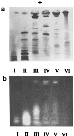
contained two proteins with chitobiosidase activity, and five proteins with endochitinase activity (Broadway et al., 1995). Strong anion exchange perfusion chromatography separated the endochitinases from the chitobiosidases, as shown by the elution profile of the column (Figure 1). Polyacrylamide gel
electropho-Time (sec)
FIG. 1. Strong anion exchange perfusion chromatography of semipurified chitinolytic enzymes. Total chitinolytic activity, as measured by hydrolysis of p-nitrophenyl /3-D-W,W-diacetylchitobiose, was 0.14 nkat/peak I, 1.52 nkat/peak II, 0.26 nkat/peak III,
resis enabled visualization of the number of bands of proteins in each peak (Figure 2a), and demonstrated that peaks I, II, and III had endochitinase activity, peaks III, IV, and V had chitobiosidase activity, while peak VI had no chitin-olytic activity (Figure 2b).
Biological Activity of Alkaline Chitinolytic Enzymes. The lyophilized,
chi-FIG. 2. Polyacrylamide gel electrophoresis of the peaks collected from anion exchange perfusion chromatography (Figure 1): (a) Coomassie stain to detect all proteins; (b) fluorogenic overlay to locate enzymes with chitinolytic activity. Based on previous exper-iments (Broadway et al., 1995), the upper (most acidic) two bands were chitobiosidases, while the lower (most alkaline) five bands were endochitinases.
tinolytic enzyme mixture contained 0.8-1.1 nkat of chitobiosidase activity/mg, and 154-165 units of endochitinase activity/mg. Dietary supplementation with the mixture of chitinolytic enzymes resulted in significant reductions in weight of larval Trichoplusia ni (expressed as percentage of the control) (F = 121.56,
df = 3,23, P < 0.001). However, results of each of the three bioassays are
presented (Figure 3a) rather than pooled data, because there was a significant interaction between date (bioassay number) and percent dietary intake of chi-tinase (P = 0.016). Results of each bioassay showed a similar inverse relation-ship between larval weight and dietary chitinase, although absolute values were variable at 0.25% dietary chitinase. Dietary supplementation with chitinases also resulted in significant reductions in percent pupation (F = 34.69, df = 3,20, P < 0.001), and percent adult emergence (F = 6.64, df = 3,8, P = 0.015) for Trichoplusia ni (Figure 3b and c). Due to the low number of indi-viduals surviving to adulthood, ANOVA was conducted on data pooled across the three test dates.
In addition, ingestion of the mixture of chitinolytic enzymes resulted in a dose-dependent increase in mortality for Myzus persicae at 24 and 48 hr (F = 20.33 and 20.02, respectively; df = 5,15, P < 0.001), Bemisia argentifolii at 18 and 42 hr (F = 11.23 and 25.12, respectively; df = 5,11, P < 0.001) and
Hypothenemus hampei (F = 5.84, df = 3,8, P = 0.021) at 30 days
(Figure 4).
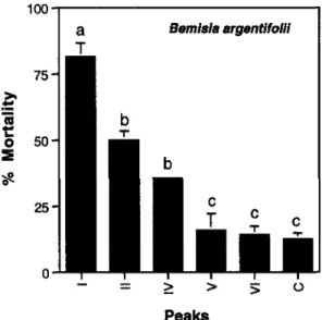
Separation of endochitinases from chitobiosidases resulted in significantly different levels of mortality for adult B. argentifolii (F = 17.77, df = 5,19, P
< 0.001). Fractions containing endochitinase (peaks I and II) resulted in 80%
and 50% mortality, respectively, after 36 hr of exposure, the fraction containing both proteins with chitobiosidase activity (peak IV) significantly elevated mor-tality to 38%, whereas mormor-tality following exposure to a single (most acidic) chitobiosidase (peak V) did not differ from the untreated controls (peak VI and C) (Figure 5).
DISCUSSION
The control of insect pests on commercially grown crops relies predomi-nantly upon the use of synthetic insecticides. However, in recent years, the use of these insecticides is increasingly regulated and limited because of their poten-tial hazards to human health and the environment. As a result, there is an active search for alternative approaches to insect control that are target-specific and environmentally benign. One approach that is gaining significant attention is the use of agricultural cultivars that are resistant to pests. These cultivars can be developed by the transgenic introduction of target-specific natural resistance factors. However, to enhance host-plant resistance, we must first identify and characterize target-specific factors that will significantly reduce the population(s) of herbivorous insect(s).
chitinolytic enzymes from Streptomyces albidoflavus, (a) Larval weight as a percent of the mean weight of untreated controls. Solid bars indicate weight of larvae in bioassay 1, striped bars are larvae in bioassay 2, hatched bars are larvae in bioassay 3. (b) Percent larvae that pupated, (c) Percent pupae that molted to adults. Vertical lines indicate ±1 SEM. Columns associated with an insect growth stage having similar letters are not significantly different (LSD test).
Peaks
FIG. 5. The effect of dietary endochitinases or chitobiosidases on survival of Bemisia
argentifolii. Endochitinase treatments I and II contained 10% sucrose and 0.5% peak I
or II, respectively, from the anion exchange perfusion chromatography (Figure 1), while chitobiosidase treatments IV and V contained 10% sucrose and 0.5% peak IV or V, respectively, from the anion exchange column (Figure 1). Two controls were included: treatment VI contained 10% sucrose and 0.5% peak VI (no chitinolytic activity), and treatment C, which contained 10% sucrose. Vertical lines indicate ±1 SEM. Columns with similar letters are not significantly different (LSE test).
Only a limited number of natural products have been characterized and identified as effective defensive agents against herbivorous insects. Few of these are proteins (e.g., proteinase inhibitors, arcelin, a-amylase inhibitors, lectins, endotoxin from Bacillus thuringiensis, and lipoxygenases), and even fewer are target-specific (Duffey and Felton, 1989; Gill et al., 1992; Hedin, 1983; Rosen-thai and Janzen, 1979). Identification and characterization of proteins as resis-tance factors) enables the isolation of gene(s) that encode(s) these proteins.
FIG. 4. The effect of dietary chitinolytic enzymes on survival of Myzus persicae, Bemisia
argentifolii, and Hypothenemus hampei. For B. argentifolii, solid bars indicate percent
mortality after 18-hr exposure, stippled bars indicate percent mortality after 42-hr expo-sure. For M. persicae, solid bars indicate percent mortality after 24-hr exposure, while stippled bars indicate percent mortality after 48-hr exposure. For H. hampei, solid bars indicate percent mortality after 30-day exposure. Vertical lines indicate ±1 SEM. Col-umns associated with a single time of observation and having similar letters are not significantly different (LSD test).
These genes can be transgenically inserted into agricultural crops, which may enhance the resistance of these crops against herbivorous insects without altering desirable characteristics of the cultivar(s) (Fraley et al., 1988; Hilder et al., 1987; Ryan, 1989; Vaeck et al,, 1987).
A target-specific factor would reduce the risk to nontarget organisms (e.g., vertebrates). One target site that distinguishes arthropods from higher organisms is chitin. Chitin is a principal component of the insect's exoskeleton (and of the lining of the foregut and hindgut) and peritrophic membrane (which lines the midgut of many species of insects) and is essential for structural integrity of insects. Degredation of chitin located in the alimentary canal (i.e., foregut, hindgut, and/or peritrophic membrane) may significantly disrupt digestion and nutrient acquisition, in addition to other protective and/or physiological func-tions. For example, in some insects, the peritrophic membrane is thought to function as protection against abrasion of the gut wall (the site of absorption and digestion), in the compartmentalization of digestion in the midgut lumen, and as a barrier to pathogens and phytotoxins. Destruction of the peritrophic membrane may have a negative impact on all three of these functions that are essential for the survival of such insects.
The current study demonstrated that ingestion of an artificial diet containing a mixture of endochitinases and chitobiosidases will significantly reduce the growth and development of larval Trichoplusia ni and significantly decrease the survival of Myzus persicae, Bemisia argentifolii, and Hypothenemus hampei. Separation of the endochitinases from the chitobiosidases was accomplished by perfusion chromatography. Feeding studies indicated that the endochitinases dramatically reduced survival of Bemisia argentifolii (which may have been due to acute toxicity or repellency following ingestion), while the chitobiosidases had less of an effect on survival. These results are in contrast to previous work that demonstrated that the chitobiosidases from Streptomyces albidoflavus sig-nificantly reduced the growth and survival of the plant pathogenic fungi Botrytis
cinerea and Fusarium oxysporum (Broadway et al., 1995).
The results of this study provided fundamental information required for evaluating the impact of chitinolytic enzymes on the growth and/or develoment of herbivorous insects. We now must demonstrate that chitinolytic enzymes, in planta, will reduce the growth and/or development of herbivorous insects. This information, followed by determination of the site of action and the physiolog-ical/physical effect of these ingested chitinolytic enzymes in vivo, will allow us to predict the long-term value of chitinolytic enzymes as defensive agents in plants.
Acknowledgments—This research was supported, in part, by the US Department of Agriculture
(NRI95-37302-1904 to RMB), Cornell University, Division of Biological Sciences Honors Program in Undergraduate Research (to J.B.W. and M.P.H.), Fulbright Foundation (to J.A.M.), Federation of the National Coffee Growers of Colombia (to C.G. and R.M.B.), and Hatch funds (to R.M.B., J.P.S., and M.P.H.).
REFERENCES
ABACUS CONCEPTS, INC. 1989. Super ANOVA: Accessible General Linear Modeling. Abacus
Con-cepts, Inc., Berkeley.
BERENBAUM, M. 1980. Adaptive significance of midgut pH in larval Lepidoptera. Am. Nat. 115:138-146.
BIELKA, H., DIXON, H. B. F., KARLSON, P., LIEBECQ, C., SHARON, N., VAN LENTEN, E. J., VELICK, S. F., VLIEGENTHART, J. F. G., WEBB, E. C., CORNISH-BROWN, A., LOENINO, K., Moss, G. P ., and REEDUK, J. 1984. Enzyme Nomenclature. Academic Press, New York, 646pp.
BOLLER, T. 1985. Induction of hydrolases as a defense reaction against pathogens, pp. 247-262,
in J. L. Key and T. Kosuge (eds.). Alan R. Liss, New York.
BROADWAY, R. M., WILLIAMS, D. L., KAIN, W. C., HARMAN, G. E., LORITO, M., and LABEDA, D. P. 1995. Partial characterization of chitinolytic enzymes from Streptomyces albidoflavus.
Lett. Appl. Microbiol. 20:271-276.
BROEKAERT, W. F., VAN PARIJS, J., ALLEN, A. K., and PEUMANS, W. J. 1988. Comparison of some molecular, enzymatic and antifungal properties of chitinases from thorn-apple, tobacco and wheat. Physiol. Mol. Plant Pathol. 33:319-331.
BROGLIE, K., CHET, I., HOLLIDAY, M., CRESSMAN, R., BIDDLE, P., KNOWLTON, S., MAUVAIS, C. J., and BRODLIE, R. 1991. Transgenic plants with enhanced resistance to the fungal pathogen
Rhizoctonia solani. Science 254:1194-1197.
DUFFEY, S. S., and FELTON, G. W. 1989. Plant enzymes in resistance to insects, pp. 289-313, in J. R. Whitaker and P. E. Sonnet (eds.). Biocatalysis in Agricultural Biotechnology. American Chemical Society, Washington, D.C.
FRALEY, R. T., FREY, N. M., and SCHELL, J. 1988. Genetic Improvements of Agriculturally Important Crops. Cold Spring Harbor Laboratory, Cold Spring Harbor, New York, 120 pp. GILL, S. S., COWLES, E. A., and PIETRANTONIO, P. V. 1992. The mode of action of Bacillus
thuringiensis endotoxins. Annu. Rev. Entomol. 37:615-636.
GRAYSON, J. M. 1951. Acidity-alkalinity in the alimentary canal of twenty insect species. V.J. Sci. Jan:46-59.
HEDIN, P. A. 1983. Plant Resistance to Insects. American Chemical Society, Washington, D.C.,
375 pp.
HEDRICK, S. A., BELL, J. N., BOLLER, T., and LAMB, C. J. 1988. Chitinase cDNA cloning and mRNA induction by fungal elicitor, wounding and infection. Plant Physiol. 86:182-186. HILDER, V. A., GATEHOUSE, A. M. R., SHEERMAN, S. E., BARKER, R. F., and BOULTER, D. 1987.
A novel mechanism of insect resistance engineered into tobacco. Nature 330:160-163. HUGHES, R. K., and DICKERSON, A. G. 1991. Modulation of elicitor-induced chitinase and
b-1,3-glucanase activity by hormones in Phaseolus vulgaris. Plant Cell Physiol. 32:853-861. JOOSTEN, M. H. A. J., and DE WIT, P. J. G. M. 1989. Identification of several pathogenesis-related
proteins in tomato leaves inoculated with Cladosporium fulvum (syn. Fulvia fulva) as 1,3-glucanases and chitinases. Plant Physiol. 89:945-951.
MAUCH, F., MAUCH-MANI, B., and BOLLER, T. 1988. Antifungal hydrolases in pea tissue. II. Inhibition of fungal growth by combinations of chitinase and b-l,3-glucanses. Plant Physiol. 88:936-942.
MISHRA, S. C., and SEN-SARMA, P. K. 1987. pH trends in the gut of xylophagous insects and their adaptive significance. Mater. Org. 22:311-319.
PEGG, G. F., and YOUNG, D. H. 1982. Purification and characterization of chitinase enzymes from healthy and Verticillum albo-atrum-infected tomato plants, and from V. albo-atrum. Physiol.
Plant Pathol. 21:389-409.
ROBERTS, W. K., and SELITRENNIKOFF, C. P. 1988. Plant and bacterial chitinases differ in antifungal activity. J. Gen. Microbiol. 134:169-176.
ROSENTHAL, G. A., and JANZEN, D. H. 1979. Herbivores—Their Interaction with Secondary Plant Metabolites. Academic Press, New York, 718 pp.
RYAN, C. A. 1989. Proteinase inhibitor gene families: Strategies for transformation to improve plant defenses against herbivores. BioEssays 10:20-24.
SAHAI, A. S., and MANOCHA, M. S. 1993. Chitinases of fungi and plants: Their involvement in morphogenesis and host-parasite interaction. FEMS Microbiol. Rev. 11:317-338.
SCHLUMBAUM, A., MAUCH, F., VOOELI, U., and BOLLER, T. 1986. Plant chitinases are potent inhibitors of fungal growth. Nature 324:365-367.
VAECK, M., REYNAERTS, A., HOFTE, H., JANSENS, S., DE BEUCKELEER, M., DEAN, C., ZABEAU, M., VAN MONTAGU, M., and LEEMANS, J. 1987. Transgenic plants protected from insect attack.
Nature 328:33-27.
VILLACORTA, A., and BARRERA, J. F. 1993. Nova dieta meridica para criacao de Hupothenemus
hampei (Ferrari) (Coleoptera: Scolytidae). An. Soc. Entomol. Bras. 22:405-409.
VOGELI-LANOE, R., HANSEN-GEHRI, A., BOLLER, T., and NEINS, F. 1988. Induction of the defense-related glucanohydrolases, b-l,3-glucanase and chitinase, by tobacco mosaic virus infection of tobacco leaves. Plant Sci. 54:171-176.
VOGELSANG, R., and BARZ, W. 1990. Elicitation of b-l,3-glucanase and chitinase activities in cell suspension cultures of Ascochyta rabiei resistant and susceptible cultivars of chickpea (Cicer
arietinum). Z. Naturforsch. 45c:233-239.
VOISEY, C. R., and SLUSARENKO, A. J. 1989. chitinase mRNA and enzyme activity in Phaseolus
vulgaris (L.) increase more rapidly in response to avirulent than to virulent cells of Pseudo-monas syringae pv. pnaseolicola. Physiol. Mol. Plant Pathol. 35:403-412.
WEBB, S. E., and SHELTON, A. M. 1988. Laboratory rearing of the imported cabbageworm. N.Y.
Food Life Sci. Bull. 122:000.
ZHE-FU, L., DANQI, W., ANNUO, L., and WEFQIN, Z. 1992. Chitinases from seeds of Zea mays and Coix lachryma-jobi L. Purification and some properties. Proc. Biochem. 27:83-88.