CorrespondenCe
Introduction
Cerebral hyperperfusion syndrome is a rare but serious com-plication after revascularization of stenosis of blood ves-sels supplying the brain and has been frequently described following carotid artery stenting. Impaired cerebral auto-regulation and post-revascularization changes in cerebral hemodynamics are the most important mechanisms invol-ved in the development of the syndrome. Clinical symptoms often include the triad of headache, seizures and transient focal neurological deficits in the absence of cerebral ische-mia. The incidence of hyperperfusion injury is estimated to be 1.1–6.8%, with mortality rates ranging from 3–26% when intracranial hemorrhage (ICH) is present. However, imaging in these patients is often uneventful and edema is frequently found in the affected areas of the brain.
The literature on hyperperfusion syndrome of the pos-terior circulation and imaging findings is rare. A case of hyperperfusion syndrome following recanalization of an occluded left subclavian artery (LSA) with an extraordinary extravasation of contrast medium and delayed occurrence of ICH is presented.
Case Report
A 56-year-old female patient presented with symptoms of intermittent claudication of the left arm and subclavian steal syndrome under physical stress. The pre-interventional clinical examination using computed tomography angio-graphy (CTA), digital subtraction angioangio-graphy (DSA) and ultrasound confirmed the diagnosis of LSA occlusion proxi-mal to the origin of the left vertebral artery (LVA) without any further high-grade stenosis of the other cervical arteries supplying the brain (Fig. 1). The retrograde flow in the LVA confirmed a subclavian steal phenomenon. The pre-inter-ventional CTA and DSA ruled out any further intracranial vascular pathologies (e. g. aneurysms and arterio-venous malformation).
The arterial blood pressure measured at the right arm was 130/95 mmHg prior to the intervention. Pre-interventional antiaggregation was established by a daily dosage of 100 mg aspirin and 75 mg clopidogrel and peri-interventional anti-coagulation was induced by the additional application of 5000 U of heparin.
Under local anesthesia an 8 French (F) sheath was intro-duced into the right femoral artery and a 6 F sheath into the left brachial artery. digital subtraction angiography of the right subclavian artery (RSA) illustrates the retrograde flow in the LVA (Fig. 2a). The angiogram obtained via a 6 F diagnostic catheter (Terumo, Japan) introduced into the left brachial artery illustrates the complete occlusion of the LSA (Fig. 2b). The occlusion site was passed from the brachial approach using a straight tip 0.35 inch guidewire (Terumo, Japan). The wire was caught in the thoracic aorta by a snare introduced from the inguinal approach and a long 8 F sheath (destination sheath, Terumo, Japan) was introduced crossing the site of occlusion. A balloon-mounted stent (Scuba 6 × 28 mm, Invatec, Italy) was placed achieving a good recanalization result (Fig. 2c). A total
Clin neuroradiol (2011) 21:223–229 DOI 10.1007/s00062-010-0052-3
Excessive Contrast Medium Leakage in Hyperperfusion
Syndrome
P. Mordasini · C. Brekenfeld · C. Fung · D. D. Do · G. Schroth · J. Gralla
p. Mordasini, Md · C. Brekenfeld, Md · G. schroth, Md · J. Gralla, Md Msc ()
department of Interventional and diagnostic neuroradiology, University of Bern, Inselspital, Freiburgstrasse 4,
3010 Bern, switzerland e-mail: [email protected] C. Fung, Md
department of neurosurgery, Bern, switzerland d. d. do, Md
Department of Angiology, Inselspital, University of Bern, Bern, Switzerland
Received: 14 October 2010 / Accepted: 23 December 2010 / Published online: 4 February 2011 © Urban & Vogel 2011
224 p. Mordasini et al.
of 170 ml low osmolar iodine contrast medium (Iopamiro 300, Bracco, USA) was used.
Immediately after recanalization, the patient complained of severe headache and became agitated. The intraarterial blood pressure revealed a raised systolic blood pressure of 205 mmHg. The blood pressure was medicinally reduced and kept within narrow margins below 140 mmHg systolic blood pressure. The patient showed no neurological deficits, in particular no signs of cerebellar dysfunction. A CT scan was performed 25 min after recanalization (Fig. 3) and sho-wed an extraordinary hyperdense signal alteration bilaterally and symmetrically in the cerebellar hemispheres, following
the cortex in the temporo-occipital lobe predominantly on the left side and presumably within the adjacent subarach-noidal space. The parenchymal hyperdensity without any mass-occupying effect in conjunction with the severe hea-dache immediately after recanalization was interpreted as a massive contrast agent extravasation.
The MRI 6.5 h after the intervention including diffu-sion-weighted imaging (dWI), T2-weighted imaging, susceptibility-weighted imaging (sWI) and T1-weighted pre-contrast and post-contrast imaging demonstrated edema in the corresponding cerebellar and cerebral areas (Fig. 4). no intracranial hemorrhage and ischemia were found. The
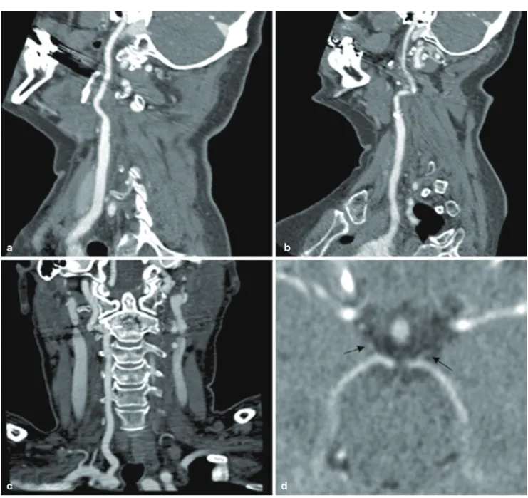
Fig. 1 Pre-interventional computed tomography angiography (CTA)
with curved reconstruction showing the right carotid artery (a), the left carotid artery (b), and the right vertebral artery (c), without any
concomitant high-grade stenosis. The intracranial pre-interventional CTA illustrates small posterior communication arteries (arrows) on both sides (d)
225 Excessive Contrast Medium Leakage in Hyperperfusion Syndrome
gadolinium-enhanced T1-weighted images did not illustrate any pathological enhancement. The patient was referred to the intensive care unit (ICU) for arterial blood pressure monitoring. The follow-up CT performed 23 h later illus-trated a complete disappearance of the parenchymal hyper-densities with some residual edema in the left occipital lobe (Fig. 5a and b).
Clopidogrel and aspirin medication was stopped after the intervention. After an uneventful clinical course in the ICU with normal blood pressure and no further antihyper-tensive medication for 2 days, the patient was returned to the ward. However, 63 h after the intervention the patient showed marked neurological deterioration and became aphasic. The control CT illustrated a mass-occupying intracerebral hemorrhage in the left occipital and temporal lobes with midline shift and intraventricular hemorrhage
(Fig. 5c and d). The patient recovered well after surgery and was sent to rehabilitation with a minor speech deficit and quadrantanopsia.
Discussion
Cerebral hyperperfusion syndrome is a serious complica-tion of craniocervical revascularizacomplica-tion and is well-descri-bed after carotid artery endarterectomy and stenting. Two synergistic mechanisms may be responsible for the deve-lopment of the syndrome: impaired cerebral autoregulation and postoperatively elevated systemic blood pressure. It is theorized that the capillary bed beyond the stenosis is prone to breakdown of the blood-brain barrier after an increase of blood flow due to impaired autoregulation. However, reports
Fig. 2 Digital subtraction angiography (DSA) of the right subcalvian
artery illustrates a subclavian steal syndrome with retrograde flow in the left vertebral artery (a), the retrograde angiography of the left sub-clavian artery shows a long occlusion of the artery, the distal guider
is placed in the origin at the aortic arch (b), postinterventional angio-graphy illustrates a good recanalization with antegrade flow in the left vertebral artery (c)
226 p. Mordasini et al.
of this phenomenon after recanalization of the subclavian artery [1] or vertebral artery [2–4] are limited.
The clinical symptoms in conjunction with increased sys-temic blood pressure confirm the diagnosis of hyperperfusion syndrome in the presented case. The extraordinary
parenchy-mal hyperdensity in CT can mimic laminar intraparenchyparenchy-mal hemorrhaging although no relevant mass-occupying effect was illustrated. The finding was consistent with a massive extravasation of iodine contrast agent immediately after stenting and is most likely the consequence of breakdown of
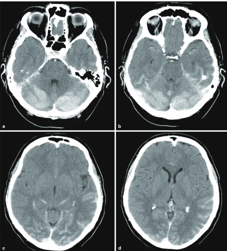
Fig. 3 Postinterventional CT performed 25 min after recanalization of
the LSA shows extensive areas of hyperdense signal symmetrically distributed in both cerebellar hemispheres including white matter and
cortex (a, b). A linear hyperdense signal is seen following the occipital and temporal cortex and subarachnoidal space (c, d)
227 Excessive Contrast Medium Leakage in Hyperperfusion Syndrome
the blood-brain barrier. The MrI ruled out any intracranial hemorrhage but illustrated brain areas affected by edema. Interestingly, at this point (6.5 h after intervention) no further extravasation of gadolinium-based contrast was found. The
reduced extravasation of contrast agent in the post-interven-tional course might be explained either by differences for contrast agents to pass the damaged blood-brain barrier or by decrease of systemic blood pressure.
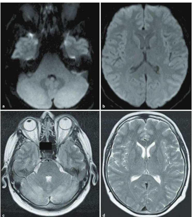
Fig. 4 The postinterventional MRI 6.5 h after intervention in
digital-weighted imaging (dWI) shows no signs of infarction or hemorrha-ge (a, b). The T2-weighted imahemorrha-ges show hyperintense signals in both
cerebellar hemispheres and subtle changes in the left occipital lobe (c, d)
228 p. Mordasini et al.
Moreover it is unlikely that the parenchymal hemorr-hage was the result of a reperfusion lesion into an area of tissue infarction as both CT and MrI imaging illustrated
no ischemic lesions. remarkably, parenchymal hemorr-hage occurred after a delay of 63 h after intervention into the left temporo-occipital lobe and despite normal systolic
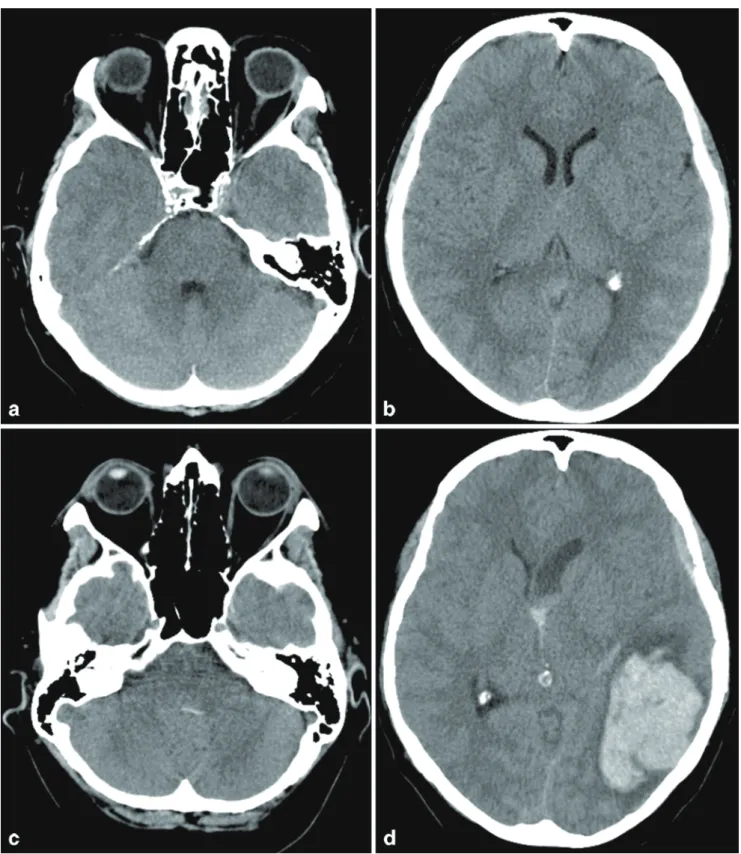
Fig. 5 The unenhanced CT performed 23 h after intervention shows
some edema in the occipital lobes, the hyperdense signal in the infra-tentorial and suprainfra-tentorial spaces however has completely diminished (a, b), the CT after acute neurological deterioration performed 63 h
after intervention illustrates some hemorrhaging in the fourth ventricle but the cerebellar parenchyma is unaffected (c), mass-occupying cerebral hemorrhage in the left occipital and temporal lobe with intra-ventricular hemorrhage and midline shift (d)
229 Excessive Contrast Medium Leakage in Hyperperfusion Syndrome
blood pressure during this period. The hemorrhage therefore occurred in a brain area previously affected by extravasa-tion of contrast agent and edema. This finding is in concor-dance with previous case reports [5–7]. It is hypothesized that contrast agent extravasation in the course of cerebral hyperperfusion syndrome indicates more serious damage of the blood-brain barrier and that these areas are at increased risk for intraparenchymal hemorrhage.
Conclusion
Cerebral and cerebellar hyperperfusion syndrome com-plicated by hemorrhage may occur after recanalization of the subclavian artery. Excessive extravasation of iodine contrast agent can mimic parenchymal hemorrhage and is likely to be a consequence of breakdown of the blood-brain barrier. despite stringent blood pressure control intracranial hemorrhage might occur.
Conflict of Interest Statement The authors declare that they have no
conflict of interest.
References
1. salerno JL, Vitek J. Fatal cerebral hemorrhage early after sub-clavian artery endovascular therapy. AJNR Am J Neuroradiol. 2005;26:183–5.
2. Zhang r, Zhou G, Xu G, Liu X. posterior circulation hyperperfu-sion syndrome after bilateral vertebral artery intracranial stenting. Ann Vasc Surg. 2009;23:686–5.
3. Rezende MT, Spelle L, Mounayer C, Piotin M, Abud DG, Moret J. Hyperperfusion syndrome after stenting for intracranial verteb-ral stenosis. Stroke. 2006;37:e12–e14.
4. Bando K, Satoh K, Matsubara S, Nakatani M, Nagahiro S. Hyper-perfusion phenomenon after percutaneous transluminal angio-plasty for atherosclerotic stenosis of the intracranial vertebral artery. Case report. J Neurosurg. 2001;94:826–30.
5. Takayama K, Nakagawa H, Iwasaki S, Taoka T, Wada T, Myou-chin K, Sakamoto M, Fukusumi A, Kichikawa K. Cerebral hemorrhage with angiographic extravasation immediately after carotid artery stenting. Radiat Med. 2007;25(7):359–63. 6. Toh CH, Wong HF, Lin TK, Ng SH. Abnormal enhancement on
imaging studies preceding hyperperfusion syndrome. Case illus-tration. J Neurosurg. 2006;105:932.
7. Sharma P, Poppe AY, Eesa M, Steffanhagan N, Hudon M, Mor-rish W. Extravasating contrast material on angiography following carotid angioplasty and stenting: not necessarily subarachnoid hemorrhage. J Neuroimaging. 2010;20:180–82.