EPIDEMIOLOGICAL, SEROLOGICAL, CLINICAL AND
RISK FACTORS OF
INFECTIOUS BURSAL DISEASE (IBD),
IN BROILERS FLOCKS IN ALGERIA
SALHI1)* Omar, Chafik-RedaMESSAI2), Khatima AIT-OUDHIA2), Aziz LOUNAS1), Amine ABDELLI1), Rachid KAIDI1), Djamel KHELEF2)
1)
University Blida 1, Institute of Veterinary Sciences, Laboratory of Biotechnology related to Animal Reproduction (LBRA), BP 270 Road of Soumaa Blida, Algeria.
2)
Higher National Veterinary School, Road Issad Abbes, Oued Smar, Algiers, Algeria. *Corresponding author: dr.salhi-omar@hotmail.com
Abstract. The present study was conducted to epidemiological, serological and clinical of Infectious Bursal Disease (IBD) on Algerian broiler chicken (45 flocks/1350 sera) using indirect Enzyme-Linked Immuno-Sorbent Assay (ELISA) method and to assess the influence of some risk factors related to this disease. Among all investigated flocks, IBD was the most seroprevalent disease (73.33%). The results show the effect of risk factors, the antibody titers were elevated in the herd recorded a high mortality (more than 10%) compared with those recorded a low mortality (less than 10%) (p = 0.009). However, more than flocks 30 days old flocks were less seropositive those less aged of 30 days (p = 0.002). Therefore, the antibody titers were elevated in herds with bad hygiene compared with good hygiene (p = 0.04) At last, when broiler chicken were not boosted by IBD vaccine, flocks appeared to be more
seropositivity (p = 0.03).This study determined that IBD is a dominant viral disease in broilers.
Many factors are responsible for the development of this disease.
Keywords: Serological; ELISA, risk factors, IBD; broilers, Algeria.
INTRODUCTION
Infectious bursal disease (IBD) is a highly contagious acute viral disease of young chickens (3-6 weeks), which causes mortality or immunosuppression following damage to the bursa of Fabricius, resulting poor growth of young chickens and significant economic losses (Khan and Dana, 2005;Abed et al., 2018;Eterradossi and Saif, 2020). The causative agent of IBD is an infectious bursal disease virus (IBDV), belonging to the Birnaviridae family. IBDV strains are classified into two distinct serotypes namely: pathogenic and non-pathogenic (Prandini et al., 2016; Eterradossi and Saif, 2020).
The disease is manifested by debilitate, dehydration and the development of depression with watery diarrhea, swollen and blood stained vent (Islam and Samad, 2004; Van den Berg et al., 1991). Infection with less virulent strains may not show obvious clinical signs but the birds may have fibrotic or cystic bursa of Fabricius that become atrophied prematurely (before six months of age) and may die of infections by agents that would not usually cause disease in immunocompetent birds (Mohammed, 2013). The postmortem findings were haemorrhages in the thigh/pectoral muscles, enlarged, edematous and hyperemic bursa or atrophic in chronic cases and hemorrhage in the junction between gizzard and proventriculus (Chettle and Wyeth, 1989; Banda, 2002). Clinical manifestations and postmortem findings of affected birds may aid to diagnose a viral disease but laboratory diagnosis is necessary for confirmation of the
diseases (Banda, 2002; Messai et al., 2019). Various diagnostic methods like enzyme linked immunosorbent assay (ELISA) have been frequently used all over the world to detect viruses from the field samples (Auvigne et al., 2013; Desingu et al., 2014, Salhi et al., 2018).
Therefore, the present study was undertaken to find out a relationship among the disease diagnostic parameters; clinical signs and postmortem lesions, serological tests for the diagnosis of IBD in broilers flocks and to assess the risk factors associated with the disease in affected farms.
MATERIAL AND METHODS
Ethical approval: Experimental procedures approved by the Institutional Committee for the Protection of Animals of the National Administration of Higher Education and Scientific Research of Algeria (98-11, Act of 22 August 1998).
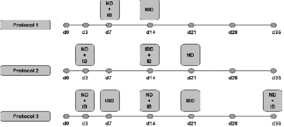
Animals: The experiment was carried out at commercial farms in the central, east and west of northern Algeria (longitude 36° and latitude 3°), from July 2018 to September 2019 on forty five (45) broiler flocks with different strains (Arbor acres, Cobb 500, Hubbard F15) aged between four to seven weeks and containing 3,000 to 10,000 birds/farm. The studied flocks had been initially vaccinated for IBD with live vaccines through different protocols (Fig. 1). For vaccination protocols 1, 2 and 3, the vaccines used against IBD are as follows: the intermediate D78 strain was used for primary vaccination, and for the recall, the strain was E228 Intermediate+. The analysed flocks were suspected to acquire a viral disease (IBD) after showing the characteristic clinicaland necropsic signs.
Fig. 1. Schematic diagram of protocols vaccine used in the flocks (d: day of vaccine)
Blood collection procedures: A total of 1350 birds were sampled randomly from 45 broiler flocks (15 samples/flock), according to our protocol, two samples were taken from each farm; the first was performed the first days after the appearance of the first clinical signs. The second one was done, two to three weeks later. Blood samples were collected from the wing vein, in dry tubes and centrifuged (5000 rpm for 10 min) at the same day to recover the sera that were stored in test tubes 'Epperndorf’ and frozen at -20 ° C until analysis.
Clinical diagnosis: Clinical diagnosis was made on the basis of clinical history from the responsible persons of the farms including veterinarians in charge of monitoring, recorded clinical signs and gross lesions of affected chickens at autopsy. Serological Methods: An indirect ELISA technique was carried out using ID.vet Innovative Diagnostics kits (Montpellier, France): ID Screen® IBDV Indirect. The sera were diluted to 1 / 500th, then loaded to ELISA plates to start immuno-sorbent
reaction as guided by manufacturer’s manuals. ELISA plates were read by ELx800 spectrophotometer (DIALAB GmbH, Wiener Neudorf, Austria) equipped with the 450 nm filter; where the measured optical density (OD) was transformed into titrated 'antibody. The averages of the titers and the coefficient of variation (CV) were automatically calculated by band and by series of samples with the software provided by the laboratory (IDSoftTM, Montpellier, France).
Interpretation of the ELISA results: To interpret the ELISA results, the following parameters were taken: The presence of clinical signs and postmortem lesion during the autopsy, the antibodies kinetics; titers between the first and the second sampling. Moreover, mainly according to the Interpreting Poultry Baselines provided by the manufacturer of IDvet ELISA kits:
Observation of risk factors: A standardized survey was used to assess risk factors associated with the mortality observed. The survey covered the following parameters: flock characteristics, strain, hygiene, vaccination programs, mortality and morbidity rates, age of occurrence, clinical and necropsy lesions, stocking density, season, area and climate.
Statistical analysis: Firstly, descriptive statistics were used to characterize flocks according the different factors. Thus, statistical analyses were performed with SAS (Version 9.1.3; SAS Institute Inc., Cary, NC).
Before fitting statistical analysis, examination of the distributions of antibody titers indicated using (PROC UNIVARIATE, Shapiro–Wilk test) that most could not be considered normally distributed.
Antibody titer of each disease through the time were analyzed by fitting the fixed effects of day, group and the interaction of day*group in a repeated measures variance analysis using a PROC MIXED models with the random effect of herd (SAS Inst. Inc. 9.1). Covariance structure used [compound symmetry or autoregressive (AR1)] was chosen based on the Akaike information criterion.
The layout of our model can be summarized as follows: Yijk = µ + Gi + Tk + GTik+ ɛijk
Where Yijk =Antibody titer, μ=overall mean, Gi = effect of group, Tk = effect of time of
sampling (k=1 and 2), GTik =effect of group × timeand ɛijk = random residual error.
A Stacked line plots of Antibody titer changes were generated using Prism 5.01 (GraphPad Software, Inc. La Jolla, CA USA).
RESULTS AND DISCUSSIONS
Table 1 presents the results of antibody titers for IBD. Among total of 45 flocks, 33 (73.33%) were tested positive to IBD. For this mentioned disease, it has been shown a low CV (CV= 33-48%) and a significant difference (p <0.0001) in
antibody titer between the first and the second sample (LSM± SE, 2062.20 vs 46168.00 ± 313.03).
Table 1 Serological results
Pathology Antibody titers CV
(%)
SE P Seropositivity (%)
Mean 1 Mean 2
IBD 2062.20 4168.00 33-48 313.03 <0.0001 73.33
For that, immune status in response to viral diseases is estimated by measuring the serological response objectified by detection of specific antibodies produced either in response to infection or following vaccination (Picault et al., 1993; Brigitte et al., 1997). On the other hand, the protected farms must have a higher average of titers than the protection threshold for all the analysis dates without being very high compared to the titer resulting from the vaccination and this in the absence of specific clinical signs (Gardin et al., 2002). Our samples herds were suspected to be infected with a viral disease such as IBD and showed typical clinical signs and necropsy signs with high morbidity and mortality; the vaccines used for these were live vaccines for all the farms. Clinical and necropsic manifestations of affected birds can help diagnose a disease, but a laboratory diagnosis is needed to confirm it (Hasan et al., 2010; Girma et al., 2017). However, outbreaks have been reported in the vaccinated populations despite the fact that vaccination is widely applied (Van Boven et al., 2008). Although the ELISA test does not distinguish post-vaccine antibodies from post-infectious antibodies when vaccinated with an inactivated vaccine, the absence or presence of clinical signs and the type of vaccine used should be taken into account (Van den Berg et al., 2000). For this, we took paired samples; the first sample is taken at the beginning of the disease and the second, two to three weeks later. In fact, since the concentration of antibodies increases between the 02 sera collected, this indicates that we had a stimulation of the immune system that could be due to a recent infection or to a symptomatic viral reactivation or not (Salhi et al., 2018; Messai et al., 2019).
Clinically, the most common clinical signs were: digestive signs (white diarrhea). Most commonly observed postmortem lesions were: inflammation of the bursa of Fabricius, nephritis, petechiae in the muscles, hemorrhage in the proventriculus and isthmus (Fig. 2).
inflammationof the bursa of Fabricius nephritis petechiae in the muscles
Thus, using the necropsic and clinical signs to detect the three diseases, we observed a very high specificity (75%). In other words, all birds suspected of having ND had specific antibodies. However, the sensitivities were 67.0%, so for this disease, necropsy and clinical diagnosis were particularly reliable (Table 2).
Table 2 Diagnostic sensitivity (%) and specificity (%), with 95 percent confidence intervals (CI) and
true Prevalence of test based on lesional signs of detecting IBD.
Pathology Sensitivity (%)
(95%CI)
Specificity (%)(95%CI) True Prevalence (%)
(95%CI)
IBD 67.0 (36.8,90.5) 75.0 (47.6, 96.3) 44.5 (37.9, 71.6)
Clinically, clinical signs were high mortality, unsteady gait, ruffled feathers, uratecontaining diarrhea and sudden death which correspond with the findings of Lukert and Saif (2003), Islam and Samad (2004). The postmortem findings were hemorrhages in the thigh/pectoral muscles, enlarged, edematous and hyperemic bursa with bloody or mucoid contents or atrophic in chronic cases and hemorrhage in the junction between gizzard and proventriculus which support the findings of Chettle & Wyeth (1989), Mera and Sirajo, 2019), Islam and Samad (2004); Hasan et al (2010), Mohammed et al (2013) and Abed et al (2018).
Table 3 Comparison of least square of means (LSMs) and standard errors (SEs) of Antibody titer
anti-IBDamong area, climate, season, age, density, mortality, hygiene, strainand protocols of
vaccination groups
Traits Group Time 1 Time2 SEM P1 P
Group Time G*T Area East 194.38 141.24 764.29 0.53 0.13 0.08 0.92 Center 843.89 1412.33 408.70 0.26 West 561.39 1165.39 459.86 0.15 Climate Dry 1062.62 1687.12 304.46 0.40 0.25 0.08 0.65 Wet 803.72 1171.38 461.27 0.08 Season Autumn 1103.33 1444.67 439.77 0.58 0.18 0.06 0.66 Summer 1166.65 1616.15 240.87 0.19 Spring 1539.25 2693.00 538.60 0.13 Age (day) ≤30 1655.71 2765.14a 377.69 0.04 0.002 0.02 0.21 >30 1066.09 1409.00b 208.36 0.24 Density (birds/m2) ≤10 1278.00 1737.50 556.83 0.56 0.78 0.14 0.96 >10 1116.64 1554.09 335.78 0.36 Mortality <10 1495.67 2527.67a 430.22 0.09 0.04 0.04 0.35 ≥10 1130.67 1524.87b 215.11 0.20 Hygiene Good 1482.18 2781.32a 393.93 0.01 0.009 0.02 0.26 Intermediate 1082.17 1401.89b 292.48 0.62 Bad 940.69 1172.13b 361.99 0.40 strain Arboracres 1257.29 1935.00 290.54 0.10 0.27 0.17 0.73 Cobb 500 935.60 985.20 486.16 0.94 ISA 1257.27 1795.18 327.77 0.25 Vaccination protocol 1 1813.12a 3406.42a 554.73 0.02 0.03 0.08 0.92 2 3065.21ab 4216.65ab 618.19 0.03 3 4398.09b 5416.10b 742.32 0.18
1 Difference between times for the same group
a, b : Different letters showing significant difference between groups within the same time sampling. Vaccination protocol, 1and 2: primo vaccine without booster vaccine; 3: primo vaccine with booster vaccine.
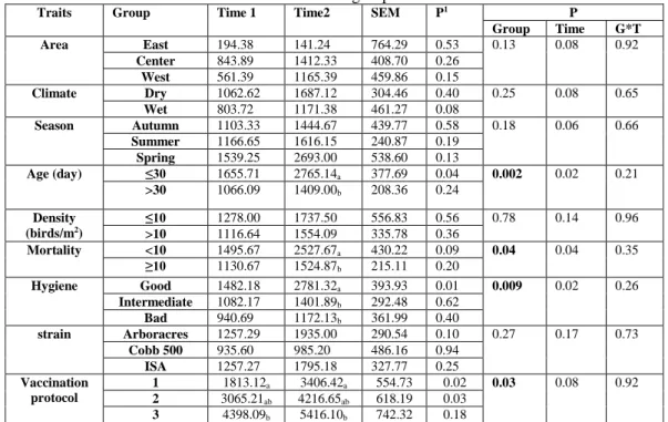
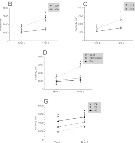
The table 3 shows the effect of risk factors (area, climate, season, age, density, mortality, hygiene, strain and protocols of vaccination groups) on the amount of antibody titers among time sampling.
There was a significant effect of the mortality, hygiene, age and vaccination groups on antibody titers in time 2. The antibody titers were elevated in the herd recorded a high mortality (more than 10%) compared with those recorded a low mortality (less than 10%) (p = 0.009). However, more than flocks 30 days old flocks were less seropositive those less aged of 30 days (p = 0.002). Therefore, the antibody titers were elevated in herds with bad hygiene compared with good hygiene (p = 0.04) At last, when broiler chicken were not boosted by IBD vaccine, flocks appeared to be more seropositivity (p = 0.03).
However there was no significant effect of climate, season, age, density, strain groups on the amount of antibody titers among time sampling. There was a significant effect of sampling time in all the groups.
Time 1 Time 2 0 1000 2000 3000 4000 30 >30 *
B
A n ti b o d y t it e r Time 1 Time 2 0 1000 2000 3000 4000 10 10 *C
A n ti b o d y t it e r Time 1 Time 2 0 1000 2000 3000 4000 Good Intermediate *D
Bad A n ti b o d y t it e r Time 1 Time 2 0 2000 4000 6000 8000 P1P2 *G
P3 * A n ti b o d y t it e rFor factors affecting IBD. The virus of IBD has marked increased pathogenecity characterized by 80% mortality in layer pullets and 25% in broilers. Infections before 3 weeks of age are usually subclinical (Michel and Jackwood, 2017). According to Khan and Dana (2005), it is considered as the most important form of the disease because of the significant economic losses that it causes, including impairment of growth and immunosuppression. Infectious Bursal Disease (IBD) is a highly contagious acute viral disease of young chickens of 3-6 weeks old (Hasan et al., 2010)
Infectious Bursal Disease IBD, also known as Gumboro disease, is an acute contagious, immunosuppressive disease of young chickens caused by the IBD virus (IBDV). The disease has a worldwide distribution and prevalence and is responsible for major economic losses in the commercial poultry industry (Ladjel, 2015). Vaccination of the birds is a major tool for the prevention and control of the disease; however, traditional inactivated and live attenuated vaccines suffer from drawbacks due to either incomplete inactivation or reversion of the attenuated pathogen into the
virulent form (Muniz et al., 2018).
Gumboro disease is extremely contagious. In infected herds, it results in very high morbidity rate after infection reaching mortality rates of up to 25% to 30% on broilers have been observed. The severity of the disease depends on the age and sensitivity of the type of poultry infected, the virulence of the strain and the extent of passive immunity transmitted by the parents (Van Den Berg et al., 2000).
Therefore, the prevention of Gumboro disease is based on both hygiene and medical prophylaxis. It should be stressed that no vaccine will solve the problem of Gumboro disease unless important health precautions are taken. These include, in particular, compliance with all-in/all-out farming methods, cleaning and disinfection of the premises and compliance with a crawl space (Dey et al., 2017). Vaccination In addition to strict compliance with hygiene and disinfection rules, the success of vaccination also depends on the choice of vaccine strain and vaccination schedule. (Muniz et al., 2018)
CONCLUSIONS
The serological study conducted in this experimentation provided an important scope about IBD as much as a dominant viral disease on broiler chickens, and found that the seropositivite of IBD were 73.33%. Clinical manifestations and postmortem findings of affected birds may aid to diagnose a disease but laboratory diagnosis is necessary for confirmation of the diseases. Further to that, the findings also suggest that risk factors related to biosecurity and farm practices appear to have a significant role in the severity of the disease observed in affected farms. If those factors are alleviated, the severity of the IBD problems in farms would be greatly reduced.
REFERENCES
1. Abed, M., Soubies, S., Courtillon, C., Briand, F. X., Allée, C., Amelot, M., Kara, R.
(2018). Infectious bursal disease virus in Algeria: Detection of highly pathogenic reassortant viruses. Infection, Genetics and Evolution, 60, 48-57.
2.Auvigne, V., Gibaud, S., Léger, L., Malher, X., Currie, R., Riggi, A. (2013). A longitudinal study of the incidence of Avian Infectious Bronchitis in France using
strain-specific haemagglutination inhibition tests and cluster analysis. Revue Méd. Vét, 164, 8-9, 417-424.
3.Banda, A. (2002). Characterization of field strains of Infectious bursal disease virus (IBDV) using molecular techniques. Dissertation (Doctor of Pholosophy).
4.Brigitte, A., Jean François, D. J., Nadia, M., Yalacé, K. (1997). Study of vaccine programs carried out in poultry farming in Senegal. Second Days of the Poultry Research, Tours.
5.Chettle, N. J., & Wyeth, P. J. (1989). Failure of maternally derived infectious bursal disease antibodies to serotypes 1 and 2 to protect against heterologous virus. British Veterinary Journal, 145(2), 165-169.
6. Desingu, P. A., Singha, S. D., Dhamaa, K., VinodhKumarb, O. R,. Singhc R., Singh, R. K. (2014). Development of slide ELISA (sELISA) for detection of four poultry viralpathogens by direct heat fixation of viruses on glass slides. Journal of Virological Methods, 209, 76-81.
7.Dey, S., Pathak, D. C., Ramamurthy, N., Maity, H. K., & Chellappa, M. M. (2019). Infectious bursal disease virus in chickens: prevalence, impact, and management strategies. Veterinary Medicine: Research and Reports, 10, 85.
8.Eterradossi, N., & Saif, Y. M. (2020). Infectious bursal disease. Diseases of poultry, 257-283.
9.Gardin, Y., Soleil, S., Rippa, I. (2002). Use of serology for monitoring Epidemiology of poultry herds. Interprofessional meetings of pathology of avian diseases, Rennes.
10.Girma, M., Kebede, B., & Megarsa, B. (2017). Seroprevalence of Infectious Bursal Disease in Backyard Chickens of Six Districts of North Shewa Zones of Oromia and Amhara Regions, Ethiopia.
11.Hasan, R. A. K. M., Ali, M. H., Siddique, M. P., Rahman, M., Islam, M.A. (2010). Clinical and laboratory diagnoses of Newcastle and infectious bursal diseases of chickens. Bangl. J. Vet. Med, 8(2), 131-140.
12.Islam, M. T., & Samad, M. A. (2004). Clinico-pathological studies on natural and experimental infectious bursal disease in broiler chickens. Bangladesh Journal of Veterinary Medicine, 2(1), 31-35.
13.Khan, C. M., Dana, A. (2005). The Merck Veterinary Manual. 9th ed.; New Jersey, USA: Merck and Co.,Inc. p: 2255-2257.
14.Ladjel, T. (2015). Enquête séro-épidémiologique post-vaccinale de la maladie de Gumboro en élevage avicole en région centre.
15.Lukert, P. D and Saif Y. M. (2003). Infectious bursal disease. Ames, Iowa, Iowa State University Press.
16.Mera, M. U., & Sirajo, G. (2019). Outbreak of infectious bursal disease in a flock of 14 weeks old ISA brown pullets, Sokoto State, Nigeria. GSC Biological and Pharmaceutical Sciences, 9(2), 001-008.
17.Messaï, C. R., Salhi, O., Khelef, D., Lounas, A., Mohamed-Cherif, A., Kaidi, R., & Aït-Oudhia, K. (2019). Serological, clinical, and risk factors of the Newcastle disease on broilers flocks in Algeria, Veterinary World, 12 (7): 938-944. Abstract.
18.Michel, L. O., & Jackwood, D. J. (2017). Classification of infectious bursal disease virus into genogroups. Archives of virology, 162(12), 3661-3670.
19.Mohammed, M. H., Zahid, A. A. H., Kadhim, L. I., Hasoon, M. F. (2013). Conventional and Molecular Detection of Newcastle Disease and Infectious Bursal Disease in Chickens J. World's Poult. Res, 3(1), 05-12.
20.Muniz, E. C., Verdi, R., Jackwood, D. J., Kuchpel, D., Resende, M. S., Mattos, J. C. Q., & Cookson, K. (2018). Molecular epidemiologic survey of infectious bursal disease viruses
in broiler farms raised under different vaccination programs. Journal of Applied Poultry Research, 27(2), 253-261.
21.Picault, J. P., Lecoq, H., Guittet, M., Bennejean, G. (1993). Poultry technical science, 4, 3749.
22.Prandini, F., Simon, B., Jung, A., Pöppel, M., Lemiere, S., Rautenschlein, S. (2016). Comparison of infectious bursal disease (IBD) live vaccines and a HVT-IBD vector vaccine and their effects on the immune system of commercial layer pullets. Avian Pathology, 45, 114-125.
23.Salhi, O., Khelef, D., Messai, C. R., Lounas, A., Mohamed-Cherif, A., Kaidi, R., Ait-Oudhia, K. (2018). Serological Survey of Dominant Viral Diseases (Newcastle Disease (ND), Infectious Bronchitis (IB) and Infectious Bursal Disease (IBD)), in Broilers Flocks in Northern Algeria. Bulletin of University of Agricultural Sciences and Veterinary Medicine Cluj-Napoca. Veterinary Medicine, 75(2), 155-162.
24.Van Boven, M., Bouma, A., Fabri, T. H. F., Katsma, E., Hartog, L., Koch, G. (2008). Herd immunity to Newcastle disease virus in poultry by vaccination. Avian Pathology, 37(1), 1-5.
25.Van den Berg, T,P., Gonze, M., Meulemans, G. (1991). Acute infectious bursal disease of poultry: isolation and characterization of a highly virulent strain. Avian Pathology, 20, 133-143.
26.Van den Berg, T. P., Eterradossi, N., Toquin, D., Meulemans, G. (2000). Infectious bursal disease (Gumboro disease). Revue Scientifique Technique, 19, 509-543.