HAL Id: hal-01901846
https://hal.sorbonne-universite.fr/hal-01901846
Submitted on 23 Oct 2018
HAL is a multi-disciplinary open access
archive for the deposit and dissemination of sci-entific research documents, whether they are pub-lished or not. The documents may come from teaching and research institutions in France or abroad, or from public or private research centers.
L’archive ouverte pluridisciplinaire HAL, est destinée au dépôt et à la diffusion de documents scientifiques de niveau recherche, publiés ou non, émanant des établissements d’enseignement et de recherche français ou étrangers, des laboratoires publics ou privés.
Distributed under a Creative Commons Attribution| 4.0 International License
Livedoid Vasculopathy: A French Observational Study
Including Therapeutic Options
Emma Gardette, Philippe Moguelet, Jean David Bouaziz, Dan Lipsker,
Olivier Dereure, François Le Pelletier, Catherine Lok, Thierry Maisonobe,
Didier Bessis, Jacqueline Conard, et al.
To cite this version:
Emma Gardette, Philippe Moguelet, Jean David Bouaziz, Dan Lipsker, Olivier Dereure, et al.. Live-doid Vasculopathy: A French Observational Study Including Therapeutic Options. Acta Dermato-Venereologica, Society for Publication of Acta Dermato-Dermato-Venereologica, 2018, 98 (9), pp.842 - 847. �10.2340/00015555-2965�. �hal-01901846�
A
cta
DV
A
cta
DV
A
dvances in dermatology and venereology
A
ctaD
ermato-V
enereologicaCLINICAL REPORT
SIGNIFICANCELivedoid vasculopathy is a chronic thrombotic disease of the skin microcirculation resulting in painful ulcers mainly af-fecting the lower legs. This study presents a French cohort of patients with livedoid vasculopathy. It describes clinical, histological characteristics and outcome of patients. It no-tably shows a frequent thrombophilia background. Nerve damage can be associated with cutaneous manifestations. These data also confirm that heparin or oral anticoagulants are able to achieve a complete response. Refractory cases can be treated with intravenous immunoglobulins.
Livedoid vasculopathy is a rare thrombotic cutaneous disease. This observational study aimed to assess the clinical and biological features of livedoid vasculopa thy and the efficacy of treatments. Patients enrolled had typical livedoid vasculopathy both clinically and histologically. Investigation of thrombophilia was per formed. Electromyography was undertaken in the pre sence of symptoms suggesting peripheral neuro pathy. Eighteen women and 8 men were included, with a mean age of 35.5 years at onset. Twenty patients had at least one thrombophilia factor. Ten patients had a peripheral neuropathy with 2 of these patients demon strating a specific thrombo-occlusive vasculopathy on muscle biopsy. Anticoagulation with low molecular weight heparin was the most prescribed therapy and was associated with the best outcome (effective in 14 patients). Eight patients had severe disease refractory to anticoagulation and required intravenous immuno globulins, producing a good response in 6 patients.
Key words: livedoid vasculopathy; peripheral neuropathy;
throm bosis of dermal vessels; thrombophilia; low molecular weight heparin; intravenous immunoglobulins.
Accepted May 4, 2018; Epub ahead of print May 8, 2018 Acta Derm Venereol 2018; 98: 842–847.
Corr: Dr. Stéphane Barete, MD, PhD, Department of Dermatology, Hôpital
Pitié-Salpêtrière, 47-83 Boulevard de l’Hôpital, FR-75013 Paris, France. E-mail: [email protected].
L
ivedoid vasculopathy (LV) is a chronic disease mani-festing as recurrent necrotic and painful lower limb ulcerations. These resolve leaving atrophic porcelain-white scars with surrounding telangiectasias known as atrophie blanche (AB). The estimated incidence is 1:100,000 individuals, predominantly affecting young to middle-aged females, with a sex ratio of 3:1 (1, 2).Histopathology reveals a vaso-occlusive disorder with intraluminal thrombosis of dermal vessels without leu-kocytoclastic vasculitis (1, 3). The exact mechanism of this entity is unknown but underlying thrombophilia with abnormalities of coagulation or fibrinolysis, is observed in up to 50% of patients (2, 4). Plasminogen activator inhibitor-1 (PAI-1) is a major inhibitor of fibrinolysis. The 4G/5G polymorphism of the PAI-1 promoter gene results in enhanced transcription and the 4G allele is
associated with increased PAI-1 levels. Impaired fib-rinolysis through PAI-1 involvement was observed in several studies, related to increased levels of PAI-1 antigen (5), enhanced activity (6) or 4G polymorphism of the promoter gene (7).
Recently, a few cases have reported peripheral neuro-pathy in association with LV, most often mononeuritis multiplex (8–15).
Most treatments are based on anticoagulation (1, 3). Several retrospective studies and case reports have shown a good response to intravenous immunoglobulins (IVIG) in refractory patients (16–19). However, in the absence of large prospective controlled studies, there is no recom-mendation on the dose or duration of medication and as such the optimal therapeutic regimen is unknown.
We performed an observational multicenter study in France reviewing the diagnosis and management of pa-tients with LV. We aimed to assess the clinical and histo-logical features of LV, to identify related coagulopathies including plasma antigenic levels and 4G polymorphism of PAI-1, neurological involvement, therapeutic mana-gement and patient outcome.
METHODS
Study design and inclusion criteria
This observational study was conducted in 6 different French dermatology departments between 2006 and 2015. Patients included in the study presented with typical LV based on both clinical symptoms (recurrent ulcers of the leg, livedo reticularis, AB) and histology (thrombosis of dermal vessels). The exclusion
Livedoid Vasculopathy: A French Observational Study Including
Therapeutic Options
Emma GARDETTE1, Philippe MOGUELET2, Jean David BOUAZIZ3, Dan LIPSKER4, Olivier DEREURE5, François LE PELLETIER6,
Catherine LOK7, Thierry MAISONOBE8, Didier BESSIS5, Jacqueline CONARD9, Camille FRANCES1 and Stéphane BARETE10,11
Departments of Dermatology: 1CHU Tenon, APHP, 3CHU Saint Louis, APHP, Paris, 4CHU de Strasbourg, Strasbourg, 5CHU de Montpellier,
Montpellier, 7CHU d’Amiens, Amiens, Departments of Histopathology: 2CHU Tenon, APHP and 6Groupe Hospitalier Pitié-Salpêtrière, APHP,
8Department of Neurophysiology, Groupe Hospitalier Pitié-Salpêtrière, APHP, Paris, 9Department of Hemostasis and Vascular Biology,
CHU Cochin, APHP, 10Unit of Dermatology, Groupe Hospitalier Pitié-Salpêtrière, APHP, and 11Inflammation-Immunopathology-Biotherapy
A
cta
DV
A
cta
DV
A
dvances in dermatology and venereology
A
ctaD
ermato-V
enereologica 843Livedoid vasculopathy: a French cohort study
Acta Derm Venereol 2018
criteria were severe venous insufficiency of the lower limbs do-cumented by Doppler ultrasound and vasculitis (leukocytoclasia) in skin biopsies. Data was collected according to a standardized case-report form (CRF) which included clinical, pathological and biological parameters focusing on known characteristics of VL patients. Data regarding neurological abnormalities and electro-myography where also included when performed. Patients were informed about this observational study and written consent was obtained for all patients who underwent genetic investigations. Twenty-one patients had prospective follow-up, 4 patients had data collected retrospectively and one patient was lost to follow-up.
Clinical and laboratory assessments
Collected data included sex, age at onset of symptoms and at diag-nosis of LV, body mass index (BMI), previous oral medication for LV, clinical features and location of skin lesions. Assessment of pain with a visual analog scale (VAS) was not regularly reported in our patients. The impact on quality of life (QoL) was quantified with the 36-item Short Form Survey (SF-36) as previously descri-bed by Polo Gascon et al. (20). Laboratory assessment included full blood count, fibrinogen level, protein electrophoresis, autoimmune screen with antinuclear antibodies, antiphospholipid antibodies and lupus anticoagulant, antineutrophil cytoplasmic antibodies (ANCA), cryoglobulin and cryofibrinogen, homocysteine level, antithrombin, protein C and S activity, prothrombin gene muta-tion, factor V (Leiden) mutation and methylenetetrahydrofolate reductase (MTHFR) mutation.
All skin biopsies were reviewed independently by two dermato-pathologists. The tissue was embedded in paraffin and stained with hematoxylin and eosin (HES). The slides were screened for dermal vessel thrombosis (superficial, mid or deep dermis), endothelial proliferation, intravascular fibrin deposition, segmental hyaliniza-tion of the vessel wall, and extravasahyaliniza-tion of red blood cells.
PAI-1 antigenic plasma level and genotype of the promoter (4G/5G or 4G/4G, 5G/5G) were assessed when the technologi-cal equipment was available in the center’s laboratory. Patients with abnormal neurological examination or neuropathic pain underwent an electromyogram (EMG) and a muscle and nerve biopsy if indicated. Muscle biopsies were analyzed by the same neuropathologist.
The principal medications used in this cohort were:
Antip-latelet drugs, aspirin (Aspegic®, Laboratoire Sanofi-Aventis,
Paris, France) 75–100 mg/day or clopidogrel (Plavix®,
Labora-toire Sanofi-Aventis) 75 mg/day; Unfractionated heparin (UFH); Low molecular weight heparin (LMWH), enoxaparin sodium
(Lovenox®, Laboratoire Sanofi-Aventis) or tinzaparin sodium
(Innohep®, Laboratoires LEO, Voisins-le-bretonneux, France);
Factor Xa inhibitor, fondaparinux (Arixtra®, Aspen Pharma
Trading Ltd, Munich, Germany); Vitamin K antagonist, warfarin
(Coumadin®, Bristol-Myers Squibb, New York, USA) with INR
target of 2–3; Pulsed intravenous immunoglobulins (IVIG) 2 g/kg per cycle. All anticoagulant agents were used at a curative dose (UFH, LWMH and fondaparinux). Other medications were used occasionally including colchicine, dapsone, hydroxychloroquine
(Plaquenil®, Laboratoire Sanofi-Aventis), topical or oral steroids,
and pentoxifylline.
Outcome and response to treatment
Patients were entered into the observational study at the time of a LV flare. Response to treatment for each flare was evaluated after 3 months as follows: complete healing of ulcers (complete response [CR]), improvement > 50% compared to baseline (partial response [PR]) or no improvement (no response [NR]). Within this period, a first evaluation was done after one month of treatment,
with the possibility to switch to another treatment if pain relief was not obtained.
Due to the multiplicity of treatments depending on practitioner’s choice and the variable frequency of flares from one patient to another, 3 groups were defined retrospectively in order to get a clearer picture of the different disease profiles: long-term remission was defined as the absence of flare for a least 4 years, short-term remission defined as the absence of flare for at least 2 years and active disease when a flare had occurred within the last 2 years.
RESULTS
Clinical characteristics
Eighteen women (70%) and 8 men (30%) were included. 42% of patients were smokers. Comorbidities included systemic lupus erythematosus in one patient, familial hypertriglyceridemia in one patient, renal transplant in 3 patients (2 of whom were HIV positive). There was no history of deep vein thrombosis or pulmonary embolism. Median age at onset of LV was 35.5 years (range 6–67 years). The median time between first skin lesions and histological diagnosis was 3.4 years (range 2.2 months–29.5 years). Median follow-up time was 5 years (range 1.2–9.4 years). Flares consisted of purpura and necrotic ulcers in all cases and livedo reticularis in 85% cases. Sequelae consisted of hyperpigmentation and atrophie blanche in 80–100% of cases with peripheral te-langiectasia in 62% of cases. Lesions were affecting both legs in all but one patient, located mainly on the ankle (96%), shin (73%) and foot (dorsum 58%, sole 50%).
Mean duration of flares was 55 days and mean time between flares was 8 months (range 2–36 months). A total of 94 flares occurred during the follow-up period considering all patients together. Fourteen patients (54%) had flares triggered by warm temperatures (summer flares) and one patient by physical exertion. Pain was a constant feature. Mean score of SF-36 for 7 patients was 52/100 and the impact was maximal (score > 50/100) for body pain and physical functioning and the lowest score (44/100) was for the perception of general health.
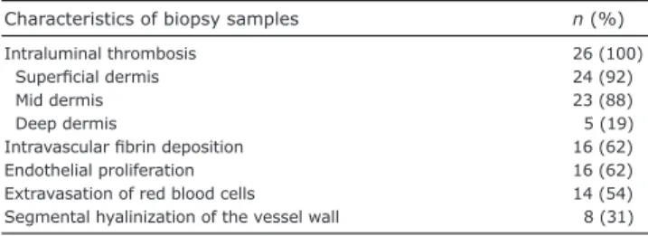
Histological characteristics
The histopathological findings in skin biopsies are in-dicated in Table I. Dermal thrombosis was found in all biopsy specimens, mostly in the papillary dermis, but
Table I. Histopathological characteristics of livedoid vasculopathy
Characteristics of biopsy samples n (%)
Intraluminal thrombosis 26 (100)
Superficial dermis 24 (92)
Mid dermis 23 (88)
Deep dermis 5 (19)
Intravascular fibrin deposition 16 (62)
Endothelial proliferation 16 (62)
Extravasation of red blood cells 14 (54)
A
cta
DV
A
cta
DV
A
dvances in dermatology and venereology
A
ctaD
ermato-V
enereologicaalso in the deep dermis around the dermal-hypodermal junction. A scattered perivascular lymphocytic infiltrate was present in 19 patients (73%) without signs of vascu-litis. One patient had a cutaneous “pseudo-polyarteritis nodosa” appearance with circumferential fibrin deposits in the intima, thrombosis of a small artery from the dermis-subcutis junction with perivascular inflammation in the adventitia.
Coagulation disorders
Multiple thrombophilia factors were detected in patients and are detailed in Table SI1. Twenty patients (77%) had at least one positive thrombophilia factor; 14 patients (54%) had one factor, 4 patients (15%) 2 factors, one patient demonstrated 3 factors, and one patient had 5 dif-ferent factors detected. Only one patient had an increased PAI-1 antigen, with a 5G/5G genotype. The 4G/4G (22%) and 4G/5G (45%) genotypes among 9 patients were not associated with high level PAI-1.
Neurological involvement
Of the 20 patients in whom neurophysiological investi-gations were available, 10 (50%) had a peripheral neuro-pathy, attributed to LV. Time between the first onset of cutaneous manifestations of LV and the diagnosis of neuropathy ranged from 6 months to 30 years with a median time of 11.4 years. Nine patients had an abnor-mal EMG, including 3 cases of mononeuritis multiplex and 6 cases of sensory polyneuropathy. One patient had a normal EMG but was diagnosed with small fibre neuropathy confirmed by laser-evoked potentials. Three patients had a sensory deficit and 7 patients had subjec-tive symptoms but a normal neurological examination. There were no cases of motor deficit. The main causes of peripheral neuropathy were excluded (diabetes, alcohol consumption, vitamin deficiency, neurotoxic drugs). Muscle-nerve biopsies were performed in 2 patients (one mononeuritis multiplex and one axonal neuropathy) and revealed a severe loss of myelinated axons associated with vasculopathy made of enlarged and thickened ves-sels of the epineurium, alterations of the endoneurium with endothelial cell damage and necrosis of capillaries. There was no sign of necrotizing vasculitis, granuloma or pathological deposits.
Therapeutic management
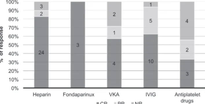
Among a total of 94 flares, 64 (68%) were treated with the following drugs: unfractionated heparin (UFH), low molecular weight heparin (LMWH), fondaparinux, vita-min K antagonist (VKA), antiplatelet agents or IVIG. 16 (17%) other flares were treated with colchicine, dapsone,
hydroxychloroquine, oral or topical steroids, or pentoxi-fyllin. Fourteen flares (15%) were not treated. The most prescribed first-line therapy was heparin-based anticoa-gulation (UFH or LMWH). In a majority of patients, one or two treatments were tried before an effective response was obtained but 40% of patients required 3 different treatment modalities or more (Fig. S11). First-line therapy was based on heparin anticoagulation or antiplatelet drugs. VKA was never used as a first-line treatment, nor was IVIG which was prescribed after 1 to 3 other treatment failures. According to the treatment used (Fig. 1), the mean CR for flares was 83% for heparin (UFH or LMWH), 59% for VKA and only 33% for antiplatelet drugs. IVIG led to CR in 63% of cases and produced at least a PR in 94% of flares.
Patient outcome
Nine patients (36%) achieved long-term remission with the absence of flares for at least 4 years. The median time since the last flare was 6 years. Skin flares were treated successfully with anticoagulation for 7 patients and LV disappeared spontaneously in 2 patients. Five patients had no maintenance treatment; among them, 2 patients achieved remission after smoking cessation and 2 patients with end-stage renal failure achieved re-mission after renal transplantation. Four patients had a maintenance treatment for secondary prophylaxis: VKA (n = 2), aspirin (n = 1), aspirin + VKA (n = 1).
Four patients (16%) achieved short-term remission with the absence of flare for 2 years. All patients respon-ded to anticoagulation with LMWH and they all received a maintenance treatment after the last flare based on antiplatelet agents: aspirin (n = 2), clopidogrel (n = 1) or dual antiplatelet treatment (n = 1).
Among the 12 other patients in short-term remis-sion, 4 patients had regular flares which responded to anticoagulation in 3 patients and combination of pen-toxifylline and hydroxychloroquine in one patient. The other 8 patients (32%) had severe disease with healing taking over 3 months, and no response to conventional
24 3 4 10 3 2 1 5 2 3 2 1 4 0% 10% 20% 30% 40% 50% 60% 70% 80% 90% 100%
Heparin Fondaparinux VKA IVIG Antiplatelet
drugs
% of response
CR PR NR
Fig. 1. Evaluation of response according to treatment used for livedoid vasculopathy flares. Stacked graph bars represent the cumulated
% of response for flares (number of flares reported in each bar area) treated with different drugs; complete response (CR), partial response
A
cta
DV
A
cta
DV
A
dvances in dermatology and venereology
A
ctaD
ermato-V
enereologica 845Livedoid vasculopathy: a French cohort study
Acta Derm Venereol 2018 antiplatelet or anticoagulation drugs. They all received
IVIG. One patient did not respond, and a further patient stopped treatment following an adverse event during the first infusion. For the other 6 patients, a total of 15 flares were treated with IVIG with success: 10 cases of CR and 5 of PR.
Toxicity and adverse events with treatments
Two serious adverse events occurred with fondaparinux: one patient was hospitalized for a thigh hematoma and one patient died of hemorrhagic stroke at the age of 56 years. Two patients treated with IVIG had adverse events; one was diagnosed with aseptic meningitis and another developed neutropenia.
DISCUSSION
In this multicenter observatory study, cutaneous manifes-tations of LV were similar to those reported previously (1, 3), confirming the typical clinical presentation of this entity. However, as evidenced by the delay of 3.4 years before diagnosis, LV remains underdiagnosed by gene-ral practitioners and dermatologists. Pain is a constant feature impacting on quality of life as observed by SF36 scores. Similarly, Polo Gascon et al. (20) identified a high impact on QoL showing a rise of mean DLQI from 11.37 to 17.83 during active disease, confirming a significantly higher impact with LV compared to other common skin diseases. The use of a specific LV-designed score of QoL would be of benefit in monitoring these patients.
In our study, thrombotic occlusion of small dermal vessels with intraluminal fibrin deposits was often as-sociated with segmental hyalinization of the vessel wall, endothelial proliferation or red blood cell extravasation. We observed thrombosis in the deep dermis in 19%, which is a location scarcely reported in other studies (1, 3). We recommend a surgical incisional biopsy in-stead of a punch biopsy to confirm the diagnosis of LV, as it should be large and deep enough to analyze the dermo-hypodermal junction to assess hypodermis vessel thrombosis and rule out PAN or any other necrotizing vasculitis. The association of LV and PAN has been re-ported previously (21). In our study, only one patient had a pseudo-PAN appearance without any extra cutaneous clinical symptoms of PAN. However, the histological selection of our patients was strict, excluding all cases with signs of vasculitis.
Pathophysiology is not clearly understood but an un-derlying hypercoagulable state is the cornerstone of the disease. Our thrombophilia screen revealed abnormal coagulation factors in 77% of our patients, a frequency superior to previous studies: 64% in Tran et al. (22), 52% in Di Giacomo et al. (2) and 50% in Hairston et al. (4). Hyperhomocysteinemia was frequent (50% of patients)
but there were no cases of severe hyperhomocysteinemia (> 50 µmol/l).
PAI-1 has a central role in the inhibition of fibrinolysis through the inhibition of the activators of plasminogen (tissue plasminogen activator tPA and urokinase uPA). PAI-1 has been implicated in LV in 3 previous studies (5–7). In our study, only one patient had an elevated PAI-1 antigen and he also presented with severe hypertrigly-ceridemia. This association has been reported previously (23, 24). Of note, we were not able to study PAI-1 antigen for every patient because of the lack of availability of technological equipment in some laboratories. The 4G allele in the promoter region of the PAI-1 gene has been shown to be more transcriptionally active and associated with higher levels of PAI-1. Studies have been discordant about the exact thrombotic risk associated with 4G/5G polymorphism but a recent meta-analysis by Wang et al. (25) has concluded that there was a significantly higher risk of thrombosis with the 4G homozygosity in venous thrombosis. In LV, one patient has been reported with 4G/4G homozygous genotype (7). In our LV population, we found that the distribution of the 4G/4G; 4G/5G and 5G/5G genotypes (respectively 22%, 45%, 33%) was not different to the control samples in the same laboratory or other studies (respectively 30%, 50%, 20%) (26, 27). We conclude that the 4G/4G polymorphism in the promoter region of the PAI-1 gene was not particularly associated with LV in our patients.
In this observational study we identified different risk factors for LV at each step of the coagulation cascade: disturbance of hemostasis, fibrinolysis defect, autoim-mune conditions and rheological disorders. Alone, such abnormalities are not always pathogenic. The combined effect of multiple thrombophilia factors induces an im-balance between coagulation and fibrinolysis, leading to small vessel thrombosis and skin ulceration. The presence of these different hypercoagulable factors can vary over time, explaining the chronic evolution with flares and periods of remission. Additional superficial venous insufficiency can occur, explaining location on the lower legs and heat as a trigger factor. Of note, smo-king prevalence rate was higher in the LV cohort than in the general French population: 42% in LV patients vs 32 to 35% of smokers in France (data from the French National Institute for Education and Health). We also noticed a long-term remission after smoking cessation alone in 2 patients. Interestingly, 3 patients received a kidney transplant and 2 of them, HIV-infected, have been recently reported by our team (28).
To date, peripheral neuropathy is the only known extra-cutaneous manifestation of LV known. Our study has revealed a striking incidence of peripheral neuropathy in 50% of patients. In two patients, muscle and nerve biop-sies identified similar findings of vasculopathy, without signs of vasculitis, as previously documented in 2015
A
cta
DV
A
cta
DV
A
dvances in dermatology and venereology
A
ctaD
ermato-V
enereologica(29). Peripheral neuropathy has been rarely described with a total of 9 detailed cases in the literature: one sensory ganglionopathy, one axonal neuropathy and 7 cases of mononeuritis multiplex (8, 10–15). Among them, 5 nerve biopsies revealed the same images of axonal loss with a lymphocytic infiltrate, without signs of vasculitis. Gan et al. (9) has reported 6 cases of peripheral neuropathy in their cohort of 70 patients but without electromyographic details. This is concordant with an ischemic process and confirms that the same thrombotic disease occurs in small vessels of the skin and nerves, without any vasculitic process. Interestingly, we describe the first case of small fibre neuropathy associated with LV. It requires special in-vestigations because the conventional techniques (EMG, nerve biopsy) only explore large nerve fibres. This entity is probably underestimated and would explain the high percentage of patients with neuropathic pain despite a normal EMG, persisting after ulcer healing of ulcers.
Finding an effective therapy is a real challenge for clinicians and a consensus is difficult to achieve (30). Indeed, both response to treatments and outcome remain variable from one patient to another. To date, there are no predictive clinical or biologic indicators for severity or frequency of flares. Evaluation of treatment efficacy is complex because duration of flares can vary and patients might heal spontaneously. However, pain relief seems to be the earliest sign of the healing process.
A recent review of LV treatment by Micieli & Alavi (31) have confirmed that the most prescribed treatment was anticoagulation in 98% of cases. Among anticoa-gulant therapies rivaroxaban was the most prescribed (54%), followed by anabolic steroids, IVIG and antiplate-let drugs. Rivaroxaban might be an interesting alternative to heparin and easier to manage than VKA (32–34). A recent phase 2 study has revealed good efficacy and tolerability for this anticoagulant (34). Nevertheless, 30% patients required LMWH as a back-up treatment.
In our study, LMWH and antiplatelet agents were the most often used as a first-line therapy and anticoa-gulation by heparin was the most successful treatment. Fourteen of our patients (56%) had a good response to anticoagulants. Tinzaparin sodium was used with a varying duration from 15 days to 3 months until ulcer healing. The convenience of a subcutaneous daily injec-tion promoted compliance. Antiplatelet drugs and VKA appeared to be less effective in our cohort and should not be recommended as a first-line treatment for flares. It is difficult to assess the efficacy of fondaparinux, as only two patients were treated with this medication and both developed major hemorrhagic events.
However, 32% had severe disease with regular flares not responding to anticoagulants. These patients were successfully treated with IVIG: 94% had at least > 50% improvement with 63% going to complete remission. Our data were concordant with previous studies and justified
the use of IVIG as a second-line treatment after unsuc-cessful use of heparin anticoagulation (16–19).
Limitations
The question of a maintenance treatment to prevent flares was very difficult to address. We recommend venous compression, especially during summer time to limit venous stasis and edema, which can promote thrombo-sis (3). As far as possible, thrombophilia factors should be corrected. Some of our patients received antiplatelet drugs or VKA as a long-term therapy. We acknowledge several limitations to this study of a rare disease: small sample population, difficult follow-up with some retro-spective data collection, variable frequency of flares both in an inter- and intra-individual way and large diversity of treatments, which made the impact of a maintenance treatment difficult to analyze. It is worth mentioning that 2 patients had LV flares when INR was low and healed with VKA dose-adjustment. The need for maintenance treatment with anticoagulation or antiplatelet drugs or others agents has not been studied in the literature. We propose two treatment algorithms as to illustrate the way we manage patients with LV for initial diagnosis and recurrent flares respectively (Figs S2 and S31).
Conclusion
Our national observational study confirms that livedoid vasculopathy is a rare disease, justifying an incisional surgical skin biopsy for diagnosis, to rule out vasculitis. Patients should undergo full thrombophilia screening. Interestingly, PAI-1 was exceptionally elevated and 4G/4G polymorphism was not associated with a high level of PAI-1 in our cohort. Peripheral neuropathy was associated with LV with an incidence of 50% and nerve biopsies revealed an ischemic process, confirming that LV is not only a cutaneous but also a peripheral neurolo-gical disease. Curative anticoagulation by LMWH seems to be the most efficient treatment, whereas antiplatelet drugs are less effective. IVIG can be an interesting alter-native treatment for the most severe patients. However, in this small size study, the benefit of anticoagulation maintenance therapy as secondary prevention has not been well evaluated. Rivaroxaban, with a good efficacy reported by several studies, may be increasingly recom-mended in the near future.
ACKNOWLEDGEMENTS
We are indebted to the EMSED (Etude des maladies systémiques en dermatologie) and GAD (Groupe angiodermatologie) thematic groups from the French society of dermatology (SFD) for their support and clinical cases. We thank Dr Jessica Gale and Dr Ca-therine H Orteu for their kind proofreading.
A
cta
DV
A
cta
DV
A
dvances in dermatology and venereology
A
ctaD
ermato-V
enereologica 847Livedoid vasculopathy: a French cohort study
Acta Derm Venereol 2018
REFERENCES
1. Criado PR, Rivitti EA, Sotto MN, de Carvalho JF. Livedoid vasculopathy as a coagulation disorder. Autoimmun Rev 2011; 10: 353–360.
2. Di Giacomo TB, Hussein TP, Souza DG, Criado PR. Frequency of thrombophilia determinant factors in patients with livedoid vasculopathy and treatment with anticoagulant drugs – a prospective study. J Eur Acad Dermatol Venereol 2010; 24: 1340–1346.
3. Alavi A, Hafner J, Dutz JP, Mayer D, Sibbald RG, Criado PR, et al. Livedoid vasculopathy: an in-depth analysis using a modified Delphi approach. J Am Acad Dermatol 2013; 69: 1033–1042 e1031.
4. Hairston BR, Davis MD, Pittelkow MR, Ahmed I. Livedoid vas-culopathy: further evidence for procoagulant pathogenesis. Arch Dermatol 2006; 142: 1413–1418.
5. Castillo-Martinez C, Moncada B, Valdes-Rodriguez R, Gon-zalez FJ. Livedoid vasculopathy (LV) associated with sticky platelets syndrome type 3 (SPS type 3) and enhanced activity of plasminogen activator inhibitor (PAI-1) anomalies. Int J Dermatol 2014; 53: 1495–1497.
6. Agirbasli M, Eren M, Eren F, Murphy SB, Serdar ZA, Seckin D, et al. Enhanced functional stability of plasminogen activator inhibitor-1 in patients with livedoid vasculopathy. J Thromb Thrombolysis 2011; 32: 59–63.
7. Deng A, Gocke CD, Hess J, Heyman M, Paltiel M, Gaspari A. Livedoid vasculopathy associated with plasminogen activator inhibitor-1 promoter homozygosity (4G/4G) treated suc-cessfully with tissue plasminogen activator. Arch Dermatol 2006; 142: 1466–1469.
8. Alix JJ, Hadjivassiliou M, Ali R, Slater D, Messenger AG, Rao DG. Sensory ganglionopathy with livedoid vasculopathy con-trolled by immunotherapy. Muscle Nerve 2015; 51: 296–301. 9. Gan EY, Tang MB, Tan SH, Chua SH, Tan AW. A ten-year re-trospective study on livedo vasculopathy in Asian patients. Ann Acad Med Singapore 2012; 41: 400–406.
10. Kim JE, Park SY, Sinn DI, Kim SM, Hong YH, Park KS, et al. Ischemic neuropathy associated with livedoid vasculitis. J Clin Neurol 2011; 7: 233–236.
11. Malaguti MC, Cavallaro T, Speziali L, Zorzi MG, Marangoni S, Morini A. Mononeuritis multiplex associated with primary li-vedoid vasculopathy: neuropathological evidence of ischemic nerve damage. J Neurol Sci 2015; 351: 214–215.
12. Osada S, Kimura Y, Kawana S. Case of livedoid vasculopathy with peripheral neuropathy successfully treated with low-dose warfarin. J Dermatol 2010; 37: 98–101.
13. Pai BS, Pai K. Livedoid Vasculopathy and Mononeuritis Mul-tiplex, with a Fulminant Hepatic Failure which was caused by Herpes Simplex Hepatitis: A Case Report. J Clin Diagn Res 2013; 7: 921–923.
14. Toth C, Trotter M, Clark A, Zochodne D. Mononeuropathy multiplex in association with livedoid vasculitis. Muscle Nerve 2003; 28: 634–639.
15. Tubone MQ, Escobar GF, Peruzzo J, Schestatsky P, Maldo-nado G. Livedoid vasculopathy associated with peripheral neuropathy: a report of two cases. An Bras Dermatol 2013; 88: 227–229.
16. Bounfour T, Bouaziz JD, Bezier M, Petit A, Viguier M, Rybojad M, et al. Intravenous immunoglobulins in difficult-to-treat ulcerated livedoid vasculopathy: five cases and a literature review. Int J Dermatol 2013; 52: 1135–1139.
17. Kim EJ, Yoon SY, Park HS, Yoon HS, Cho S. Pulsed intrave-nous immunoglobulin therapy in refractory ulcerated livedoid vasculopathy: seven cases and a literature review. Dermatol
Ther 2015; 28: 287–290.
18. Kreuter A, Gambichler T, Breuckmann F, Bechara FG, Rotter-dam S, Stucker M, et al. Pulsed intravenous immunoglobulin therapy in livedoid vasculitis: an open trial evaluating 9 con-secutive patients. J Am Acad Dermatol 2004; 51: 574–579. 19. Monshi B, Posch C, Vujic I, Sesti A, Sobotka S, Rappersber-ger K. Efficacy of intravenous immunoglobulins in livedoid vasculopathy: long-term follow-up of 11 patients. J Am Acad Dermatol 2014; 71: 738–744.
20. Polo Gascon MR, de Carvalho JF, de Souza Espinel DP, Barros AM, Alavi A, Criado PR. Quality-of-life impairment in patients with livedoid vasculopathy. J Am Acad Dermatol 2014; 71: 1024–1026.
21. Mimouni D, Ng PP, Rencic A, Nikolskaia OV, Bernstein BD, Nou-sari HC. Cutaneous polyarteritis nodosa in patients presenting with atrophie blanche. Br J Dermatol 2003; 148: 789–794. 22. Tran MD, Becherel PA, Cordel N, Piette JC, Frances C.
[“Idio-pathic” white atrophy]. Ann Dermatol Venereol 2001; 128: 1003–1007.
23. Crutchley DJ, McPhee GV, Terris MF, Canossa-Terris MA. Le-vels of three hemostatic factors in relation to serum lipids. Monocyte procoagulant activity, tissue plasminogen activator, and type-1 plasminogen activator inhibitor. Arteriosclerosis 1989; 9: 934–939.
24. Kobayashi Y, Fukuo Y, Shibuya T, Terashi A. [The correlation between the activity of tissue plasminogen activator (TPA), levels of tissue plasminogen activator inhibitor (PAI-1) an-tigen and serum lipids in healthy subjects]. Nihon Ronen Igakkai Zasshi 1990; 27: 578–583.
25. Wang J, Wang C, Chen N, Shu C, Guo X, He Y, et al. Associa-tion between the plasminogen activator inhibitor-1 4G/5G polymorphism and risk of venous thromboembolism: a meta-analysis. Thromb Res 2014; 134: 1241–1248.
26. Parpugga TK, Tatarunas V, Skipskis V, Kupstyte N, Zaliaduo-nyte-Peksiene D, Lesauskaite V. The effect of PAI-1 4G/5G po-lymorphism and clinical factors on coronary artery occlusion in myocardial infarction. Dis Markers 2015; 2015: 260101. 27. Sartori MT, Danesin C, Saggiorato G, Tormene D, Simioni P,
Spiezia L, et al. The PAI-1 gene 4G/5G polymorphism and deep vein thrombosis in patients with inherited thrombophi-lia. Clin Appl Thromb Hemost 2003; 9: 299–307.
28. Hurabielle C, Sebille G, Barrou B, Moguelet P, Frances C, Ba-rete S. Livedoid Vasculopathy Associated with HIV Infection in Two Patients: A Causal Relationship? Acta Derm Venereol 2016; 96: 844–845.
29. Allenbach Y, Tourte M, Stenzel W, Goebel HH, Maisonobe T, Frances C, et al. Expanding the spectrum of livedoid vasculo-pathy: peculiar neuromuscular manifestations. Neuropathol Appl Neurobiol 2015; 41: 849–852.
30. Frances C, Barete S. Difficult management of livedoid vas-culopathy. Arch Dermatol 2004; 140: 1011.
31. Micieli R, Alavi A. Treatment for Livedoid Vasculopathy: A Systematic Review. JAMA Dermatol 2018; 154: 193–202. 32. Drabik A, Hillgruber C, Goerge T. A Phase II Multicenter Trial
With Rivaroxaban in the Treatment of Livedoid Vasculopathy Assessing Pain on a Visual Analog Scale. JMIR Res Protoc 2014; 3: e73.
33. Lee JM, Kim IH. Case series of recalcitrant livedoid vascu-lopathy treated with rivaroxaban. Clin Exp Dermatol 2016; 41: 559–561.
34. Weishaupt C, Strolin A, Kahle B, Kreuter A, Schneider SW, Gerss J, et al. Anticoagulation with rivaroxaban for livedoid vasculopathy (RILIVA): a multicentre, single-arm, open-label, phase 2a, proof-of-concept trial. Lancet Haematol 2016; 3: e72–79.