HAL Id: hal-02402073
https://hal.archives-ouvertes.fr/hal-02402073
Submitted on 9 Dec 2020
HAL is a multi-disciplinary open access
archive for the deposit and dissemination of
sci-entific research documents, whether they are
pub-lished or not. The documents may come from
teaching and research institutions in France or
abroad, or from public or private research centers.
L’archive ouverte pluridisciplinaire HAL, est
destinée au dépôt et à la diffusion de documents
scientifiques de niveau recherche, publiés ou non,
émanant des établissements d’enseignement et de
recherche français ou étrangers, des laboratoires
publics ou privés.
induction of the Crabtree effect
Mónica Rosas Lemus, Elodie Roussarie, Noureddine Hammad, Alexis
Mougeolle, Stéphane Ransac, Razanne Issa, Jean-Pierre Mazat, Salvador
Uribe-Carvajal, Michel Rigoulet, Anne Devin
To cite this version:
Mónica Rosas Lemus, Elodie Roussarie, Noureddine Hammad, Alexis Mougeolle, Stéphane Ransac, et
al.. The role of glycolysis-derived hexose phosphates in the induction of the Crabtree effect. Journal
of Biological Chemistry, American Society for Biochemistry and Molecular Biology, 2018, 293 (33),
pp.12843-12854. �10.1074/jbc.RA118.003672�. �hal-02402073�
Me´xico, Mexico City, Me´xico 04510 Edited by Jeffrey E. Pessin
Evidence for the Crabtree effect was first reported by H. Crabtree in 1929 and is defined as the glucose-induced decrease of cellular respiratory flux. This effect was observed in tumor cells and was not detected in most non-tumor cells. A number of hypotheses on the mechanism underlying the Crabtree effect have been formulated. However, to this day, no consensual mechanism for this effect has been described. In a previous study on isolated mitochondria, we have proposed that fructose-1,6-bisphosphate (F1,6bP), which inhibits the respiratory chain, induces the Crabtree effect. Using whole cells from the yeast
Saccharomyces cerevisiae as a model, we show here not only that
F1,6bP plays a key role in the process but that glucose-6-phos-phate (G6P), a hexose that has an effect opposite to that of F1,6bP on the regulation of the respiratory flux, does as well. Thus, these findings reveal that the Crabtree effect strongly depends on the ratio between these two glycolysis-derived hexose phosphates. Last, in silico modeling of the Crabtree effect illustrated the requirement of an inhibition of the respi-ratory flux by a coordinated variation of glucose-6-phosphate and fructose-1,6-bisphosphate to fit the respiratory rate decrease observed upon glucose addition to cells. In summary, we conclude that two glycolysis-derived hexose phosphates, G6P and F1,6bP, play a key role in the induction of the Crabtree effect.
The Crabtree effect was first described by H. Crabtree in 1929 and is defined as the glucose-induced decrease of cellular
respiratory flux (1). This effect was observed in tumor cells and
had no occurrence in most non-tumor cells (1). Consequently,
studies aiming to decipher the molecular mechanism leading to the Crabtree effect were mostly conducted by comparing tumor
cell lines and their non-tumorigenic counterpart. The origin of the glucose-induced repression of cellular respiratory flux has long been sought and even though a number of hypotheses have been formulated, its triggering mechanism(s) is still unknown. It is possible that its induction may be due to a combination of
several factors (2). In some tumor cells, a drastic decrease in
phosphate (Pi) was observed upon glucose addition and the
Crabtree effect was eliminated by adding an excess of Pi. This
led the authors to propose that a decreased cytosolic Pi
concen-tration was the actual trigger of this phenomenon (3). Another
hypothesis is that the glycolytic enzymes (phosphoglycerate kinase and pyruvate kinase) compete with mitochondria for
free cytoplasmic ADP (2, 4). Addition of glucose would trigger
an overactive glycolysis that would outcompete mitochondria for ADP uptake. ADP being one of the substrates of the mito-chondrial ATP synthase, the activity of this enzyme would decrease and so would mitochondrial respiration. However, this hypothesis might not hold true in vivo because the
mito-chondrial adenine nucleotide translocase Km is almost 100
times lower than that of the glycolytic enzymes (5).
Conse-quently mitochondria would still import cytosolic ADP regard-less of glycolysis activation. In the same line of thought, it has been shown that the thermodynamic phosphate potential (i.e.
[ATP/ADPxPi]) changes in response to glucose addition to
sarcoma ascites tumor cells (6), due to a decrease in both
ADP and Pi.
Cytoplasmic Ca2⫹ levels have also been proposed as being
responsible for the Crabtree effect. Indeed, one study showed
that glucose addition increased mitochondrial Ca2⫹ uptake
inhibiting the ATP synthase (7). However, such a regulation
cannot be proposed as an unequivocal mechanism of induction
of the Crabtree effect because in a hepatoma cell line Ca2⫹
levels did not change in response to glucose (2).
It has also been proposed that because the mitochondrial outer membrane regulates the access of substrates to the inter-membrane space, it could regulate the oxidative
phosphoryla-tion rate (8). Indeed if ADP or the respiratory substrates were
kept in the cytoplasm this would induce a decrease in respira-tory flux. In agreement with this hypothesis, it has been shown that, in proteoliposomes, physiological concentrations of NADH close reconstituted porin (the external mitochondrial
membrane main permeability barrier) (9). It has also been
shown that within normal adult cardiomyocytes and in the This work was supported by the CNRS (Conseil National de la Recherche
Sci-entifique), the Comite´ de Dordogne and Gironde de la Ligue Nationale Contre le Cancer, The Fondation ARC pour la recherche sur le Cancer, the Plan Cancer 2014 –2019 number BIO 2014 06, the French Association against Myopathies, and the European Commission (to H. N.). The authors declare that they have no conflicts of interest with the contents of this article. This publication reflects the view only of the authors and the Euro-pean Commission cannot be held responsible for any use which may be made of the information contained therein.
1These authors contributed equally to the results of this work.
2To whom correspondence should be addressed: Institut de Biochimie et
Ge´ne´tique Cellulaires, CNRS UMR 5095, 1, rue Camille Saint Sae¨ns, 33077 Bordeaux Cedex, France. Tel.: 33-556-999-035; Fax: 33-556-999-040; E-mail:
anne.devin@ibgc.cnrs.fr.
J. Biol. Chem. (2018) 293(33) 12843–12854
12843
at UNIV BORDEAUX on October 28, 2019
http://www.jbc.org/
HL-1 cardiac cell line, intracellular local diffusion restrictions of adenine nucleotides and metabolic feedback regulation of respiration via phosphotransfer networks are different, most probably as a result of differences in structural organization of
these cells (10). Cardiomyocytes contain tight complexes where
mitochondria and Ca2⫹/Mg2⫹-ATPases are organized to
ensure effective energy transfer and feedback signaling via spe-cialized pathways. In contrast, these complexes do not exist in HL-1 cells, which exhibit less organized energy metabolism
(11). Hence diffusion restrictions are most likely not involved in
the induction of the Crabtree effect.
Some years ago work from our laboratory pointed to a pos-sible induction of the Crabtree effect by glycolysis-derived
hexose phosphate, namely F1,6bP3(12). We showed that on
isolated mitochondria, F1,6bP inhibits the mitochondrial res-piratory chain, whereas G6P stimulates it. More precisely, at physiological levels, F1,6bP inhibits mitochondrial complexes
III and IV (12). This inhibition was shown on mitochondria
isolated from the yeast Saccharomyces cerevisiae and on mito-chondria isolated from rat liver (mammalian mitomito-chondria). However, no inhibition from F1,6bP was observed on mito-chondria isolated from Crabtree negative yeast–yeast that do not harbor an inhibition of cellular respiration when glucose is added to the culture medium. These results led us to propose that F1,6bP that accumulated within the cell upon glucose addi-tion was responsible for the inducaddi-tion of the Crabtree effect.
We show here on whole cells of S. cerevisiae that glycolysis hexose phosphate, namely glucose-6-phosphate that activates the respiratory chain and fructose-1,6-bisphosphate that inhib-its the respiratory chain, play a crucial role in the induction of the Crabtree effect. We show that not only the F1,6bP-induced inhibition of the respiratory chain plays a role in the induction of the Crabtree effect but also, the activation of the respiratory chain by G6P is important. In comparable stoichiometries, both hexose phosphates have opposite effects on the respiratory chain activity. Thus, the induction of the Crabtree effect depends on the ratio between these two hexose phosphates. Last, in silico modeling of the Crabtree effect taking into account our experimental results shows that the hexose phos-phates-induced kinetic regulation of respiratory chain activity is mandatory to observe the Crabtree effect.
Results
The Crabtree effect is due to an inhibition of the mitochondrial respiratory chain
We investigated the induction of the Crabtree effect in our
model S. cerevisiae. As previously shown (12), glucose addition
to yeast cells grown on non-fermentative substrate induced a
decrease in respiratory rate (Fig. 1A). To determine whether
this inhibition was due to a respiratory chain inhibition or to a decrease in the phosphorylation processes at the mitochondrial level, we assessed the respiratory rate under uncoupled condi-tions, where the respiratory chain flux is at a maximal rate. Upon glucose addition, the uncoupled respiratory rate of WT
cells decreased (Fig. 1A), pointing to respiratory chain
inhibi-tion. Glucose addition did not modify significantly adenine
nucleotides or Pi(Fig. 1C). These results rule out an
involve-ment of ADP or Pion the induction of the Crabtree effect in the
S. cerevisiaeWT strain.
To this day, the only mechanism proposed for the glucose-induced respiratory chain inhibition is the effect of fructose-1,6-bisphosphate, a glycolysis intermediary that accumulates
upon glucose addition to cells (12). We thus measured this
intermediate in WT cells upon glucose addition.Fig. 1Bshows
that although in cells grown on lactate as a carbon source fruc-tose-1,6-bisphosphate concentration is quite low, it signifi-cantly rises up eight times upon glucose addition.
Deletion of hexokinase 2 prevents the induction of the Crabtree effect
To further investigate the role of F1,6bP on the induction of the Crabtree effect we explored means to alter the accumula-tion of glycolysis metabolites in response to glucose addiaccumula-tion. Hexokinase is the first enzyme of the glycolysis pathway. There are a number of isoenzymes in both yeast and mammalian cells. However, hexokinase2 (hxk2) is the predominant hexokinase during growth on glucose. It is the yeast homolog of glucoki-nase, which is overexpressed in tumor cells and it is not inhib-ited by its product, glucose-6-phosphate. As a consequence its activity should result in high accumulation of glycolysis
inter-mediates in response to glucose addition (13). We thus studied
the induction of the Crabtree effect in a hxk2-deletion strain. In
the ⌬hxk2 strain, no inhibition of the respiratory rate was
observed upon glucose addition (Fig. 2A). In the⌬hxk2 strain
neither adenine nucleotides nor Piconcentrations were
signif-icantly affected upon glucose addition (Fig. 2D).
To ensure that in⌬hxk2 cells the absence of respiratory rate
inhibition was not due to no variation or decreased F1,6bP accumulation in response to glucose addition, we measured the
fructose-1,6-bisphosphate concentration. Fig. 2B shows that
whereas the fructose-1,6-bisphosphate concentration is low in cells grown on lactate as a carbon source, upon glucose addition
its concentration rises significantly in the⌬hxk2 cells.
How-ever, upon glucose addition the overall concentration remains
slightly lower in the⌬hxk2 cells as compared with the WT cells
(seeFig. 1). It has previously been shown that glucose-6-phos-phate antagonizes the fructose-1,6-bisphosglucose-6-phos-phate effect on
res-piratory chain activity: it activates the resres-piratory chain (12).
We thus determined the glucose-6-phosphate concentration
change in response to glucose addition both in WT and⌬hxk2
cells. Glucose addition induced a significant increase in
glu-cose-6-phosphate in⌬hxk2, whereas the increase of G6P in WT
cells is lower and not statistically significant (Fig. 2C).
Not only F1,6bP but also G6P plays a crucial role in the induction of the Crabtree effect
HAP4p (master regulator of the HAP transcription complex) plays a crucial role in the orientation of fermentative
meta-bolism in the yeast S. cerevisiae (14). Hap4p is the activator
subunit of the transcriptional complex involved in carbon
source-dependent regulation of respiratory function (15, 16).
Transcription of this subunit is glucose repressible (16),
sug-3The abbreviations used are: F1,6bP, Fru-1,6-P; G6P, Glc-6-P; hxk2,
hexoki-nase2; OXPHOS, oxidative phosphorylation; LAC, lactate.
at UNIV BORDEAUX on October 28, 2019
http://www.jbc.org/
gesting that Hap4p is the key component of the complex in terms of its control of transcriptional activity in response to carbon source. Thus a strain overexpressing HAP4p should exhibit an increased mitochondrial content and a strong orien-tation of energetic metabolism toward respiratory metabolism, altering the relationship between glycolysis intermediates and respiratory rate. In this strain, glucose addition did not inhibit
the respiratory rate (Fig. 3A), whereas the Crabtree effect was
present in the WT cells (control cells harboring an empty
plas-mid). Measurements of both adenine nucleotides and Pi
showed that these parameters were mostly unaffected upon glucose addition in both the WT strain and the strain
overex-pressing the HAP4p subunit (Fig. 3D).
Whereas fructose-1,6-bisphosphate concentration is quite low in cells grown on lactate as a carbon source, it increases upon glucose addition in both the WT and HAP4p
overex-pressing cells (Fig. 3B). The absence of an induction of the
Crabtree effect in the HAP4p-overexpressing cells is thus not due to an absence of fructose-1,6-bisphosphate accumulation upon glucose addition even though in the HAP4p-overexpress-ing cells, F1,6bP never reaches a concentration comparable
with the one in the WT cells. We next investigated the effect of glucose on glucose-6-phosphate (which stimulates the respira-tory chain) concentration in WT cells and in cells overexpress-ing HAP4p. For each strain, glucose-6-phosphate does not vary
significantly upon glucose addition (Fig. 3C). However, its
con-centration is significantly doubled in the
HAP4p-overexpress-ing cells as compared with the WT cells (seeFig. 3C). This result
could imply an increase in the antagonist effect of glucose-6-phosphate on the activity of the respiratory chain in HAP4p-overexpressing cells.
The sensitivity of mutant cells to F1,6bP-mediated inhibition is maintained
The results described above raised the question of whether
the respiratory chain from⌬hxk2 and HAP4p-overexpressing
cells remained sensitive to F1,6bP. To address this question, we studied the respiratory chain F1,6bP-mediated inhibition on the respiratory activity of nystatin-permeabilized spheroplasts
from each of the strains of interest (17).
Fig. 4, A and B,clearly shows that there are no significant differences in the F1,6bP-mediated inhibition between the WT
Figure 1. Study of the Crabtree effect in WT cells. A, glucose-induced decrease in respiratory rate. Glucose concentrations were as follows: none (black), 30
mM(gray), 60 mM(red), and 90 mM(white). B, fructose-1,6-bisphosphate accumulation. C, ATP (black), ADP (gray), and Pi(white). Respiratory rates,
fructose-1,6-bisphosphate, adenine nucleotides, and Pidetermination were performed as described under “Experimental procedures.” Results are mean⫾ S.E. of at least
three separate experiments.
at UNIV BORDEAUX on October 28, 2019
http://www.jbc.org/
and mutant cells. Thus the absence of Crabtree effect induction
in the⌬hxk2 and HAP4p-overexpressing cells was not due to a
modulation of the respiratory chain response to F1,6bP. A similar experiment was performed with G6P to ensure that respiratory chains in mutant cells were indeed stimulated by this hexose phosphate (data not shown). However, upon its addition to spheroplasts, G6P was steadily metabolized into F1,6bP making such a control experiment impossible.
Both glucose-6-phosphate and fructose-1,6-bisphosphate play a crucial role in the induction of the Crabtree effect
As mentioned above, whereas F1,6bP has been shown to inhibit mitochondrial respiratory chain, G6P stimulates it. In
our previous paper, on isolated mitochondria (12), we showed
that, in the presence of G6P, F1,6bP inhibited the mitochon-drial respiratory chain only above the point where a stoichio-metric amount of each hexose was required, i.e. the respiratory chain is inhibited when the G6P/F1,6bP ratio drops below 1. To determine whether in whole cells the G6P/F1,6bP ratio rather than the F1,6bP concentration alone was the key inducer of the
Crabtree effect, we assessed this ratio in WT, ⌬hxk2, and
HAP4-overexpressing cells.Fig. 5clearly shows that whereas
the G6P/F1,6bP ratio drops well below 1 in WT cells upon
glucose addition, it stays pretty close to this value in the⌬hxk2
and HAP4-overexpressing cells.
The G6P/F1,6bP ratio is the key player for the induction of the Crabtree effect
If our hypothesis stands true, i.e. the induction of the Crabtree effect relies upon the G6P/F1,6bP ratio, this ratio may be manipulated in our mutant cells to trigger the Crabtree effect. To perform this experiment, yeast cells were grown in galactose medium, where the glycolysis pathway is active and the glycolytic enzymatic content is higher than in a non-fer-mentable medium (used in our previous experiments). The rea-soning behind this is that to accumulate more F1,6bP in our mutant cells, and thus decrease the G6P/F1,6bP ratio, active glycolytic enzymes are required. Consequently, WT and mu-tant cells were grown on galactose medium and the Crabtree
effect was induced by the addition of 60 mMglucose.Fig. 6A
shows that upon glucose addition, WT and mutant (⌬hxk2 and
HAP4-overexpressing) cells exhibit a statistically significant Crabtree effect as well as a consequent increase in F1,6bP
con-centration in WT and mutant cells (Fig. 6B), whereas variations
of the G6P concentration are not statistically significantly
dif-ferent (Fig. 6C). So in galactose-grown WT or mutant cells,
glucose induced both a Crabtree effect and a decrease in the G6P/F1,6bP ratio, even if the effect of glucose addition was lower than the one observed in cells grown in non-fermentative
medium (Fig. 1A). It should be stressed here that the respiratory
Figure 2. Study of the Crabtree effect in⌬hxk2 cells. A, respiratory rate. Glucose concentrations were as follows: none (black), 30 mM(gray), 60 mM(red), and 90 mM(white). B, fructose-1,6-bisphosphate accumulation in⌬hxk2 cells upon glucose addition. C, glucose-induced glucose-6-phosphate modulations in WT (white) and⌬hxk2 (black) cells and D, ATP (black), ADP (gray), and Pi(white) in⌬hxk2 cells. Respiratory rates and fructose-1,6-bisphosphate,
glucose-6-phosphate, adenine nucleotides, and Pidetermination were performed as described under “Experimental procedures.” Results are mean⫾ S.E. of at least three
separate experiments.
at UNIV BORDEAUX on October 28, 2019
http://www.jbc.org/
rate inhibition upon glucose addition when cells are grown on galactose medium is lower (but still statistically significant) than the one assessed when cells are grown on
non-fermenta-tive medium (Fig. 1A); this stands true for all three strains.
However, in these cells and in the absence of glucose, the G6P/
F1,6bP ratio is already below 1 (Fig. 6D), due to an active
gly-colysis. Thus in this experimental setup, the respiratory chain is most likely already slightly inhibited due to a concentration of F1,6bP a bit higher than the G6P concentration.
An in silico study of the Crabtree effect confirms a role for glycolysis-derived hexose phosphate
There are good models of yeast metabolism with an accurate representation of the kinetics of glycolytic enzymes. It was thus tempting to see whether these models, developed for other purposes, would reproduce the experimental features of the Crabtree effect. We choose the recent model of Smallbone et al.
(21) developed to fit the experimental glycolytic metabolites
concentrations and to calculate the control coefficients of the glycolytic enzymes on the glycolytic flux. Two questions were addressed here: (i) do we reproduce in silico the experimental
variations of F1,6bP and G6P and (ii) is a regulation by these metabolites necessary to obtain the Crabtree effect or could the Crabtree effect be obtained independently of such a regulation? To do so, as described under “Experimental procedures” and
considering our previous experimental results (12), we
intro-duced a regulatory function that is a Hill function of the G6P/ F1,6bP ratio. Because the ATP and ADP concentrations did not appreciably change experimentally, we fixed their values in the model to the experimental values.
Concerning the effect on respiratory rate (VO2⫽ OXPHOS_
NADH⫹ OXPHOS_LAC), in the absence of regulation of the
OXPHOS rate by the G6P/F1,6bP ratio, the addition of a high
concentration of glucose (60 mM) slightly increases the
respira-tory rate about 4%. This increase is entirely due to an increase in OXPHOS_NADH as expected (NADH increases upon glucose addition to cells). OXPHOS_LAC is fixed due to the fixed ratio
of ATP/ADP (Table 1andFig. 7). In the presence of a regulation
of the OXPHOS rate by the G6P/F1,6bP ratio, both OXPHOS_ NADH and OXPHOS_LAC decrease when glucose increases
(up to 60 mM) (Table 2). OXPHOS_NADH shows a lower value
at a high glucose concentration due to a low G6P/F1,6bP value,
Figure 3. Study of the Crabtree effect in HAP4p-overexpressing cells. A, respiratory rate. Glucose concentrations were as follows: none (black), 30 mM(gray), 60 mM(red), and 90 mM(white). B, fructose-1,6-bisphosphate accumulation in WT (white) and HAP4p-overexpressing (black) cells upon glucose addition. C, glucose-induced glucose-6-phosphate modulations in WT (white) and HAP4p-overexpressing cells (black) cells. D, ATP (black), ADP (gray), and Pi(white) in WT
and HAP4p-overexpressing cells. Respiratory rates, fructose-1,6-bisphosphate, glucose-6-phosphate, adenine nucleotides, and Pidetermination were
per-formed as described under “Experimental procedures.” Results are mean⫾ S.E. of at least three separate experiments. HAP4OX: ⌬hap4 strain harboring a plasmid overexpressing HAP4p.
at UNIV BORDEAUX on October 28, 2019
http://www.jbc.org/
despite the higher concentration of NADH. The respiratory
rate (VO2⫽ OXPHOS_NADH ⫹ OXPHOS_LAC) decreases
by about 20% (Table 2andFig. 8), in accordance with
experi-mental values (seeFig. 4, for example).
Fig. 9, A and B, shows that the concentrations of F1,6bP and G6P obtained in the model are close to the experimental values. They do not depend much upon the presence of the regulatory Hill function of the G6P/F1,6bP ratio. At low glucose concen-trations, F1,6bP is equal to 0.6 as compared with an experimen-tal value of 1 and the G6P value is equal to 1.8 as compared with an experimental value of 2, which gives a G6P/F1,6bP ratio of 2.6 as compared with the experimental value of 2. In the pres-ence of high concentrations of glucose, both concentrations
reach values between 7.8 and 8.7 mMfor F1,6bP and between
3.12 and 3.16 for G6P (the experimental values being between 8 and 9 and between 3.3 and 3.7, respectively, which gives G6P/ F1,6bP values around 0.4 as compared with experimental values
between 0.3 and 0.4). Consequently, the results ofFig. 9show that
the model actually fits the experimental values of F1,6bP and G6P and their increase in the presence of glucose. Thus, modeling illus-trates the need to consider the G6P- and F1,6bP-mediated modu-lation of oxidative phosphorymodu-lation to simulate the decrease in the respiratory rate observed after glucose addition.
Last, we plotted the relationship between the rate of respi-ration and the G6P/F1,6bP ratio with the values obtained
throughout this work (Fig. 10). The points, coming from
Figs. 1–3(summarized inFig. 5) andFig. 6are remarkably gath-ered, which substantiate our hypothesis of an apparent regula-tion of the Crabtree effect inducregula-tion by the G6P/F1,6bP ratio. In optimizing the parameters K of the model to fit the
experimen-tal results drawn inFig. 10, we found that K⫽ 0.27 gives a good
fit of the experimental results, perfectly describing the inhibi-tion of respirainhibi-tion for G6P/F1,6bP values below 0.7– 0.8. As stressed previously, we show that when this ratio is above 0.8, no inhibition of the respiratory rate is observed. Furthermore, the calculation of the G6P/F1,6bP ratio from the literature on
ascites tumor cells (2, 6) shows that it decreases upon glucose
addition to cells and the plot of the first values corresponding to
ascites tumor cells is in accordance with our values in yeast (Fig.
10). Of note, these authors respiratory rate inhibition is higher
than what is seen in yeast, most likely due to the synergistic effects of hexose phosphate and phosphate potential.
Discussion
The Crabtree effect was first evidenced by H. Crabtree in
1929 (1). Ever since then a number of studies were conducted
on different models to decipher the molecular mechanisms responsible for the inhibition of the respiratory rate upon glucose addition. A number of hypotheses were formulated, depending on the cellular model and the experimental condi-tions used. However, to this day, no unifying mechanism has been evidenced. We have previously shown that glycolysis-de-rived hexose phosphates were able to stimulate (G6P) or inhibit (F1,6bP) the mitochondrial respiratory chain. This previous work was done on mitochondrial isolated either from yeast or rat liver. The F1,6bP-induced inhibition of the respiratory chain led us to propose that the accumulation of this metabolite upon glucose addition to cells was a key player in the induction of the Crabtree effect. However, these experiments were mostly con-ducted on isolated mitochondria as a model system. In this paper, we used whole yeast cells, which are a more physiological model to study the molecular events leading to the Crabtree effect.
We assessed a number of parameters upon induction of the Crabtree effect. Parameters that were proposed to be
responsi-ble for this effect, such as ADP, Pi, and the phosphate potential
did not vary appreciably under our experimental conditions, regardless of the glucose concentration used to induce the Crabtree effect. Furthermore, the fact that the uncoupled res-piratory rate is decreased upon glucose addition clearly points to a respiratory chain inhibition as the source of the Crabtree
effect. Should ADP and Pibe responsible for this effect, they
would decrease the cellular respiratory rate and yet have no effect on the uncoupled respiratory rate. G6P concentration varied only marginally upon glucose addition in WT cells. In contrast, F1,6bP accumulated widely, whereas the respiration is inhibited. To strengthen our hypothesis that the increase in F1,6bP and G6P are one of the causes of the Crabtree effect induction, we used a different means to vary their concentra-tions, including culture media and mutations. The assessment of G6P and F1,6bP concentrations in the WT as well as in the mutant strains demonstrated that not only was F1,6bP accumu-lation mandatory for the induction of the Crabtree effect but that the G6P intracellular concentration plays a key role too. Indeed, G6P stimulates the mitochondrial respiratory chain,
Figure 4. Fructose-1,6-bisphosphate-induced inhibition of respiration on permeabilized spheroplasts. A, fructose-1,6-bisphosphate-induced inhibition
of respiration on permeabilized spheroplasts isolated from WT (black) and⌬hxk2 (red) cells. B, fructose-1,6 bisphosphate-induced inhibition of respiration on per-meabilized spheroplasts isolated from WT (black) and HAP4p-overexpressing (red) cells. Respiratory rates were measured as described under “Experimental procedures.” Results are mean⫾ S.E. of at least three separate experiments. The values at F1,6bp⫽ 10 mMare statistically significantly different from the value at F1,6bP⫽ 0 mMfor each strain (WT, hxk2, and⌬HAP4OX).
at UNIV BORDEAUX on October 28, 2019
http://www.jbc.org/
antagonizing the F1,6bP-mediated inhibition in such a way that the ratio between G6P and F1,6bP has to be below 0.7– 0.8 to
trigger a Crabtree effect (Fig. 10), i.e. the cytoplasmic
concen-tration of F1,6bP has to be slightly higher than that of G6P. It should be stressed here that on isolated mitochondria the ratio between these hexoses also has to be below 1 to inhibit the
respiratory chain (12). Finally an in silico model developed (21)
predicts an increase in the hexose phosphate concentrations following the addition of glucose and the simulated G6P/ F1,6BP ratio changes in response to glucose addition were quantitatively in good agreement with our experimental results. However, the main interest of the model was to show that it is mandatory to implement the G6P/F1,6bP ratio-in-duced modulation of oxidative phosphorylation to reproduce
the glucose-mediated decrease in respiratory rate observed after the addition of glucose to cells.
To summarize our study we plotted the rate of respiration as a function of the G6P/F1,6bP ratio for all our experimental
conditions (seeFig. 10). The fact that all points are remarkably
gathered drawing a smooth curve with a threshold around 0.7– 0.8 indicates that the G6P/F1,6bP ratio is a rather good variable to express the antagonist effect of G6P and F1,6bP on the
res-piratory chain (12). Moreover, tumor cell lines exhibit the
Crabtree effect (1, 2) and previous work from the literature on
ascites tumor cells have shown that the concentrations of
hexose phosphate increase upon glucose addition (2, 6). The
calculation of the G6P/F1,6bP ratio from these data shows that it decreases upon glucose addition to cells and the plot of the
Figure 5. G6P/F1,6bP ratio in WT and⌬hxk2 (A) and WTp and HAP4OX (B) overexpressing cells. The ratios were calculated from the date ofFigs. 1–3. WT (white) and mutants (black) are represented.
Figure 6. Study of the Crabtree effect in galactose-grown cells without (white) or with (black) glucose. A, glucose-induced decrease in respiratory rate. B,
fructose-1,6-bisphosphate accumulation. C, glucose-6-phosphate modulation in cells. D, data from B and C were used to calculate the G6P/F1,6bP ratio. Respiratory rates, fructose-1,6-bisphosphate, and glucose-6-phosphate were measured as described under “Experimental procedures.” Results are mean⫾ S.E. of at least three separate experiments.
Table 1
Simulation results without modulation of the respiratory rate by the ratio G6P/F1,6bP
Glucose mM G6P F1,6bP G6P/F1,6bP OXPHOS_NADH OXPHOS_LAC VO2 ATPase NADH Pyr
0.5 1.81 0.61 2.96 2.94 1.04 3.98 14.29 0.015 1.99
60 3.16 7.49 0.42 3.10 1.04 4.13 14.29 0.08 5.13
at UNIV BORDEAUX on October 28, 2019
http://www.jbc.org/
first values corresponding to ascites tumor cells is in
accord-ance with our values corresponding to yeast cells (seeFig. 10).
This further reinforces both our results and our hypothesis of a role of the G6P/F1,6bP ratio in the induction of the Crabtree effect. We propose that hexose phosphates that arise from glycolysis, namely G6P and F1,6bP, play a key role in the induction of the Crabtree effect. Further studies will be necessary to determine whether such a mechanism applies to a number of cell types. Experimental procedures
Yeast strains, culture medium, and growth conditions
The following yeast strains were used in this study BY4742
(MAT␣; his3⌬1; leu2⌬0; lys2⌬0; ura3⌬0), BY4742 ⌬hap4
(MAT␣; his3⌬1; leu2⌬0; lys2⌬0; ura3⌬0; hap4::kanMX4), and
BY4742 ⌬Hxk2 (MAT␣; his3⌬1; leu2⌬0; lys2⌬0; ura3⌬0;
Hxk2::kan MX4). Cells were grown aerobically at 28 °C in the following medium: 0.175% yeast nitrogen base without sulfate
(Difco), 0.2% casein hydrolysate (Merck), 0.5% (NH4)2SO4,
0.1% KH2PO4, 2% lactate (w/v) (Prolabo), pH 5.5, 20 mg liter⫺1
ofL-tryptophan (Sigma), 40 mg liter⫺1of adenine
hydrochlo-ride (Sigma), and 20 mg liter⫺1of uracil (Sigma). When cells
carried a plasmid (pTET-HAP4 (18)), the relevant amino acid
was omitted from the medium. Growth was measured at 600 nm in a Safas spectrophotometer (Monaco). Dry weight de-terminations were performed on samples of cells harvested throughout the growth period and washed twice in distilled
Figure 7. Theroretical fluxes in the metabolic network with 0.5/60 mMexternal glucose in the absence of G6P/F1,6BP modulation. The first value of flux
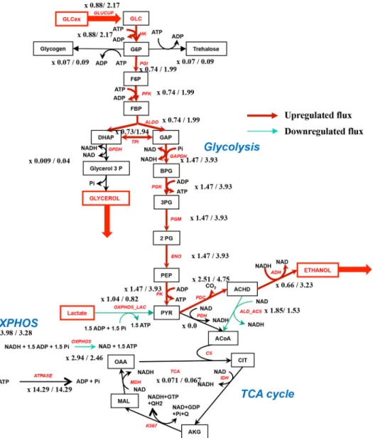
corresponds to 0.5 mMexternal glucose and the second value to 60 mMexternal glucose. The red steps have their flux increased after addition of 60 mMglucose and the green ones decreased. The ratio G6P/F1,6bP goes from 2.95 (0.5 mMglucose) to 0.42 (60 mMglucose). A slight OXPHOS activation is observed in this case. Table 2
Simulation results with OXPHOS modulation by the ratio G6P/F1,6bP
Glucose mM G6P F1,6bP G6P/F1,6bP OXPHOS_NADH OXPHOS_LAC VO2 ATPase NADH Pyr
0.5 1.81 0.61 2.96 2.94 1.04 3.98 14.29 0.015 1.99
60 3.15 8.50 0.37 2.46 0.82 3.28 14.29 0.09 4.78
at UNIV BORDEAUX on October 28, 2019
http://www.jbc.org/
water. Cellular volume determination was performed measur-ing the median cell volume of asynchronous yeast cell cultures using a Coulter counter apparatus (Beckman-Coulter). The
volumes stipulated inTable 3were determined.
Oxygen consumption assays
Oxygen consumption was measured polarographically at 28 °C using a Clark oxygen electrode in a 1-ml thermostatically
controlled chamber. Respiratory rates (JO2) were determined
from the slope of a plot of O2concentration versus time.
Respi-ration assays of growing cells were performed in the growth
medium except in the case of uncoupled respiration (10 M
carbonyl cyanide p-chlorophenylhydrazone), where 100 mM
ethanol was added to avoid any kinetic control upstream the respiratory chain.
Adenine nucleotide measurements
Cellular extracts were prepared by an ethanol extraction
method adapted from the one described in Ref.19. Briefly, cells
were harvested by rapid filtration on nitrocellulose filter (1
m). The filter was immediately dropped into a glass tube
con-taining 5 ml of ethanol, 10 mMHepes, pH 7.2 (4/1), and the tube
was then incubated at 80 °C for 3 min. The mixture was cooled down on ice for at least 3 min, and the ethanol/Hepes solution was eliminated by evaporation using a rotavapor apparatus.
The residue was suspended in 500l of water. Insoluble
parti-cles were eliminated by centrifugation (12,000 ⫻ g, 10 min,
4 °C) and adenine nucleotide content was determined on the supernatant. ATP and ADP were measured using a luciferin/ luciferase enzymatic kit (ATPlite One-step, PerkinElmer Life Sciences). For ADP content determination, the extract was incubated for one-half hour at 28 °C in the following buffer: 75
mMKH2PO4, 15 mMMgSO4, 0.1 mMphosphoenolpyruvate,
and 1 unit of pyruvate kinase to transform ADP into ATP, then quantified using the ATPlite kit.
Pidetermination
Pi was determined according to the method of Sumner
(20).
Figure 8. Theroretical fluxes in the metabolic network with 0.5/60 mMexternal glucose in the presence of G6P/F1,6BP modulation. The red steps have
their flux increased after addition of glucose and the green ones decreased. An OXPHOS inhibition is observed in this case.
at UNIV BORDEAUX on October 28, 2019
http://www.jbc.org/
Hexose phosphate measurements
Glycolysis hexose phosphates were measured in the
fol-lowing buffer: 50 mMtriethanolamine, 7.5 mMMgCl2, 3.75
mM EDTA. G6P was measured in the presence of 1 mM
NAD⫹and 0.3 unit/ml of glucose-6-phosphate
dehydroge-nase. NADH absorbance was followed at 340 nm and the signal was calibrated with standard NADH of known concentration.
F1,6bP was measured in the following buffer: 50 mM
trietha-nolamine, 7.5 mMMgCl2, 3.75 mMEDTA, 1 mMNAD⫹, 1 mM
Pi, 1 mMADP, 0.6 units/ml of phosphoglycerate kinase, and 0.3
unit/ml of aldolase. The reaction was started by adding 0.3 unit/ml of glyceraldehyde-3-phosphate dehydrogenase. NADH absorbance was followed at 340 nm and the signal was cali-brated with standard NADH.
Spheroplast preparation
Spheroplasts were obtained according to Ave´ret et al. (17)
and were suspended in the following buffer: 1Msorbitol, 1.7 mM
NaCl, 0.5 mMEGTA, 10 mMKCl, 1 mMpotassium phosphate,
10 mMTris-HCl, 10 mMNH4Cl, 6 mMiodoacetate, whenever
required and 1% BSA, pH 6.8. Protein determination was done using the biuret method with BSA as a standard.
Model description
To further investigate the role of glycolysis hexose phos-phates in the induction of the Crabtree effect, we performed an in silico study of the induction of this effect. We started
from a yeast glycolysis model as developed in Ref.21. This
model involved a detailed description of all glycolysis re-actions as well as the pathway from pyruvate to ethanol through acetaldehyde (pyruvate decarboxylase and alcohol dehydrogenase) and two branches: one toward acetate
(ace-tate branch: acetaldehyde 3 acetate) and the other toward
succinate thus summarizing most of the TCA cycle
(succi-nate branch: pyruvate3 succinate).
We adapted this model to our experimental conditions by suppressing the acetate branch and replacing the succinate branch by a more detailed representation of the TCA cycle involving pyruvate dehydrogenase and the TCA cycle
reac-tions: acetyl-CoA⫹ 4 NAD ⫹ ADP ⫹ Pi3 4 NADH⫹ ATP,
both modeled by an irreversible mass action equation. For the sake of simplicity, the reduction of quinone Q within the TCA cycle was replaced by the reduction of an extra molecule of NAD, which has the same proton stoichiometry in yeast because S. cerevisiae does not contain a proton pumping com-plex I. Instead of the acetate branch we added a reaction called ALD_ACS (acetaldehyde dehydrogenase and acetyl-CoA syn-thase) that represents the production of acetyl-CoA from
acet-aldehyde: acetaldehyde ⫹ NAD ⫹ ATP 3 acetyl-CoA ⫹
NADH ⫹ ADP ⫹ Pi, also modeled by an irreversible mass
Figure 9. Experimental (blue, values are from the WT) and simulated (orange, without inhibition and green, with inhibition) values of F1,6bP (A) and G6P (B) at various glucose concentrations.
Figure 10. Respiratory rate as a function of the [G6P]/[F1,6bP] ratio. Black filled circles correspond to the WT,⌬hxk2, and HAP4p-overexpressing cells
grown on lactate medium (Figs. 1–3and5). Blue filled squares correspond to the same strains grown on galactose medium (Fig. 6). The red triangles correspond to values from the literature on ascites tumor cells. Red circles are derived from Ref.2
and the red triangles are derived from Ref.6. The full curve is drawn according to the model described under “Experimental procedures” with K⫽ 0.27 in the reg-ulatory Hill’s curve as a function of the G6P/F1,6bP ratio (Hill’s n⫽ 4). The dotted
line corresponds to an absence of regulatory Hill’s function.
Table 3
Cellular volumes of the different strains used in this study
Cellular volume determination was performed as described under “Experimental procedures.” Results are means⫾ S.D. of at least three separate experiments.
Strain Volume (fentoliter/cell) ⴞ WT 32 1 ⌬Hxk2 33 1 WTp 37 2 HAP4 63 4
at UNIV BORDEAUX on October 28, 2019
http://www.jbc.org/
OXPHOS_NADH⫽
Vmax_NADH⫻ GF_inhib ⫻ INH_ATP ⫻ NADH ⫻ ADP
KNADH⫻ KADP⫻
冉
1⫹ NADH KNADH冊
⫻冉
1⫹ADP KADP冊
(Eq. 1) OXPHOS_LAC⫽Vmax_lactate⫻ GF_inhib ⫻ INH_ATP ⫻ lactate ⫻ ADP
Klactate⫻ KADP⫻
冉
1⫹ lactate Klactate冊
⫻冉
1⫹ADP KADP冊
(Eq. 2) with INH_ATP⫽ 2⫻ KA KA⫹ ATP ADP (Eq. 3) GF_Inhib⫽冉
G6P F1,6bP冊
4 K4⫹冉
G6P F1,6bP冊
4 (Eq. 4)where Vmax_NADH⫽ 50 mM/s, Vmax_lactate⫽ 33 mM/s and
KADP⫽ 1 mM; and Klactate⫽ 1 mM; KNADH⫽ 1M.
INH_ATP function is a decreasing hyperbola representing
the OXPHOS inhibition at high ATP concentrations (KA⫽ 1).
The GF_Inhib term summarizes the activation and
inhibi-tion by G6P and F1,6bP, respectively (K ⫽ 0.005). It is a
sigmoid function of the ratio G6P/F1,6bP taking values between 0 and 1.
The concentration and flux values at steady-state are
obtained using Copasi (http://copasi.org/)4(23).
Data analysis
Data are presented as mean⫾ S.E. Differences between
sub-strate conditions were analyzed by t test, with the following
code in the figures: (*) for p⬍ 0.05, (**) for p ⬍ 0.01, (***) for p ⬍
0.001, and (****) for p ⬍⬍ 0.001. When comparing different
values obtained in the same experiment (typically the value without glucose and the value with glucose added in the same sample) the mean of the differences was compared with 0 (matched pairs differences) with the same representation code in the figures.
References
1. Crabtree, H. G. (1929) Observations on the carbohydrate metabolism of tumours. Biochem. J. 23, 536 –545CrossRef Medline
2. Rodríguez-Enriquez, S., Jua´rez, O., Rodríguez-Zavala, J. S., and Moreno-Sa´nchez, R. (2001) Multisite control of the Crabtree effect in ascites hep-atoma cells. Eur. J. Biochem. 268, 2512–2519CrossRef Medline
3. Koobs, D. H. (1972) Phosphate mediation of the Crabtree and Pasteur effects. Science 178, 127–133CrossRef Medline
4. Weinhouse, S. (1972) Glycolysis, respiration, and anomalous gene expres-sion in experimental hepatomas: GHA Clowes memorial lecture. Cancer
Res. 32,2007–2016Medline
5. Veech, R. L., Lawson, J. W., Cornell, N. W., and Krebs, H. A. (1979) Cytosolic phosphorylation potential. J. Biol. Chem. 254, 6538 – 6547Medline
6. Sussman, I., Erecin´ska, M., and Wilson, D. F. (1980) Regulation of cellular energy metabolism: the Crabtree effect. Biochim. Biophys. Acta. 591, 209 –223CrossRef Medline
7. Wojtczak, L., Teplova, V. V., Bogucka, K., Czyz˙, A., Makowska, A., Wie˛ckowski, M. R., Duszyn´ ski, J., and Evtodienko, Y. V. (1999) Effect of glucose and deoxyglucose on the redistribution of calcium in Ehrlich ascites tumour and Zajdela hepatoma cells and its consequences for mitochondrial energetics. Eur. J. Biochem. 263, 495–501 CrossRef Medline
8. Saks, V., Belikova, Y., Vasilyeva, E., Kuznetsov, A., Fontaine, E., Keriel, C., and Leverve, X. (1995) Correlation between degree of rupture of outer mitochondrial membrane and changes of kinetics of regulation of respi-ration by ADP in permeabilized heart and liver cells. Biochem. Biophys.
Res. Commun. 208,919 –926CrossRef Medline
9. Zizi, M., Forte, M., Blachly-Dyson, E., and Colombini, M. (1994) NADH regulates the gating of VDAC, the mitochondrial outer membrane chan-nel. J. Biol. Chem. 269, 1614 –1616Medline
10. Anmann, T., Guzun, R., Beraud, N., Pelloux, S., Kuznetsov, A. V., Koger-man, L., Kaambre, T., Sikk, P., Paju, K., Peet, N., Seppet, E., Ojeda, C., Tourneur, Y., and Saks, V. (2006) Different kinetics of the regulation of respiration in permeabilized cardiomyocytes and in HL-1 cardiac cells: Importance of cell structure/organization for respiration regulation.
Biochim. Biophys. Acta 1757,1597–1606CrossRef
11. Eimre, M., Paju, K., Pelloux, S., Beraud, N., Roosimaa, M., Kadaja, L., Gruno, M., Peet, N., Orlova, E., Remmelkoor, R., Piirsoo, A., Saks, V., and Seppet, E. (2008) Distinct organization of energy metabolism in HL-1 cardiac cell line and cardiomyocytes. Biochim. Biophys. Acta
1777,514 –524CrossRef
12. Díaz-Ruiz, R., Ave´ret, N., Araiza, D., Pinson, B., Uribe-Carvajal, S., Devin, A., and Rigoulet, M. (2008) Mitochondrial oxidative phosphorylation is regulated by fructose 1,6-bisphosphate: a possible role in Crabtree effect induction? J. Biol. Chem. 283, 26948 –26955CrossRef Medline
13. Aiston, S., Trinh, K. Y., Lange, A. J., Newgard, C. B., and Agius, L. (1999) Glucose-6-phosphatase overexpression lowers glucose 6-phosphate and inhibits glycogen synthesis and glycolysis in hepatocytes without affecting glucokinase translocation: evidence against feedback inhibition of glu-cokinase. J. Biol. Chem. 274, 24559 –24566CrossRef Medline
14. van Maris, A. J., Bakker, B. M., Brandt, M., Boorsma, A., Teixeira de Mat-tos, M. J., Grivell, L. A., Pronk, J. T., and Blom, J. (2001) Modulating the
4Please note that the JBC is not responsible for the long-term archiving and
maintenance of this site or any other third party hosted site.
at UNIV BORDEAUX on October 28, 2019
http://www.jbc.org/
distribution of fluxes among respiration and fermentation by overexpres-sion of HAP4 in Saccharomyces cerevisiae. FEMS Yeast Res. 1, 139 –149
CrossRef Medline
15. de Winde, J. H., and Grivell, L. A. (1993) Global regulation of mitochon-drial biogenesis in Saccharomyces cerevisiae. Prog. Nucleic Acids Res. Mol.
Biol. 46,51–91CrossRef
16. Forsburg, S. L., and Guarente, L. (1989) Identification and characteriza-tion of HAP4: a third component of the CCAAT-bound HAP2/HAP3 heteromer. Genes Dev. 3, 1166 –1178CrossRef Medline
17. Ave´ret, N., Fitton, V., Bunoust, O., Rigoulet, M., and Gue´rin, B. (1998) Yeast mitochondrial metabolism: from in vitro to in situ quantitative study. Mol. Cell. Biochem. 184, 67–79CrossRef Medline
18. Chevtzoff, C., Yoboue, E. D., Galinier, A., Casteilla, L., Daignan-Fornier, B., Rigoulet, M., and Devin, A. (2010) Reactive oxygen species-mediated regulation of mitochondrial biogenesis in the yeast Saccharomyces
cerevi-siae. J. Biol. Chem. 285, 1733–1742CrossRef Medline
19. Loret, M. O., Pedersen, L., and Franc¸ois, J. (2007) Revised procedures for yeast metabolites extraction: application to a glucose pulse to car-bon-limited yeast cultures, which reveals a transient activation of the purine salvage pathway. Yeast 24, 47– 60CrossRef Medline
20. Sumner, J. B. (1944) A method for the colorimetric determination of phos-phorus. Science 100, 413– 414CrossRefCrossRef Medline
21. Smallbone, K., Messiha, H. L., Carroll, K. M., Winder, C. L., Malys, N., Dunn, W. B., Murabito, E., Swainston, N., Dada, J. O., Khan, F., Pir, P., Simeonidis, E., Spasic´, I., Wishart, J., Weichart, D., et al. (2013) A model of yeast glycolysis based on a consistent kinetic characterisation of all its enzymes. FEBS Lett. 587, 2832–2841CrossRef Medline
22. Cornish-Bowden, A., Mazat, J.-P., and Nicolas, S. (2014) Victor Henri: 111 years of his equation. Biochimie 107, 161–166CrossRef Medline
23. Hoops, S., Sahle, S., Gauges, R., Lee, C., Pahle, J., Simus, N., Singhal, M., Xu, L., Mendes, P., and Kummer U. (2006) COPASI–a COmplex PAthway SImulator. Bioinformatics 22, 3067–3074CrossRef Medline
at UNIV BORDEAUX on October 28, 2019
http://www.jbc.org/
Alerts:
When a correction for this article is posted •
When this article is cited •
to choose from all of JBC's e-mail alerts Click here
http://www.jbc.org/content/293/33/12843.full.html#ref-list-1
This article cites 23 references, 10 of which can be accessed free at
at UNIV BORDEAUX on October 28, 2019
http://www.jbc.org/

![Figure 10. Respiratory rate as a function of the [G6P]/[F1,6bP] ratio.Black filled circles correspond to the WT, ⌬ hxk2, and HAP4p-overexpressing cells grown on lactate medium (Figs](https://thumb-eu.123doks.com/thumbv2/123doknet/14519365.531104/11.891.61.433.87.545/figure-respiratory-function-black-circles-correspond-overexpressing-lactate.webp)
