HAL Id: hal-02374328
https://hal.archives-ouvertes.fr/hal-02374328
Submitted on 21 Nov 2019
HAL is a multi-disciplinary open access archive for the deposit and dissemination of sci-entific research documents, whether they are pub-lished or not. The documents may come from teaching and research institutions in France or abroad, or from public or private research centers.
L’archive ouverte pluridisciplinaire HAL, est destinée au dépôt et à la diffusion de documents scientifiques de niveau recherche, publiés ou non, émanant des établissements d’enseignement et de recherche français ou étrangers, des laboratoires publics ou privés.
Alterations of redox dynamics and desmin
post-translational modifications in skeletal muscle
models of desminopathies
Florence Delort, Bertrand-David Segard, Coralie Hakibilen, Fany
Bourgois-Rocha, Eva Cabet, Patrick Vicart, Meng-Er Huang, Guilhem Clary,
Alain Lilienbaum, Onnik Agbulut, et al.
To cite this version:
Florence Delort, Bertrand-David Segard, Coralie Hakibilen, Fany Bourgois-Rocha, Eva Cabet, et al.. Alterations of redox dynamics and desmin post-translational modifications in skeletal mus-cle models of desminopathies. Experimental Cell Research, Elsevier, 2019, 383 (2), pp.111539. �10.1016/j.yexcr.2019.111539�. �hal-02374328�
Research Article 1
Alterations of redox dynamics and desmin post-translational
2
modifications in skeletal muscle models of desminopathies
3
Florence Delorta, Bertrand-David Segarda, Coralie Hakibilena, Fany Bourgois-4
Rochaa, Eva Cabeta, Patrick Vicarta, Meng-Er Huangb, Guilhem Clary c, Alain 5
Lilienbauma, Onnik Agbulutd , and Sabrina Batonnet-Pichona 6
a
Université de Paris, Unité de Biologie Fonctionnelle et Adaptative, CNRS UMR 7
8251, F-75013 Paris, FRANCE. 8
9
b
Institut Curie, PSL Research University, CNRS UMR3348, Université Paris-Sud, 10
Université Paris-Saclay, Orsay, 91405, France. 11
12
c
Inserm U1016, Institut Cochin, CNRS UMR8104, Université Paris-Descartes, 13
Sorbonne Paris Cité, Plateforme Protéomique 3P5, Paris, France. 14
15
dSorbonne Université, Institut de Biologie Paris-Seine (IBPS), CNRS UMR 8256,
16
Inserm ERL U1164, Biological Adaptation and Ageing, 75005, Paris, France. 17
18
19
20
Abbreviations: IF, intermediate filament; ROS, reactive oxygen species; WT, 21
wildtype; PTM, post-translational modifications; NAC, N-acetyl-L-cysteine; GSSG, 22
oxidized glutathione; GSH, reduced glutathione; AAV, adeno-associated-virus; TA, 23
tibialis anterior; DMEM, Dulbecco’s Modified Eagle’s medium; HE, hematoxylin/eosin; 24
SDH, succinate dehydrogenase 25
2 27
Abstract: 28
Desminopathies are a type of myofibrillar myopathy resulting from mutations in DES, 29
encoding the intermediate filament protein desmin. They display heterogeneous 30
phenotypes, suggesting environment influences. Patient muscle proteins show 31
oxidative features linking oxidative stress, protein aggregation, and abnormal protein 32
deposition. To improve understanding of redox balance in desminopathies, we further 33
developed cellular models of four pathological mutants localized in 2B helical domain 34
(the most important region for desmin polymerization) to explore desmin behavior 35
upon oxidative stress. We show that the mutations desQ389P and desD399Y share 36
common stress-induced aggregates, desR406W presents more scattered 37
cytoplasmic aggregative pattern, and pretreatment with N-acetyl-L-cysteine (NAC), 38
an antioxidant molecule, prevents all type of aggregation. Mutants desD399Y and 39
desR406W had delayed oxidation kinetics following H2O2 stress prevented by
N-40
acetyl-L-cysteine pretreatment. Further, we used AAV-injected mouse models to 41
confirm in vivo effects of N-acetyl-L-cysteine. AAV-desD399Y-injected muscles 42
displayed similar physio-pathological characteristics as observed in patients. 43
However, after 2 months of N-acetyl-L-cysteine treatment, they did not have reduced 44
aggregates. Finally, in both models, stress induced some post-translational 45
modifications changing Isoelectric Point, such as potential hyperphosphorylations, 46
and/or molecular weight of human desmin by proteolysis. However, each mutant 47
presented its own pattern that seemed to be post-aggregative. In conclusion, our 48
results indicate that individual desmin mutations have unique pathological molecular 49
mechanisms partly linked to alteration of redox homeostasis. Integrating these 50
mutant-specific behaviors will be important when considering future therapeutics. 51
3 Highlights
53
►Desmin mutations trigger variable aggregative patterns and NAC pretreatment 54
avoids it. 55
►Desmin mutations induce delayed oxidation kinetic with H2O2 stress, which are 56
prevented by NAC. 57
►Stresses induce own post-aggregative post-translational modifications, which are 58
prevented by NAC. 59
►Distinct pathological molecular mechanisms of desmin mutations weigh on 60
therapeutics. 61
Keywords: desmin, oxidative stress, aggregation, N-acetyl-L-cysteine, myopathies, 62
intermediate filaments. 63
Introduction
: 64Desmin is a muscle-specific type III intermediate filament (IF). All IFs share a 65
common tripartite organization characterized by a central alpha-helical coiled coil-66
forming region (rod-domain) and two non-alpha-helical regions (“head” and “tail”) with 67
variable lengths and sequences (1) as represented for desmin in figure 1A. This 68
structure allows protein monomers to assemble (2,3) and ultimately form networks 69
within cells. Thus, desmin participates in cytoskeleton formation, crosslinking 70
myofibrils and connecting them to mitochondria (4), nuclei, membrane desmosomal 71
proteins in cardiac cells (2,3), or costamers in skeletal muscle (4,5). 72
Currently, ~70 mutations within the human desmin gene, DES (HUGO Gene 73
Nomenclature Committee database #2770), lead to desminopathies, a subcategory 74
4 of myofibrillar myopathies that mostly arise between the second and fifth decade of 75
life (6,7). Individuals with desminopathies present skeletal or cardiac muscle defects 76
(8) that exhibit heterogeneous features, such as progressive skeletal myopathy (9), 77
different types of cardiomyopathy (10), or both (11–14). The same point mutation can 78
produce different phenotypes within a family, suggesting that genetic factors as well 79
as environmental (such as intensive sport practice, alimentation, anxiety…) influence 80
disease progression (15). 81
Most desmin mutations are missense and affect the 2B end of the protein rod central 82
dimerization domain, leading to desmin network disorganization (16). The main 83
histological characteristics are myofibrillar disorganization, sarcomere misalignment, 84
and desmin-containing protein aggregates in the sarcoplasm. Many studies have 85
investigated how desmin mutations affect the capacity of recombinant proteins to 86
self-assemble in filaments in vitro, as well as in transient cell experiments with or 87
without another IF (e.g., vimentin) (17). Indeed, several mutant monomers 88
polymerize normally in vitro, while some present irregular-diameter filaments, such as 89
desD399Y (17,18). For other mutants, the assembly process stops at intermediate 90
steps, such as tetramer, unit-length filament, filament elongation, or IF maturation 91
(17). 92
Thus, desmin assembly and/or desmin aggregation seem to depend on sequence, 93
without obvious links to mutation position. Moreover, some post-translational 94
modifications (PTMs) of desmin or other IFs have been linked to 95
assembly/disassembly (17). Modifications of PTMs pattern have also recently been 96
highlighted in myofibrillar (19,20) or cardiac pathology (21) context. 97
5 In addition, mouse Des-/- muscle cell mitochondria exhibit an increase in size and 98
number, loss of correct positioning, and pronounced degeneration after work 99
overload (22,23). Further, desL345P transgenic (24) or desR349P knock-in (KI) mice 100
also present mitochondria mislocalization or enzyme dysfunction (25). These 101
alterations indicate a link between oxidative metabolism and desmin. 102
Skeletal muscle fiber metabolism constantly generates reactive oxygen species 103
(ROS) witch level depending to exercise intensity and regularity (26). Redox 104
homeostasis continually neutralizes ROS with antioxidant protective molecules that 105
belong to three main types: enzymes that can metabolize ROS and environmental 106
oxygen peroxide (H2O2), endogenous non-enzymatic compounds, and exogenous
107
non-enzymatic compounds (e.g., provided by food). Among endogenous 108
antioxidants, glutathione appears to be most important in the context of muscular 109
fatigue—the ratio between oxidized glutathione [disulfide glutathione (GSSG)] and 110
reduced glutathione (GSH) is an indicator of redox status in cells or tissues (27). 111
Among exogenous antioxidants, N-acetyl-L-cysteine (NAC) is known to counteract 112
the increased levels of ROS and then to provide effects within muscle cells. In fact, 113
this precursor of endogenous antioxidant, increases levels of the reduced glutathione 114
before, during, and after exercise (28). 115
Imbalanced redox homeostasis is a pathogenic mechanism and thus potential 116
therapeutic target in myopathies. Indeed, SEPN1-related myopathy, RYR1-related 117
myopathy, or Duchenne muscular dystrophy have direct defects in redox regulation 118
systems or have secondary redox abnormalities (29). Previously, pretreatment with 119
NAC before H2O2 exposure significantly improved cell survival in SelN‐devoid
120
myoblasts, as well as in controls, whereas other food antioxidants did not (30). 121
6 Further, a link between oxidative stress and desmin protein aggregation has been 122
shown in patient biopsies. Indeed, main glycoxidation marker (advanced glycation 123
end product, N-carboxy-methyl-lysine, or N-carboxy-ethyl-lysine) levels are increased 124
in muscle samples from individuals with desminopathies (31). Moreover, desmin is a 125
major target of these oxidation and nitration modifications (32). 126
As patient cells are rarely available, we previously constructed isogenic cell lines 127
expressing inducible wildtype (desWT) or mutant desmin—with mutations located in 128
the head, rod, and tail domains (desS46Y, desD399Y, and desS460I, respectively)— 129
to explore protein aggregation mechanisms. Only desD399Y is sensitive to stresses, 130
such as heat shock or redox environmental changes (H2O2 or cadmium treatment). In
131
addition, some antioxidant molecules, such as N-acetyl-L-cysteine (NAC) (33) and 132
alpha-tocopherol (34), can prevent stress-induced aggregation. 133
Here, we first further expanded our inducible cell lines to other 2B mutations. Indeed, 134
mutations in the end of the 2B domain of desmin are predominant (~66%) in human 135
patients with desminopathies and lead to musculoskeletal and cardiac phenotypes of 136
variable severity (35). This area also contains a stutter region, a conserved sequence 137
important for filament assembly and stabilization (36). Thus, the 2B region seems 138
essential for polymerization of the desmin network, perhaps making it susceptible to 139
stress. Moreover A357P, Q389P, and R406W mutations share with D399Y a 140
common skeletal muscle phenotype in patients (distal and proximal myopathy). 141
Patients also mainly present respiratory insufficiency (except for Q389P) and cardiac 142
defects (except for A357P) (for review see 17). R406W leads to the most severe 143
phenotype and presents particular subsarcolemmal desmin accumulation in skeletal 144
muscle. 145
7 Mutations introducing proline in the C-terminal part of the 2B domain are the most 146
frequent (12 out of 24) (35). They lead to desmin aggregation and affect cellular 147
assembly of IFs (37). However, not all mutations are dominant—A360P is recessive 148
(13). We chosed to study at least two mutations carrying a proline: A357P and 149
Q389P. These two mutations present a phenotype, especially on skeletal muscles, in 150
agreement with our model of myoblasts. In addition to these two mutations, R406W 151
substitution affects the YRKLLEGEE region, another extremely conserved and 152
important domain in the regulation of network assembly (38–40). 153
Thus, we investigated whether the aggregative behaviors in stress conditions and the 154
response to NAC pretreatment depend on the 2B location or on the specific mutation. 155
Furthermore, we also analyzed the link between 2B mutations presenting stress 156
induced aggregation and redox dynamics. To confirm the effect of NAC in a more 157
physiological context, we performed experiments with injection of adeno-associated-158
virus (AAV), expressing WT or mutated desmin, in mouse tibialis anterior (TA) 159
muscle, with or without antioxidant treatment. Finally, to understand these behaviors, 160
we analyzed post-translational modifications (PTMs) of desmin in cell or in muscle. 161
Indeed, as described for other intermediate filaments, some PTMs, such as 162
phosphorylation, ubiquitination, sumoylation, glycosylation, and/or ADP ribosylation, 163
are known to modify their behavior (41). Thus, they could also regulate desmin in 164
response to stress. 165
Altogether, desmin cytoplasmic redox dynamic and PTMs alterations in our models 166
provide new information on the variability and importance of oxidative redox balance 167
in skeletal muscular cells containing a pathological variant of desmin. 168
8
Materials and methods
: 170Molecular model and secondary structure predictions 171
The crystal structure of human vimentin coil 2B fragment (CYS2) [residues 328 172
(Cys328)–411; PDB entry 1GK4] was used to construct desmin 2B end domain 173
models (WT and mutant) with PYMOL software (DeLano Scientific LLC, Palo Alto, 174
CA, United States). For energy minimization, computations were done in vacuo with 175
GROMOS96 43B1 parameters set, without reaction field, from Swiss-PdbViewer 176
4.0.1 (http://spdbv.vitalit.ch/download.html). We used Pubmed Blast 177
(http://blast.ncbi.nlm.nih.gov/Blast.cgi) for sequence alignment of CYS2 (residues 178
328–411) and desmin (residues 333–416). 179
Cell lines and culture 180
New stable cell lines from murine C2C12 myoblasts (ATCC, NY, USA) were 181
generated as previously described (33). Briefly, mutagenesis was performed on 182
pPuro-Myc-human-Desmin vector to introduce human mutations desA357P, 183
desQ389P, and desR406W, following manufacturer’s instructions (Quick Change-XL 184
mutagenesis kit, Stratagene, San Diego, CA, United States). Plasmids were 185
electroporated (Genepulser II, BioRad, Hercules,CA, United States; 500 µF, 350 V, 186
with 10 µg of DNA) into clone 21 (tet-on) cells (33). Stable clones were selected after 187
one week of puromycin selection. Doxycycline induction of human desmin variants 188
was verified by western blotting (using c-Myc epitope antibody (1/1000, 9E10, Santa 189
Cruz Biotechnologies, Dallas, TX, United States)) and immunostaining. Double stable 190
cell lines were grown at 37°C and 5% CO2 in Dulbecco’s Modified Eagle’s medium
191
(DMEM, Life Technologies, Carlsbad, CA, United States) supplemented with 20% 192
9 fetal calf serum (Eurobio, Les Ulis, France), 1% penicillin/streptomycin (Life 193
Technologies, Carlsbad, CA, United States), 1 mg/mL G418 (Euromedex, 194
Souffelweyersheim, France), and 2 μg/mL puromycin (Euromedex, 195
Souffelweyersheim, France). 196
Satellite cells were extracted from 2 gastrocnemius and 2 plantaris muscles of 1-197
month-old C56Bl6(N) knock-in mice for the R405W mutation (murine homologue of 198
human desmin R406W; named after KI-R405W) (12). Following previous work (42), 199
muscles were incubated 4 x 10 min in a solution of DMEM-10 HamF12/Glutamax 200
(Life Technologies, Carlsbad, CA, United States) containing 1.5 mg/mL of protease 201
XIV (Sigma-Aldrich, Saint-Louis, MO, United States) and 1/500 primocin (Invitrogen, 202
Carlsbad, CA, United States) at 37°C under regular agitation. After each incubation, 203
tubes were centrifuged at 400 g for 30 seconds at room temperature. Floating cells 204
were eliminated, and 3 other fractions containing satellite cells were diluted in 20% 205
FBS medium and sieved with a 40-µm filter. Filtrate containing satellite cells was 206
centrifuged for 5 min at 1400 g at room temperature. Cells in DMEM-207
HamF12/Glutamax containing 20% FBS, 2% Ultroser (Pall Life Sciences, 208
Portsmouth, United Kingdom), 8.6 ng/mL of FGF2 (Life Technologies, Carlsbad, CA, 209
United States), 1/100 N2 (Life Technologies, Carlsbad, CA, United States), and 210
1/500 primocin were seeded onto 1/20 Matrigel-coated 6-well plates (Corning, 211
Corning, NY, United States) at 1000 cells/well. After activation and proliferation, 212
satellite cells were maintained or analyzed by seeding at 1000 cells/cm² on plastic 213
covered with 1/20 Matrigel. 214
Oxidative and heat stress procedures 215
10 As previously described, stable cell lines were seeded at 3 x 10³ cells/cm2 in 6-well 216
plates and induced with doxycycline (10 μg/mL, Sigma-Aldrich, Saint-Louis, MO, 217
United States) after 24 h (28). Hydrogen peroxide and heat stresses were performed 218
48 h after induction as follows: 0.2 mM H2O2 (Sigma-Aldrich, Saint-Louis, MO, United
219
States) was added to the cell culture for 2 h or cells were heat shocked at 42°C for 2 220
h. Cells were immediately analyzed (T0 time point) or media was replaced and cells 221
were treated 24 h later (T24 time point). For pre-treatment, NAC (10 mM, #A7250 222
Sigma-Aldrich, Saint-Louis, MO, United States) was added to cells 16 h before 223
stress. 224
Western blotting 225
Myoblast proteins were extracted by scraping cells in RIPA buffer without SDS [50 226
mM Tris pH 7.5, 150 mM NaCl, 5 mM EDTA, 1% NP40, 1 mM Na3VO4, 10 mM NaF,
227
1 mM PMSF, protease inhibitor cocktail 1X (complete mini, EDTA free, Roche, Bâle, 228
Switzerland)] for 1D SDS-PAGE or urea buffer (8 M urea, 2 M thiourea, 4% CHAPS, 229
50 mM DTT) for 2D SDS-PAGE. After migration, proteins were transferred to 230
nitrocellulose membranes (0.45 µm, Macherey Nagel, Düren, Germany), which were 231
saturated with 5% non-fat milk in 0.5% Tween/PBS. Membranes were incubated with 232
the primary antibody at the appropriate dilution for 1 h at room temperature or 16 h at 233
4°C. Primary antibodies used were: 1- mouse monoclonal anti-c-Myc (1/1000, 9E10, 234
Santa Cruz Biotechnologies, Dallas, TX, United States); 2- rabbit polyclonal anti-235
desmin (1/1000, # C3956, Sigma-Aldrich, Saint-Louis, MO, United States); 3- mouse 236
monoclonal anti-alpha-actin (1/2000, #MAB1501R, Millipore, Burlington, MA, United 237
States); or 4- rabbit polyclonal anti-GFP (1/2500, A11122, Life Technologies, 238
Carlsbad, CA, United States). Isotype-specific anti-mouse or anti-rabbit secondary 239
11 antibody coupled with horseradish peroxidase (1/10000, #31430 or #31460, Pierce, 240
Thermo Scientific, Waltham, MA, United States) was detected by incubating with 241
Clarity Western ECL (BioRad, Hercules, CA, United States) and visualized with a 242
CCD camera (FUJI Las 4000 or Ai600, GE Healthcare, Chicago, IL, United States). 243
Cell immunofluorescence 244
Cells were fixed with 2% paraformaldehyde (Santa Cruz Biotechnologies, Dallas, TX, 245
United States) for 15 min at room temperature, permeabilized 5 min with 0.5% Triton 246
X-100, and incubated with mouse monoclonal anti-c-Myc primary antibody (1/100, 247
9E10 #sc40 Santa Cruz, Biotechnologies, Dallas, TX, United States) for 1 h at room 248
temperature. After 3 washes with PBS, cells were incubated 45 min with isotype-249
specific anti-mouse secondary antibody labeled with Alexa-488 (#A11001, Molecular 250
Probes, Eugene, OR, United States). DNA was stained with Hoechst 33258 (1 251
µg/mL, Sigma-Aldrich, Saint-Louis, MO, United States) for 10 min. Finally, cells were 252
washed in PBS and mounted in Fluoromount medium (Interchim, San Diego, CA, 253
United States). Images were acquired with confocal microscopy (LSM700 Zeiss, 254
Iena, Germany) at the imaging facility of the Functional and Adaptive Biology (BFA) 255
unit. 256
For aggregation measurements, images were taken as 5x5 tile scans on 3 random 257
nuclear fields chosen in Hoechst-stained areas. Total cell number was calculated by 258
counting nuclei with an ImageJ software in-house macro. Cells with aggregates were 259
visually counted on the images. Aggregation percentage and ratio between stressed 260
versus unstressed aggregated cells were calculated. Experiment was repeated at 261
least 3 times. 262
12 Redox dynamics
263
Mammalian cell vector expressing cytoplasm-targeted HyPer (cyto-HyPer) was 264
constructed by amplifying HyPer sequence (Evrogen, Moscow, Russia, (43)) and 265
introducing a Nuclear Export Signal (NES). The PCR product, fusing NES in frame to 266
the 3’ end of HyPer, was then cloned in pCMV/myc/cyto vector (Life Technologies, 267
Carlsbad, CA, United States). Vector expressing cytoplasm-targeted Grx1-roGFP2 268
(cyto-Grx1-roGFP2) was constructed in a similar way using pEIGW-Grx1-roGFP2 269
(Addgene, Cambridge, MA, United States (44)) as template. Expression vector 270
encoding HyPer targeted to mitochondrial matrix (mito-HyPer) was purchased from 271
Evrogen (Moscow, Russia). Stable cell lines or satellite cells were seeded at 11,500 272
cells/cm2 or 22,000 cells/cm2, respectively, in 6-well plates. After 24 h, cells were 273
transfected with cyto-HyPer, mito-HyPer, or cyto-Grx1-roGFP2 in DMEM medium 274
without antibiotics using JetPRIME (Polyplus-transfection, Illkirch-Graffenstaden, 275
France) according to manufacturer’s instructions. Eight hours after transfection, 276
antibiotics were added, including doxycycline for desmin induction. Transfection 277
efficiency was evaluated on an inverted fluorescence microscope with a FITC filter 278
(Zeiss, Iena, Germany) at the imaging facility of the BFA unit. 279
Oxidative kinetics were started by adding 0.1 mM H2O2 to cells in complete media
280
and incubating for 5, 15, 30, 60, or 120 min. Medium was aspirated and cells were 281
washed with PBS and then overlaid with 15% trichloroacetic acid. After 30 min, cells 282
were scraped and pelleted in 1.5-mL microtubes. After two washes with ice-cold 283
acetone and air drying, pellets were resuspended in TES buffer (100 mM Tris pH 8.8, 284
10 mM EDTA, 1% SDS). Loading buffer without reducing agent was added to 285
samples. Equal volumes were loaded on 12% SDS acrylamide gels and results were 286
analyzed by western blotting. 287
13 Intramuscular injection of AAV vectors
288
Plasmids encoding c-Myc-tagged human desmin (desWT and desD399Y) under the 289
regulation of the CMV promoter were used to prepare AAV vectors. 290
AAV2/2.CMV.Myc-desWT (AAV-desWT) or Myc-desD399Y (AAV-desD399Y) were 291
provided by the Vector Production Center of the Therapeutic Research Institute at 292
the Health Research Institute of the University of Nantes (UMR1089, Nantes, 293
France). Final viral preparations were split in 50-μL aliquots in D-PBS (with Ca2+
and 294
Mg2+) and stored at −80°C. Number of viral genomes (vg) was titrated by polyA 295
quantitative PCR and was 5.2 x 10¹².mL-1 for AAV-desWT and 5 x 10¹².mL-1 for AAV-296
desD399Y. 297
The injection procedure was previously described by Joanne et al. (45) Briefly, for 298
intramuscular injections, 11 week-old male C57BL/6J mice (Janvier Labs, Le Genest-299
Saint-Isle, France) were anesthetized by intraperitoneal injection of an analgesic and 300
sedative drug mix [100 mg.kg-1 ketamine (Merial, Lyon, France) and 10 mg/kg 301
xylazine (Bayer, Leverkusen, Germany)]. Each TA muscle was injected with 70 μL of 302
AAV diluted in PBS (~3 x 10¹¹ vg/70 μL) using an insulin syringe. Animals were split 303
into 4 groups of n=5 mice: desWT with NAC, desWT without NAC, AAV-304
desD399Y with NAC, and AAV-desD399Y without NAC. NAC was diluted to 20 mM 305
in drinking water. Bottles were changed every 2–3 days and weighed before and 306
after replacement to calculate the absorption volume of NAC by mice. 307
Animal experiments were conducted with respect to animal health and well-being, 308
and all procedures were approved by our institutional ethics committee (authorization 309
APAFIS#5441 -20 16052314036983 v7, for AAV-injected mice; CEB-16-2016, for KI-310
R405 mice). All procedures were also conducted according to French and European 311
14 laws, directives, and regulations on animal care (European Commission Directive 312
86/609/EEC; APAFIS#5441 -20 16052314036983 v7). For AAV experiments, the 313
animal facility (Centre d’expérimentation Fonctionnelle (CEF), Pitié Salpétrière 314
Hospital, Paris) is fully licensed by French competent authorities and has animal 315
welfare insurance. 316
Preparation of TA muscles 317
TA muscles were removed 1 or 2 months following injection after euthanizing mice by 318
cervical dislocation. For immunohistochemical analysis, muscles were embedded in 319
tragacanth gum (Fisher Scientific, Hampton, NH, United States) and frozen by 320
plunging in isopentane precooled in liquid nitrogen for at least 1 minute. For western 321
blotting, muscles were directly frozen in liquid nitrogen in microtubes and later 322
pulverized in a cryogenic mortar (Dutscher, Issy-les-Moulineaux, France) and 323
resuspended in urea buffer for 2D gels. 324
Muscle sections and histological staining 325
Serial sections of 6 μm thickness were sliced using a CM1950 cryostat, (Leica, 326
Wetzlar, Germany) recovered on Superfrost Plus microscope slides (Thermo 327
Scientific, Waltham, MA, United States), and stored at –80°C. Muscle sections were 328
kept at room temperature for 20 min before staining with either hematoxylin/eosin 329
(HE), succinate dehydrogenase (SDH), or fluorescent antibodies as follows. 330
HE: Transverse muscle sections were incubated with 0.7% hematoxylin solution 331
(Harris modified, Sigma-Aldrich, Saint-Louis, MO, United States) for 6 min at room 332
temperature, washed 5X with water, and stained with 0.5% eosin in acidified 90% 333
ethanol (Sigma-Aldrich, Saint-Louis, MO, United States) for 1 min at room 334
15 temperature. Muscle sections were dehydrated by incubating in successive baths of 335
gradually increasing ethanol concentrations (30%, 50%, 70%, 85%, 95%, and 100%, 336
repeated twice) for 2 min each, followed by two baths in clearing solution (Histo-337
Clear, National Diagnostics, Atlanta, GA, United States) for 10 min and 15 min, and 338
rapidly mounted in VectaMount medium (Clinisciences, Nanterre, France). Images 339
were acquired on a stereomicroscope (Leica, Wetzlar, Germany) at the BFA unit. 340
SDH: Transverse muscle sections were stained by immersion in SDH buffer (0.2 M 341
phosphate buffer containing 5.4% sodium succinate and 0.1% nitro blue tetrazolium) 342
for 2 h at 37°C and then washed with water. Finally, muscle sections were either 343
mounted directly in VectaMount medium (Clinisciences, Nanterre, France) or 344
processed for further immunofluorescence. Images were captured on a 345
stereomicroscope (Leica, Wetzlar, Germany) at the BFA unit. 346
Immunostaining: Warmed muscle sections were fixed with 4% paraformaldehyde for 347
10 min at room temperature. Endogenous fluorescence was prevented with 50 mM 348
NH4Cl for 30 min. Sections were permeabilized in 0.5% triton/PBS for 10 min,
349
blocked in 1% bovine serum albumin/PBS, and incubated with rabbit anti-Myc 350
(1/1000, # C3956, Sigma-Aldrich, Saint-Louis, MO, United States) or rabbit anti-351
laminin (1/100, #L9393, Sigma-Aldrich, Saint-Louis, MO, United States) primary 352
antibodies for 1 h at room temperature or overnight at 4°C. Primary antibodies were 353
detected by incubating sections with suitable secondary antibodies for 45 min. 354
DNA was stained with 1 µg.mL-1 Hoechst 33258 (Sigma-Aldrich, Saint-Louis, MO, 355
United States) for 10 min. Cells were washed in PBS and mounted in Fluoromount 356
medium (Interchim, San Diego, CA, United States). All images were captured using a 357
16 digital camera mounted to a confocal laser scanning microscope (LSM700 Zeiss, 358
Iena, Germany) at the imaging facility of the BFA unit. 359
Analysis of TA muscle characteristics 360
TA fiber amount and diameter (minimum Feret values) were calculated on whole 361
muscle transverse sections stained with laminin. Images were analyzed with ImageJ 362
software. Percent transduction was calculated by measuring the number of fibers that 363
were positively stained for Myc compared to the total number of fibers in the section. 364
For aggregation rate, number of fibers containing aggregates was reported in relation 365
to total transduced fibers. 366
2D gel electrophoresis 367
2D gel electrophoresis was performed at the proteomics platform at the Cochin 368
Institute (Paris, France). Briefly, protein separation was performed by 369
isoelectrofocusing on 7-cm pH 4–7 strips in the first dimension and SDS-PAGE on 10 370
or 12% polyacrylamide gels in the second dimension. Proteins were transferred to 371
nitrocellulose membranes and analyzed by western blotting. Calculation of theoretical 372
isoelectric points (pI) for Myc-labeled desWT and mutant desmin proteins provided 373
different values according to the website used (Table 1). 374
17 376
Isoelectric point
(pI) calculation ExPASy
Protein Calculator v3.4 Protein isoelectric point calculator Scansite WT murine desmin 5.21 5.28 5.05 5.21 WT human desmin 5.21 5.28 5.06 5.21 WT Myc-desmin 5.11 5.17 5.07 5.11 D399Y Myc-desmin 5.15 5.20 5.03 5.15 377
Table 1: Algorithmic isoelectric point (pI) calculations from different web sites:
378
ExPASy (https://web.expasy.org/compute_pi); Protein Calculator v3.4 (http://protcalc.sourceforge.net);
379
Protein isoelectric point calculator (http://isoelectric.org);
380
Scansite (https://scansite4.mit.edu/4.0/#calcMolWeight).
381 382
One site also indicates phosphorylated peptide molecular weights and 383
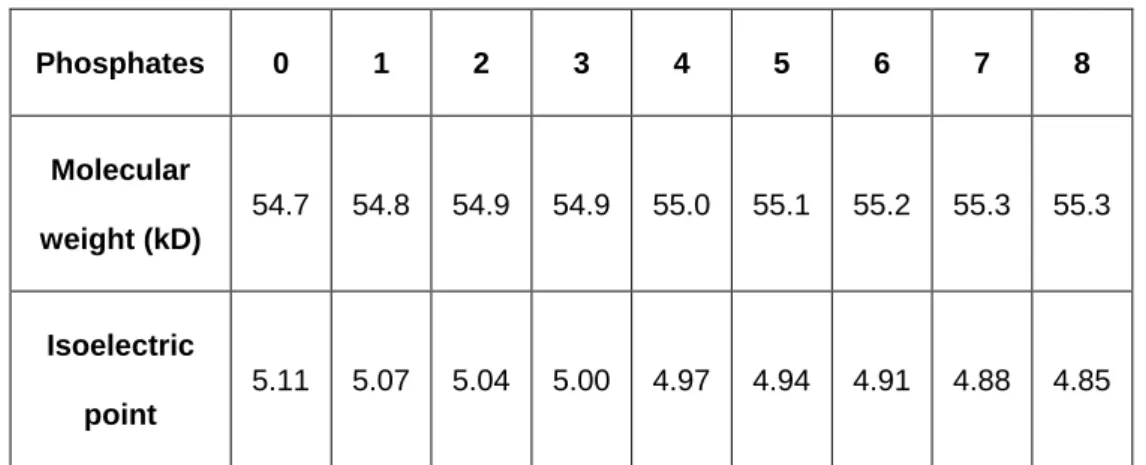
phosphorylation that regulate desmin network assembly. For tagged desWT, the 384
obtained values are detailed in Table 2. 385 Phosphates 0 1 2 3 4 5 6 7 8 Molecular weight (kD) 54.7 54.8 54.9 54.9 55.0 55.1 55.2 55.3 55.3 Isoelectric point 5.11 5.07 5.04 5.00 4.97 4.94 4.91 4.88 4.85
Table 2: Predictions of molecular weight evolutions and isoelectric points according to the number of
386
phosphorylation of desWT (https://scansite4.mit.edu/4.0/#calcMolWeight).
387
Statistical analysis 388
Box plots were created with GraphPadPrism software or R free software, and results 389
were statistically analyzed with non-parametric tests—“nparcomp” function with 390
18 Dunnet comparison to WT, or Tukey comparison for others. Curves, scatter plots, 391
and histograms as well as corresponding graphic analyses were done with 392
GraphPadPrism software—either 2-way ANOVA corrected by Bonferroni non-393
parametric function, or Kruskall-Wallis tests. Significant differences were considered 394
at p < 0.05. 395
Results
: 396DesA357P, desQ389P, desD399Y, and desR406W mutants located in the 2B 397
desmin rod domain exhibit in silico distinctive structural interactions. 398
DesD399Y is located distally in the 2B desmin rod domain. We previously 399
demonstrated that it exhibits stress-induced aggregation, whereas mutations located 400
in the head (desS46Y) or tail (desS460I) do not display this aggregation (33). To 401
further investigate mechanisms leading to stress-induced aggregation, we chose 402
three other pathological mutations—desA357P, desQ389P, and desR406W (Figure 403
1A)—also located in the end of this domain between stutter and C-terminal regions, 404
which is a notorious mutation hot spot (35). 405
First, we constructed desmin 2B end domain mutant and WT models with PYMOL 406
software and compared in silico the impact of each substitution (Figure 1B). A357P is 407
located in the stutter region that is considered critically important for proper filament 408
assembly. Further, it is in the "e" position in the heptad (internal residue repeats 409
conserved in IF, as described by Strelkov et al.(46). On our structural representation, 410
it is well inside the dimer and phenylalanine lateral chains face in both directions. We 411
can see the position of alanine in the helix/dimer and its replacement with proline. 412
Representation of structural modifications occurring with Q389P substitution shows 413
19 loss of a large negative side chain residue and gain of a small neutral one. D399Y 414
and R406W substitutions provide insight about ionic bridging over two disrupted 415
domains—in both, ionic residues are replaced with aromatic amino acids. 416
417
418
Figure 1: Mutation locations within desmin structure and in silico modeling of mutation effects on
419
structure of desmin coiled coil 2B domain. (A) Overview of desmin structure showing locations of
420
individual mutations in the 2B domain and myc tag. (B) Graphic representation of tripartite desmin,
421
based on vimentin crystal structure (1gk4), with mutations in central rod 2B domain of each monomer,
422
with amino acids of interest represented by molecular structures. WT helical regions are magenta and
423
mutants are cyan or green. A357P and Q389P mutations are dark blue. A357P is located in the stutter
424
region, so conserved phenylalanine is also in molecular structure. D399 and R406 interactions
(salt-425
bridge) with side chain residue K395 or E401 are in dark blue and are represented with dotted lines.
426
New stable cell lines expressing desmin mutations exhibit the same expression 427
level as desD399Y-expressing cells. 428
20 Second, we generated six new inducible stable cell lines (2 clones per mutation) 429
expressing desA357P, desQ389P, and desR406W according to the same protocol 430
that we used for desD399Y and desWT (25). Expression in all clones was analyzed 431
by western blot to confirm the level of exogenous human desmin compared to 432
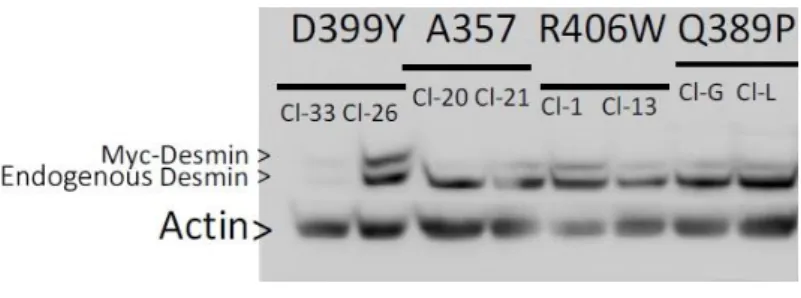
desD399Y (Figure 2). 433
434
Figure 2: Steady-state desmin expression levels. Western blot of endogenous and human
Myc-435
desmin in inducible stable cell lines 72 h after induction (T24 without stress). Actin represents a
436
loading control. “Cl-X” represents name of the selected clone for each desmin mutation.
437
For each mutation, one construct expressed low levels of desmin and another 438
expressed higher levels. At steady state, endogenous desmin was always more 439
highly expressed than exogenous protein (maximum ratio exogenous/endogenous of 440
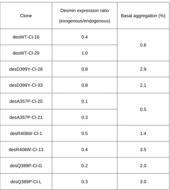
1:1 for desWT clone 29 and 0.8:1 for des D399Y clone 26; Table 3). Overall, clones 441
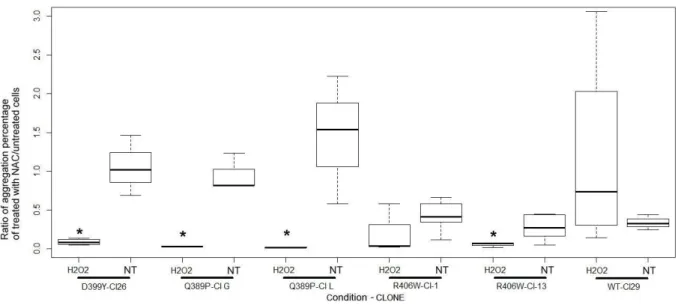
had a similar ratio of ~0.4:1, permitting us to compare new inducible clones to each 442
other in basal conditions or upon stress. Moreover, as demonstrated in table 3, the 443
basal protein aggregation in these cells remained below 3.5% (Table 3). 444
21 446
Clone
Desmin expression ratio (exogenous/endogenous) Basal aggregation (%) desWT-Cl-16 0.4 0.6 desWT-Cl-29 1.0 desD399Y-Cl-26 0.8 2.9 desD399Y-Cl-33 0.8 2.1 desA357P-Cl-20 0.1 0.5 desA357P-Cl-21 0.3 desR406W-Cl-1 0.5 1.4 desR406W-Cl-13 0.4 3.5 desQ389P-Cl-G 0.2 2.0 desQ389P-Cl-L 0.3 3.0 447
Table 3: Steady-state desmin expression levels and aggregation percentages. Western blot
448
analysis of endogenous and human Myc-desmin in inducible stable cell lines 72 h (T24) after
449
induction. Quantification was realized with ImageJ software. Protein amounts were normalized with
450
anti-alpha-actin antibody (Actin). Basal aggregation percentage was calculated as described in
451
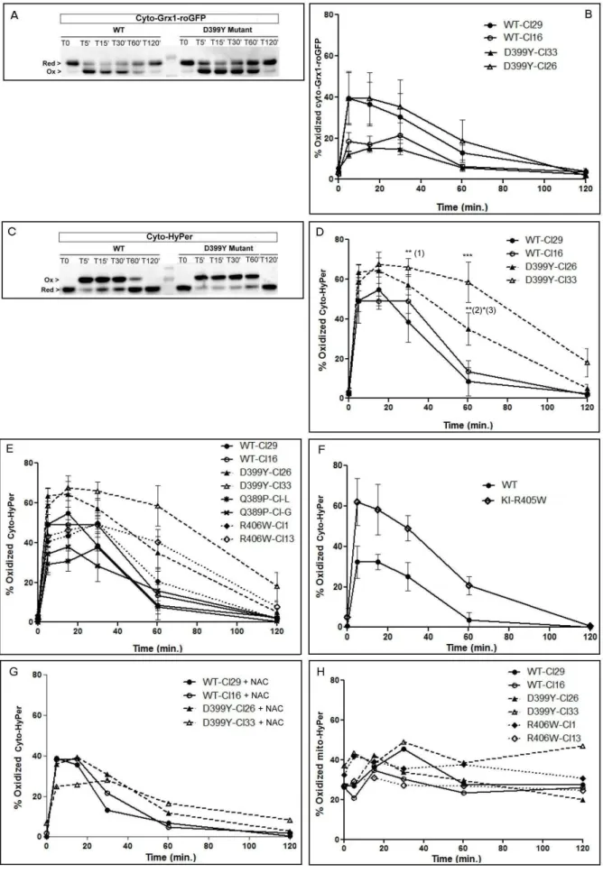
material and methods.
452
Stresses induce protein aggregation only in desQ389P, desD399Y, and 453
desR406W, with varying aggregate morphologies. 454
We measured desmin aggregation in all stable cell lines in response to oxidative or 455
heat shock stresses after 24h. In response to stress, a normal desmin network was 456
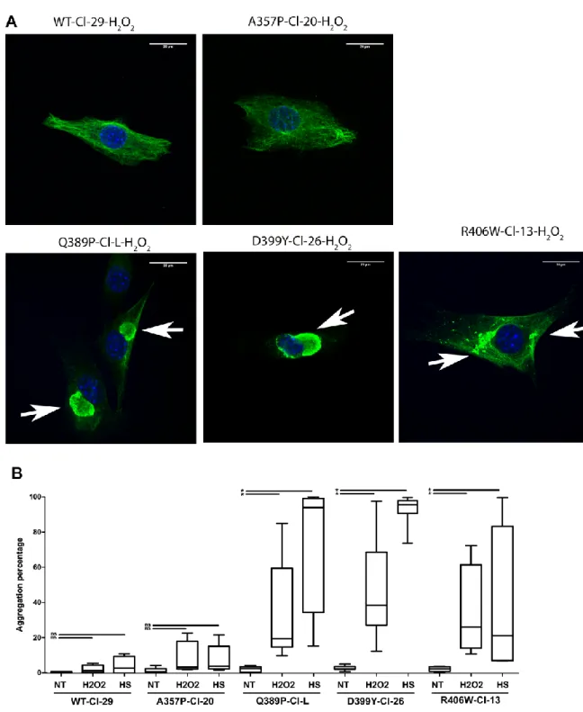
present in desWT- or desA357P-expressing cell lines (Figure 3A, first row). 457
22 458
Figure 3: Increased aggregation upon stresses in desD399Y, desQ389P, and R406W cells, but
459
not desA357P cells. (A) Representative confocal micrographs showing immunostaining at 24 h after
460
oxidative stress (T24) for Myc-desmin (green) and Hoechst dye (blue). DesWT and desA357P form
461
normal networks. DesQ389- and desD399Y-expressing cells form large aggregates near the nucleus
462
(arrows, left and middle panels). DesR406W-expressing cells form smaller accumulations distributed
463
throughout the cytoplasm (arrows, right panel). White bar = 20 μm, 40x magnification. (B) Box plot
464
representing the aggregation percentages 24 h after stress (T24) compared to untreated cells. Three
465
different treatments are shown: heat shock (HS), oxidative stress (H2O2), and no stress (NT). Cells
466
were counted in at least 3 independent experiments (>100 cells each) (ns: not significant; *p < 0.05,
467
compared to unstressed).
23 As previously described, stress of desD399Y-expressing cells induced large 469
aggregates very close to the nucleus (Figure 3A, middle in second row). DesQ389P 470
displayed similar characteristics (Figure 3A, left in second row). DesR406W also 471
exhibited stress-induced, variably-shaped aggregates, although they had weaker 472
staining, maybe due to small size. These aggregates were also more numerous and 473
more dispersed in the cytoplasm (Figure 3A, right in second row). We quantified 474
aggregation rates for each stable clone, representing cells with any aggregates, as 475
compared to the total cell population (Figure 3B, Supp. Data 1). DesA357P-476
expressing cells presented the same pattern as desWT, with no modification of 477
aggregation rate, indicating that desA357P cells are not sensitive to oxidative or heat 478
shock stress. In contrast, heat shock significantly induced a high aggregation rate for 479
desQ389P and desD399Y (~20–40-fold increase), while oxidative stress led to 480
significant, but less, aggregation (~10-fold increase). DesR406W cells also had 481
significant aggregation in response to stress, although with more dispersed values. 482
The anti-aggregative effect of NAC in the oxidative condition was confirmed for 483
desQ389P, desD399Y, and desR406W (Figure 4). Indeed, ratios of aggregate 484
percentages comparing NAC-treated and untreated cells showed a noticeable 485
decrease. For all cell lines sensitive to oxidative stress, NAC pretreatment 486
significantly reduced stress-induced aggregation. 487
24 488
Figure 4: Effect of NAC as an anti-aggregative molecule on stable cell lines expressing
489
desQ389P, desD399Y, and desR406W. First, aggregate-containing cells were quantified, untreated
490
or pretreated with NAC, 24 h after stress or control conditions (NT). Then, box plots represent the ratio
491
of percentage of cells with aggregates in the presence of NAC, reported to untreated cells. Cell
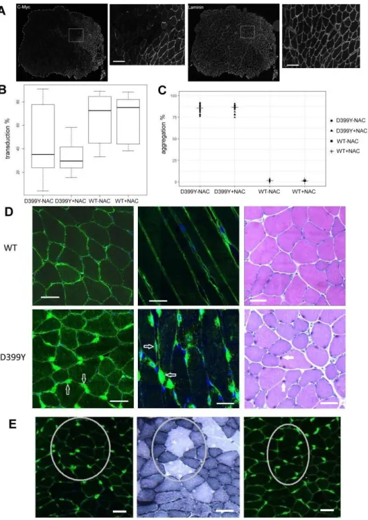
492
counting was performed on three independent experiments (~100 cells each). For each clone,
493
compared to its control NT, *p < 0.05, as calculated with Kruskall-Wallis non-parametric test.
494
Cytoplasmic redox dynamic in desmin mutant cell lines exhibits various 495
behaviors. 496
To further investigate how stress induces aggregation in all responsive cell lines and 497
how NAC can prevent this effect, specific redox reporters were used to monitor redox 498
balance in cells expressing desmin mutants following oxidative stress. We first used 499
the cyto-Grx1-roGFP2 to monitor cytoplasmic redox dynamic. This reporter 500
specifically reacts with GSSG/GSH in the targeted compartment, so the percentage 501
of the oxidized form reflects the redox state of glutathione (44) (Figure 5A and 5B). A 502
second probe, HyPer, reacts specifically with H2O2 and is targeted to the cytoplasm
503
(cyto-HyPer). Therefore, the percentage of the oxidized form of cyto-HyPer reflects 504
the quantity of cytoplasmic H2O2 over time (Figure 5C and 5D).
25 506
Figure 5: desD399Y and desR406W trigger alteration of redox balance during stress.
26
(A) Representative western blots of the oxidized and reduced forms of cyto-Grx1-roGFP2 reporter
508
following oxidative stress in stable cell lines expressing desWT or desD399Y (B) Quantification of
509
oxidation kinetics of the cyto-Grx1-roGFP reporter over time. (C) Representative western blots of the
510
oxidized and reduced forms of cyto-HyPer reporter following oxidative stress in stable cell lines
511
expressing desWT or desD399Y with the cyto-HyPer reporter. (D) Quantification of oxidation kinetics
512
of the cyto-HyPer reporter over time. (E) Percentage of oxidized cyto-HyPer in stable cell lines
513
expressing desWT, desD399Y, desQ389P, or desR406W, without pretreatment. (F) Percentage of
514
oxidized cyto-HyPer in satellite cells from KI-R405W mice. (G) Percentage of oxidized cyto-HyPer in
515
stable cell lines expressing desWT or desD399Y after NAC pretreatment (10 mM). (H) Percentage of
516
oxidized mito-HyPer in stable cell lines expressing desWT, desD399Y, desQ389P, or desR406W,
517
without pretreatment. Each point represents the mean of three independent experiments, with bars
518
indicating SEM. Prism software was used for 2-way ANOVA, corrected by Bonferroni non-parametric
519
function (*p < 0.05; **p < 0.01; ***p < 0.001).
520
Both cyto-Grx1-roGFP2 and cyto-HyPer probes were first compared in experiments 521
with desWT and desD399Y clones. There were no significant differences in cells 522
expressing desWT or desD399Y with Cyto-Grx1-roGFP (Figure 5B). Using cyto-523
HyPer, however, we detected significantly delayed oxidation kinetics in the cytoplasm 524
of desD399Y clones (Figure 5D). Indeed, more H2O2 remained in desD399Y clones
525
at 60 min after the beginning of oxidative stress compared to desWT clones, 526
although both constructs reached near-zero values after 120 min. 527
We thus expanded cyto-HyPer probes analysis to the two other mutants with stress-528
induced aggregation, desR406W and desQ389P. Similar to desD399Y, both clones 529
expressing desR406W had similar significant differences in reporter oxidation 530
depending on the duration of oxidative stress (Figure 5E). We confirmed these 531
results using the more physiological model of satellite cells extracted from our new 532
KI-R405W animal model. These cells showed very comparable delayed recovery of 533
basal reporter reduction (Figure 5F). In contrast, unexpectedly considering the 534
aggregative pattern seen in Figure 3A, desQ389P cell lines have a similar behavior 535
to desWT (Figure 5E). 536
To better understand the effect of NAC pretreatment on the evolution of cytoplasmic 537
content during H2O2 incubation, we made the same measurements in the presence
27 of NAC. NAC especially reduced the percentage of oxidized reporter in cells 539
expressing desD399Y (Figure 5G). Differences in the averages between untreated 540
and treated showed that this decrease was ~16%–25% for both. 541
Finally, we investigated the mitochondrial effect of H2O2 incubation using the 542
mitochondrial matrix-targeted HyPer reporter. Redox balance is distinct in this 543
organelle, as the reporter was more oxidized even without treatment and remained 544
unchanged in all experiment conditions (Figure 5H). This indicates that mitochondria 545
are not affected as cytoplasm in response to oxidation. 546
Expression of desD399Y in vivo induces aggregation with slightly altered SDH 547
repartition in adult mouse muscle fibers. 548
Heat-shock and oxidative stress are two types of environmental variations that can 549
be found in muscle physiology (47). DesD399Y induces aggregation following these 550
environmental stresses, as do other mutations, such as desQ389P or desR406W. 551
We used a specific desD399Y mouse model with AAV-injected TA muscle to confirm 552
the anti-aggregative effect of NAC in vivo. We initially injected AAV containing 553
desWT or desD399Y in TA muscle of C57BL/6J mice and then analyzed resulting 554
desmin expression and aggregation with or without NAC treatment after 1–2 months. 555
We calculated transduction efficiency by counting anti-Myc-stained fibers compared 556
to all fibers stained with laminin (Figure 6A and B). Surprisingly, transduction 557
efficiency was variable and higher for desWT (mean: 75%) than desD399Y (~50%), 558
regardless of the duration before analysis (1 or 2 months). 559
28 560
Figure 6: AAV-injected muscles overexpressing desD399Y mimic some important features of
561
human desminopathies: an in vivo model to test the effect of NAC as a putative
anti-562
aggregative molecule. (A) Representative images of immunohistochemical staining of muscle
cross-563
sections with anti-Myc antibodies to visualize transduced fibers (left) and laminin to visualize all fibers
564
(right). Results are quantified in plots of (B) transduction percentage and (C) aggregation percentage
565
from 4 injected mouse TA muscles for each condition. (D) Confocal micrographs of transversal (left)
566
and longitudinal (middle) sections of muscle expressing desWT or desD399Y stained with anti-Myc
567
antibodies (green). Nuclei are counterstained with Hoechst dye (blue). Right panels show HE-stained
568
transverse sections imaged with a stereomicroscope. White arrows indicate aggregates. Images taken
569
with a 40x objective; white bar = 25 µm. (E) Confocal micrographs of a series of desD399Y-injected
570
muscle cross-sections stained with anti-Myc antibodies (green) and Hoechst dye (blue) (left and right
571
panels) or SDH (middle panel). Circles highlight area with aggregation and the absence of SDH
572
staining. Images taken with a 40x objective.
29 As expected, expression of ectopic desmin was detected in muscle protein extracts 574
after western blotting with anti-Myc or anti-desmin antibodies (Supp. Data 2). 575
Aggregation percentages were evaluated by counting fibers with aggregates, 576
regardless of the number of aggregates, compared to all fibers (Figure 6C). In AAV-577
desWT-injected TA, <5% of fibers presented aggregates, while ~95% of AAV-578
desD399Y-injected fibers were positive for aggregates. As observed in stable cell 579
lines, aggregates were always localized around nuclei (Figure 6D). Disorganization of 580
surrounding z-disks, as observed in patient biopsies with aggregates, was not noted. 581
In a few cases, fibers with aggregates had some areas without SDH staining (in 582
circles in Figure 6E). Those small spots indicated absence of enzyme activity and 583
were smaller than rubbed-out fibers observed in patient biopsies (i.e. where, in some 584
muscle fibers, SDH stain looks as though it has been rubbed away inside the fiber). 585
In addition, we quantified fiber size in injected muscles. Minimum Feret diameters 586
(min Feret) is commonly considered a better marker than area measurement for 587
muscle physiology comparisons, because it minimizes the effects of oblique 588
sectioning. AAV-desD399Y-injected muscles have slightly decreased min Feret 589
diameter of fibers compared to AAV-desWT-injected muscles (Supp. Data 3). 590
Altogether, data indicate that AAV-desD399Y injected muscle mimics some main 591
human desminopathy features and could serve as a model to test the antioxidative 592
effects of NAC. 593
594
The day following injection, we split mice into two groups: one with untreated drinking 595
water, and one with 20 mM of NAC solution in the drinking water. Despite results with 596
cell lines expressing desD399Y, aggregation was similar with or without NAC 597
treatment (Figure 6C). However, unexpectedly and independently of the desD399Y 598
30 mutant, NAC treatment increased the total number of fibers (Supp. Data 3A) and 599
seemed to be associated with a slight decrease in min Feret diameter (Supp. Data 600
3B).
601
2B desmin mutants exhibit different PTM patterns in vitro and in vivo. 602
To understand some molecular alterations of desminopathies upon stress, we 603
investigated PTMs in our cell and mouse models of pathological mutations under 604
different environmental factors. We hypothesized that PTM differences can be linked 605
to stress-induced aggregation, as highlighted previously. We thus used bidirectional 606
gels to look for differences between exogenous desmin PTMs in stable clones 607
expressing desWT versus mutants 24 h after heat shock or oxidative stress, or in 608
injected muscles. 609
As expected, human Myc-tagged desmin presented a pI of ~5.2 (coherent with Table 610
1). However, we observed several patterns. Untreated desWT cells (clone 29) had 611
four spots, whereas heat-shocked cells had at least eight spots with the same 612
molecular weight (Figure 7, first line, two first columns). DesD399Y cells showed the 613
same pattern, although they presented several additional spots with higher molecular 614
weights (Figure 7, second line, two first columns). We found similar results after 615
oxidative stress by H2O2 (Figure 7, fourth column), although treated
desWT-616
expressing clones did not have as many spots as expected (Figure 7, WT/H2O2 T24).
Figure 7: Stress induces various desmin PTMs, depending on the mutation. For cell models
619
(left): 2D-PAGE analysis of total protein extracts from stable clones expressing desWT (WT-Cl29) or
620
mutant desD399Y (D399Y-Cl26), desQ389P (Q389P-Cl-L), or desR406W (R406W Cl-1) or satellite
621
cells from KI-R405W mice. Samples represent different time points (T0, just after stress; T24, 24 h
622
after stress) and stresses [thermal, HS, or oxidative (H2O2)] with or without NAC pretreatment. For
623
muscles (right): 2D-PAGE analysis of total protein extracts of TA muscles, either AAV-injected (with
624
desWT or desD399Y), treated with NAC for 2 months or untreated, or from KI-R405W mice.
625
After exposure to heat shock or oxidative stress, extracts from the stable clone 626
expressing desQ389P presented two forms of desmin with close molecular weights 627
(Figure 7, third line). One is the expected size of desmin (53 kDa) with several 628
additional spots that could represent desD399Y, and the second is close to 49 kDa. 629
The day after oxidative stress, desR406W also had a different migration profile from 630
desWT. We observed four spots, with some additional lower-molecular-weight spots 631
close to 49 kDa, similar to Q389P but with a higher pI (~5.4). 632
Because aggregates are already present 24 h after stress, we wanted to determine 633
whether the observed PTMs take place just after the stress, when no aggregation is 634
detectable. For all analyzed cell lines, desmin patterns at T0 were the same as for 635
untreated cells (four spots) (Figure 7, third column). We also examined the patterns 636
at T4h, when aggregation begins for the three previously studied cell lines (33). 637
Profiles were similar to those obtained at T0—four spots of different pIs but similar 638
molecular weights (data not shown). Thus, these results suggest that PTMs are 639
probably a post-aggregative process. 640
We also analyzed desmin patterns by 2D-PAGE after stressing cells that were 641
pretreated with NAC. Except for desR406W, all mutant patterns in the presence of 642
NAC were similar to desWT without stress (Figure 7, fifth column). For desR406W, 643
spots corresponding to potential proteolysis remained present, potentially with one 644
additional higher-molecular-weight spot. 645
33 To further analyze our in vivo results, we compared these patterns to protein extracts 646
from AAV-desWT-injected or AAV-des-D399Y-injected muscles. None of the 647
modifications observed in cells could be detected in muscle given the high presence 648
of aggregates and morphological alterations presented above (Figure 7, last two 649
columns). Indeed, in AAV-desWT-injected as well as AAV-D399Y-injected muscles in 650
the absence of NAC, four forms of desmin appeared at the expected molecular 651
weights. After pretreatment with NAC, the profile appeared to be globally identical, 652
although only three spots seemed to remain, with probable disappearance of the 653
highest-pI form. 654
Finally, patterns obtained with muscles or satellite cells from KI-R405W (Figure 7, 655
last lines) displayed many PTMs around the values in Tables 1 and 2, indicating both 656
molecular weight and potential known phosphorylation modifications. 657
Discussion
: 658Usually, no proline residue is present in the central domain in desmin alpha helices. 659
This residue is known to create a bend of ~25° in the helix (48). However, with Pymol 660
software, it is only possible to visualize that the alpha amine of this residue no longer 661
participates in alpha helix stabilization. Helix 2B length and rigidity may be sufficient 662
to attenuate this deformation. In the case of replacement of alanine with a proline 663
(A357P), this position supposedly stabilizes the dimer with ionic interactions. 664
Substitution of proline at least alters the alpha helix and should be enough to drive 665
the pathological phenotype. Size and bulkiness of the side chain has a moderate 666
effect, as shown in Figure 2B, and affects hydrophobicity. Pymol software indicates 667
that the helix deformation for Q389P comes with a moderate potential change 668
outside the coiled coil, which can affect interactions between several dimers and may 669
34 impact polymerization. Proline extends less beyond the helix than glutamine, which 670
may affect assembly of the dimers in tetramer and/or radial compaction. In fact, 671
Q389P could be compared to other mutations leading to replacement of a residue by 672
a proline, such as R350P (b in heptad as Q389P), A357P (e in heptad as D399Y), 673
and L370P (inside the heptad, leading to a lost hydrophobic interaction). However, 674
R350P is upstream of the stutter, A357P is in the stutter, and L370P creates a bend 675
in a conserved sequence, similar to Q389P, but at different coiled coil locations. It is 676
therefore possible that proline destabilization of the alpha helix is not equivalent for 677
these four positions, even if their pathogenic effect is established (37). On the other 678
hand, abolishing interactions between side chains [C–O of aspartate (399) or 679
glutamate (401) with NH of lysine (395) or arginine (406)] may destabilize the dimer. 680
In addition, tyrosine spreads much more than aspartate and changes the charge at 681
this position. For tryptophan, its orientation and size are also very different from those 682
of arginine. These two mutations could also interfere with dimer assembly in the 683
tetramer and/or with radial compaction. For R406W, modeling has already been used 684
to study the interactions of desmin and synemin (49), which strengthens the desmin 685
network. These anchors are not permitted by the mutation, even though the 406 686
residue is not in charge of the interaction. Again, this indicates that the effect of 687
substitution on the secondary structure might not be predominant, that side chain 688
size could disturb tetramer or later assembly, and that accumulation affects 689
construction of the network, which could have a disturbing influence. However, the 690
dimeric structure does not allow us to see this cumulative process. 691
Our inducible cell lines allow comparisons between desmin mutants in an isogenic 692
environment. The basic properties of the cell lines (proliferation and inducibility) were 693
similar to cells expressing desWT or desD399Y. In addition, expression levels of the 694
35 exogenous proteins are close to or below a ratio of 1:1 with respect to endogenous 695
murine desmin, minimizing the effect of overexpression by maintaining closer-to-696
physiological conditions. These isogenic contexts enable comparisons of only 697
mutation effects and allow analysis of their behavior in parallel. Random insertion 698
matter could be bypassed with new gene technologies. However, we tested several 699
clones for each mutation to ensure that the observed effect is indeed dependent on 700
mutation and not insertion. Moreover, for R406W mutation, redox effects have been 701
validated in satellite cells from R405W-KI mice. Additionally, these clones present an 702
inducible expression that avoid deleterious effects of overexpression. Finally, it would 703
be interesting to have cells or muscles from patients to confirm these results; 704
unfortunately, they are extremely rare and no samples are currently available for the 705
mutations studied. 706
Currently to detect desmin point mutations, there is only one specific antibody for 707
R349P. To sense specifically exogenous human desmin, we therefore used a Myc-708
tag. Indeed, it allows first to quantify (via a shift with desmin antibody) the exogenous 709
desmin vs endogenous and second to detect specifically the human desmin carrying 710
the 6 mutations studied. In addition, we have previously shown that the Myc-tag in N-711
terminal position, does not cause any alteration in the formation of the WT desmin 712
network in cellulo, with or without environment changes (31). Surprisingly, stress did 713
not induce similar aggregation patterns for all 2B mutations as it did in cells 714
expressing the desD399Y mutation. These results suggest that, despite the presence 715
of proline in the rod domain, desA357P-expressing cells are not so sensitive to 716
external stress, unlike desQ389P. These results are similar to those for desS46Y and 717
desS460I, located respectively in the head and tail domains, which were previously 718
shown to have no stress-induced aggregation (33). Our results here show that 719
36 desQ389P behaves like desD399Y in terms of aggregate shape and formation upon 720
stress, while desR406W responds to stress to a lesser extent than desQ389P or 721
desD399Y, indicating desR406W may be more sensitive to environmental changes in 722
each experiment. Transient experiments show that all mutations have the same 723
dominant negative effect and lose the ability to form a network without IFs 724
(16,17,37,50). However, our stable clones reveal specific characteristics for each 725
mutation in response to stress. This sensitivity to stress cannot be related to the type 726
of mutation (e.g., proline substitution) or region in the protein (e.g., 2B domain), 727
confirming the variability of aggregate induction mechanisms. 728
To decipher the underlying molecular mechanisms, we constructed compartment-729
specific genetically encoded redox reporters. The cyto-Grx1-roGFP2 reporter did not 730
reveal any difference of cytoplasmic GSH/GSSG redox state between C2C12 cells 731
expressing desWT and desD399Y. It is possible that desD399Y expression induces 732
only a slight change of GSH/GSSG ratio that is beyond detection by cyto-Grx1-733
roGFP2 reporter. Consistently, we were unable to detect any GSSG in C2C12 cells 734
and in our clones by high performance liquid chromatography (data not shown). In 735
contrast, cyto-HyPer reacted differently in cells expressing desWT and in some 736
desmin mutants to oxidative stress-induced aggregation. A significant shift was 737
observed at 60 min, with a greater amount of the oxidized probe found in clones 738
expressing desD399Y and desR406W (Figure 5B). In addition, the delayed return to 739
a normal redox ratio took up to 120 min, especially for the desD399Y clone. Delayed 740
oxidation kinetics related to H2O2 metabolism could reflect an imbalanced redox
741
homeostasis in mutant cells, although no H2O2 increase was detectable at
steady-742
state. Furthermore, satellite cells extracted from KI-R405W mice exhibited similar 743
results, confirming the implication of redox balance pathways. Finally, observations 744
37 with mito-HyPer reporter imply that it is the cytoplasmic redox homeostasis that is 745
mostly affected in desmin mutants. 746
Cytoplasmic redox dynamics in response to oxidative stress varied in various 747
mutants. Cells expressing desWT and desQ389P could rapidly restore the redox 748
balance as indicated by HyPer reporter, while cells expressing desD399Y and 749
desR406W required more recovery time. On the other hand, desQ389P and 750
desD399Y displayed similar cellular aggregation. Therefore, these mutants may rely 751
on different altered enzymatic pathways and each mutation may affect specific redox 752
mechanism. 753
Finally, NAC pretreatment induced WT-like results in desD399Y and desR406W 754
cells. As in the case of SEPN1- Knock Out (KO) cells, NAC pretreatment reduces 755
abnormal sensitivity to oxidative stress induced by H2O2 (30). Cytoplasmic redox
756
balance related to H2O2 content is therefore weakened in the context of D399Y or
757
R406W mutation. We have previously shown that, in cells expressing desD399Y, 758
cadmium chloride (non-oxidative, heavy metal) disturbs the redox dynamic and 759
induces moderate aggregation (33). Therefore, redox homeostasis seems to be 760
fragile in these mutant desmin-expressing cells, so reducing treatment can cause 761
destabilization. This may have deleterious effects on desmin network stability in the 762
context of mutants and NAC would indirectly reduce this sensitivity through 763
glutathione synthesis. 764
Therefore, it may be important for trials of therapeutic molecules to test a wider range 765
of mutations expressed in murine models. Our laboratory has recently experimented 766
with a new type of mouse model that is faster to produce than the KI model. We 767
introduced two desmin mutations (R406W and E413K) into AAV vectors and injected 768