HAL Id: tel-02488929
https://tel.archives-ouvertes.fr/tel-02488929
Submitted on 24 Feb 2020HAL is a multi-disciplinary open access archive for the deposit and dissemination of sci-entific research documents, whether they are pub-lished or not. The documents may come from teaching and research institutions in France or abroad, or from public or private research centers.
L’archive ouverte pluridisciplinaire HAL, est destinée au dépôt et à la diffusion de documents scientifiques de niveau recherche, publiés ou non, émanant des établissements d’enseignement et de recherche français ou étrangers, des laboratoires publics ou privés.
organic films triggered by electrochemistry
Clement Maerten
To cite this version:
Clement Maerten. Bio-inspired self-construction and self-assembly of organic films triggered by elec-trochemistry. Theoretical and/or physical chemistry. Université de Strasbourg, 2016. English. �NNT : 2016STRAE045�. �tel-02488929�
Institut Charles Sadron CNRS-UPR22
THÈSE
pour obtenir le grade de :
Docteur de l’université de Strasbourg
Discipline
: Chimie-Physique
présentée et soutenue le 20 septembre 2016 par :
Clément MAERTEN
Bio-inspired construction and
self-assembly of organic films triggered by
electrochemistry
THÈSE dirigée par :
BOULMEDAIS Fouzia Chargée de recherches, HDR, CNRS
RAPPORTEURS :
WOISEL Patrice Professeur, Université de Lille 1
LAKARD Boris Professeur, Université de Besançon
AUTRES MEMBRES DU JURY :
ROUCOULES Vincent Professeur, Université de Mulhouse
i
SUMMARY ... I
LIST OF ABREVIATIONS ... IV
GENERAL INTRODUCTION ... 1
1. BIBLIOGRAPHIC REVIEW ... 5
1.1 Phenolic and Polyphenolic biomimetism ... 7
1.1.1 Nature: our source of inspiration ... 8
1.1.1.1 Marine adhesive proteins in mussels ... 8
1.1.1.2 Polyphenols in plants ... 11
1.1.2 Bio-inspired phenolic based materials ... 13
1.1.2.1 Mussel-inspired catecholic functional 3D materials ... 13
1.1.2.2 Mussel-inspired catecholic functional surfaces... 18
1.1.2.3 Tannic acid coatings and hydrogels ... 28
1.2 Electrodeposition of Macromolecules towards the morphogenic approach ... 35
1.2.1 Electrodeposition through macromolecule precipitation ... 35
1.2.1.1 Electrodeposition of paints ... 35
1.2.1.2 Electrochemical deposition of chitosan hydrogels ... 37
1.2.1.3. Electrochemical deposition of other polymers ... 42
1.2.1.4 Electro-precipitation of enzymes ... 43
1.2.1.5 Electrogelation of silk ... 44
1.2.2 Electrochemical deposition through polyelectrolytes self-assembly ... 46
1.2.2.1 Electric-field assisted Layer-by-Layer ... 46
1.2.2.2 Electro-self-assembly of polyelectrolytes... 47
1.2.2.3 Electro-self-assembly through ionic activation ... 49
1.2.3 Electrochemical deposition of polymers through covalent bond formation ... 51
1.2.3.1 Electropolymerization ... 51
1.2.3.2 Electrochemical-Coupling Layer-by-Layer ... 52
1.2.3.3 Electro-crosslinking of polymers ... 52
References ... 54
2. MATERIAL AND METHODS ... 91
2.1 Material and sample preparation ... 93
2.1.1 Low molecular weight molecules ... 93
2.1.1.1 Commercial molecules ... 93 2.1.2.2 Synthesized molecules ... 95 2.1.2 Polymers ... 101 2.1.3 Enzyme ... 102 2.2 Methods ... 102 2.2.1 Electrochemical Methods ... 102
ii
2.2.1.3 Capacitive and faradic currents ... 106
2.2.2 Quartz Crystal Microbalance with Dissipation coupled to an electrochemical modulus (EC-QCM-D) .. 108
2.2.2.1 Quartz Crystal Microbalance basics ... 109
2.2.2.2 Coupling of the QCM-D with an electrochemical modulus: EC-QCM-D ... 111
2.2.2.3 EC-QCM-D working principle ... 112
2.2.2.4 Experimental protocol ... 113
2.2.3 Atomic Force Microscopy ... 115
2.2.4 X-Ray Photoelectron Spectroscopy (XPS) ... 117
References ... 119
3. MORPHOGEN ELECTROCHEMICALLY TRIGGERED SELF-CONSTRUCTION OF POLYMERIC FILMS BASED ON MUSSEL-INSPIRED CHEMISTRY ... 121
3.1 Abstract ... 123
3.2 Introduction ... 123
3.3 Synthesis of bis-catechol ... 125
3.4 Electro-triggered self-construction of PAH/bis-catechol films... 126
3.5 AFM characterization of self-constructed PAH/bis-catechol films ... 131
3.6 Chemical analysis of the self-constructed PAH/bis-catechol films ... 132
3.7 Influence of the physico-chemical conditions on the self-construction ... 134
3.8 Conclusions ... 138
References ... 138
4. ELECTROTRIGGERED SELF-ASSEMBLY OF METAL-POLYPHENOLS NANOCOATINGS USING A MORPHOGENIC APPROACH ... 143
4.1 Abstract ... 145
4.2 Introduction ... 145
4.3 Fe(II) and TA electrochemical characterization ... 147
4.4 Electro-triggered self-assembly of TA/Fe(III) films ... 149
4.5 Chemical analysis of the self-assembled TA/Fe(III) film ... 151
4.6 Influence of the physico-chemical conditions on the self-assembly ... 154
4.7 Stability of the coating ... 157
iii
5. ELECTROTRIGGERED SELF-CONSTRUCTION OF ENZYMATIC FILMS FOR
BIOSENSING APPLICATIONS ... 165
5.1 Abstract ... 167
5.2 Introduction ... 167
5.3 Electrocrosslinking of enzyme through catechol/gallol oxidation ... 168
5.3.1 Introduction and concept ... 168
5.3.2 Electrocrosslinking using bis-catechol ... 169
5.3.3 Electrocrosslinking using others phenol molecules ... 171
5.4 Entrapment of enzymes in electrodeposited TA/Fe(III) films ... 174
5.4.1 Introduction concept ... 174
5.4.2 Entrapment of alkaline phosphatase into TA/Fe(III) films ... 175
5.5 Outlook ... 176
References ... 176
iv
Molecules
AP : alkaline phosphatase
BMP : bone morphogenetic protein
BSA : bovine serum albumin
DNA : Deoxyribonucleic acid
DHPAA : 3,4-dihydroxypehenylacetic acid
Dopa : 3,4-dihydroxyphenyl-L-alanine
DXS : dextran
EDTA : ethylenediaminetetraacetic acid disodium salt dehydrate
EGCG : epigallocatechin-3-gallate
GA : gallic acid
GO : glucose oxidase
HA : hyaluronic acid
Hap : Hydroxyapatite (Hap)
HEPES : 4-(2-Hydroxyethyl) piperazine-1-ethanesulfonic acid
ITO : indium tin oxide
NTA : nitrilotriacetic
PAA : polyacrylic acid
PAH : poly(allylamine hydrochloride)
PDA : polydopamine
PDADMA : poly(diallyldimethylammonium chloride)
PDMS : polydimethylsiloxane
PEG : polyethylene glycol
PEI : polyethylenimine
PLL : poly(-L-lysine)
v PSS : poly(styrenesulfonate) PVP : poly(N-vinlypyrrolidone) RA : rosmarinic acid Rh6G-LEDA : N(rhodamine-6G)lactam-ethylenediamine TA : tannic acid
TUDA : 3,6,9-trioaundecandioic acid
VEGF : vascular endothelial growth factor
Methods
AFM : atomic force microscopy
CE : counter electrode
CV : cyclic voltammetry
EC-QCM-D : quartz crystal microbalance with dissipation coupled to an electrochemical modulus
FTIR : fourier transform infrared spectroscopy
NMR : nuclear magnetic resonance
QCM : quartz crystal microbalance
RE : reference electrode
WE : working electrode
XPS : X-ray photoelectron spectroscopy
Others
ECC-LbL : electrochemical-coupling layer-by-layer
EFDLA
:
electric-field directed layer-by-layer assemblyLbL : layer-by-layer
1
General introduction
Surface coatings are studied since the first quarter of the XXth century and are largely present in our day life conferring to materials new functionalities such as magnetic, electrical, optical properties, biocompatibility, protection towards corrosion or catalytic properties, etc. Many processes were developed in the last decades to functionalize all types of materials (metals, ceramics, polymers) depending on the desired property.
Electrodeposition of soft matter, a process in which an electrical “signal” is used to direct the assembly of thin films, attracts an increasing attention because it offers broad opportunities for a diverse range of applications. Compared to other polymer coating processes, it presents the advantage of being fast, easily applicable on conductive materials with complex geometries. Moreover, the electrical signal can be applied with a perfect spatiotemporal selectivity, therefore, allowing sequential assembly of different components at separate electrodes.
Five years ago, our team introduced a new concept based on the electrochemical generation of a catalyst gradient triggering the covalent assembly of polymer films: the morphogenic self-construction of films. In biology, morphogenesis, from the Greek morphê (shape) and genesis (creation), encompasses the rules describing the formation of the shape and the structure of a biological organism. In 1952, the mathematician Alan Turing published a seminal article called “The Chemical Basis of Morphogenesis” (Philosophical Transactions of the Royal Society, Vol
237, 1953) where he introduced a model to explain the formation of patterns during the creation
of living organisms. According to this model, the conjunction of chemical reactions and molecular diffusion of reactive species, called morphogens, leads spontaneously to spatial variations of the concentrations of these species resulting in the formation of stripe or stain patterns. To illustrate this model, Turing used the examples of cheetah coat spots, hydra’s tentacles positioning as well as a phase of embryo development called gastrulation.
In chemistry, a morphogen can be defined as a molecule or ion generated at an interface, that diffuses into the solution leading to a gradient and which locally induces a chemical process. This concept was applied to build polymer films composed of two polymers interacting through covalent bonding. Copper (I), the morphogen here, electrochemically produced at an electrode from copper (II) reduction, catalyzed the click reaction between alkyne and azide bearing polymers leading to their assembly, localized at the electrode. (Angewandte Chemie
2
The purpose of this PhD was to extend this strategy to other systems using a bio-inspired approach. The one-pot morphogen concept was applied to electro-triggered self-construction of polymer and polyphenols films based on mussel-inspired and polyphenols biochemistry. In comparison to the previously described system, the morphogen in this work is not a catalyst that induces a buildup but is an integral part of the final assembly.
The PhD manuscript is divided in five chapters. The first chapter gives a review of the state-of-the-art of the mussel-inspired and polyphenols-based materials and surface coatings as well as the different electrodeposition processes described in the literature. The second chapter presents the material and methods used for this work. The third chapter introduces the self-construction of covalent polymer films triggered by mussel-inspired molecule oxidation. The fourth chapter deals with the electro-induced self-assembly of polyphenols films based on ionic bonds coordination. The application to biosensing of both systems developed is exposed in the last chapter.
The first chapter is subdivided into two parts. A first part aims to present our inspiration which comes from the mussel adhesion phenomenon and the polyphenols role in plants, both based on the phenolic chemistry through catechol or gallol moieties. Materials and coatings that came out from the understanding of these phenomena are also introduced. The second part reviews the different electrodeposition processes, which can be separated on three categories based on the deposition mechanism: electrodeposition through macromolecules precipitation, polyelectrolytes self-assembly and covalent bonds formation between polymers.
The materials and methods used during this PhD are introduced in the second chapter. The commercial and synthesized molecules are described. The characterization and experimental techniques (electrochemical quartz crystal microbalance, cyclic voltammetry, atomic force microscopy and X-ray photoelectrons spectroscopy) are detailed.
The third chapter concentrates on the morphogen electrochemically triggered self-construction of polymer films based on mussel-inspired chemistry. A bis-catechol spacer, acting as the morphogen, was used to electro-crosslink a polyamine. Indeed, catechols can be oxidized into quinone moieties which are reactive towards amine functions. Thus, bis-quinone active molecules diffuse from the electrode reacting on both sides with polyamine leading to the buildup of a film on the surface. The influence of some relevant parameters such as the morphogen concentration or the scan rate was studied. The chemical composition and the morphology of the films are also discussed.
3
The fourth chapter deals with the electrotriggered self-assembly of metal-polyphenol films. Tannic acid, already widely used to functionalize surfaces, was electro-crosslinked through coordination bonding with Fe(III). Indeed, tannic acid is known to form complexes with metal ions, such as Fe(III) thanks to its gallol moities. Fe(III), the morphogen here, is generated by the oxidation of Fe(II) and induces the assembly of tannic acid from the surface. The influence of some relevant parameters, the chemical composition and the morphology of the films are analyzed. The resulting coatings appear to be stable even at reductive potential. Moreover, the concept is generalized to other phenolic molecules: gallic acid and rosmarinic acid.
Application to biosensing of the systems developed in third and fourth chapter is discussed in the last chapter. Our goal was to immobilize an enzyme on an electrode by a morphogenic process and keep its activity. In a first part, we try to electro-crosslink alkaline phosphatase through quinone/amine covalent bonding. Different phenolic molecules were used: bis-catechol, tannic acid, and a gallol-modified dendrimer. The second part introduces a different strategy: the physical entrapment of alkaline phosphatase in the electrotriggered assembly of metal-polyphenols films.
MICROSOFT
CHAPTER 1 : BIBLIOGRAPHIC REVIEW
Summary
1.1 PHENOLIC AND POLYPHENOLIC BIOMIMETISM ... 7
1.1.1NATURE: OUR SOURCE OF INSPIRATION ... 8
1.1.1.1 Marine adhesive proteins in mussels ... 8
1.1.1.2 Polyphenols in plants ... 11
1.1.2BIO-INSPIRED PHENOLIC BASED MATERIALS ... 13
1.1.2.1 Mussel-inspired catecholic functional 3D materials ... 13
1.1.2.2 Mussel-inspired catecholic functional surfaces ... 18
1.1.2.3 Tannic acid coatings and hydrogels ... 28
1.2 ELECTRODEPOSITION OF MACROMOLECULES TOWARDS THE MORPHOGENIC APPROACH ... 35
1.2.1ELECTRODEPOSITION THROUGH MACROMOLECULE PRECIPITATION ... 35
1.2.1.1 Electrodeposition of paints ... 35
1.2.1.2 Electrochemical deposition of chitosan hydrogels ... 37
1.2.1.2.1 Principle ... 37
1.2.1.2.2 Chitosan co-deposition and applications ... 38
1.2.1.3. Electrochemical deposition of other polymers ... 42
1.2.1.4 Electro-precipitation of enzymes ... 43
1.2.1.4.1 Electrochemical deposition of enzymes ... 43
1.2.1.4.2 Electrophoretic deposition of enzymes ... 43
1.2.1.5 Electrogelation of silk ... 44
1.2.2ELECTROCHEMICAL DEPOSITION THROUGH POLYELECTROLYTES SELF-ASSEMBLY ... 46
1.2.2.1 Electric-field assisted Layer-by-Layer ... 46
1.2.2.2 Electro-self-assembly of polyelectrolytes ... 47
1.2.2.3 Electro-self-assembly through ionic activation ... 49
1.2.3ELECTROCHEMICAL DEPOSITION OF POLYMERS THROUGH COVALENT BOND FORMATION ... 51
1.2.3.1 Electropolymerization ... 51
1.2.3.2 Electrochemical-Coupling Layer-by-Layer ... 52
1.2.3.3 Electro-crosslinking of polymers ... 52
7
1.1 Phenolic and Polyphenolic biomimetism
Biomimetism aims at understanding natural phenomena in order to not only broaden our knowledge of Nature but also to find ideas to create new concepts particularly in materials science. Even if tremendous technological advances were made by mankind during the past centuries, nature is still far way beyond us. Therefore, it has been ages since man has been trying to mimic nature for his advantage. Leonardo da Vinci was already trying to design a flying machine mimicking flying birds. Since da Vinci, numerous examples of biomimetic concepts were created. Spiker silk which presents unique mechanical properties has drawn great attention in order to replace Kevlar. Lotus flea’s microstructure allowed to create superhydrophobic surfaces. Geckos are able to walk on every surface without falling, leadings
to the design of new adhesives. Velcro© was inspired by the tiny hooks on the surface of burs.
In this work, our inspiration comes from two observations: the unique capacity of mussels to adhere on all type of surfaces in water in extremely harsh conditions, and the fact that polyphenols are able to coordinate metals contributing to the coloration of plants. Both phenomena are based on the diversified chemistry of catechol and gallol moieties (Figure 1.1). Catechols are benzene derivatives with two ortho-hydroxyl groups and are widespread in nature. For example, dopamine is a well-known neurotransmitter, and Dopa is the amino-acid responsible of the strong adhesive properties of the proteins involved in mussel adhesion. Gallols have a chemical structure very similar to catechol, with three neighboring hydroxyl groups instead of two. Along with catechols, they are present in the plant polyphenols.
Figure 1.1: Chemical structure of catechol and gallol moieties with examples of natural phenomena
8
Firstly, we will give a simplified understanding of these two natural phenomena that inspired us and the biomolecules implied. Secondly, we will overview the functional materials inspired by them.
1.1.1 Nature: our source of inspiration
1.1.1.1 Marine adhesive proteins in mussels
Mussels are bivalve molluscs of the marine family Mytilidae. These organisms have the property to stick to all kind of materials (glass, wood, organic surface, metal…) under extremely harsh conditions: they endure huge changes of temperature in seawater, fluctuations of salinity, and mechanical solicitation due to waves and currents.
In order to attach to surfaces, mussels secrete a bundle of radially distributed threads called byssus (Figure 1.2). The byssus can be divided into four parts: the attachment plaques in direct contact with the surface, the threads, the stem and the root. The threads are joined to the stem, which inserts into the base of the foot. The byssal tension and movement are controlled by 12 byssal retractor muscles.
Figure 1.2: Adhesion in the marine mussel Mytilus californianus. a) Adult mussel (5cm length)
displaying an extensive byssus. b) Schematic representation of a mussel on a half-shell. Each byssus is a bundle of threads tipped with adhesive plaques.1
In order to understand how the mussel can adhere to any kind of surfaces, we will focus on the analysis of the chemical composition and the structure of the byssal plaques which are responsible for the strong adhesion of the organism. Around seven different proteins are present
in the plaque, five of them are specific: mefp-2, -3, -4, -5, and -6.1 Having different peptidic
9
(Figure 1.3b). These proteins are generally called marine adhesive proteins and were observed
in other organisms such as tube worms,2and barnacles.3
Figure 1.3: a) Distribution of the mepfs in the attachment plaque or pad. b) The amino acid sequences
of mefp-3 and mefp-5, have the highest known Dopa content. The insight shows the chemical structure of Dopa as it appears in the tri-Dopa sequence of mefp-5.4
The mefp-3, -5 proteins, localized in the adhesive part of the plaque in contact with the surface,
have the highest Dopa content.4 Dopa amino acids present catechol moieties (Figure 1.3) which
are responsible for the strong adhesion of the plaque. Indeed catechol can interact with atoms or chemical functions in different ways (Figure 1.4): it can form hydrogen bonds, metal-ligand
complexes especially with Fe(III) ions,5 charge-transfer complexes, and covalently react with
nucleophiles (amines, thiol) when oxidized.6
Figure 1.4: Chemical pathways of different interactions that catechol moieties (red square) are capable
of forming.6
Because of the numerous possible interactions of catechols with various atoms and chemical functions, and the diversity of the adhesive proteins contained in the byssus, the mechanism of mussel adhesion has not been fully understood. Yet, important advances were made during the
10
past decades notably with the pioneering works of Herbert Waite. Different methods such as
gel electrophoresis,7 high-performance liquid chromatography8-9 and MALDI-TOF,9-10 were
used to determine the chemical structure of the adhesive proteins and the chemical composition of the interface attachment plaque/surface:
It has been highlighted that catechol moieties in marine adhesive proteins can be oxidized whether by auto-oxidation or by a catechol oxidase enzyme which converts catechol into
ortho-quinones.6 Converted quinones contribute to adhesion in two ways. They enhance the
insolubility of the plaque mainly by reacting covalently with amine functions from lysine residues or with catechol moieties (through radical reaction) of other marine proteins (Figure
1.5).2, 10-11 Quinones also enhance adhesion of the plaque on organic surfaces mainly by Michael
addition on nucleophilic groups of the surfaces.4
Figure 1.5: Proposed pathway of Dopa-based cross-linking in plaque proteins. 1) Dopa is converted to
Dopa-orthoquinone by catalysis or auto-oxidation. 2) Dopa-quinone reacts with Dopa by a reverse dismutation process to form semiquinone free radicals. 3) Free radicals couple through a ring-ring C-C bond. 4) Di-Dopa is reoxidized once or twice to a diquinone. 5) Schiff-base addition of Dopa-quinone on amine function of lysine.10
11
The ability of catechols to complex metals also highly contributes to the capacity of catechol bearing molecules to adhere to a surface in a reversible way. Mussel are known to capture high concentrations of metal ions, such as Fe(III) or Ca(II), from seawater12. Firstly, ions complexation by Dopa-catechol allows adhesion of the mussel on a surface containing metallic
atoms.4 Secondly, complexation of metallic ions by Dopa or oxidation of Dopa-catechol into
Dopa-quinone by the ions induces reinforcement of the matrix byssus whether it is in the plaque,13or in the thread.14
The interaction Dopa-surface will be different depending on the nature of the surface. For
example on TiO2 surfaces, Dopa interacts with the surface through Ti atom complexation while
on mica surfaces the interaction is made through H-bonding.15 This adhesion can be defined as
a “chemical adhesion” because it is due to covalent bonds or coordination bonding. It can be opposed to a “physical adhesion” where adhesion is based on a nanostructuration of the surface which adheres, and weak interactions.
It should be noted that catechols are also involved in other natural phenomena such as
neurotransmission with dopamine and others catecholamines.16
1.1.1.2 Polyphenols in plants
Polyphenols are a wide class of biomolecules which are present in huge amounts in plants.
Nowadays plant polyphenols are known because of their beneficial effect on human health17-19
and of their presence in fruits, vegetables and in derived beverages or food (wine, chocolate…). It can be explained roughly by their strong antioxidant property and are thus, widely used in the industry. A simple definition would be to include all molecules with several phenolic functions, yet in 1962 E.C Bate Smith, Tony Swain and Edwin Haslam gave a clear definition of the term “polyphenols” as water-soluble plant phenolic compounds having molecular masses ranging
from 500 to 3000-4000 Da and possessing 12 to 16 phenolic hydroxyl groups on five to seven aromatic rings per 1000 Da or relative molecular mass.20 More recently, the definition has been enlarged to other plant phenols. According to Haslam, polyphenols can be divided into
three classes depending on the reactions from which they derive21 (Figure 1.6): the
proanthocyanidins also called condensed tannins (1), the gallo- and ellagitannins also called hydrolyzable tannins (2), and the phlorotannins (3).
12
Figure 1.6: Representative examples of (1) condensed tannins, (2) hydrolyzable tannins and (3)
phlorotannins.21
Polyphenols are secondary plant metabolites, which means that they don’t contribute directly to the living of the organism’s cells but they ensure important ecological functions at different levels. They are involved in the coloration of the leaves and fruits through their phenolic
moieties. For example, the color of some blue flowers is attributed to delphinidin.22 Delphinidin
glycosides have a spectral maximum of 535 nm. They thus require a copigment to shift the spectrum to blue (580 nm). The interaction between delphinidin and its copigment is based on hydrogen bonding. More recently, Kondo et al highlighted the fact that blue coloration of flowers is mainly attributed to the formation of anthocyanins/metals complexes through
phenol/metal complexation.23Anthocyanins are a class of polyphenols and more specifically a
class of flavonoids. As other polyphenols, they are able to coordinate metals such as magnesium, iron or aluminum. Figure 1.7 represents the anthocyanin called commelinin responsible for the color of a plant called Commelina communis.
13
Figure 1.7: a) Picture of the Commelina communis. b) Chemical structure of the commelinin. c) X-ray
crystallographic structure of the commelinin complex with Mg2+.23
Polyphenols have also a role of protection against ultraviolet radiation of the sun. UV radiation is divided into three bands, with different energies and different ecological impacts. UV-B (280-315 nm) is the band of highest energy. It can penetrate through the ozone layer and causes damage to plant life. Polyphenols and more specifically flavonoids shield plant from UV-B because, they generally absorb in the 280-315 nm region and are capable of radical
scavenging.22, 24 Polyphenols ensure other functions in the plant metabolism such as enhancing
nutrients intake25-26, ensuring protection against microbial attacks and insects22, 27, and interaction with other organisms.
1.1.2 Bio-inspired phenolic based materials
Phenomena associated with mussel adhesion and with the presence of polyphenols are based on phenol properties of catechol and gallol moieties leading to an increasing interest of the scientific community on these functions.
1.1.2.1 Mussel-inspired catecholic functional 3D materials
Inspired by the exceptional ability of mussels to adhere on any kind of surface in water and by the catechol based biochemistry behind this adhesion, a huge community of researchers designed different functional materials.
Adhesives are an integral part of our lives but are still limited by one main factor: working in water or in moist air.28 In order to overcome this limitation, understanding how a simple
14
mollusk can overpass this drawback is a real challenge. Mussel-inspired gels were mainly developed for adhesive applications. In order to create adhesives with properties similar to the mussel “glue”, the first studies were devoted to find synthesis pathways to create polypeptide
mimics, pionnered by Hiroyuki Yamamoto.29-30 In 1998, Deming et al reported the synthesis of
water soluble copolymers of Dopa and L-Lysine, amino-acids present in marine adhesive
proteins, with a controlled composition and molecular weight.31 These copolymers were able
to form cross-linked gels which displayed moisture-resistant properties upon oxidation with air
(oxygen), NaIO4, H2O2 and mushroom tyrosinase. Payne and co-workers improved the
mechanical properties of chitosan hydrogels by enzymatically oxidizing catechols
functionalized chitosan.32 More precisely, tyrosinase was added to a dopamine-modified
chitosan hydrogel to oxidize catechols moieties of dopamine leading to their reaction with amine groups of chitosan (process of tanning) increasing the viscosity and the mechanical properties of the hydrogel.
Messersmith’s group greatly contributed to this field during the past twenty years. Many strategies were elaborated to create gels with high Dopa content to enhance their adhesive properties. For example, adhesive copolymers with Dopa amino acids which are able to photopolymerize under UV irradiation in less than one minute were synthesized. Once exposed to UV irradiation, these copolymers form quickly a gel with high Dopa content and thus which exhibits good adhesion properties on titanium.33 More recently, new mimic hydrogels were designed by photopolymerization of dopamine methacrylamide, 2-methoxyethyl acrylate and
ethylene glycol dimethacrylate monomers.34 Ethylene glycol dimethacrylate was used as a
cross-linker to create gels with catechol moieties, presenting good mechanical and adhesion properties.
In order to obtain new materials with magnified adhesion properties, combined mussel and
gecko inspired surfaces were developed.35 These hybrid surfaces were obtained in two steps:
electron-beam lithography was used to create gecko-foot-mimetic nanopillar arrays and a marine adhesive protein mimic was deposited on the nanostructured polydimethylsiloxane (PDMS) (Figure 1.8). A hybrid nanoadhesive surface was obtained with combined chemical (mussel) and physical (gecko) adhesion.
15
Figure 1.8: Schematic representation of the fabrication of a hybrid wet/dry nanoadhesive.35
In 2007, on the contrary to the previous examples, Wilker and co-workers designed a new polymer (copolymer from styrene and 3,4-dihydroxystyrene monomers) based on catechol
functions.36In order to obtain gels with relevant mechanical and adhesion properties, oxidizing
agents such as Fe(III), NaIO4 and Cr2O72- have to be used to crosslink those polymers. An extend of this study was to add a third ammonium-containing monomer in order to make the
copolymer cationic.37 Doing so, electrostatic adhesion was added to the catechol adhesion with
enhancement of underwater bonding. The same authors have optimized the 3,4-dihydroxystyrene/styrene ratio of the copolymer in order to get the best adhesion properties on various type of surfaces.38
Injectable composite hydrogels were designed by mixing catechol-grafted hyaluronic acid (HA-DN on Figure 1.9a) with thiol-modified pluronic (Plu-SH on Figure 1.9a).39 Indeed as mentioned above, quinones (oxidized catechol moieties) are known to react with various functional groups such as thiol (Figure 1.9b). These gels have the property to be slightly
cross-16
linked at room temperature keeping an almost liquid state and making them injectable (Figure 1.9a). When brought to the human body temperature (37°C), the cross-linking is enhanced and the mixture goes through a sol-gel transition, leading to gel formation in the organism. In this case, no oxidizing agents are needed to trigger the gelation.
Figure 1.9: Schematic representation of HA/Pluronic hydrogels. a) Slightly cross-linked 3-D network
formation of HA/Pluronic hydrogels and their sol-gel transition behavior. b) The mechanism of Michael-type addition between a quinone on HA and a thiol group on Pluronic.39
A very similar strategy was developed by the same authors with a catechol-grafted chitosan
instead of hyaluronic acid for the same application.40 Mussel mimetic gels based on
catechol-modified PEG were also used for biomedical applications such as islet transplantation in the context of diabetes diseases.41
A novel approach was designed lately based on the pH-triggered cross-linking of a
Dopa-modified PEG by Fe(III) ions (Figure 1.10a).42 Indeed, Fe(III)/catechol complexes are pH
dependent (Figure 1.10b). At low pH values, mono-complexes (catechol/iron ratio 1:1) are formed. Going through more basic pHs, bis-complexes (ratio 2:1) and then tris-complexes (ratio
17
3:1) are observed. These complexes are notably characterized by green, blue and red colors respectively. In order to avoid the precipitation of iron hydroxide at basic pH, the mixture of the Dopa-modified PEG and Fe(III) was prepared at pH 5. The resulting mixture is a dark green solution (Figure 1.10c). A drop of this solution was deposited on a surface near a drop of alkaline solution. By mixing both droplets, the viscosity of the mixture drastically increased leading to first a dark blue/purple viscous liquid (bis-complexes) and then a dark red gel (tris-complexes). Based on the same principle, gels were assembled with titanium instead of iron ions.
Figure 1.10: a) Chemical structure of the Dopa-modified polyethylene glycol. b) The pH-dependent
stoichiometry of Fe(III)-catechol complexes. c) Physical state and color of Dopa-PEG gels in mono- (green), bis- (purple), and tris-complexes (red). 42
pH responsive PEG gels were also formed through boronate-catechol complexation.43 Catechol
can form complexes with boron atoms at basic pH. Thus, by mixing branched catechol-modified PEGs with 1,3-benzenediboronic acid, gels can be obtained at pH =9. If the pH is brought at 3, these gels go back to a liquid state. These gels present self-healing properties. PEGs were also modified with nitrodopamine.44 The originality of this work is that the o-nitrophenyl ethyl moieties are photocleavable (Figure 1.11a). Thus, these nitrodopamine-modified PEGs can form photo-destructible gels by adding Fe(III) ions or oxidizing agents to the mixture (Figure 1.11b). Patterned gels were formed by using an UV light combined with a template of the desired pattern.
18
Figure 1.11: Structure of nitrodopamine derivatives and their photocleavage mechanisms. a) Photolytic
reaction of the o-nitrophenyl ethyl moiety. b) Different strategies used to trigger bonding (through oxidation of the catechol unit to quinone and further reaction, or through formation of metal complexes) and debonding upon exposure of nitrodopamine derivatives to light. Catechol reactions may lead to other polymerization products, but the photoreaction cleaves the linked chains in every case.44
1.1.2.2 Mussel-inspired catecholic functional surfaces Polydopamine and its properties
In 2007, Messersmith and co-workers introduced a mussel-inspired coating based on the
polymerization of dopamine.45-46 Obtained by dip coating of a substrate in an alkaline solution
of dopamine, dopamine polymerization into polydopamine occurs at the surface of the object and in solution according to the mechanism described in Figure 1.12a. The coating deposited reached a maximum thickness of 50 nm after 24h (Figure 1.12c). This technique can be applied on any type of surfaces (metal, ceramic, polymers) even with complex geometries. The resulted coatings present free catechol/quinone groups that can be used for electroless metallization through catechol/metal complexes formation and for functionalization with organic species through quinone reaction with thiol or amine functions. Some examples will be given page 22.
19
Figure 1.12: a) Proposed structural evolution and polymerization mechanisms of dopamine. Under an
oxidative condition, e.g. alkaline pH, dihydroxyl group protons in dopamine are deprotonated becoming dopamine-quinone, which subsequently rearranges via intramolecular cyclization to leukodopaminechrome. Further oxidation and rearrangement leads to 5,6-dihydroxyindole, whose further oxidation causes inter-molecular cross-linking to yield polydopamine. b) A schematic illustration of thin film deposition of polydopamine by dip-coating an object in an alkaline dopamine solution. c) Thickness evolution of polydopamine coating on Si measured by AFM.45
Recently, polydopamine was used as an ink through microcontact printing to create patterned coatings of polydopamine.47 A PDMS substrate with the desired pattern was dipped into a solution of dopamine which polymerizes according to the mechanism introduced before (Figure 1.13). Then, the polydopamine-modified PDMS substrate was removed from the solution and printed on a surface. The resulted patterned polydopamine coating can then be used for spatially
controlled cell adhesion, protein adsorption and metal deposition.47
Figure 1.13: a) Schematic illustration of microcontact printing of polydopamine (PDA) patterns. b)
20
Polydopamine coatings are hydrophilic and this property was used to deposit by spray silver
nanowires on stretchable polydopamine-modified elastomeric substrate.48 The hydrophilic
character of polydopamine allows to transform superhydrophobic surfaces into hydrophilic
ones.49 Moreover by using soft lithography combined with microfluidic apparatus, patterned
superhydrophobic surfaces can be designed. To do so a patterned PDMS mold was applied on a superhydrophobic surface (Figure 1.14a). The empty spaces between surface and the PDMS were filled with a dopamine solution which then was polymerized into polydopamine. When the PDMS was removed, only the polydopamine pattern remained on the surface. These coatings allowed to spatially localize droplets onto the surface which kept their position even when the surface was tilted (Figure 1.14c and d).
Figure 1.14: Micropatterning of superhydrophobic surfaces and wetting properties. a) Schematic
description for preparing polydopamine-patterned superhydrophobic surfaces. b) ToF-SIMS images of the polydopamine-patterned superhydrophobic surface: CN- (left) and C8H5N2O- (right). Scale bar is 100 µm. c,d) Rolling vs. stationary water droplets: c) the water droplet on the unmodified superhydrophobic rapidly rolled down when the surface is tilted (4°). d) The water droplet on the polydopamine-micropatterned superhydrophobic surface remained attached even when the surface is tilted at 90° and 180°.49
Li-ion batteries mainly use polyethylene as separator but polyethylene still lacks compatibility with conventional liquid electrolytes due to its hydrophobic character. Therefore, polyethylene
separators were coated with polydopamine making the modified surface hydrophilic.50
Moreover, polydopamine improved the electrolyte uptake and the ionic conductivity allowing enhanced battery performance compared to batteries with uncoated separators. Furthermore, polydopamine films have an amphiphilic character: it can be negatively charged by deprotonation of catecholic hydroxyl groups or positively charged by protonation of indolic nitrogen atoms. This aspect was used for the creation of electrodes with pH tunable ion
21
block cations at pH<3 and anions at pH>11. Recently, Lu and coworkers used
polydopamine-modified clay in order to incorporate it in an epoxy resin.52 The polydopamine coating allows
first to promote dispersion of clay into the epoxy matrix through hydrogen bonding between catechol and epoxy, but also to boost its thermomechanical properties (viscosity, storage
modulus). Dopamine was also used as reducing agent for graphene oxide reduction.53
Dopamine not only reduces graphene oxide but also polymerizes on the surface of graphene oxide sheets allowing for further functionalization. This process was also achieved with
norepinephrine.54
Polydopamine can also be used to promote cell adhesion in surface-mediated drug delivery
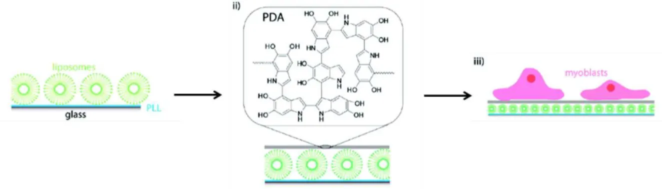
systems.55 Surfaces coated by poly(L-lysine) and liposomes were coated with polydopamine
(Figure 1.15). Polydopamine promotes cell adhesion and viability and depending on the thickness of the polydopamine layer, allows to control the drug release from lyposome.
Figure 1.15: Schematic illustration of adsorption of liposomes, on PLL precoated glass slides (i)
followed by the deposition of a polydopamine PDA layer (ii) and the adhesion of myoblast cells on these surfaces (iii).55
As mentioned previously, polydopamine coatings are extremely versatile platforms for secondary functionalization in order to confer diverse functions to a surface. As quinone moieties are reactive towards nucleophiles, biomolecules such as trypsin or growth factors (vascular endothelial growth factor (VEGF) and bone morphogenetic protein (BMP)) can be easily immobilized on polydopamine-coated surfaces.56-58 This was achieved for trypsin on
various types of surfaces.56 VEGF functionalized surfaces allowed to enhance human dermal
microvascular endothelial cells adhesion and viability making them suitable for bone healing
and regeneration applications.57 BMP functionalized TiO2 nanotubes via polydopamine
promoted cell growth, differentiation and mineralization with potential application for coated
titanium implants with bone regeneration properties.58 Neural interfaces are devices allowing
to monitor a neural signal and transform it into an electric signal that can be read by a computer. Microelectrode arrays of different types (gold, platinum, ITO…) were coated with
22
polydopamine to functionalize them first with poly-D-lysine and then with neural cells.59 These
neuron-adhesive modified-electrodes, allowed to record neural activity from the neuronal network making them suitable for neural interfaces applications.
In biomedicine, an Au/polypyrrole actuator was covered with polydopamine to enhance
bacterial adhesion and accumulation from physiological media.60Polydopamine was also used
as the basis for triazine-based polymers cross-linked through thioether linkages in order to
create a fiber-reinforced adhesive patch for fixation of bones fractures.61
As polydopamine appears to be biocompatible, it was used to encapsule yeast cells in order to protect them.62 In this case, the surface of the cell wall has the role of initiator of polydopamine coating by covalent bonding between polydopamine and amine or thiol functions of proteins present in the cell wall. The entrapped cells maintain their biological activity while being protected. Moreover by using the ability of polydopamine to be functionalized with organic molecules, coated cells were modified with streptavidin for specific immobilization on biotin-modified surfaces. Polydopamine was also used to coat magnetite nanoparticles in the presence
of a protein.63 By removing the protein with an acetic acid rinsing, a coating with the protein
print was created making these particles suitable for protein recognition. Recently, Woisel and coworkers used polydopamine as a platform for cyclodextrin-modified polymer immobilization
to create drug-release coatings on CoCr vascular stents64 and TiO2 implants.65 Polydopamine
occurs also to be a good reducer for silver ions. For example, fibrous structured bacterial cellulose was coated with polydopamine in order to incorporate silver nanoparticles to form an
antimicrobial network.66 Silver nanoparticles were also immobilized on cotton fabrics through
polydopamine cotton functionalization to create antibacterial textiles.67
The “dopamine concept” can be applied with other derivatives of dopamine as norepinephrine68
or catechol/amine synthetic polymers such as a copolymer from N-(3,4-dihyroxyphenethyl) methacrylamide and aminoethylmethacrylamide monomers which was used as a DNA
immobilization platform for DNA hybridization.69
Mussel-inspired layer-by-layer assemblies
Marine adhesive mimic polymers were also used in Layer-by-Layer (LbL) assemblies. Introduced by Decher, the LbL process consists in the exposure of a substrate alternatively to a polycation and polyanion solution with rinsing steps between each deposition step.70 The buildup process relies on the charge overcompensation observed at each deposition step. A marine adhesive mimic polymer was used in a LbL process in order to create hybrid composite
23
films with nacre structuration.71 These films were based on an alternate deposition of inorganic
nanometer-sized sheets of Montmorillonite clay and a Dopa-Lys-PEG polymer (Figure 1.16a). Fe(III) post treatment induced a cross-linking, and allowed to obtain a removable film with exceptional mechanical properties compare to the classic nacre inspired assembled coatings (Figure 1.16c). LbL assemblies were also realized with a positively charged copolymer bearing catechol moieties and colloidal synthetic layered silicate.72 These assemblies were used for anticorrosion applications: catechols are corrosion redox inhibitors and clay gives barrier properties to the films.
Figure 1.16: a) Molecular structure of Dopa-PEG-Lys polymer. b) Digital photograph of 300 bilayers
Dopa-PEG-Lys/Clay films on microscope glass slide with (left) and without (right) Fe(III) crosslinking. c) Digital photograph of a Fe(III) crosslinked 300 bilayers Dopa-PEG-Lys/Clay free-standing film.71
Mussel-inspired LbL assemblies were also notably used for antibacterial coating design. For example, catechol grafted poly(ethyleneimine) (PEI) and hyaluronic acid (HA) LbL allowed to
obtain well controlled assemblies on any kind of substrate, even polymeric ones.73 Using the
redox activity of the catechol moieties, silver nanoparticles were deposited on these coatings conferring them anti-microbial properties. Mussel-inspired LbL films containing silver nanoparticles for antimicrobial applications were also built with polystyrene sulfonate and a
24
Detrembleur and coworkers used quinone-grafted poly(methacrylate) with poly(allylamine hydrochloride) (PAH) allowing cross-linking between quinones and amine functions of the PAH ensuring a long time stability to the LbL film.75 Going further Nisin, an anti-bacterial peptide, was used instead of PAH creating a robust anti-bacterial coating.
Catechol-mediated anchoring for surfaces functionalization
As catechol moieties are known to interact with different type of surfaces, they were widely used as anchors for surface modification both for macroscopic planar surfaces, or for nanoscale surfaces of particles. In 2005, Messesmith and coworkers introduced a surface-initiated atom transfer radical polymerization (SI-ATRP) by using a biomimetic initiator.76 Starting from dopamine (1 in Figure 1.17), they designed a bifunctional molecule (2 in Figure 1.17) with a catechol on one side (mussel mimetism) for anchoring on the surface and an alkyl bromine to initiate ATRP (3 to 4 on Figure 1.17). The obtained surfaces were anti-fouling towards cell adhesion. By using a Ti substrate coated with pattern photoresist through standard photomask lithography, the biomimetic initiator was selectively anchored on the bare Ti area allowing to spatially control the SI-ATRP. More recently, PEG dendrimers with a catecholic anchor were
immobilized on TiO2 surfaces.77 The obtained films were less hydrated that the ones build from
linear PEGs and showing good non-fouling properties for proteins.
Figure 1.17: Synthesis and anchoring of the biomimetic initiator and subsequent SI-ATRP.76
Other bio-inspired antifouling surfaces were designed. An antifouling peptidomimetic polymer
was synthesized to be immobilized on metallic surfaces.78 It was composed of two parts: a short
functional peptide domain made of alternate Dopa and Lysine residues which allows anchoring of the polymer on a metallic surface and an N-substituted glycine oligomer of different size,
25
resistant to protein and cell adhesion. Saxer et al. designed a copolymer poly(L-lysine) grafted with 3,4-dihydroxypehenylacetic acid (DHPAA) and PEGs that was assembled in monolayer
on TiO2 surfaces.79 Immobilization operates via the catechol moiety of DHPAA (strong
binding) and lysine residues (long range electrostatic interactions) while bacterial and cellular adhesion were prevented by PEG chains. Dopamine modified surfaces via catechol anchoring
were used to further immobilize the tripeptide Arg-Gly-Asp (RGD) and collagen on a surface.80
These functional surfaces were used to study the effect of the type of adhesive ligand (RGD or collagen) on the bacterial and fibroblast adhesion in order to create a selective antifouling surface that inhibits bacterial activity while promoting cell adhesion.
Catechol-driven anchoring was used to design antimicrobial coatings. For example, a hybrid molecule was developed by Gademann and coworkers with an anachelin chromophore linked
to vancomycin through a PEG linker.81 The anachelin chromophore presents a catechol moiety
to immobilize the molecule on a surface and vancomycin is an antibiotic giving the desired antimicrobial property against Bacillus subtilis. Antimicrobial functional surfaces were also created by dip-coating of a surface in a solution of a copolymer synthesized by free-radical polymerization from dodecyl quaternary ammonium, catechol and methoxyethyl containing
monomers.82
Catechol-driven anchoring allows to functionalize not only macroscopic planar surfaces but also nano-scaled surfaces. Its ability to coordinate metals make it a suitable candidate for metallic nanoparticle functionalization. Magnetite nanoparticles (Fe3O4) were functionalized
with different catechol-modified PEG to improve their biocompatibility,83-84 catechol–modified
porphyrin for cancer therapy,85 catechol-conjugated bisphosphonate for uranyl ions removal
treatment in blood,86 catechol-anchoring mediated luminescent terbium complexes for imaging
applications,87 and dopamine-grafted biotin for the formation of heterodimers of magnetite and
silver nanoparticles.88 PEG functionalized magnetite nanoparticles were used as reducers of
non-specific uptake by cell macrophages.89 Catechol-stabilized iron oxide nanoparticles were
used as MRI contrast and anticancer agents.90 HA modified with dopamine was immobilized
on magnetite nanoparticles.91HA is known to interact with specific receptors giving a targeting
functionality to the coated particles. Dopamine-modified magnetite nanoparticles were used to create magnetic and transparent composites by covalently grafting it (through amine functions
of dopamine) in a polymethylhydrosiloxane network modified with crystal liquids.92 Dumbbell
nanoparticles are two nanoparticles of different chemical nature that are linked to each other. Magnetite-gold dumbbell nanoparticles were functionalized with an antibody through catechol
26
anchoring for magnetite and with PEG through thiol anchoring for gold.93 Through the antibody
functionality, these particles acted both as magnetic and optical probes. Recently, Sun and coworkers coupled a rhodamine derivative, N(rhodamine-6G)lactam-ethylenediamine (Rh6G-LEDA, 1a on Figure 1.18) to PEG-dopamine modified magnetite nanoparticles (1b on Figure 1.18).94 Rh6G-LEDA were bound to metal ions such as Fe(III) (1c on Figure 1.18) which enhanced greatly its fluorescence. Thus, these particles were used for Fe(III) sensing applications.
Figure 1.18: Structural illustration of Rh6G-LEDA (1a), its coupling with PEG-Fe3O4NP (1b), and its complex with Fe(III) (1c) along with the structural change that enhances its fluorescence property.94
TiO2 nanoparticles were functionalized with dopamine in a first step and then biotin in a second
step by covalent reaction of the valeric acid chain of biotin with the amine function of
dopamine.95 These modified TiO2 nanoparticles were deposited on an ITO electrode to further
immobilize avidin on the coated electrode. TiO2-entrapped Chlorella cells were also
functionalized through catechol chemistry.96 TiO2 nanorods were functionalized with a diblock
polymer with a catechol anchor to form hairy rod mesogens (Figure 1.19) which can be aligned under application of an electric field leading to liquid crystals formation.97
27
Figure 1.19: Buildup of defined diblock copolymers with an anchor and a soluble block for the
functionalization of TiO2 nanorods to form the hairy rod mesogen.97
Along the same idea, ZnO nanorods were functionalized with dopamine-modified
polystyrene.98 In order to functionalized MoS2, nanoparticles Tahir et al. synthesized a polymer
grafted with nitrilotriacetic (NTA) and dopamine groups acting as two different anchor
functions.99 A Ni atom act as an “anchoring ion”, it is partially coordinated by NTA on one side
and by the surface S atoms of the external part of the MoS2 nanoparticles. The modified nanoparticles, soluble in polar solvents, were immobilized through catechol groups of the ligand on surfaces to create hybrid materials with diverse functionalities. Gold nanoparticles were coated with dopamine-modified fluorescein-hyaluronic acid in order to probe reactive oxygen
species formation in macrophage cells.100 Indeed, reactive oxygen species induce cleavage of
HA chains and thus a fluorescence recovery signal which can be measured. Alumina nanoparticles were also functionalized with PEGs but through gallol anchoring instead of catechol, allowing to disperse them in a solution at higher concentrations.101 Recently, Hirsemann et al. modified kaolinite with catechol (covalent grafting) and a Ruthenium complex
(ion exchange).102 This bi-modification allows to enhance the difference in chemical nature of
the two opposited basal surfaces of kaolinite expressing the Janus character of the mineral. As it was recalled before, dopamine promotes reduction of graphene oxide into graphene.
28
Dopamine modified with an alkyne function on the amine side was used to reduce graphene oxide into graphene and further functionalize it through click reaction between alkyne and azide
functions (Cu(I) catalyzed)103or alkyne and thiol functions.104Azide-modified dopamine were
used for ZnO nanoparticle functionalization with alkyne-modified poly(3-hexylthiophene),105
magnetite nanoparticle functionalization,106 nanodiamond particle modification with
poly-N-isopropylacrylamide.107 Magnetite nanoparticles were also modified with maleimide-grafted
dopamine for functionalization through click reaction between maleimide and thiol functions.108
Catecholic polymers were also used as adhesive re-inforcers of carbon nanotube fibers.109 A
star-branched catechol-grafted (PEI) was infiltrated in the carbon nanotube fibers during the post spinning treatment. Then by evaporation of the solvent and curing at 120°C a final densification of the carbon nanotubes through the PEI-catechol was obtained. This process allows to produce carbon nanotubes fibers with greatly improved tensile and mechanical strength.
The mussel-inspired chemistry and the new materials described here are far from being
exhaustive. Ss some recent reviews give a more complete state of the art on this topic.110-111
1.1.2.3 Tannic acid coatings and hydrogels
Tannic acid (TA) is a polyphenol present in abundance in nature and easily accessible. Mankind used the properties of TA and others polyphenols for a very long time. Polyphenols are called “tannins”, a word that comes from the action “to tan”, a process already used by the antique greek civilization. This process consists in the use of natural substances issued from plants in order to turn animal skins into leather (Figure 1.20).
29
During the past decade, TA draw a great attention from the scientific community in the
nanoscience field. In 2005, Lvov and coworkers were the first to used TA in LbL assemblies.113
TA was used as negatively charged molecule in combination with two different polycations: poly(dimethyldiallylamine) and PAH in order to build nanofilms on planar surfaces. The interactions between TA and polycations are based on electrostatic interactions and hydrogen bonding. Two years later, it was shown that tea polyphenols interact with polymers via different types of interactions: hydrogen bonding, hydrophobic effect (Van der walls) and π-π stacking
depending on the chemical structure of the polymer.114
TA was used to create hydrogen-bonded LbL assemblies with neutral polymers. The resulted coatings were stable at high pH in contrary to others H-bond based LbL.115 Hammond and coworkers exploited this ability to create LbL assemblies through H-bonding to design a drug
delivering surface.116 Block copolymer micelles of poly(ethylene
oxide)-block-poly(2-hydroxyethylemethacrylate) were conjugated with a chemotherapeutic agent: doxorubicin. These micelles were assembled with TA at pH=7.4 through H-bonding mediated LbL assembly (Figure 1.21). The carbamate linkage between doxorubicin and the polymer backbone is pH sensitive and is cleaved at acidic pHs. Thus when micelles film were brought in contact with an acidic medium, doxorubicin release was triggered. Doxorubicin was also entrapped in
TA-poly(N-vinlypyrrolidone) (PVP) H-bonded multilayer capsules.117
Figure 1.21: Schematic representation of hydrogen-bonding LbL assembly of doxorubicin-containing
micelles with TA.116
Hammond and co-workers showed that as other H-bond based LbL assemblies, TA-containing LbL assemblies can also be dissolved electrochemically when deposited on electrodes by
electrochemically producing OH- ions.118 Oxygen reduction at the coated-electrode leads to the
30
triggers the dissolution of the film. H-bonded TA LbL were also realized with
poly(N-isopropylacrylamide) and poly(2-isopropyl-2-oxazoline).119 TA/thrombin LbL were deposited
onto porous gelatin sponges which were shown to promote hemostasis making these functional
materials potential coatings to prevent hemorrhage.120 Hydrogen-bonded TA LbL assemblies
were built on silica microparticles in order to create microcapsules by removing the silica.121
The permeability of the resulting capsules was pH responsive and thus could be tuned, making them suitable for the loading or the releasing of functional molecules. The mechanical
properties of these capsules were studied in dry and liquid states.122 They had an elastic modulus
of 700 MPa in dry state and around 0.5 MPa when swollen. The value in liquid could be enhanced and tuned by adding a pre-layer of PEI to the construction and by using a PVP of
greater molecular weight. PVP/TA LbL films were deposited on cell surfaces.123 These coatings
are biocompatible and the entrapped cells showed good viability. Moreover, gallol groups of tannic acid allowed surface immobilization of the cells.
In 2013, Caruso and coworkers introduced a new concept: the one-pot assembly of tannic acid/Fe(III) films and capsules.124 As catechol moieties, gallols coordinate metals such as Fe(III) in a pH dependent manner. Thus by mixing Fe(III) and TA in solution, dipping any substrate (planar surface, microparticles) in this solution and then increasing the pH, a film of TA cross-linked by Fe(III) is assembled on the surface (Figure 1.22). As TA has an affinity with various types of materials, this process is versatile and can be applied to any kind of substrate. The concept was extended to others metallic ions such as aluminum, vanadium,
chromium, cobalt…125 and to small phenolic molecules: gallic acid, pyrogallol and
pyrocatechol.126 As mono-complexes are formed at acidic pH, these capsules can be
diassembled in acidic medium. TA/Fe(III) capsules are permeable to macromolecules. Thus, it is possible to load them with drugs and then diassemble them at acidic pH for drug release
31
Figure 1.22: Schematic illustration of the one-step assembly of coordination complexes on various types
of substrates.128
LbL were developed based on the same principle.129In that case, Fe(III) and TA are not mixed
together in solution and the deposition occurs by dipping a substrate alternatively in TA and Fe(III) solutions resulting in a layered organized coating compared to the randomly distributed one-step coating. Recently, Jia and coworkers studied the influence of different parameters on
the behavior of TA/Fe LbL films.130 They showed that the order of addition of TA and Fe(III)
greatly changes the film growth. When NaOH is added to the mixture, no significant effect on the thickness of the films was observed. However, the morphology of the films changes going from large aggregates to smaller ones, and the stability of the films was greatly improved. This effect is attributed to the formation of tris-complexes (ratio Fe/gallol: 1/3). Finally, they observed that surfaces with low wettability promote film formation due to hydrophobic effect between the surface and TA. Another study showed that when films are assembled at pH 5, the interaction between Fe(III) layers and TA layers is dominated by electrostatic interactions and not metal-ligand interactions.131
TA/Fe(III) were mixed with graphene oxide to improve the late one’s mechanical properties.132
Indeed, this process creates cross-links between the graphene oxide sheets, enhancing their cohesion. TA/Fe(III) films were also deposited on living cells.133 The films present good cytocompatibility, and cytoprotection towards UV-irradiation, bacterial binding and silver nanoparticles. Moreover, the shell can be easily removed (Figure 1.23) if needed as TA/Fe(III) are easily degradable.
32
Figure 1.23: Schematic representation for controlled formation and degradation of the TA–Fe(III)
shell on individual S. cerevisiae.133
A melamine sponge was also functionalized with TA/Fe(III) films.134 Free gallol groups were
used for a second step functionalization of the sponge with 1-dodecanethiol through covalent bonding between thiol and oxidized catechol conferring superhydrophobic and fire-resistant properties to the material.
Tannic acid was also used to create 3D gels, by mixing it with Fe(III) and PAH.135 The process
is very similar to the one introduced by Messersmith in 2011 (see above).42 All three compounds
are mixed at acidic pH and the mixture is then brought to more basic pH by adding NaOH. As mentioned before, basification of the solution triggers formation of bis- and tris-gallol/Fe(III) complexes, but also enhances catechols oxidation into quinones which further react on the amine functions of PAH. Thus, the resulting 3D-network is composed of PAH chains covalently cross-linked by TA while TA itself is cross-linked by Fe(III). TA is mixed only with PAH or
Fe(III), no gel can be obtained. DNA-TA gels can also be designed.136 Indeed by simply adding
TA to a DNA solution, a hydrogel is spontaneously formed by cross-linking of DNA by TA through hydrogen bonding (Figure 1.24). These gels can be dissolved by TA’s ester bond
33
hydrolysis at basic pH, creating a DNA release. TA was also used as an intermolecular
cross-linker through hydrogen bonds in PEG hydrogels.137
Figure 1.24: Schematic description of the formation and degradation of DNA-TA hydrogels.
Polyphenols, in particular TA, can form coatings.138 The principle is quite simple and consists
in the dip coating of a substrate in a solution of polyphenol during several hours without stirring followed by a rinsing step. TA, pyrogallol, epicatechin gallate, epigallocatechin gallate and epigallocatechin, as well as crude extracts of red wine, cacao bean and green tea, were used on various kinds of substrates (metals, polymers etc) and with different shapes (planar surface, particles) (Figure 1.25). Even if the mechanism is not fully understood, it seems to occur through self-polymerization of polyphenols by radical reaction between oxidized phenolic moieties. These coatings allow to immobilize PEG chains for antifouling applications and metals for plasmonic tuning applications. The redox properties of polyphenols can also be used
for the design of anti-oxidant coatings.138 It appears that not all polyphenols can form coatings
34
Figure 1.25: Schematic illustration of compositions, deposition conditions and potential applications
of plant polyphenol inspired multifunctional coatings.138
Other polyphenols were used to create functional materials. For example, epigallocatechin-3-gallate (EGCG) was grafted on collagen fibers used as support for palladium nanoparticles
through catechol-metal complexation, creating a catalytic network of collagen.140 More
recently, EGCG was used to create assemblies for cancer therapy.141 More precisely, Herceptin
(an anticancer protein) forms nano-micelles through complexation with EGCG. This core was functionalized with EGCG-modified PEG chains to form a protective shell. Herceptin carriers showed greater anticancer impact than the protein alone. Tea polyphenol were also used to
create hybrid metal-organic nanostructures.142 To do so, tea polyphenols were directly extracted
from tea leaves, and were mixed with either silver or gold ions. Self-assembly was then triggered by microwave-assisted oxidation leading to covalent cross-linking between tea polyphenols. The resulting nanostructures were made of metallic core and self-polymerized tea polyphenols around the metallic core. Ag/tea polyphenols structures display good antibacterial activity without expressing cell toxicity.