HAL Id: tel-00843015
https://tel.archives-ouvertes.fr/tel-00843015
Submitted on 10 Jul 2013
HAL is a multi-disciplinary open access archive for the deposit and dissemination of sci-entific research documents, whether they are pub-lished or not. The documents may come from teaching and research institutions in France or abroad, or from public or private research centers.
L’archive ouverte pluridisciplinaire HAL, est destinée au dépôt et à la diffusion de documents scientifiques de niveau recherche, publiés ou non, émanant des établissements d’enseignement et de recherche français ou étrangers, des laboratoires publics ou privés.
Development and characterization of a bi-functional
vector for cancer targeting
Christiane Wenk
To cite this version:
Christiane Wenk. Optical-guided surgery of the feline fibrosarcoma & Development and characteri-zation of a bi-functional vector for cancer targeting. Agricultural sciences. Université de Grenoble, 2012. English. �NNT : 2012GRENV063�. �tel-00843015�
Université Joseph Fourier / Université Pierre Mendès France / Université Stendhal / Université de Savoie / Grenoble INP
THÈSE
Pour obtenir le grade de
DOCTEUR DE L’UNIVERSITÉ DE GRENOBLE
Spécialité : Chimie et Biologie Arrêté ministériel : 7 août 2006
Présentée par
« Christiane Hanna Frederike WENK »
Thèse dirigée par « Jean-Luc COLL » et « Pascal DUMY »
codirigée par « Didier BOTURYN » et « Véronique
JOSSERAND »
préparée au sein du Laboratoire Institut Albert Bonniot INSERM/UJF U823 & Département Chimie Moléculaire UJF-CNRS UMR5250
dans l'École Doctorale Chimie et Science du Vivant
CHIRURGIE
GUIDEE
PAR
FLUORESCENCE
DU
FIBROSARCOME
FELIN
ET
DEVELOPPEMENT
ET
CARACTERISATION D’UN VECTEUR
BI-FONCTIONNEL POUR LE CIBLAGE DU
CANCER
Thèse soutenue publiquement le « 17 décembre 2012 », devant le jury composé de :
M Jürgen, BORLAK
Professeur, Hanovre, Rapporteur M Gilles, SUBRA
Professeur, Montpellier, Rapporteur Mme Frédérique, PONCE Professeur assitant, Lyon, Membre Mme Anne, IMBERTY
Professeur, Grenoble, Membre M Jean-Luc, COLL
Directeur de recherche, Grenoble, Membre M Pascal, DUMY
University of Grenoble
Ecole Doctorale Chimie et Sciences du Vivant
THESIS
For obtaining the degree
DOCTOR OF THE UNIVERSITY OF GRENOBLE
Specialty: PhD in Chemistry - Biology
Public defended by
Christiane Hanna Frederike WENK
The 17th of December 2012
Optical-guided surgery of the feline fibrosarcoma
&
Development and characterization of
a bi-functional vector for cancer targeting
Committee members:
Professor Jürgen BORLAK, Hannover Reporter
Professor Giles SUBRA, Montpellier Reporter
Doctor Frédérique PONCE, Lyon Examiner
Professor Anne IMBERTY, Grenoble Examiner
Doctor Jean-Luc COLL, Grenoble Thesis director
I
R
EMERCIEMENTS
Au terme de ma thèse, je tiens à adresser mes remerciements à tous ceux qui ont contribué de près ou de loin à l’aboutissement de ce travail.
Je remercie,
Mes directeurs et co-directeurs de thèse, qui ont dirigé ces travaux et m’ont permise de découvrir le monde de la recherche. Un grand merci pour les critiques et corrections de ce manuscrit. Veuillez trouver ici l’assurance de mon profond respect.
Je remercie
Jean-Luc Coll pour m’avoir acceptée et accueillie dans son équipe, étant vétérinaire et débarquée d’Allemagne.
Merci pour ton encadrement, ta bonne humeur, ta patience, le soutien que tu m’as apporté, avec une humanité pareille…..
Le Professeur Pascal Dumy, pour m’avoir accueillie dans son laboratoire, d’avoir dirigé ces travaux et d’avoir rendu une thèse en cotutelle possible.
Didier Boturyn, pour m’avoir encadrée et fait découvrir les mystères de la synthèse peptidique, conseillée avec une grande disponibilité ainsi que pour sa confiance tout au long de mes travaux. Merci pour ta patience et pour toute l’aide que tu m’as apportée.
Véronique Josserand, pour sa disponibilité et le partage de son savoir, surtout en imagerie. Je te remercie pour ta compréhension et ton aide.
I thank Professor Jürgen Borlak and Professor Gilles Subra for giving me the honor of judging my work as reporters. Find within these words the expression of my truly gratefulness.
I thank Professor Anne Imberty and Frédérique Ponce for accepting to evaluate my work as members of my thesis committee. Therefore, I express my sincere thankfulness.
Je remercie Florence Ruggiero et Pierre Labbé pour leur temps et leurs conseils comme membres de mon comité de suivi de la thèse.
Je remercie Frédérique Ponce, encore une fois, et Claude Carozzo, pour leur collaboration dans les études « chat ». C’était un plaisir de faire avancer un tel projet avec vous. Cela m’a apporté beaucoup plus qu’une expérience scientifique.
Merci aux membres de leurs équipes pour leur participation fructueuse à ce projet, je tiens particulièrement à remercier Dorothée pour son aide et sa disponibilité.
Dans ce contexte je tiens à remercie Odile Allard et Phillipe Rizo de Fluoptics, pour leur collaboration, leurs réflexions et leur confiance tout au long cette étude. Particulièrement je remercie Stéphanie pour son soutien et sa présence dans ce projet. Je te remercie pour les discussions, les jours de « ouf » inoubliables et ta bonne humeur.
Merci à tous les membres des équipes 2 et 5 de l’Institut Albert Bonniot.
A mes collègues de bureau, Julien V, Julien G, Maxime, Mélanie, Jonathan, Daphné et Victor, Sandrine et Anastassia, Benoit et Lucie, et à ceux des bureaux voisins, Amandine, Claire, Michelle, Morgane, Laetitia,
II
Sylvie, Béatrice, Asma, Delphine, Céline, Pascal, Sana, Gabriele, Ihab, Blaise, Floriane, Sophie, Tudor et Maxime. Merci à vous tous pour votre gentillesse, votre bonne humeur, votre « follitude », vos conseils et votre aide…. Entre autre j’ai appris les mots les plus importants en français grâce à vous, en tombant dans tous les pièges de cette langue vive. C’était un plaisir d’être membre de « cette équipe » magnifique !
Egalement un grand merci à Christian, Mylène et Alexeï pour les heures passées ensemble à faire marcher les appareils fluos!
Je ne saurais pas oublier les gens indispensables, Denise, Dalenda, Michaelis, Jamila et Johanna !
Et puis un grand merci à Coco, au CHU maintenant, pour son travail et sa gentillesse tout au long de ces travaux.
Tous les membres de l’équipe « Ingénierie et Interactions Biomoléculaires » pour leur serviabilité, leur bonne humeur et tout spécialement aux étudiants et techniciennes qui ont participées de manière très active à rendre tous les jours au DCM agréables. Merci à mes collègues et amis Corinne, Audrey, Isabelle et Abdel, et à Emilie, Romaric, Pierre, Mathieu, Julien, Nathalie, Julien G, Rémy et Cédric. Mais également à Régine et Véronique.
Plus particulièrement je remercie Sandrine et Anastassia, qui représentent beaucoup plus « que » des collègues, mais des amies formidables. Merci pour les moments passés ensembles, pour votre soutien, vos sacrifices, votre présence, vos « services de taxi » dans les pires moments,… votre partage de savoir-faire mais surtout des bons et mauvaises moments, nos rigolades, nos galopades….merci pour tout ! Et je ne pourrai pas oublier mon amie Asma, avec qui j’ai découvert le monde arabo-andalou. Merci pour tes écoutes, les discussions et le temps des « h’s» et beaucoup plus…. !
J’adresse également mes remerciements aux personnes que j’ai rencontrées au cours de ma thèse et qui l’ont rendue par leurs présences, discussions et sourires, très plaisante. Plus particulièrement je m’adresse à Sebastian A et Agnès qui ont assuré aux derniers moments, Monsef, Ester, Houda, Thanh, Vietnam et Noël. Merci pour votre amitiè.
Danke an Aliki und Christina, die mir seit Jahren teure Freundinnen sind.
L’Université Joseph Fourier, pour avoir financé mes travaux de recherche durant trois ans.
Un dernier grand merci aux personnes qui m’ont encouragée et soutenue dans mon parcours, espérant de n’avoir pas oublié quelqu’un. Qu’il me pardonne.
Ein großes Dankeschön an meine Familie, für Ihre Unterstützung in jeder Hinsicht.
Danke an meine Eltern, die immer für mich da sind, immer ein offenes Ohr haben und mir ermöglicht haben, meinen Weg trotz aller Obstakel zu meistern. Danke für all die Erfahrung, Ratschläge, Taten und vor allem Liebe, die ihr mir gegeben habt.
Danke an meine Brüder, Florian und Bastian, ohne die meine Familie nie komplett wäre und natürlich an Sonja, die kleine Sophia und den süßen Felix, Onkel Charly und Karla.
Enfin, j’aimerais bien laisser une pensée en mémoire de Sandrine, une amie rayonnante, avec qui j’adorais rêver de projets futurs et avec qui j’ai partagé ma passion : le cheval. Tu me manques.
III
P
REFACE
This work has been accomplished in co-tutelle during the last three years of my PhD studies in team 5 “Cancer Targets and Experimental Therapeutics” of the research center INSERM/UJF U823 in the Institute Albert Bonniot, and in the team “Engineering and Interactions of BioMolecules (I2BM)” of the research unity UMR CNRS-UJF 5250 in the facilities of the Department of Molecular Chemistry at Grenoble. It was realized under the supervision of Jean-Luc Coll and Pascal Dumy, and co-supervised by Didier Boturyn and Véronique Josserand.
The studies on cats have been realized in collaboration with the Veterinarian University of Lyon (VetAgro-SUP) at Marcy l’Etoille.
This work gave reason for the following publications and presentations:
PUBLI CATI ONS
articles
Keramidas, M; Josserand, V.; Righini, C.; Wenk, C.H.F.; Faure, C; Coll, J.L. 2010. “Intraperoperative near-infrared image-guided surgery for peritoneal
carcinomatosis in a preclinical experimental model.” British Journal of Surgery, Vol 97; ISSUE 5; pages 737-743.
[Presented in: APPENDICES]
Christiane H F Wenk & A. Briat, Mitra Ahmadi, Michael Claron, Didier Boturyn, Véronique Josserand, Pascal Dumy, Daniel Fagret, Jean-Luc Coll, Catherine Ghezzi, Lucie Sancey, Jean-Philippe Vuillez. 2012. “Reduction of renal uptake of 111In-DOTA-labeled and A700-labeled RAFT-RGD during integrin αvβ3 targeting using single photon emission computed tomography and optical imaging“. Cancer science, 103, Nr. 6: 1105–1110.
[Presented in: RESULTS & CONCLUSIONS, chapter II]
Pascal Dumy, Christiane Wenk, Michael Claron, Jean-Luc Coll, Bénédicte Allard, Didier Boturyn 2012. « Des molécules fluorescentes au service de la chirurgie. »
L'Actualité Chimique, N°366 Pagination : 23-28
[Presented in: APPENDICES]
Christiane H. F. Wenk, Véronique Josserand, Stéphanie Guillermet, Corinne Tenaud, Didier Boturyn, Pascal Dumy, Dorothée Watrelot-Virieux, Claude Carozzo, Frédérique Ponce and Jean-Luc Coll 2012. “Near-infrared optical guided surgery of highly infiltrative fibrosarcomas in cats using an anti-alpha v ß3 integrin molecular probe.” Cancer letters - doi:10.1016/j.canlet.2012.10.041
IV
Christiane H. F. Wenk, Pascal Dumy, Véronique Josserand, Jean-Luc Coll and Didier Boturyn 2012. “Integrin and Matrix Metalloprotease Dual-Targeting with MMP substrate-RGD conjugate.” Organic & Biomolecular Chemistry 11, pages 448–452
[Presented in: RESULTS & CONCLUSIONS, chapter III]
review
Dufort, S.; Sancey, L.; Wenk, C.; Josserand, V., Coll, J.L. 2010. “Optical Small Animal Imaging in the Drug Discovery Process” Biochim. Biophys. Acta Biomembrans, 8. doi:10.1016/j.bbamem.2010.03.016
[Presented in: APPENDICES]
POSTER COMMUNIC ATION
NEAR-INFRARED GUIDED SURGERY OF CANCER: FROM MODELS TO CLINICAL CASES
Wenk C; Keramidas M; Josserand V.; Righini C. A.; Faure C.; Boturyn D.; Rizo P; Dumy P and Coll JL.
442. WE-Heraeus-Seminar “Molecular Imaging”, October 2009, Bad Honnef, Germany
NEAR-INFRARED GUIDED SURGERY OF CATS AND DOGS
Wenk C; Keramidas M.; Josserand V.; Righini C. A.; Faure C; Boturyn D.; Rizo P.; Dumy P. and Coll JL
5èmes Journées Scientifiques du CLARA ; March 2010, Villeurbanne, France
IMPROVING TUMOR SURGERY AND DEVELOPING CANCER-TARGETED THERAPY BY MEANS OF REAL TIME FLUORESCENCE IMAGING
Wenk C and Keramidas M, Josserand V, Righini C, Boturyn D, Dumy P and Coll JL Journée pour la Recherche Respiratoir (J2R), Nantes 2010, France
SYNTHESES ET EVALUATIONS DE VECTEURS PEPTIDIQUES ACTIVABLES POUR L’IMAGERIE TUMORALE
Wenk C H F, Josserand V, Coll JL, Dumy P, Boturyn D
17ième congrès du groupe français des peptides et des protéins (GFPP); February 2011, Aussois, France
NEAR-INFRARED IMAGING OF CANCER: OPTICAL GUIDED SURGERY AND SMART PROBES DEVELOPMENT
Wenk C H F, Josserand V, Guillermet S, Ponce F, Dumy P, Boturyn D, Coll JL 1er Journée Scientifique du médicament ; June 2011, Grenoble, France
ACTIVATABLE-TARGETING-PROBES FOR NEAR-INFRARED IMAGING OF CANCER Wenk C H F, Josserand V, Dumy P, Boturyn D, Coll JL
Cancer Microenvironment - XIth Topic Day Institut Albert Bonniot; October 2011, Grenoble, France
SMART-PROBES FOR NEAR-INFRARED IMAGING OF CANCER Wenk C H F, Josserand V, Dumy P, Boturyn D, Coll JL
Journées Thématiques GDR « Imageries in vivo » (IMAGIV) - Des nouveaux concepts en imagerie àl’application clinique; December 2011, Paris, France
V
SMARGETING PROBES FOR OPTICAL IMAGING OF CANCER Wenk C H F, Josserand V, Dumy P, Boturyn D, Coll JL
7th Cancer Scientific Forum of the Canéropôle CLARA, March 2012, Lyon, France
OPTICAL-GUIDED SURGERY OF FIBROSARCOMA ON CAT PATIENTS. A VETERINARY CLINICAL STUDY
Wenk C H F, Josserand V, Guillermet S, Tenaud C, Watrelot-Virieux D, Boturyn D, Carozzo C, Ponce F, Coll JL
World Molecular Imaging Congress (WMIC), September 2012, Dublin, Ireland
SMART PROBES FOR OPTICAL IMAGING OF CANCER Wenk CH. F.,Josserand V, Dumy P, Boturyn D, Coll JL
World Molecular Imaging Congress (WMIC), September 2012, Dublin, Ireland
ORAL C OMMUNICA TION
Near-infrared guided surgery of cancer: from models to clinical cases
Wenk C H F; Keramidas M; Josserand V.; Righini C. A; Boturyn D.; Rizo P.; Dumy P. and Coll JL
Hot Topics in Molecular Imaging (TOPIM) – ESMI winter conference “Emerging imaging methods in medicine” - January 2011, Les Houches, France
Smartprobes for near-infrared imaging of cancer
Wenk CH. F.,Josserand V, Dumy P, Boturyn D, Coll JL
Comité de these, Institut Albert Bonniot, December 2010, Grenoble, France
Smart-probes for optical imaging of cancer and optical guided surgery of cats
Wenk CH. F.,Josserand V, Dumy P, Boturyn D, Coll JL
Comité de these, Institut Albert Bonniot, October 2011, Grenoble, France
Optical guided-surgery of the feline fibrosarcoma & design of SMARgetting probes
Wenk CH. F.,Josserand V, Dumy P, Boturyn D, Coll JL
Comité de these, Institut Albert Bonniot, June 2012, Grenoble, France
HONOURS A ND AWARDS
Best communication/poster award:
Wenk C; Journée pour la Recherche Respiratoir (J2R), Nantes 2010, France
Wenk C H F; Cancer Microenvironment - XIth Topic Day Institut Albert Bonniot; October 2011, Grenoble, France
Wenk C H F; World Molecular Imaging Congress (WMIC), September 2012, Dublin,
VII
T
ABLE
O
F
C
ONTENTS
INTRODUCTION _______________________________________________________________________________ 1
PREAMBLE ________________________________________________________________________________ - 1 - I. Tumorigenesis ______________________________________________________________________ - 3 - I.1. Characteristics of tumorigenesis ____________________________________________________ - 3 - II. Angiogenesis _______________________________________________________________________ - 7 - II.1. Integrins _____________________________________________________________________ - 10 -
II.1.1. Integrin composition __________________________________________________ - 10 - II.1.2. Integrin conformation & substrate recognition ______________________________ - 12 - II.1.3. Integrin turn-over ____________________________________________________ - 13 - II.1.4. Roles of integrin _____________________________________________________ - 13 - II.2. The integrin αvβ3 ______________________________________________________________ - 15 - III. Proteases and cancer ________________________________________________________________ - 17 - III.1. Matrixmetalloproteases (MMPs) __________________________________________________ - 19 -
III.1.1. MMP structure ______________________________________________________ - 19 - III.1.2. MMP tuning ________________________________________________________ - 21 - III.1.3. Role of MMPs in angiogenesis __________________________________________ - 22 - III.1.4. Gelatinases and solid cancers ___________________________________________ - 22 - III.1.5. The controversial role of gelatinases in tumorigenesis ________________________ - 24 - IV. Integrin αvβ3 and Gelatinases __________________________________________________________ - 27 -
IV.1. Integrin-Gelatinases interactions __________________________________________________ - 27 - IV.1.1. Integrin-Gelatinases interaction mediated through SIBLINGs __________________ - 27 - IV.1.2. Integrin-Gelatinases interaction mediated through PEX binding ________________ - 28 - IV.1.3. Other interaction mechanisms of integrin and Gelatinases_____________________ - 28 - V. Molecules for targeting and optical imaging of cancer ______________________________________ - 31 -
V.1. Choice of the Near-infrared window for in vivo optical imaging __________________________ - 31 - V.2. Basic principles of Fluorescence __________________________________________________ - 34 -
V.2.1. Extinction coefficient (EC) and quantum yield (QY) _________________________ - 35 - V.2.2. Quenching and FRET _________________________________________________ - 35 - V.3. Imaging probes for NIR imaging of cancer __________________________________________ - 40 -
V.3.1. Non-specific agents __________________________________________________ - 40 - V.3.2. Specific agents ______________________________________________________ - 40 - V.3.3. Double targeting _____________________________________________________ - 47 - V.3.4. The integrin ligand RGD ______________________________________________ - 48 - V.3.5. Integrin αvβ3 and the RGD _____________________________________________ - 48 -
VI. Peptidic molecular probes for cancer targeting – the MMP smart approach ______________________ - 54 - VI.1. MMP Inhibitors (MMPIs) _______________________________________________________ - 54 -
VI.1.1. Natural inhibitors – TIMPs _____________________________________________ - 54 - VI.1.2. Synthetic inhibitors ___________________________________________________ - 56 - VI.2. MMP and “Smart” contrast agents and therapeutics ___________________________________ - 59 -
VI.2.1. Drug delivery _______________________________________________________ - 61 - VII. Imaging and cancer _________________________________________________________________ - 63 -
VII.1. General presentation of the methods used in Medical Imaging ___________________________ - 63 - VII.1.1. X-ray computed tomography (CT) _______________________________________ - 64 - VII.1.2. Magnetic resonance imaging (MRI) ______________________________________ - 65 - VII.1.3. Ultrasonography (US) ________________________________________________ - 66 - VII.1.4. Nuclear imaging: PET and SPECT ______________________________________ - 66 - VII.2. Optical Imaging _______________________________________________________________ - 68 - VIII. Imaging and surgery ________________________________________________________________ - 72 - VIII.1. Surgical approaches in cancer __________________________________________________ - 73 - IX. NIR Fluorescence imaging for intra-operative guidance _____________________________________ - 78 -
RESULTS AND CONCLUSIONS _________________________________________________________________ 85
VIII
I.1. Preliminary safety observation studies ______________________________________________ - 90 - I.1.1. Introduction ________________________________________________________ - 90 - I.1.2. Results ____________________________________________________________ - 91 - I.2. Fluorescecnce kinetic distribution of AngioStamp _____________________________________ - 92 -
I.2.1. Fluorescence in the plasma _____________________________________________ - 93 - I.2.2. Fluorescence in the blood ______________________________________________ - 94 - I.2.3. Fluorescence in the urine ______________________________________________ - 95 - I.2.4. Fluorescence in the kidney _____________________________________________ - 96 - I.2.5. Fluorescence in the shoulder ___________________________________________ - 97 - I.3. Conclusion and Discussion _______________________________________________________ - 98 - I.4. Surgery of the feline fibrosarcoma _________________________________________________ - 99 -
I.4.1. Surgery of group 6 __________________________________________________ - 133 - I.4.2. Dealing with the autofluorescence and “auto-LUT” during surgery ____________ - 134 - I.4.3. Good positioning of the camera during surgery ____________________________ - 135 - I.5. General conclusion ____________________________________________________________ - 136 - II. GELOFUSIN STUDY ________________________________________________________________ - 137 - III. DESIGN &CHARACTERIZATION OF ACTIVATABLE PROBES FOR CANCER IMAGING & THERAPY ____ - 147 - III.1. Results and discussion _________________________________________________________ - 149 -
III.1.1. MMP-2 AND MMP-9 CLEAVAGE TEST – SEQUENCE SELECTION ________________ - 149 - III.1.2. Synthesis of activatable probes _________________________________________ - 152 - III.1.3. Synthesis of final compound (RAFT-RGD-MMP) _________________________ - 154 - III.2. Fluorescent characteristics and cleavage properties of the RAFT-RGD-MMP ______________ - 155 -
III.2.1. Conclusion ________________________________________________________ - 167 - III.3. In vitro characterization ________________________________________________________ - 169 -
III.3.1. Analysis of the RAFT-RGD-MMP binding on cells using flow cytometry _______ - 170 - III.3.2. Analysis of the RAFT-RGD-MMP binding on cells using confocal microscopy __ - 182 - III.3.3. Conclusion and discussion ____________________________________________ - 183 - III.4. Double targeting effect in vivo ___________________________________________________ - 184 -
III.4.1. Results ___________________________________________________________ - 184 - III.4.2. Conclusion ________________________________________________________ - 189 - III.5. General conclusion ____________________________________________________________ - 190 - III.6. Supplementary data RAFT-RGD-MMP ____________________________________________ - 191 -
III.6.1. Enzyme activity control and inhibitor test ________________________________ - 191 - III.6.2. MMP-2 and MMP-9 expression of different cell lines _______________________ - 192 - III.6.3. Flow cytometry analysis of the integrin αvβ3 expression _____________________ - 194 -
III.6.4. Level of autofluorescence of mouse organs ex vivo _________________________ - 195 -
DISCUSSION AND PERSPECTIVES ____________________________________________________________ 199
I. Near-infrared fluorescence guided surgery in the feline fibrosarcoma _________________________ - 201 - I.1. Feasibility of NIRF-guided surgery in veterinary clinic conditions _______________________ - 201 - I.2. Continuation of the work _______________________________________________________ - 202 - I.3. RGD-motif, integrin and the study ________________________________________________ - 203 - I.4. Clinical set-up of instrumentation ________________________________________________ - 204 - I.5. Surgeon’s impression __________________________________________________________ - 206 - II. Reduction of renal retention of RAFT-RGD by pre-injection of gelofusin ______________________ - 208 - III. Introduction of a bi-functional RAFT-RGD derivative: the RAFT-RGD-MMP __________________ - 210 - III.1. Additional properties of the activatable dual-molecule ________________________________ - 211 - III.2. In vivo studies _______________________________________________________________ - 212 - III.3. Possible complementary studies __________________________________________________ - 213 - III.4. Double-targeting approach with other enzymes ______________________________________ - 216 - III.5. SMARgeting molecule as prodrug and theranostic ___________________________________ - 216 - [Don’t worry about progressing slowly, but only about standing still.] _____________________________ 221
MATERIALS AND METHODS __________________________________________________________________ 223
I. Preliminary safety observation studies __________________________________________________ - 225 - I.1. Animals ____________________________________________________________________ - 225 - I.2. Fluorescent tumor-targeting probe ________________________________________________ - 225 - I.3. General examination of the cats __________________________________________________ - 225 - I.4. Physical and biochemical parameters ______________________________________________ - 225 - I.5. Calculation for the dose 1X for cats _______________________________________________ - 226 -
IX
I.6. Image analysis and unit definition ________________________________________________ - 227 - I.7. Portable clinical fluorescence imaging device _______________________________________ - 228 - II. Intra-operative surgery ______________________________________________________________ - 228 - II.1. Patient recruitment ____________________________________________________________ - 228 - II.2. Fluorescent tumor-targeting probe ________________________________________________ - 229 - II.3. Surgical fibrosarcoma resection and imaging ________________________________________ - 229 - II.4. Histo-pathological and immuno-histological analyses _________________________________ - 229 - II.5. Analysis of NIR fluorescence data ________________________________________________ - 230 - II.6. Statistical analysis ____________________________________________________________ - 230 - III. Protocols for peptide synthesis________________________________________________________ - 231 - III.1. Material and Equipment ________________________________________________________ - 231 - III.2. General Peptide synthesis procedures _____________________________________________ - 231 - III.3. Compounds __________________________________________________________________ - 236 - IV. Cleavage studies of linear peptide sequences ____________________________________________ - 245 - V. Cell culture _______________________________________________________________________ - 246 - VI. Fluospectrometry __________________________________________________________________ - 247 - VII. MMP -2 and -9 expression study ______________________________________________________ - 248 - VIII. Flow cytometry analysis of integrin αvβ3 expression _______________________________________ - 249 -
IX. Cell – molecule-interaction __________________________________________________________ - 249 - IX.1. Flow cytometry analysis (FACS) of the molecule – cell interaction ______________________ - 249 - IX.2. Fluorescence microscopy analysis of the molecule – cell interaction _____________________ - 250 - X. In vivo fluorescence imaging _________________________________________________________ - 251 -
APPENDICES _______________________________________________________________________________ 255
I. Supplementary Data – Preliminary Safety Observation Study In Cats _________________________ - 257 - II. Table Of Amino Acids ______________________________________________________________ - 260 - III. Supplementary - Articles ____________________________________________________________ - 261 -
XI
I
NDEX OF
F
IGURES
INTRODUCTION
Figure I-1: Stages of tumorigenesis. 4
-Figure I-2: The Cells of the Tumor Microenvironment. 6
-Figure II-1: Blood vessel formation. 7
-Figure II-2: Tumor development and angiogenesis. 9
-Figure II-3: The members of the human integrin superfamily and how they combine
to form heterodimeric integrins. 11
-Figure II-4: The extracellular region of a human integrin. 11 -Figure II-5: Intergin formation: A) inactive, bend form; B) active form 12
-Figure II-6: Roles of the αvβ3 integrin in angiogenesis. 16
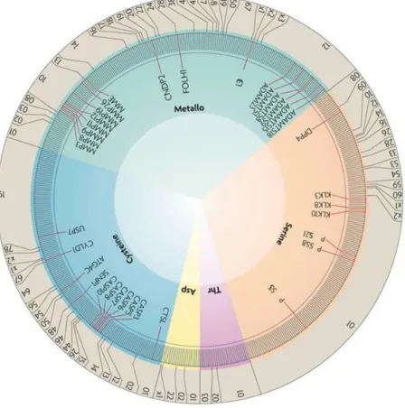
Figure III1: Distribution of tumourprotective proteases in the human degradome. 18
-Figure III-2: Table: Classes of the MMP family 19
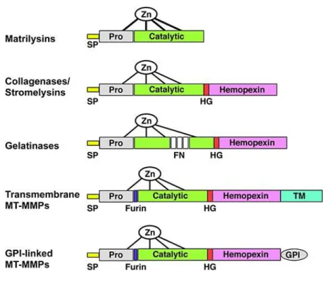
-Figure III-3: Domain structure for the major classes of MMPs. 20
-Figure III-4: MMPs in tumor progression. 23
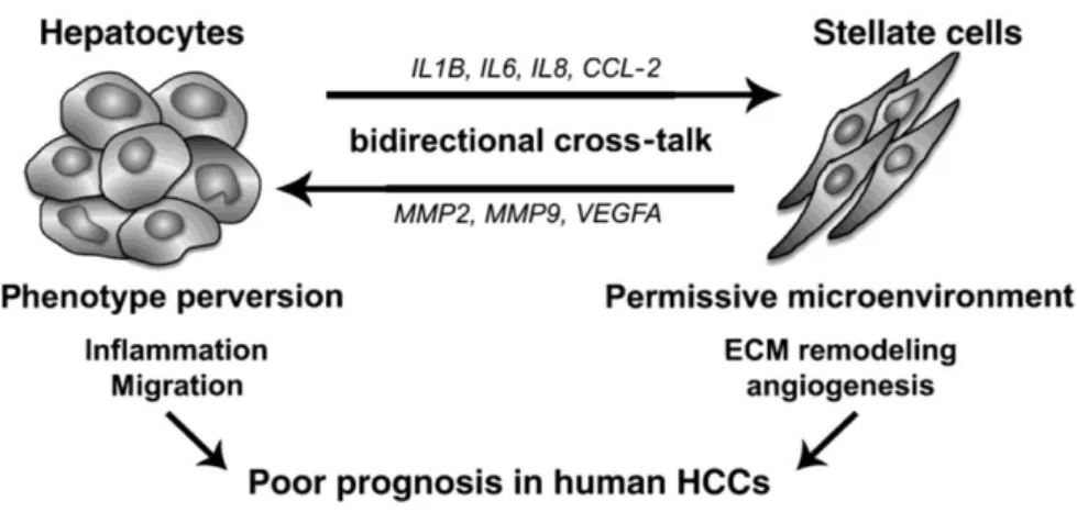
-Figure III-5: Proposed model for molecular cross-talk between hepatocytesand
activated HSCs in HCCs. 26
-Figure IV-1: SIBLINGs 28
-Figure V-1: Absorption in tissue. 33
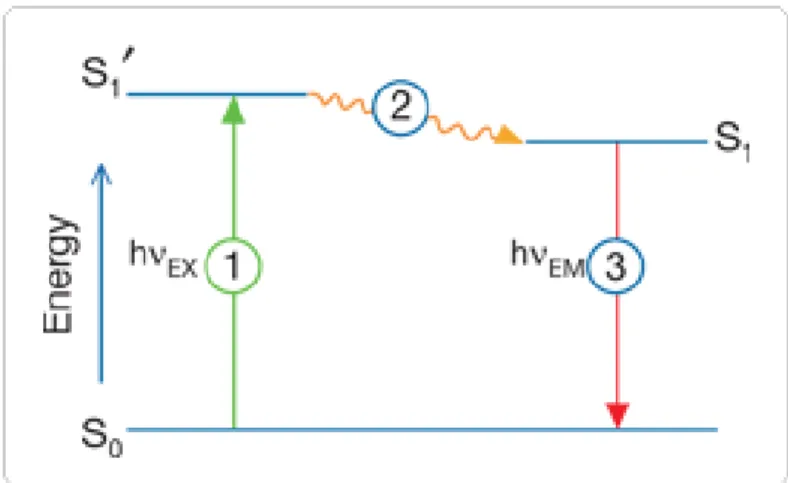
-Figure V-2: Autofluoresence of mouse organs under different optical conditions. 33 -Figure V-3: The Jablonski diagram illustrates the processes of optical absorption
and subsequent emission of fluorescence. 34
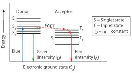
-Figure V-4: Protease-activatable fluorescence imaging probe. 36 -Figure V-5: Jablonski diagram - Principle of the Förster Resonance Energy
Transfer (FRET). 37
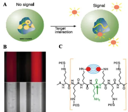
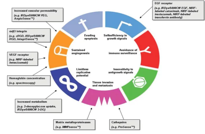
-Figure V-6: Structure of activatable fluorescent KLA-containing conjugate. 37 -Figure V-7: Hallmarks of cancer and their targets for optical imaging. 41 -Figure V-8: Principle of enzyme detection via the disruption of intramolecular
self-quenching. 42
-Figure V-9: Five classes of molecular-specific optical contrast agent. 43 -Figure V-10: Hallmarks of cancer and their targets for optical imaging. 45
-Figure V-11: Chemical structures based on the RGD motif. 50
-Figure V-12: Representation of the extracellular domain structure of the integrin αvβ3, associated to the ligand c[-RGDf(N-Me)V-] in presence of Mn2+
. 50
-Figure V-13: Improved αvβ3 Integrin targeting. 52
-Figure V-14: The RAFT-c(RGD)4 scaffold. 53
-Figure VI-1: The complex of proMMP-2 and TIMP-2. 55
-Figure VI-2: Schematic structure of branched triple-helical peptides. 60 -Figure VII-1: Representation of different medical imaging methods. 63
-XII
Figure VII-3: Near-infrared intra-operative camera systems. 69 -Figure VII-4: 2D-Fluoresence-reflectance imaging systems. 71 -Figure IX-1: Metastatic cancer cells invade lymph vessels and blood vessels near a
tumor and migrate to other parts of the body. 78
-Figure IX-2: NIR fluorescence labeling of lymphatic channels and SNL by ICG. 79 -Figure IX-3: Tumor-specific fluorescence intra-operative imaging in ovarian
cancer. 81
-RESULTS & CONCLUSIONS
Figure I-1: The imaging device: Fluobeam™ 88
-Figure-I-2: Laboratory cats used for preliminary experiments with doses 1X, 3X
and 5X. 90
-Figure I-3: Schedule of sampling and fluorescence imaging 91 -Figure I-4: Fluorescence kinetic of plasma samples (A & B, enlargement) and of
the three cats, compared to mouse data. 93
-Figure I-5: Fluorescence kinetic of blood samples (A & B, enlargement) and of the
three cats, compared to mouse data. 94
-Figure I-6: Fluorescence kinetic of urine samples of cat 1 to 3. 95 -Figure I-7: A) Fluorescence kinetic of the kidney region (B enlargement) of the
three cats, compared to mouse data. 96
-Figure I-8: A) Fluorescence kinetic of the shoulder region (B enlargement) of the
three cats. 97
-Figure I-9: X-ray scanner images of case #5. 99
-Figure I-10: Intra-operative imaging by using the Fluobeam™. 102 -Figure I-11: Fluorescence intensity compared during surgery of all 6. 133 -Figure I-12: Tumor-to-healthy tissue ratio of all 6 groups during surgery. 134 -Figure I-13: Tumor-signal normalized LUT of different tissues compared to their
Auto-LUT images. 135
-Figure I-14: Difference in measured fluorescence intensity dependent on the
placement towards the central laser beam. 135
-Figure III-1: Structure of the scaffold RAFT presenting the ligand « RGD ». 148 -Figure III-2: Chromatograms of 1, 2, 3 and 4 at 214 nm and their structure. 149 -Figure III-3: Grafting of Dansyl fluorescent dye on resin. 150 -Figure III-4: RT-HPLC analysis at 633nm of the degradation of compound 3 by
MMP-9, with structure of molecule. 151
-Figure III-5: Percentage of compound 3 cleavage after incubation with MMP-9. 151 -Figure III-6: Molecule structure of compound 5 showing orthogonal and N-terminal
protection groups. 152
-Figure III-7: RP-HPLC analysis of 5 (top) and 6a (bottom). 153
-Figure III-8: Mass spectrometry analysis of 6a. 153
-Figure III-9: Total deprotection of compound 7a; (a) TFA 95%, H2O 2.5%, TIS
-XIII
Figure III-10: Fluorescence imaging of RAFT-RGD-MMP drops before and after
incubation with 155
-Figure III-11: Fluorescence emission spectra and corresponding absorption spectra
of RAFT6RGD-MMP before and after incubation with MMP-9. 156
-Figure III-12: Measured quenching of the compound RAFT-RGD-MMP before and
after incubation with MMP-9. 157
-Figure III-13: Fluorescence emission spectra of RAFT-RGD-MMP: effect of A)
MMP inhibition and B) Plasma stability. 157
-Figure III-14: Interaction of RAFT-RGD-MMP and RAFT-RGD-Cy5 (control)
with HEK 293(β3) cells in vitro. 171
-Figure III-15: Evolution of pa- MFI (%) in HEK293(ß3) treated with either
RAFT-RGD-MMP (0.2µM) or RAFT-RGD-Cy5 (0.4µM) at 37°C. 171
-Figure III-16: FACS analysis of HEK 293(β3) pre-treated with RAFT-RGD and incubated with RAFT-RGD-MMP (0.2µM) and RAFT-RGD-Cy5 (0.4µM)
respectively, at 37°C at T0, 5 min and 2 h. 172
-Figure III-17: Interaction of RAFT-RGD-MMP and RAFT-RGD-Cy5 (control)
with A375 cells in vitro. 173
-Figure III-18: Evolution of pa- MFI in A375 treated with either RAFT-RGD-MMP
(0.2µM) or RAFT-RGD-Cy5 (0.4µM) at 37°C. 173
-Figure III-19: FACS analysis of A375 pre-treated with RAFT-RGD and incubated with RAFT-RGD-MMP (0.2µM) and RAFT-RGD-Cy5 (0.4µM) respectively, at
37°C at T0, 5 min and 2 h. 174
-Figure III-20: Interaction of RAFT-RGD-MMP and RAFT-RGD-Cy5 (control)
with TS/A-pc cells in vitro. 175
-Figure III-21: Evolution of pa-MFI in TS/A treated with either RAFT-RGD-MMP
(0.2µM) or RAFT-RGD-Cy5 (0.4µM) at 37°C. 175
-Figure III-22: Interaction of RAFT-RGD-MMP and RAFT-RGD-Cy5 (control)
with HT1080 cells in vitro. 177
-Figure III-23: Evolution of pa- MFI in HT1080 treated with either
RAFT-RGD-MMP (0.2µM) or RAFT-RGD-Cy5 (0.4µM) at 37°C. 177
-Figure III-24: Interaction of RAFT-RGD-MMP and RAFT-RGD-Cy5 (control)
with Colo829 cells in vitro. 178
-Figure III-25: Evolution of pa-MFI in Colo829 treated with either
RAFT-RGD-MMP (0.2µM) or RAFT-RGD-Cy5 (0.4µM) at 37°C. 179
-Figure III-26: Interaction of RAFT-RGD-MMP and RAFT-RGD-Cy5 (control)
with PC-3 cells in vitro. 179
-Figure III-27: Evolution of pa-MFI in PC-3 treated with either RAFT-RGD-MMP
(0.2µM) or RAFT-RGD-Cy5 (0.4µM) at 37°C. 179
-Figure III-28: Interaction of RAFT-RGD-MMP and RAFT-RGD-Cy5 (control)
with Hek293(β1) cells in vitro. 180
-Figure III-29: Evolution of pa- MFI in HEK(β1) treated with either
RAFT-RGD-MMP (0.2µM) or RAFT-RGD-Cy5 (0.4µM) at 37°C. 180
Figure III30: FACS data of RAFTRGDCy5 and RAFTRGDMMP compared. 181 -Figure III-31: Biphoton confocal microscopy of living cells. 182
-XIV
Figure III-32: Biodistribution of RAFT-RGD-MMPin vivo. 184
-Figure III-33: Compared tumor-to-skin ratio during time in vivo. 186 -Figure III-34: Distribution of RAFT-RGD-MMP in different organs. 187 -Figure III-35: Excised tumors (HEK293(β3), TS/A-pc and A375) of
RAFT-RGD-MMP injected mice. 188
-Figure III-36: Tumor-to-muscle ratio of subcutaneous tumor bearing mice at 3 h
post injection of 2 nmol RAFT-RGD-MMP. 188
-Figure III-37: Control of MMP-9 activity and inhibitor by means of
Fluospectrometry. 191
-Figure III-38: Zymography of 10-times concentrated serum-free supernatant at 24 h of the cell lines A375, HEK293(β3), TS/A-pc, HEK293(β1) and HT1080 (A)and
A375, PC-3, Colo829, TS/A-pc and HT1080 (B). 193
-Figure III-39: Gelatinases expression of different cell lines used with HT1080 as
reference. 193
-Figure III-40: Fluorescence intensity in non treated mouse organs ex vivo. 195
-DISCUSSION & PERSPECTIVES
Figure III-1: Highly simplified diagram of intergin-gealtinase interaction. 212 -Figure III-2: Illustration of RAFT-RGD-MMP and FRET via Cy3 & Cy5
engraftment. 215
-Figure III-3: Illustration of RAFT-RGD-MMP and Quenching via QSY & Cy5
engraftment. 215
-Figure III-4: Example of prodrug design based on the RAFT-RGD-MMP. 218
-MATERIALS & METHODS
Figure I-1: Estimation of AngioStamp™ dose for cats. 227
-Figure III-1: Scheme of the removal of Fmoc with Piperidine 232
-Figure III-2 Scheme of the removal of Dde 234
-XV
I
NDEX OF
T
ABLES
INTRODUCTION
Table II1: Role of integrins in the tumor progression. ... 14 Table V1: Spectroscopic properties of fluorescent dyes (adapted after invitogen.com). ... 39 Table VI1: Classification of protease inhibitors according to their mechanism ... 56 Table VIII1: Characteristics of benign and malignant tumors. (Baba et al., 2007) ... 72 Table VIII2: Surgery Techniques. ... 73 -Table VIII-3: Advantages and disadvantages of medical imaging modalities used with molecularly targeted or nontargeted contrast agents in a clinical setting. ... 77
-RESULTS & CONCLUSIONS
Table I1: Compared fluorescence data of cat plasma. ... 93 Table I2: Compared fluorescence data of cat blood. ... 94 Table I3: Compared fluorescence data of cat urine. ... 95 Table I4: Compared fluorescence data of cat kidney. ... 96 Table I5: Compared fluorescence data of cat shoulder. ... 97 Table I6: Conditions tested. ... 99 -Table I-7: Patient data, showing dose and time of surgery after injection of AngioStamp™, region of the tumor, notable complications for surgery (health status, other than primary tumor), weight, age, sex, color and followup until august 2012. ... 101 -Table III-1: RT-HPLC measured retention times (RT) of compounds 1, 2, 3 and 4of crude peptide (control) and of peptides after incubation with MMP-2 and MMP-9 (2 h, 37°C). ... 150 Table III2: Degree of integrin αvβ3 and MMP9 expression of different cell lines. ... 169 Table III3: FACS analysis of integrin expression in different cell lines. ... 194
-MATERIALS & METHODS
Table I1: Species specific dose of AngioStamp™... 227 Table III1: Procedures for cleavage from the resin ... 233
-APPENDICES
-XVII
L
IST OF
A
BBREVIATIONS
AcOH acetic acid Alloc Allyloxycarbonyl AP-1 activator protein-1 Boc Tertio-butyloxycarbonyl CT Computed Tomography Cy5 Cyanine 5 Cy3 Cyanine 3 DiPEA N,N-Diisopropylethylamine DMF N,N-Dimethylformamide Dns Dansyl DNP dinitrophenyl ECM extracellular matrix equiv. molar equivalent EtOH Ethanol
EPR enhanced penetration and retention ERK extracellular signal-regulated kinase
F50 50 %Fluorescence (time of reducing the maximal fluorescence intensity by half)
FACS Fluorescence-Activated Cell Sorting FAK focal adhesion kinase
FGF-2 fibroblast growth factor-2 FITC fluorescein isothiocyanate Fmoc 9-Fluorenylmethyloxycarbonyl
XVIII
FN fibronectine
h hour/-s
HAS human serum albumin HCC hepatocellular carcinoma
Her2/neu human epidermal growth factor receptor 2
ICG indocyanine green
IL-8 Interleukin-8
JNK c-Jun N-terminal Kinases
LUT Lookup table (for transforming measured values (input data) in a visible grayscale image (output format))
mAb monoclonal antibody
Mca 7-methoxycumarin
MeOH Methanol
MFI mean fluorescence intensity
min minute/-s
MMP Matrixmetalloprotease
MRI Magnetic Resonance Imaging
MS Mass Spectrometry
ms millisecond
NF-κB nuclear factor kappa-light chain-enhancer of activated B cells NHS N-hydroxysuccinimide
NIR Near Infrared
NSCLC Non small cell lung cancer
Pbf 2,2,4,6,7-Pentamethyl-dihydrobenzofurane-5-sulfonyl PDGF Platelet-derived growth factor
XIX PET Positron Emission Tomography PI3K Phosphoinositole 3-Kinase
PyBop (Benzotriazol-1-yloxy)tris(pyrrolidino)phosphonium hexauorophosphate
QSY Quencher
RAFT Regioselectively Addressable Functionalized Template RFA radiofrequency ablation
RGD cyclic R-G-D-f-K
RT-HPLC Reverse Phase High Performance Liquid Chromatography
RT Retention Time
SASRIN Super Acid Sensitive Resin SBR signal-to-background ratio SLN sentinel lymph node
SPECT single photon emission computed tomography SPPS Solid Phase Peptide Synthesis
TFA Triuoracetic acid TIS Triisopropylsilane
TNBS Trinitrobenzensulfonic acid TNF-α tumor necrosis factor alpha Trt trityl / triphenylmethyl
US ultrasound imaging
„Ich kann freilich nicht sagen, ob es besser werden wird, wenn es anders wird; aber soviel kann ich sagen: es muss anders werden, wenn es gut werden soll.“
Georg Christoph Lichtenberg 1742–1799
- 1 -
P
REAMBLE
In most cancers, tumor resection is the first therapeutic indication before chemotherapy or radiotherapy assuming that the tumor is removable. Since the patient survival strongly depends on the exhaustiveness of the tumor resection, innovative methods improving the quality of cancer surgery are needed to lower the risks of postoperative local recurrence.
Actually, several approaches for image-guided surgery are in development for clinical application, thereunder the design of optical imaging devices. This technology, working in an optical window with improved information out-come, the near-infrared (NIR), showed an improvement in the quality of the surgery in preclinical studies. First studies in clinics have been done by different research groups, but the translation from preclinics into clinics needs the adaptation of the imaging device for the operation block to warrant correct surgery.
Nevertheless, for a real clinical employment these systems are restricted to one imaging probe being the only one approved for use in human (FDA; ANSM) with the imaging properties needed. This is a critical point for the development and translation of NIR-optical imaging systems in clinical application, especially as the tracer used is non-specific and thus limited in its application.
Relating to this, a portable clinical imaging device (FluobeamTM) for optical guided surgery has been developed, working with a tumor targeting probe (AngioStamp™) in the NIR window. The combination of imaging device and specific tracer allowed speeding up high quality surgery in preclinical studies.
One part of my work was the transfer of this technology into a clinical veterinary phase, that is to say the clinical surgery of spontaneous tumors in cats, in order to evaluate this system regarding clinical surgery in humans.
In parallel, cancer diagnosis and therapy lack of specificity towards the tumor cells, limiting a precocious detection and safe treatment. Conventional cytostatic and cytotoxic treatments lead to adverse effects, which limit the dose of administration.
Consequently, clinical cancer research claim the development of new sensitive and performing diagnostic methods and of efficient therapeutics with reduced adverse effects.
- 2 -
Therefore, cancer research is pushed towards the development of targeting strategies. This corresponds to the use of new molecules emerging from biological knowledge of cancer and reacting specifically with the cancer cell.
Among the specific physiopathological phenomenon of cancer, targeting of tumor angiogenesis represents a promising strategy to limit tumorigenesis. Tumor neo-vessels differ from quiescent vessels concerning their structure but also in their functioning by overexpressing membrane receptors and other specific recognition elements on the surface of endothelial cells. At the same time these receptors are over-expressed by certain tumor cells themselves, which allows a targeting of the tumor directly. In order to efficiently target these receptors synthetic ligands have been developed with very high specificity and selectivity. Other potential targets are enzymes over-expressed in the tumoral tissue. A group of these enzymes are amongst others in correlation with the receptors of angiogenesis and necessary for tumor cell invasion and spreading. Due to their cleavage activity they incorporate a target for activatable molecules in the sense of pro-drugs or quenched imaging agents.
In this context my laboratories have developed the vector “RAFT-(cRGD)4”, in the following simply called RAFT-RGD. This molecule is based on decapeptide scaffold with two independent domains, allowing the combination of two different functions. One domain presents a ligand in a multivalent fashion, whereas the other domain carries the vectorization agent: therapeutics or imaging agents.
The RAFT-RGD showed its capacity of targeting tumor and endothelial cells expressing the receptor integrin αvβ3 in vitro and in vivo, which allowed the fluorescence imaging in vivo of integrin αvβ3 positive tumors and serves also as the already mentioned tracer AngioStamp™.
My second project was the design of an innovative fluorescent molecule for cancer diagnosis and therapy, thus a pharmacodiagnosticum (or theranostic). The molecule constructed is a derivative of the RAFT-RGD combined with a cassette, cleavable by enzymes overexpressed in tumors. A fluorophore, quenched by another molecule in the cassette, is set free, when the peptide sequence connecting these molecules, is cleaved. The result is an increase in the fluorescence signal. The molecule, afterwards called RAFT-RGD-MMP, was synthesized and tested in first studies in vitro and in vivo using different tumor cell lines in order to study the capacity of dual-targeting compared to a mono-target approach.
- 3 -
I. Tumorigenesis
I.1.
Characteristics of tumorigenesis
Cancer can be considered as a general term covering a plethora of different malignancies. These pathogenic conditions are characterized by changes in certain physical properties normally preserving cell homeostasis.
Cell homeostasis consists in the maintenance of a balance between the loss and repair of cells. In the case of tumors, an imbalance occurs between reduced cell loss and excessive cell growth.
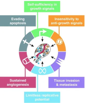
Hanahan and Weinberg1,2 described eight biological capabilities acquired by cancer cells called hallmarks of cancer. Mentioned are the sustaining of proliferative signaling, evading growth suppressors, resisting cell death, enabling replicative immortality, inducing angiogenesis, activating invasion and metastasis, reprogramming of energy metabolism, and evading immune destruction.
These mechanisms are needed for and lead to the multistep development called tumorigenesis. The term tumorigenesis describes the process of initiating and promoting the development of a tumor. In general it consists of three consecutive steps: 1) initiation of cell changes to be able to form tumors, 2) promotion, which is the growth of existing tumors and 3) progression, which is characterized by increased growth speed and invasiveness of the tumor cells (Figure I-1).
1
Hanahan, D., und R. A. Weinberg. „The hallmarks of cancer“. Cell 100 (2000): 57–70.
2
Hanahan, Douglas, und Robert A. Weinberg. „Hallmarks of Cancer: The Next Generation“. Cell 144, Nr. 5 (March 4,
2
Hanahan, Douglas, und Robert A. Weinberg. „Hallmarks of Cancer: The Next Generation“. Cell 144, Nr. 5 (March 4, 2011): 646–674.
- 4 -
Due to genomic instability and the inflammatory state of premalignant and malignant lesions, generation of the listed characteristics occurs. This is due to exposition to a carcinogen, either of chemical, physical or viral origin. Leading to activation of certain oncogenes and/or the inhibition of tumor suppressor genes, initiation and progression of cancer takes place.
After all, normal cells transform into “immortal” cells with higher activity in cell growth and cell-division. They possess means of modifying their environment and deregulating the physiological fine-tuned balance in order to support their development and spreading.
This is reflected for example by defect negative-feedback loops, like the oncoprotein Ras or the kinase mTOR, whose inhibition leads to enhanced proliferation. Other differences, compared to normal cells, is the evasion of growth suppressors, like
retinoblastoma-associated and TP53 proteins, the enhancement of proliferation signaling and the resisting to physiological cell death, as well as inducing angiogenesis.
Hanahan et al. describe also that necrotic cells can recruit inflammatory cells of the immune system. In the context of neoplasia, this can actively promote tumor progression,
Figure I-1: Stages of tumorigenesis.
11-- 5 11--
because such cells are capable of enhancing angiogenesis, cancer cell proliferation, and invasiveness3.
Necrotic cells can release bioactiveregulatory factors, which can directly stimulate neighboring viable cells to proliferate, with the potential, to facilitate neoplastic progression. It is known that some tumors are densely infiltrated by cells of the immune system and thereby mirror inflammatory conditions arising in non-neoplastic tissues.
Inflammation is in some cases evident at the earliest stages of neoplastic progression and is demonstrably capable of facilitating the development of neoplasias into cancers.
Additionally, inflammatory cells can release chemicals, notably reactive oxygen species that are actively mutagenic for nearby cancer cells, accelerating their genetic evolution towards states of heightened malignancy.
Apart from dysregulation of physiological mechanisms, cancer cells have certain morphological properties which make them histologically identifiable. The dedifferentiated cells are presenting rounded cytoskeleton, with reduced cell-to-cell and cell-to-extracellular matrix adhesion. The nucleolus is characterized by hypertrophy, macro- and
microsegregation4 and the neoplastic cell population is highly genotypically and phenotypically heterogeneous.
Besides, tumor cells are more or less organized in a supporting tissue. This so called tumor stroma consists of fibroblasts, smooth muscle cells, blood and lymphatic vessels and infiltrated immune cells, such as macrophages, mastocytes and lymphocytes, which are all embedded in an extracellular matrix (ECM)5 (Figure I-2). Furthermore the tumor
microenvironment consists of soluble factors such as cytokines and growth factors.
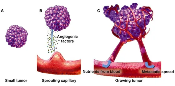
The vascularization and sprouting of new blood vessels in the tumor stroma is important for tumor growth and invasion of the surrounding tissue and metastasis. It represents not only the supplying system for nutrients, oxygen and the elimination pathway of waste but also a transport system for metastatic cells to reach the organs of metastasis manifestation. Thus, it is also an access point for molecules, either diagnostic or therapeutic ones.
3
Hanahan, Douglas, und Robert A. Weinberg. „Hallmarks of Cancer: The Next Generation“. Cell 144, Nr. 5 (March 4, 2011): 646–674.
4
Baba, Alecsandru Ioan A I, und Cornel C Câtoi. Comparative Oncology. Bucharest: The Publishing House of the Romanian Academy, 2007.
5
Li, Zhi-Wei, und William S Dalton. „Tumor microenvironment and drug resistance in hematologic malignancies“. Blood
- 6 -
I am going now to give a brief description of the process of angiogenesis in order to introduce the interest in targeting certain structures or enzymes for specific tumor imaging and therapy.
Figure I-2: The Cells of the Tumor Microenvironment.
(Upper) An assemblage of distinct cell types constitutes most solid tumors. Both the parenchyma and stroma of tumors contain distinct cell types and subtypes that collectively enable tumor growth and progression. (Lower) The distinctive
microenvironment of tumors.
(hatched background represents the extracellular matrix)
- 7 -
II. Angiogenesis
The formation of blood vessels is an important physiological process during embryogenesis and development.
Angiogenesis itself is the process of forming new blood vessels from existing ones. Therefore vascular basement membrane degradation is needed and remodeling of the ECM for migration of endothelial cells invasion of surrounding tissue and lumen formation is necessary.
Establishment of an arterial network requires vascular remodeling by perivascular mural cells including pericytes and vascular smooth muscle cells (Figure II-1).
Bone marrow-derived stem cells have been reported to play a critical role in contribution to growing vessels by the mechanism of differentiation into vascular endothelial cells6.
Physiological angiogenesis in adults does only occur in certain cases like wound healing or during the menstrual cycle of a woman. Therefore it is a symptom of pathology like rheumatoid arthritis, retinopathy, psoriasis, arteriosclerosis or cancer7.
While the tumor does not exceed 1-2 mm3, the tumor cells do get enough oxygen and nutrition by simple diffusion8. Exceeding this size, the inhibitors of the angiogenesis are
6
Lyden, D, K Hattori, S Dias, C Costa, P Blaikie, L Butros, A Chadburn, u. a. „Impaired recruitment of bone-marrow-derived endothelial and hematopoietic precursor cells blocks tumor angiogenesis and growth“. Nature medicine 7, Nr. 11 (November 2001): 1194–1201.
7
Carmeliet, Peter. „Angiogenesis in health and disease“. Nature medicine 9, Nr. 6 (Juni 2003): 653–660.
Figure II-1: Blood vessel formation.
- Cao, Yihai. “Therapeutic Angiogenesis for Ischemic Disorders: What Is Missing for Clinical Benefits?“ Discovery Medicine 9, Nr. 46 (March 3, 2010): 179–184.-
- 8 -
oppressed and the pro-angiogenetic factors, like vascular endothelial growth factor (VEGF), platelet-derived growth factor (PDGF) and tumor necrosis factor-α (TNF-α), are secreted by the tumor cells, the tumor stroma, inflammatory cells or the ECM9. This is a process, which takes normally place due to stress (metabolic like hypoxia, hypoglycemia, low pH; mechanic, inflammation and/or gene dysregulation), and is called “angiogenic switch”. This implies the shift of the balance to more pro- than anti-angiogenic signals and often occurs at an early, premalignant stage10.
The endothelial cells that form existing blood vessels, respond to angiogenic signals in their vicinity by proliferating and secreting proteases. These proteases break open the blood-vessel wall to enable migration towards the site of the angiogenic stimuli. Then proliferating endothelial cells organize themselves into new capillary tubes by altering the arrangement of their adherence-membrane proteins. Finally, through the process of anastomosis, the capillaries, emanating from the arterioles and the venules, join to provide a continuous blood flow. This sustains tumor cell metabolism and sets up escaping avenues for metastatic tumor cells (Figure II-2).
Thus the tumor induces its proper vascularization, which allows its development and spreading11,12.
8
Folkman, J. „Endothelial cells and angiogenic growth factors in cancer growth and metastasis. Introduction“. Cancer
metastasis reviews 9, Nr. 3 (November 1990): 171–174.
9 Carmeliet, P, und R K Jain. „Angiogenesis in cancer and other diseases“. Nature 407, Nr. 6801 (September 14, 2000): 249–
257.
10
Rundhaug, Joyce E. „Matrix Metalloproteinases and Angiogenesis“. Journal of Cellular and Molecular Medicine 9, Nr. 2 (2005): 267–285.
11
Gimbrone, M A, Jr, S B Leapman, R S Cotran, und J Folkman. „Tumor dormancy in vivo by prevention of neovascularization“. The Journal of experimental medicine 136, Nr. 2 (August 1, 1972): 261–276.
12
Folkman, J, und D Hanahan. „Switch to the angiogenic phenotype during tumorigenesis“. Princess Takamatsu symposia 22 (1991): 339–347.
- 9 -
Figure II-2: Tumor development and angiogenesis.
- Siemann DW., Vascular targeting agents. Horizons in Cancer Therapeutics: From Bench to Bedside. 2002;3(2):4-15.-
Compared to normal blood vessels, tumor blood vessels have a chaotic vasculature and are tortuous, leaky, and dilated or uneven in diameter, with reduced attachment of pericytes13.
Various classes of adhesion molecules are involved in tumor angiogenesis, such as members of the integrin, cadherin, selectin and immunoglobulin families.
Here I will focus on the integrins, as the αvβ3 integrin, represents our target.
13
Rundhaug, Joyce E. „Matrix Metalloproteinases and Angiogenesis“. Journal of Cellular and Molecular Medicine 9, Nr. 2 (2005): 267–285.
- 10 -
II.1.
Integrins
The integrins modulate physical processes like migration and invasion of the cell as well as intracellular pathways leading to cell survival and proliferation as well as cell death by anoïkis when cells loose contact with the ECM. The function of these receptors ranges from cell-substrate-attachment mediation to intracellular signal transduction. The signalization in all its complexity is accentuated by the capacity of the integrin to modulate the mediated signals of other membrane receptors and the other way round14.
As integrins do not have an intrinsic enzymatic activity, they recruit different enzyme partners for signal transmission.
II.1.1. Integrin composition
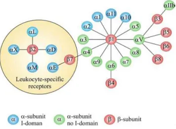
Integrins are heterodimeric cell surface receptors composed of noncovalently associated transmembrane glycoproteins, α and β, which connect adhesive proteins in the ECM to the cytoskeleton. There are at least 18 different α subunits and eight different β subunits identified, which represent - in an associated form - over 24 receptors for different spectra of extracellular ligands (Figure II-3)15.
The α and β subunits consist of a large extracellular domain (700-1100 amino acids) and a short cytoplasmic domain of 30 to 50 amino acids. The N-terminal extremities, next to the extracellular domain of the α subunit, contain regions, which are capable to bind divalent cations (Mg2+, Ca2+). The β subunit has cystein-rich regions in its extracellular domain and a folded N-terminal, forming a large loop (Figure II-4)16.
14 Porter, J C, and N Hogg. “Integrins Take Partners: Cross-talk Between Integrins and Other Membrane Receptors.” Trends
in Cell Biology 8, no. 10 (October 1998): 390–396.
15
van den Steen et al., 2006, Journal of biol. Chem.. 281, 18626-18637.
16
- 11 -
Figure II-3: The members of the human integrin superfamily and how they combine to form heterodimeric integrins.
At least 18 α subunits and eight β subunits have been identified in humans, which are able to generate 24 different integrins. Integrin subunits that bind to each other to form a heterodimer are connected by solid lines. Each integrin has
distinct ligand-binding specificity and tissue and cell distribution.
- Niu G, Chen X. Why Integrin as a Primary Target for Imaging and Therapy. Theranostics 2011; 1:30-47.-
Figure II-4: The extracellular region of a human integrin.
The crystal structure represents a net form of integrin αVβ3 with no bound RGD
- 12 -
II.1.2. Integrin conformation & substrate recognition
The association αβ determines the specificity of the integrin for its substrate. The interaction ligand-integrin is modulated by the avidity and the affinity and is strongly influenced by the ligand’s conformation and the possible multimeric formation. The transmission of the signal in the cell is guaranteed by the “key-lock-principle”. Nevertheless, as one ligand can be recognized by more than one integrin, cooperative and competitive effects between integrins lead to complex signaling. Thus precise analysis of its environment by the cell is more than a simple physical interaction of cell and ECM.
Being a dimer, different conformations of integrins can be observed, which modifies their affinity towards their ligand and their state of activation (Figure II-5). Integrin-ligand affinity is modulated by chemical allostery, mechanical allostery and integrin clustering. Several integrins are basically not active (Figure II-5, B), but expressed on the cell surface in an inactive form (Figure II-5, A). This results in a lack of ligand-receptor interaction and consequently no signal is emitted17. The cell glycocalyx is also of importance, as it occupies the cell-substrate space and thus influences the diffusion of reactants, limiting the attachment of both integrin and ligand to surfaces18.
17
Takagi, Junichi, and Timothy A Springer. “Integrin Activation and Structural Rearrangement.” Immunological Reviews 186 (August 2002): 141–163.
18
Boettiger, David. „Mechanical control of integrin-mediated adhesion and signaling“. Current opinion in cell biology (August 1, 2012).
A B
Figure II-5: Intergin formation: A) inactive, bend form; B) active form
-After: Bass, Mark D. „SHARPINing integrin inhibition“. Nature Cell Biology 13, Nr. 11 (November 2, 2011):
- 13 -
Concerning their specificity for their ligands, integrins can be classified in different groups: laminin integrins, collagen integrins, leucocytes integrins, and integrins recognizing the short peptide sequence Arg-Gly-Asp (RGD)19.
Upon engagement, integrins cluster at the attachment site and recruit multiple intracellular cytoskeletal proteins, such as α-actinin, talin, and filamin, as well as signaling proteins, including focal adhesion kinase and integrin-linked kinase20,21. The formation of focal adhesion contacts is a prerequisite for adhesion, spreading, and migration through structures of the ECM.
II.1.3. Integrin turn-over
An important aspect is also the possible internalization of integrins by endocytic routes, such as the formation of circular dorsal ruffles during macropinocytosis and clathrin-dependent or -inclathrin-dependent endocytic pathways. This implicates the rapid trafficking of integrins to early endosomes. Nevertheless the majority of these internalized integrins are rapidly recycled back to the plasma membrane (t1/2 of integrin recycling often in less than 15 min)22.
II.1.4. Roles of integrin
As playing a principal role in the maintenance of tissue-cohesion and cell migration, integrin dimers are also implicated in the progression of several pathologies, making them a potential therapeutic target16. Integrins control also diverse other cellular processes, including proliferation, apoptosis and differentiation, thus are implicated in development, immune responses and progression of diseases.
Concerning cancer, the transformation of cancer cells is inter alia characterized by the disorganization of the cytoskeleton and the loss of adherence. Analyzing the modifications of expressed integrins in healthy tissues and tumor tissue, it has been observed that the integrins
19
Takeda, Ikuko, Shin-Ichiro Maruya, Takashi Shirasaki, Hiroki Mizukami, Takenori Takahata, Jeffrey N Myers, Seiji Kakehata, Soroku Yagihashi, and Hideichi Shinkawa. “Simvastatin Inactivates Beta1-integrin and Extracellular Signal-related Kinase Signaling and Inhibits Cell Proliferation in Head and Neck Squamous Cell Carcinoma Cells.”
Cancer Science 98, no. 6 (June 2007): 890–899.
20
R.E. Seftor, E.A. Seftor, M.J. Hendrix, Molecular role(s) for integrins in human melanoma invasion, Cancer Metastasis Rev. 18 (1999) 359–375.
21
R.O. Hynes, Integrins: versatility, modulation, and signaling in cell adhesion, Cell 69 (1992) 11–25.
22
Bridgewater, Rebecca E, Jim C Norman, und Patrick T Caswell. „Integrin trafficking at a glance“. Journal of cell science 125, Nr. Pt 16 (August 15, 2012): 3695–3701.
- 14 -
do either positively either negatively participate in the phenotype of tumor transformation23 (Table II-1).
Table II-1: Role of integrins in the tumor progression.
-After: Desgrosellier, Jay S, und David A Cheresh. „Integrins in cancer: biological implications and therapeutic opportunities“. Nature reviews. Cancer 10, Nr. 1 (January 2010): 9–22.-
23
Mizejewski, G J. „Role of integrins in cancer: survey of expression patterns“. Proceedings of the Society for Experimental
Biology and Medicine. Society for Experimental Biology and Medicine (New York, N.Y.) 222, Nr. 2 (November