HAL Id: hal-02975690
https://hal.archives-ouvertes.fr/hal-02975690
Submitted on 22 Oct 2020
HAL is a multi-disciplinary open access
archive for the deposit and dissemination of
sci-entific research documents, whether they are
pub-lished or not. The documents may come from
teaching and research institutions in France or
abroad, or from public or private research centers.
L’archive ouverte pluridisciplinaire HAL, est
destinée au dépôt et à la diffusion de documents
scientifiques de niveau recherche, publiés ou non,
émanant des établissements d’enseignement et de
recherche français ou étrangers, des laboratoires
publics ou privés.
Distributed under a Creative Commons Attribution| 4.0 International License
Fur identify a promoter-binding mechanism and its role
in Francisella tularensis virulence
J. Pérard, S. Nader, M. Levert, L Arnaud, P. Carpentier, C. Siebert, F.
Blanquet, C. Cavazza, P. Renesto, D. Schneider, et al.
To cite this version:
J. Pérard, S. Nader, M. Levert, L Arnaud, P. Carpentier, et al.. Structural and functional studies of
the metalloregulator Fur identify a promoter-binding mechanism and its role in Francisella tularensis
virulence. Communications Biology, Nature Publishing Group, 2018, 1 (1),
�10.1038/s42003-018-0095-6�. �hal-02975690�
Structural and functional studies of the
metalloregulator Fur identify a promoter-binding
mechanism and its role in Francisella tularensis
virulence
J. Pérard
1
, S. Nader
1
, M. Levert
2
, L. Arnaud
1
, P. Carpentier
1
, C. Siebert
2
, F. Blanquet
2
, C. Cavazza
1
, P. Renesto
2
,
D. Schneider
2
, M. Maurin
2
, J. Coves
3
, S. Crouzy
1
& I. Michaud-Soret
1
Francisella tularensis is a Gram-negative bacterium causing tularaemia. Classified as possible
bioterrorism agent, it may be transmitted to humans via animal infection or inhalation leading
to severe pneumonia. Its virulence is related to iron homeostasis involving siderophore
biosynthesis directly controlled at the transcription level by the ferric uptake regulator Fur, as
presented here together with the
first crystal structure of the tetrameric F. tularensis Fur in the
presence of its physiological cofactor, Fe
2+. Through structural, biophysical, biochemical and
modelling studies, we show that promoter sequences of F. tularensis containing Fur boxes
enable this tetrameric protein to bind them by splitting it into two dimers. Furthermore, the
critical role of F. tularensis Fur in virulence and pathogenesis is demonstrated with a
fur-deleted mutant showing an attenuated virulence in macrophage-like cells and mice. Together,
our study suggests that Fur is an attractive target of new antibiotics that attenuate the
virulence of F. tularensis.
DOI: 10.1038/s42003-018-0095-6
OPEN
1Univ. Grenoble Alpes, CNRS, CEA, BIG-LCBM, 38000 Grenoble, France.2Univ. Grenoble Alpes, CNRS, CHU Grenoble Alpes, Grenoble INP, TIMC-IMAG,
38000 Grenoble, France.3Univ. Grenoble Alpes, CNRS, CEA, IBS, 38000 Grenoble, France. Correspondence and requests for materials should be addressed
to J.Pér. (email:julien.perard@cea.fr) or to S.C. (email:serge.crouzy@cea.fr) or to I.M-S. (email:isabelle.michaud-soret@cea.fr)
123456789
F
rancisella tularensis is a small, highly infectious
Gram-negative bacterium, causing the zoonotic disease
tular-aemia
1. This species is currently divided into three
sub-species, including subsp. tularensis (type A strains), subsp.
holartica (type B strains) and subsp. mediasiatica
2. Only type A
and type B strains of F. tularensis are known to cause tularaemia
in humans. A large number of animal species can be infected with
this pathogen, but lagomorphs and small rodents are considered
the primary sources of human infections. The disease may also be
transmitted through arthropod bites, mainly Ixodidae ticks and
mosquitoes. Francisella tularensis also survives for prolonged
periods in the environment, and humans can be infected through
contact with contaminated soil or water. Because a few bacteria
inhaled through aerosols may induce an acute severe pneumonia,
with a mortality rate of 30% or more, F. tularensis has been
classified as a potential category A biothreat agent by the Center
for Disease Control and Prevention
3. No effective vaccine is
currently licenced for human or animal use, and a few antibiotic
compounds are used as
first-line drugs in tularaemia patients.
Alternative treatments are urgently needed both to improve the
prognosis of patients with severe diseases, and also to improve
our preparedness to the intentional release of resistant strains of
this pathogen in the context of bioterrorism
4,5. Although
numerous genes have been shown to be important for the
pathogenesis and virulence of F. tularensis, there is still a blatant
lack of knowledge about central biological functions such as iron
homeostasis
6,7and metalloregulators
8,9. As a facultative
intra-cellular bacterial pathogen, F. tularensis multiplication and
viru-lence depend on the host cell iron pool
10. Indeed, a major defence
strategy used by infected eukaryotic organisms is to withhold this
metal by sequestering free iron. In reaction to iron starvation, F.
tularensis is able to secrete an iron chelator structurally similar to
the polycarboxylate siderophore rhizoferrin
11,12. The
figA gene
(also called fslA), involved in the siderophore synthesis, but also
the
figE gene (fslE), responsible for its uptake, have been
char-acterized to play an important role in the virulence and/or
intracellular replication of this pathogen
9,12,13. These genes
belong to the locus
figABCDEF (fig for Francisella iron-regulated
genes)
14,15(Fig.
1
). The
fig operon is regulated by the ferric
uptake regulator Fur, which is supposed to bind to the fur-figA
intergenic region that contains a specific sequence called a FurBox
(Fig.
1
a
), although such direct interaction has not yet been
demonstrated. The Fur protein is a global transcriptional
reg-ulator that senses iron status and controls the expression of
genes involved in iron homeostasis, virulence and oxidative
stresses
16–18.
In the present study, to go further in the in vitro and in vivo
characterization of the properties of the F. tularensis Fur (FtFur)
protein, we used a virulent F. tularensis subsp. holarctica strain, a
Type B biovar I, referred to as CHUGA-Ft6. This strain was
isolated from a blood sample from a French patient suffering
from a typhoidal form of tularaemia
19. Interestingly, comparing
FtFur to Fur from Escherichia coli (EcFur), Pseudomonas
aeru-ginosa (PaFur), Legionella pneumophila (the agent of legionellosis,
LpFur) and Yersinia pestis (the agent of plague, YpFur), we have
evidenced that these proteins can be discriminated by their
quaternary structure in solution
20. EcFur and YpFur belong to the
group of the commonly accepted dimers, while FtFur, PaFur and
LpFur belong to a group of tetramers. A structural zinc in a
cysteine-rich site (site 1) has been characterized in many Fur
proteins including FtFur
16,21,22. In addition, the Fur proteins
need metallic dications such as Co
2+, Mn
2+or Fe
2+in a
reg-ulatory site (site 2) to be activated for the binding to DNA
20.
Here, we present, to our knowledge, the
first crystal structure of
a tetrameric Fur protein in the presence of its physiological
cofactor, the ferrous ion. This structure sheds light on the
metal-binding sites and corresponds to two intertwined pre-activated
dimers. We demonstrate the direct interaction of the protein with
the promoter region controlling expression of the genes involved
in siderophore synthesis and identify essential residues in this
interaction. In addition, owing to the coupling of computer
models and free energy calculations with cross-link experimental
studies, we bring evidence for a DNA-driven tetramer splitting
mechanism mediated by specific promoter sequences, and leading
1500
a
b
c
d
fur figA figB figC figD
fig operon CHUGA-Ft6
5′ 3′ Identity/EcFurBox EcFurBox 0 100 50 25 12.5 6 3 1.5 0.7 0.3 11/19 10/19 16/19 10/19 16/19 11/19 1000 500 0 Ft6 p < 0.001 p < 0.001 p < 0.001 Expression f old Fe atoms/bact – + – + – + 5×10⁴ PfigA PpdpB PigIc 7×10⁴ 2×10⁴ Ft6Δfur Ft6Δfur+fur FtFur (nM) 2T/4D+PfigA 1T/2D+PfigA 1D+PfigA PfigA FtFur (100 nM)
Fig. 1 FtFur regulatesfig operon by recognition of DNA FurBox. a qRT-PCR showing the absence of fur transcripts in the CHUGA-Ft6Δfur strain, with a 16S RNA standard as a control, Fur (Ft6 and Ft6Δfur + fur) repressed the transcription of figA. This repression is abolished in the absence of Fur (Ft6Δfur). The data correspond to two independent experiments made in triplicate. P values were calculated using the Student's t test. Iron concentration was measured by ICP-AES (error under 1%) from 2 mL bacterial culture (see Methods section) and number of Fe atoms per bacteria has been deduced.b Organization of thefig operon and sequence of the fur-figA intergenic region (PfigA). The identical bases between PfigA and EcFurBox are indicated underneath showing overlapping FtFur binding sites.c Evaluation of the ability of FtFur to bind identified or predicted Fur boxes and estimation of the apparent Kds (for DNA seq of each promoter see Supplementary Fig.1).d EMSA of FtFur in the presence of the 43 bp PfigA sequence. The proposed stoichiometry is written on the figure: D corresponding to dimer and T to tetramer
to the formation of two Fur dimer–DNA complexes. Finally, the
critical role of FtFur in bacterial virulence and pathogenesis is
demonstrated using a fur-deleted CHUGA-Ft6 mutant (Ft6Δfur),
which
shows an attenuated
virulence, both in
murine
macrophage-like cells and in mice, reinforcing that FtFur can be
thus defined as a crucial anti-virulence target.
Results
Fur is directly involved in
F. tularensis iron homeostasis. A
Δfur mutant was already generated in the virulent Schu S4 strain
(subsp. tularensis) to demonstrate that siderophore production is
regulated by FtFur in F. tularensis
9. However, the direct
invol-vement of FtFur in virulence has never been reported to our
knowledge. We have constructed the CHUGA-Ft6Δfur strain by
the allelic exchange method and deletion was confirmed by
quantitative real-time polymerase chain reaction (qRT-PCR) and
sequencing as we did not detect any fur transcript in Ft6Δfur
(Supplementary Fig.
1
). Using this approach, we demonstrated
that the siderophore synthesis is under the direct control of FtFur
in CHUGA-Ft6 strain. CHUGA-Ft6Δfur shows an approximately
25-fold higher level of
figA transcript when cultured in
iron-replete conditions compared to the wild-type (WT) strain. The
WT phenotype, that is Fur transcriptional repression of the
fig
operon genes, is recovered when the WT fur is expressed in trans
to complement the fur deletion (CHUGA-Ft6Δfur + fur)
(Fig.
1
a). This means that siderophore production is repressed by
FtFur in the presence of iron and derepressed in the absence of
the protein. Inductively coupled plasma atomic emission
spec-troscopy (ICP-AES) quantification of the bacterial iron
con-centration showed that, under our culture conditions, the
CHUGA-Ft6Δfur strain accumulates 1.6-fold more iron than
the WT (Fig.
1
a). These data strongly suggest that FtFur can bind
the fur-figA intergenic region that contains sequences closely
related to the EcFurBox identified in E. coli (Fig.
1
b). Only a few
Fur boxes were identified in Francisella genome, compared to E.
coli, in the promoter of
figA, pdpB (coding for the pathogenicity
determinant protein PdpB) and iglC (coding for the pathogenicity
island protein IglC) both in Schu S4
14and CHUGA-Ft6. Then,
electrophoretic mobility shift assay (EMSA) with
manganese-activated FtFur have been performed on consensus EcFurBox and
on PfigA, PpbpB and PiglC sequences (Fig.
1
c and Supplementary
Fig.
2
).
FtFur binds with a very high affinity to EcFurBox when
activated with Co(II) (Kdapp
= 9 nM
20) and to the PfigA
promoter (estimated Kdapp
= 5 nM) and with a low affinity to
PpbpB (averaged estimated Kdapp
= 100 nM; Fig.
1
c and
Supplementary Fig.
2
C). In contrast, and while iglC gene
expression is also found up-regulated under iron-restricted
conditions in F. tularensis
14, no binding is detected indicating
the absence of direct regulation by Fur. The migration on EMSA
gel of the FtFur/PfigA complex shows a composite pattern with
three successive bands assigned to the binding of one to two
tetramers (or one to four dimers) (Fig.
1
d and Supplementary
Fig.
2
B) which appear as the protein concentration is increased.
This suggests that FtFur could bind to several predicted Fur boxes
in the sequence (Fig.
1
b). Indeed, using a shorter version (PfigAS)
of PfigAL, the main species detected corresponds to one dimer
bound to DNA (Supplementary Fig.
2
B).
Western blot experiments suggest that the estimated 3000
protein subunits/bacteria may be mainly present as a tetramer
in vivo (see Supplementary Fig.
2
F, G) and that hydrogen peroxide
(H
2O
2) treatment (1 mM for 4 h) does not impact this amount.
This is not surprising considering the high stability of the tetramer
in solution. This copy number is in the same range as described in
E. coli or Vibrio cholerae (5000 and 2500 subunits/bacteria
estimated, respectively, in normal growth conditions)
23,24.
Con-sidering the number of 50,000 total iron atoms/bacteria quantified
by ICP-AES (Fig.
1
a) and the volume of CHUGA-Ft6 around
10
−15L, 5 µM of FtFur subunit and 80 µM total iron are expected.
Assuming micromolar range Kd for (Fe-FtFur) as found in the
literature for E. coli (1–10 µM for Fe-EcFur
25), we can expect that
a pool of metallated Fur tetramer exists in the cell prior to
association with the few DNA target present in F. tularensis.
Fe-FtFur and Mn-FtFur contain intertwined pre-activated
dimers. Recombinant FtFur was purified as a tetramer
contain-ing one equivalent of Zn(II) per subunit
20. FtFur was crystallized
in the presence of Mn(II) as MnCl
2or Fe(II) as (NH
4)
2Fe(SO
4)
2,
the latter under anaerobic conditions. The structure of Mn-bound
FtFur was obtained from purified protein metalled with Mn at
high concentration before crystallization in the presence of Mn
(II) and was determined ab initio at 1.7 Å resolution by the
single-wavelength anomalous diffraction (SAD) method. This structure
was used to determine that of Fe(II)-bound FtFur at 1.8 Å
reso-lution by molecular replacement (see X-ray data in
Supplemen-tary Fig.
3
and Supplementary Tables
1
and
2
). Both
Mn(II)-bound and Fe(II)-Mn(II)-bound proteins have similar overall structures
appearing as a compact tetramer made of a dimer of dimers per
asymmetric unit (α-carbon root mean square deviation (r.m.s.d.)
of 0.271 Å between the two structures). The main differences
come from disordered N-terminal and C-terminal residues. Thus,
the structure of Fur containing the physiological activator metal,
namely Fe(II)-bound FtFur, the
first one described to date, will be
used for a detailed description (Fig.
2
a). Among the total 140
residues of the protein, 131 to 133 were resolved per chain. Each
subunit presents secondary structure elements similar to those
found in other Fur structures. It consists of a N-terminal
DNA-binding domain (residues 7–82) composed of a winged
helix–turn–helix motif in which α4 is the DNA recognition helix.
A short hinge connects the DNA-binding domain to the
C-terminal dimerization domain (residues 89–138). The
dimer-ization domain consists of three antiparallel
β-strands (β3 to β4)
and two
α-helices (α5 to α6), α5 intersecting between β4 and β5
(Fig.
2
a and Supplementary Fig.
4
). The dimeric interface is
mediated by
β5 from each subunit forming an antiparallel
β-sheet, part of a six-stranded β-sheet in the dimer (Fig.
2
b).
The two dimers in the tetramer structure are nearly identical
(α-carbons r.m.s.d. = 0.318 Å between the two dimers) with an
almost perfect superposition of the secondary structure elements.
The interaction between the two dimers through their
DNA-binding domains is stabilized by H-bonds involving atoms of the
DNA recognition helices (α4) of chains AB and CD (for chains A
and C: Gln61
CHε/Ser64
AO; Ser64
CHγ/Ser64
AOγ; Ser64
CO/Gln61
AHε and Arg57
CHh/Glu63
AOε salt bridge, and
equivalently for chains B and D). The two salt bridges between
Arg57 and Glu63 constitute the most important interactions
(Fig.
2
c). These interactions combined with an interface area of
2830 Å
2between the dimers AD and BC (PISA
26) explain the
high stability of the tetramer in solution (Fig.
2
d). For
comparison, in a previous work, we demonstrated that PaFur,
initially described as a crystallographic dimer
27, was tetrameric in
solution
20with a substantially lower predicted interface area of
2120 Å
2.
Each dimer is in a closed conformation with the wing in 'inside'
positions corresponding to the 'active' form in which the
DNA-binding domains are prepared to bind target DNA
20. However,
the dimer–dimer interactions naturally prevent any kind of
interaction with DNA through the recognition helices. Indeed,
Tyr56 and Arg57 (the one involved in the salt bridge stabilizing
the tetramer), both present in the recognition helix, are highly
conserved residues known to have base-specific interaction with
DNA
28–30. We thus hypothesize that the metalled FtFur tetramer
structure is a pre-activated form of the protein. The mechanism
of pre-activated tetramer disruption driven by DNA is
concep-tually of interest.
First structural description of an iron substituted Fur. X-ray
fluorescence spectra indicated that Fur crystals contain two metal
species: one is the expected Zn and the second is the metal added
during crystallization, that is, Mn(II) or Fe(II) (Supplementary
Fig.
3
A). The structures of Mn-FtFur and Fe-FtFur are similar
and confirm the presence of one Zn
2+and one Mn
2+or Fe
2+per
subunit. Zn
2+in structural site S1 is coordinated by four sulphur
atoms from two pairs of cysteines in CX
2C motifs (Cys93-Cys96
and Cys133-Cys136) (Table
1
). It connects the short C-terminal
helix
α5 to the β-sheet of the dimerization domain
(Supple-mentary Fig.
4
). The presence of S1 in Fe-FtFur but not in PaFur
demonstrates that zinc is not a prerequisite for tetramer
forma-tion. The second site S2 binds either Mn
2+or Fe
2+. The metal
ion adopts a distorted octahedral geometry with a 3N–3O
coor-dination sphere (Fig.
3
). S2 connects the DNA-binding domain
(His33 and Glu81 (bidentate) and the dimerization domain which
provides three ligands (His88, His90 and Glu101). It is described
as the essential 'regulatory' site, present in all known activated Fur
structures. H33A-H90A double mutations in FtFur S2 provoke a
total inactivation of the protein in vitro (Supplementary Fig.
5
C).
The Fur structures containing an S2 site
filled with Zn
2+show
some variation in the coordination sphere. This
flexibility may be
explained by the preference of Zn
2+for a tetrahedral geometry
compared to Fe
2+, found physiologically in S2, which favours a
hexacoordinated octahedral environment with N/O ligands. Ab
initio quantum chemical geometry optimizations of models of the
S2 site using DFT with B3LYP hybrid functional and 6–31 G(d)
basis set have been performed with bound Mn
2+or Fe
2+. The
similar optimized geometries, with a larger coordination sphere
for Mn
2+than for Fe
2+, validate the X-ray structures (Fig.
3
,
Supplementary Fig.
4
A–D and Supplementary Table
3
).
Some structures of Fur or Fur-like proteins (such as HpFur and
PaFur) revealed the presence of a third metal-binding site (S3)
involving four conserved residues 2 His, 1 Asp and 1 Glu
22,27. In
FtFur, Tyr125 is found in place of one very conserved His. The
structure shows that the phenol group makes H-bonds with the
other putative ligands preventing metal binding in the position
where a metal ion was expected. Accordingly, the structures of
FtFur do not display any S3 site (Fig.
3
and Supplementary
Fig.
4
).
In summary, the crystal structures of FtFur highlighted the
presence of two metal ions per subunit: one structural Zn
2+,
already present in the non-activated protein as purified, and
either one Mn
2+or one Fe
2+, its physiological activator, with
identical ligands in the regulatory site of similar geometry.
Metalled FtFur behaves as a dimer of pre-activated dimers with
the DNA-binding domains forming a kind of crown with
interacting recognition helices through two salt bridges between
Arg57 and Glu63 together with other weaker interactions.
Quaternary structure of FtFur in the presence of the FurBox.
Size-exclusion chromatography coupled to multi-angle laser light
scattering with online refractometer (SEC-MALLS-RI) was used
to investigate the behaviour of FtFur in the presence of DNA. As
Table 1 Metal coordination from X-ray and DFT calculations
Atoms Distance Å Site S1 Zn Zn Cys93/96/133/136S 2.3 2.3 Site S2 Fe Mn X-ray / DFT X-ray / DFT His33 NƐ2 2.3 / 2.20 2.3 / 2.26 Glu81 OƐ1 2.2 / 2.25 2.3 / 2.43 Glu81 OƐ2 2.3 / 2.36 2.3 / 2.32 His88 NƐ2 2.3 / 2.22 2.3 / 2.25 His90 NƐ2 2.2 / 2.18 2.3 / 2.22 Glu101 OƐ1 2.0 / 2.05 2.3 / 2.10
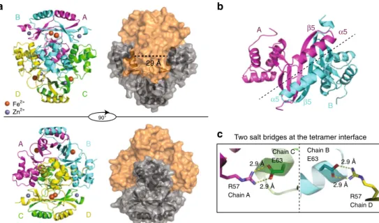
Bond distances for sites S1 and S2 ligands deduced from the X-ray structures and calculated by DFT A B D C A B D C A β5 α5 B β5 α5 R57 Chain A R57 Chain D Chain C E63 Chain B E63 2.9 Å 29 Å 2.9 Å 2.9 Å 2.9 Å
Two salt bridges at the tetramer interface
90°
a
b
c
Fe2+
Zn2+
Fig. 2 Structure of FtFur at 1.8 Å resolution in the presence of physiological iron Fe2+.a Fe-FtFur structure solved by SAD at 1.8 Å under anaerobic conditions in the presence of Fe2+. The cartoon model presents the four chains, labelled A–B–C–D. Surface representation indicates the dimer/dimer interface and the distance between two recognition helices (29 Å).b Symmetry at the dimer/dimer interface between two monomers involving helixα5 and strandβ5. c One of the most important interactions suggested by the structure is a salt bridge between Arg57 and Glu63
shown in Fig.
4
a, in the presence of FurBox the protein eluted
at a lower volume than the protein alone or the FurBox
alone. The deduced molecular weight of the corresponding peak
is 74 ± 2 kDa,
fitting with a complex between tetrameric FtFur
(64 kDa) and the FurBox duplex (15 kDa). This can be
inter-preted as the binding of FtFur to DNA as a tetramer or as two
dimers.
The evolution of the purified tetrameric FtFur in the presence of
DNA was then analysed by cross-link experiments using 0.1%
glutaraldehyde (GTA). Under denaturing conditions, in the absence
of DNA (Fig.
4
b), the main detected band corresponds to a species
with a molecular weight of approximately 62 kDa, in very good
agreement with the size of a covalently bound FtFur tetramer. After
cross-link in the presence of the EcFurBox, only two bands were
detected corresponding to the monomer and to a dimeric form of
the protein, respectively. Mutations of the FurBox (FurBox
m, see
Supplementary Fig.
2
) targeting four bases previously shown to be
crucial for the specific Fur/DNA interactions
31, three of them being
involved in interactions with Tyr56
28,29, resulted in the
conserva-tion of the tetramer without appariconserva-tion of dimers.
The monomer (M)/dimer pattern was also obtained with PfigA.
These results demonstrate that tetrameric FtFur splits into dimers
in the presence of specific DNA contrary to the dissociation of
PaFur previously observed with non-specific DNA
20. Besides, they
strongly suggest that FtFur binds the FurBox as dimers in vitro.
MALLS and SAXS data validate a two-dimer/DNA complex.
The activated Mn-bound FtFur form and the Mn-bound FtFur/
EcFurBox complex were examined by small-angle X-ray
scatter-ing (SAXS). In both cases, at three different concentrations (1–10
Y103 Y125 E108 H87 Absence of Site S3 H88 E81 E101 C93 C133 C136 C96 Site S1 (Zn2+) Site S2 (Fe2+) H33 H90 D89Fig. 3 Metal binding site in Fe-FtFur. Focus on one monomer extract on Fe-FtFur showing S1 and S2 sites simultaneously together with the unfilled S3 site
1.0×10⁵
a
b
Mn-FtFur-FurBox Mn-FtFur FurBox FurBox FurBoxm FurBoxm PfigAL PfigAL GTA 0.1% – + + + + + + + + + + 95 72 55 36 28 T D M 17 1.0 0.5 0.0 Relativ e scale FurBox 1.0×10⁴ 12.0 13.0 14.0 15.0 Volume (mL) 16.0 17.0 18.0Log molar mass (g/mol)
Fig. 4 Quaternary structure of Mn-FtFur in the presence of FurBox analysed by SEC-MALLS-RI and cross-link assay. a SEC-MALLS-RI data of Mn-FtFur and the FurBox analysed alone or as a complex in the presence of 1 mM MnCl2. Data are normalized by RI scale with sample concentrations ranging between 4 and 6 mg mL−1. FtFur/FurBox gives a MW of 78 ± 2 kDa, Mn-FtFur gives a MW of 64 ± 1 kDa and FurBox gives a MW of 15 ± 0.5 kDa.b Cross-link assay with GTA on SDS-PAGE 4–20% acrylamide in TGS buffer. A dimer (D) was trapped in the presence of specific Fur boxes (FurBox and PfigAL), while FtFur exists as a tetramer (T) in the absence of DNA or in the presence of mutated FurBoxm
mg mL
−1), the linearity of the Guinier plots indicates
mono-disperse samples. Average scattering curves of Mn-FtFur (red)
and Mn-FtFur/FurBox complex (black) in solution were recorded
(Fig.
5
a). Pair distance distribution functions (Fig.
5
b) point out
an elongation of the protein/DNA system (Dmax
= 112 Å for
DNA/protein complex against 83 Å for the protein alone) and
dramatic changes in the shape of the structure (Porod volume
=
130 nm
3, against 100 nm
3, and radius of gyration
= 32.5 Å,
against 27.4 Å). Bead molecular models of Mn-FtFur alone and in
complex with DNA complex, built by DAMMIF
32, show a
globular Mn-FtFur and a thick pancake shape for the DNA
complex (Fig.
5
c). The X-ray structure of the protein determined
in this study docks very well in the calculated envelope with a
χ
2of 1.9 (Fig.
5
d). In the absence of high-resolution structure of the
FtFur–DNA complex, a model was built based on
Mn-MgFur/EcFurBox ternary complex
29and
fits well with the
cal-culated envelope with a
χ
2of 2.2. These results are in agreement
with the conclusions of the cross-linking experiments and
sup-port the DNA-driven split of the FtFur tetramer in two dimers
sandwiching the FurBox. To better understand the mechanism of
dimer–dimer and dimer–DNA dissociation, theoretical
calcula-tions were performed.
Dimer/dimer and dimer/DNA dissociation free energy profiles.
The aim of this modelling was to evaluate precisely the difference
in binding affinity between the FtFur dimers within the FtFur
tetramers and between the FtFur dimers and DNA. Free energy
(potential of mean force) profiles for the extraction (by
transla-tion along a
fixed direction: Ox) of one FtFur dimer from the
tetramer (dimer of dimers) and of FtFur from DNA were
computed: the meticulous translation protocol is shown in
Sup-plementary Fig.
6
. The simulations include a 'moving' subsystem
(FtFur dimer, chains A and D) and a 'fixed' subsystem (FtFur
dimer, chains B and C, DNA) as shown in Supplementary Fig.
7
.
The profiles were built using the 'umbrella sampling' technique
and result from the overlapping of 26 computation windows, one
for each translation distance, and corresponding to 15 ns
mole-cular dynamics simulation each. The results of the calculations
are shown in Fig.
6
a. Binding free energies are
ΔG = 18.8, 10.5
and 8.8 kcal mol
−1for dimer from FurBox, dimer from tetramer
and dimer from mutated DNA (mutDNA containing FurBox
m),
respectively. These binding free energies correspond to
dissocia-tion constants of 17 fM, 20 nM and 0.4 µM, respectively, allowing
a thermodynamically easy separation of the tetramer into two
dimers in the close proximity of DNA, deduced from the
experiments. Statistical errors were estimated to be <1.5 kcal mol
−1with bootstrap analysis using the 'Bayesian bootstrap' method
(b-hist option in g_wham).
According to Fig.
6
b and Supplementary Fig.
8
A, the residues
mainly contributing to the stability of the tetramer are: E76, E63,
N60, R57, D37 and K14, in agreement with the experimental
results where the mutation of residues E76 and E63 into alanine
leads to easier dissociation of the FtFur tetramer into two dimers.
Close inspection shows that R57 interacts with E63, E76 with N60
and D37 with S35. For both A and D moving chains, these
residues contribute to around 30% of the total interaction energy.
The residues with the strongest contribution to the FtFur/
wtDNA complex stability are R57, Y56, T54, R19, T16 and K14,
contributing more than 50% of the total interaction energy
(Fig.
6
c and Supplementary Fig.
8
D). By homology with the
100
a
c
d
b
10 1 0 100 10 1 0 0.5 0.4 0.3 0.2 0.1 0 0 0 2 4 6 8 10 Ln (q) Ln (q) p (R) 0.5 1 q (nm–1) r (nm–1) Mn-FtFur-FurBox Mn-FtFur 1.5 2 2.5Mn-FtFur Mn-FtFur FurB0x
X2 = 1.9 0 0.5 1 q (nm–1) 1.5 2 2.5 100 90° 10 1 0 Ln (q) X2 = 22 0 0.5 1 q (nm–1) 1.5 2 2.5
Fig. 5 Comparison of small-angle X-ray scattering curves of Mn-FtFur and Mn-FtFur/FurBox complex in solution. a Average scattering curves of Mn-FtFur and Mn-FtFur/FurBox complex in solution by small-angle X-ray scattering.b Pair distance distribution functions, p(R): Mn-FtFur/FurBox (MW= 81 kDa) and Mn-FtFur (MW= 64 kDa). c Molecular models of Mn-FtFur structure (left) and of Mn-FtFur/FurBox (right) fitted in the SAXS envelope. The model of the Mn-FtFur/FurBox complex is obtained, in the absence of X-ray structure, by superposition of Mn-FtFur on the Mn-MgFur/FurBox complex (see Supplementary information).d Fits of the scattering curves
Magnetospirillum gryphiswaldense (Mg) Fur-DNA structure
29,
R57, Y56 and K14 are expected to make base-specific contacts,
whereas T54, R19 and T16 interact with phosphates. Average
interaction energy profiles between the 'moving' and 'fixed'
subsystems are shown in Supplementary information.
Accord-ing to these profiles, the dissociation of the FtFur dimers from
DNA would occur in two steps: slight unbinding of subunit D
followed by unbinding of subunit A (Supplementary Fig.
8
B, up
and down). Noticeably, the mutations in the DNA FurBox
drastically impede the binding of Fur to DNA with a 10 kcal mol
−1binding free energy decrease, explaining the selectivity of the
binding of Fur to its FurBox sequence. More precisely, three of
the four mutations face Fur chain D and their impact on the
complex dissociation is visible in Supplementary Figure
8
B
where the initial average interaction energy of FtFur chain D
with mutated DNA (−100 kcal mol
−1) is around half that with
WT DNA (−200 kcal mol
−1). Below 2.8 nm the interaction of
chain D with WT DNA remains stronger than with mutated
DNA. (Supplementary Fig.
8
C).
A summary of the FtFur dimer structure and the residues
involved in its interactions with the other dimer within the
tetramer or with DNA is shown in Fig.
6
d.
New FtFur regulation mechanism from models and mutation
data. Structural analysis suggests that the Arg57–Glu63
interac-tion plays a key role in the tetramer stabilizainterac-tion. Two of the four
Arg57 (1 per dimer) are involved in such salt bridges and the two
others are accessible to the solvent or to the DNA. Arg57 is
predicted to be one of the most important residues for the
interaction between Fur and bases in the specific DNA FurBox
(Figs.
6
,
7
). This residue is highly conserved and its importance is
in accordance with the Fur-DNA X-ray structure in M.
gry-phiswaldense where only few residues form base-specific
inter-actions: Arg57-G7, Lys15-A24′ and Tyr56-T15′/T16′
29. Similarly,
Arg65 in EcZur, a Fur-like protein, interacts with a purine DNA
base
30. In FtFur, the four Lys14 (eq. Lys15 in MgFur) and two
Arg57 are accessible for DNA interaction. Interestingly, the
electrostatic potential around the tetramer shows a clear positive
crown in the region of these residues (Fig.
7
a) where the
nega-tively charged DNA would be expected to
first interact. We
hypothesize that the specificity of the DNA-dependent tetramer
dissociation could result from the interaction of DNA with Lys14
and the accessible Arg57, which would destabilize the tetramer by
a progressive loss of the interaction of the two other Arg57 with
Glu63. Mutations of Glu63 and/or Glu76 to Ala confirmed the
15
a
b
d
c
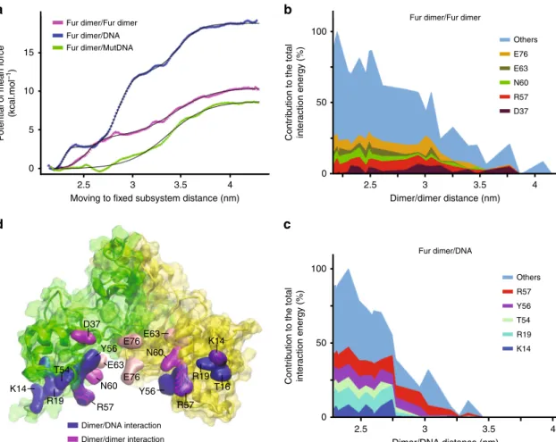
100 50 0 100 50 0Fur dimer/Fur dimer
Others E76 E63 N60 R57 D37 Others R57 Y56 T54 R19 K14 Fur dimer/Fur dimer
Fur dimer/DNA Fur dimer/DNA Fur dimer/MutDNA Dimer/DNA interaction Dimer/dimer interaction 10 P otential of mean f orce (kcal.mol –1 ) Contr ib
ution to the total
inter
action energy (%)
Contr
ib
ution to the total
inter action energy (%) 5 0 2.5 D37 E76E63 K14 R19 T16 R57 Y56 R57 N60 E76 E63 Y56 T54 R19 K14 N60
Moving to fixed subsystem distance (nm) Dimer/dimer distance (nm)
3 3.5 4 2.5 3 3.5 4
Dimer/DNA distance (nm)
2.5 3 3.5 4
Fig. 6 Computation of free energy profiles for dimer/dimer and dimer/DNA dissociation. a Potentials of mean force for the extraction of FtFur dimer from the tetramer or from DNA. The x-axis, reaction coordinate, corresponds to the average centre of mass/centre of mass distance between the 'fixed' and the 'moving' subsystems. Data points corresponding to the outputs of Wham arefitted with 1, 2 or 3 sigmoid functions with R55. Statistical errors were estimated using the bootstrap method.b Major contributors to the average interaction energy between Fur chain D and the 'fixed' dimer in the tetramer simulation. On average,five residues contribute to around 30% of the total interaction energy. c Major contributors to the average interaction energy between Fur chain D and DNA in the FtFur/wild-type DNA simulation. The x-axis corresponds to the average centre of mass/centre of mass distance between Fur and DNA. On average,five residues contribute to 54.5% of the total interaction energy. d Structure of FtFur dimer showing chain A (yellow) and chain D (green). Residues shown in blue surface are the major contributors to the average interaction energy between FtFur and DNA in the FtFur/ wild-type DNA simulation. Residues in magenta and pink surfaces are the major contributors to the interaction energy between the 'moving' dimer and the 'fixed' dimer in the FtFur tetramer simulation. The mutated residues E63 and E76 are in pink colour
importance of these residues in the stability of the tetramer since
dimeric forms were obtained, partially for the single mutants and
completely for the double mutant at high salt concentrations
(Supplementary Fig.
5
A, B). Both single mutants were still able to
bind DNA in the presence of metal ions contrarily to the double
mutant. Moreover, the Arg57–Glu63 salt bridge disruption
should leave room to interactions of the crucial Tyr56 with DNA.
Fur is involved in
F. tularensis virulence and pathogenicity.
The critical role of Fur in pathogenicity and virulence of several
pathogens is known
16,33. To investigate the putative role of FtFur
as a virulence factor we compared the phenotypes of CHUGA-Ft6
and CHUGA-Ft6Δfur using in vitro or in vivo infection models.
Three types of experiments were conducted: bacterial
multi-plication in J774-A1 murine macrophage-like cells, H
2O
2sensi-tivity assay and in vivo virulence assays in mice.
A growth defect of the CHUGA-Ft6 mutant lacking fur in
liquid medium was evidenced as shown by a longer lag time, a
longer generation time and a lower optical density at the
stationary phase as compared to the WT parental strain
(Supplementary Fig.
1
B). A similar phenotype was observed on
solid medium with a delayed onset of visible colonies and a
smaller size of colonies for CHUGA-Ft6Δfur (Supplementary
Fig.
1
C). The ability of CHUGA-Ft6, CHUGA-Ft6Δfur and
CHUGA-Ft6Δfur + fur to replicate within macrophages was then
evaluated by infecting J774-A1 murine macrophage-like cells.
One hour after infection, the host cells contained the same
number of intracellular bacteria regardless of the infecting strain
meaning that Fur is not required for macrophage infection. After
24 h incubation (Fig.
8
a), the number of intracellular bacteria was
markedly different as the WT cells were eight-fold more
abundant compared to the CHUGA-Ft6Δfur. The
fur-comple-mented strain showed an intermediate level of intracellular
macrophage multiplication. The ability of these bacterial strains
to resist an oxidative stress corresponding to the respiratory burst
set up by infected macrophages was also checked by growing
bacteria previously exposed to 1 mM H
2O
2during 4 h (Fig.
8
b).
CHUGA-Ft6 and CHUGA-Ft6Δfur + fur displayed a similar
percentage of survival while Ft6Δfur was much more sensitive to
the oxidative stress with about 50% of surviving cells.
The involvement of fur in the infectious process in vivo was
then evaluated by using mice infected with F. tularensis by
intranasal (IN) or intraperitoneal (IP) administration (Fig.
8
c, d).
The survival curves of the animals showed that regardless of the
administration route, CHUGA-Ft6 and CHUGA-Ft6Δfur + fur
caused the mice death in approximately the same delay, which is
95 h post-infection for IP and 150 to 168 h for IN inoculation. On
the other hand, mice infected with CHUGA-Ft6Δfur survived a
significantly longer time (p < 0.001 compared to CHUGA-Ft6 and
CHUGA-Ft6Δfur + fur whatever the route of infection), that is,
140 h and more than 200 h for the last animal infected by IP and
by IN routes, respectively. These results define fur as an
important virulence-associated gene in F. tularensis and are a
further example that deletion of this gene leads to an attenuated
phenotype in terms of virulence.
Discussion
Altogether, the involvement of Fur in the iron homeostasis and
the virulence of F. tularensis have been demonstrated here as well
as its direct interaction with the
figA promoter region. FtFur
belongs to the new family of tetrameric Fur proteins. It contains
the structural zinc site S1 and the regulatory site S2 and lacks the
third site S3, usually found in Fur proteins. S1 is not present in
tetrameric PaFur and S3 is absent in FtFur, which still forms a
tetramer upon deletion of S2, indicating that the tetrameric state
of the protein does not rely on such sites. To our knowledge, the
first published structure of FtFur containing the physiologically
relevant ferrous iron is presented here with a ferrous ion in an
octahedral geometry. Metalled FtFur behaves as a pre-activated
tetramer with the DNA-binding domains forming a positively
charged crown where the recognition helices interact through two
stabilizing salt bridges between two Arg57 (out of four) and two
14 Ec Pa Ft Hp Mg 16 19 37
a
c
d
b
54 56 57 R57 K14 R57Y56 K14 K14 R57 R57 Y56 R57 Y56 R57 K14 K14 R57 Y56 R57 K14Fig. 7 DNA-driven FtFur tetramer dissociation mechanism. a Electrostatic potential around FtFur calculated on parallel planes (left) and on equipotential surfaces at−0.1 (red) and 0.1 V (blue) (right). b Mn-FtFur structure with its solvent accessibility surface. The residues predicted to be involved in the DNA interaction are coloured.c Sequence alignment of the DNA-binding domains offive Fur proteins with known structure EcFur (DNA-binding domain X-ray structures only)56; PaFur27; FtFur (this work); HpFur (Helicobacter pylori)22and MgFur29. The highly conserved amino acids implicated, in site S2 (blue) and in the interactions with DNA are in bold, coloured in red for those forming base-specific interactions and black for those having interactions with the phosphates (as evidenced in the structure of MgFur in complex with DNA).d Sketch of the DNA FurBox double-strand highlighting interactions with four Fur subunits (forming two dimers). Each of them is shown in a specific colour: yellow, purple, green and cyan, corresponding to the residues shown in b. Interactions between DNA bases and Fur residues are deduced from our results and the structures of the MgFur–DNA complex (T highlighted in red interact with two subunits)
Glu63. The tetramer dissociation is driven by an interaction of the
protein with a specific DNA sequence, suggesting the
involve-ment of the two free Arg57 and Lys14, known to form
base-specific contacts with DNA. We postulate that the two H-bound
Arg57 would progressively lose their interaction with Glu63
replaced by interactions with DNA, leading to the breaking of the
salt bridge, crucial for the stability of the tetramer and its
dis-sociation into two dimers specifically bound to the FurBox. In
vivo studies reveal that FtFur is important for the virulence of F.
tularensis. Because there is no efficient vaccine and only few
poorly efficient antibiotics available to fight tularaemia, this work
shows that Fur is an attractive anti-virulence target for new
inhibitors, whose design, starting from already known inhibitors
against other Fur proteins
34, will be facilitated by the detailed
structure and mechanism of interaction with DNA.
Methods
Bacterial strains and culture media. The biovar I strain of F. tularensis subsp. holarctica used in the virulence assay, referred as CHUGA-Ft6, was isolated at Verdun Hospital (France) from a blood sample collected during routine care of a patient with typhoidal tularaemia. Identification at the species and subspecies level was obtained by PCR amplification and sequencing of the intergenic region between the 16SrRNA and 23SrRNA encoding genes35. Bacterial cultures were performed either on chocolate agar plates supplemented with PolyVitex (CPV, Biomérieux, Lyon, France) or in liquid brain heart infusion medium supplemented with 2% PolyViteX (BHI-2%PV). When necessary, kanamycin (10 µg mL−1) or sucrose (5% (w/v)) was added. Cultures were incubated at 37 °C, in a 5% CO2 -enriched atmosphere. Intracellular iron concentration was measured on stationary phase bacteria grown over 15 h in modified Mueller–Hinton medium into a shaking incubator (200 rpm at 37 °C). Briefly, the cells have been washed several times with phosphate-buffered saline-ethylenediaminetetraacetic acid (PBS-EDTA) 10 mM before hydrolysis with HNO3at 65% ON at 95 °C and measurements by ICP-AES (Shimadzu ICP 9000 instrument with Mini plasma Torch in axial reading mode20).
Construction and complementation of the Ft6Δfur mutant. A phase deletion of the fur gene was carried out in the CHUGA-Ft6 virulent isolate, by the method of
allelic exchange (Supplementary Fig.1), through the use of a suicide plasmid containing the sacB gene, pMP81236. Approximately 1000 bp adjacent regions of the Ft6 fur gene were amplified using the two primers LeftfurF-LeftfurR and RightfurF-RightfurR (Supplementary Table4). The obtained PCR products were mixed and further submitted to a second PCR using the forward primer LeftfurF and the reverse primer RightfurR, generating a PCR fragment containing the two adjacent regions of furflanked with the BamHI and EcoRV restriction sites. This fragment was digested with the two corresponding restriction enzymes and cloned into the plasmid pMP81236previously digested with the same enzymes. After
electroporation, several selection (kanamycin, then sucrose) steps were performed to obtain a delta fur mutant devoid of antibiotic resistance. In order to complement the strain Ft6Δfur, the fur gene and its promoter region were amplified using CompfurL and CompfurR primers and cloned the shuttle vector pMP828. The plasmid pMP828 containing the fur gene was then electropored into CHUGA-Ft6Δfur and complemented colonies selected on agar plates supplemented with kanamycin. The fur expression in resulting transformants was checked using a specific qRT-PCR.
Evaluation of the gene expression by qRT-PCR. Gene expression was measured from strains grown for 16 h in BHI-2%PV. Approximately 107cells were collected for total RNA extraction that was achieved using 1 mL TRIzol®reagent (Invitro-gen) and following the manufacturer’s instructions. Contaminating DNA was removed using the TURBO DNA-free™ Kit (Ambion, Life Technology). The first-strand complementary DNA (cDNA) synthesis reaction was carried out starting from 500 ng of purified RNA and using the SuperScript™ II Reverse Transcriptase Kit (Invitrogen, Life Technology). The resulting cDNA library was used as a template in combination with the specific primers for qRT-PCR, which was con-ducted using a Fast SYBR Green MasterMix in a StepOnePlus Real Time PCR Systems (Applied Biosystems). Cycling was 20 s at 95°C; 3 s at 95°C, 30 s at 60 °C, repeated for 40 cycles. The expression level of each target gene was calculated from three independent experiments and expressed as a ratio taking the expression of the housekeeping gene 16S RNA as the denominator. Primer sequences are indi-cated in Supplementary Table5.
DNA sample preparation. DNA oligonucleotides were synthesized by MWG at high purity scale. DNA duplexes werefirst annealed in water at concentration of 20 mg mL−1by heating the mixture at 95 °C for 5 min and rapid cooling on ice in buffer A (50 mM Tris-HCl, pH 8.8, 150 mM NaCl) and then stored at 4 °C. The formation and concentration of DNA duplexes were determined by SEC-MALLS-RI in binding buffer. DNA was used extemporary for biochemical experiment. 6000
a
b
c
d
100 80 Ft6 60 40 20 100 80 60 40 20 0 0 Ft6 Ft6ΔfurIntraperitoneal injection Intranasal injection
Ft6 Ft6Δ fur Ft6Δ fur +fur Ft6 Ft6Δ fur Ft6Δ fur +fur Ft6Δfur +fur Ft6Δfur +fur Ft6Δfur NS p < 0.001 p < 0.001 p < 0.001 p < 0.001 p < 0.001 5000 4000 Fold replication 3000 2000 1000 0 100 80 % survival % survival % survival 60 40 20 0 80 90 100
Post-infection time (h) Post-infection time (h)
110 120 130 140 140 160 180 200 220
Fig. 8 Fur is directly involved in F. tularensis virulence and pathogenicity. a Bacterial multiplication in J774-A1 murine macrophage-like cells. The data correspond to two independent experiments made in triplicate. P values were calculated using the Student's t test.b Hydrogen peroxide sensitivity assay expressed as the percent survival of each strain exposed 4 h to 1 mM H2O2. Bacterial suspensions were incubated 4 h with 1 mM H2O2before enumeration of the CFU onto chocolate agar plates. Data are expressed as mean ± SEM of three distinct experiments. The data correspond to two independent experiments made in triplicate. P values were calculated using the Student's t test.c, d In vivo virulence assay in mice inoculated IP with 5e2 CFU in 500µL of physiological serum or IN with 2e3 CFU in 50µL of physiological serum. Five mice were used for each condition and experiments were performed twice. Survival curves were compared using the Kaplan–Meier test. P values were <0.001 for CHUGA-Ft6 and CHUGA-Ft6Δfur + fur vs. CHUGA-Ft6Δfur, for both the IP and IN routes. No significant difference was found between CHUGA-Ft6 and CHUGA-Ft6Δfur + fur
Protein expression and purification
Apo-FtFurWT. Recombinant FtFur of F. tularensis FSC198 ref: NC-008245.1, 100% identical in sequence to the CHUGA-Ft6 protein, was purified as a tetramer containing one equivalent of Zn per subunit, as previously described20. It was
over-produced in BL21 DE3 R2 E. coli strain in Luria–Bertani (LB) medium after an overnight induction with 1 mM isopropylβ-D-1-thiogalactopyranoside (IPTG) at 18 °C. Cell were resuspended in buffer A, lysed by sonication and purified suc-cessively on several columns (GE Healthcare): (1) ion-exchange chromatography on DEAE Sepharose with linear gradient between buffer A and buffer A supple-mented with 1 M NaCl, (2) hydrophobic interaction chromatography on Butyl fast flow Sepharose with linear gradient between buffer A containing 1 M of ammo-nium sulphate and buffer A and (3) size-exclusion chromatography Superdex-200 (10/60) equilibrated with buffer A.
Apo-FtFur mutants. E63A, E76A and E63A-E76A mutants were cloned in pET-TEV (based on pET28a) vector to produce N-terminal 6×HisTag cleavable pET-TEV fusion proteins. PCR was done in the presence of appropriate primer with Phusion polymerase-HF at recommended Tm. PCR samples were incubated with a reaction buffer containing 2 UI of DpnI, 10 UI of T4 DNA ligase, 1 mM of ATP and 2 UI polynucleotide kinase (PNK) in PNK buffer from NEB for 15 min at room tem-perature (RT) before transformation in Top10 ultracompetent cells. Each mutant has been DNA-sequenced before expression and purification as described. H33A-H90A double mutant (FtFurΔS2) was obtained from pET30b-FtFurWT before cloning in pET-TEV, over-produced and purified like FtFurWT. The other mutants were over-produced in in BL21 (DE3) R2 E. coli strain LB medium after induction with 1 mM IPTG at 37 °C for 4 h. Purification was done by using Ni-NTA resin (Qiagen) in batch mode in buffer A with 10 mM imidazole and 10% glycerol. Pure protein fractions were pooled and mixed with homemade HisTag-TEV protease (1% of the protein concentration to purify) together with 1 mM dithiothreitol (DTT) and 1 mM EDTA. The solution was then dialysed using a 3 kDa cut-off membrane against 2 L buffer A containing 1 mM DTT and 1 mM EDTA at 4 °C overnight, followed by a second 2 L dialysis against buffer A to remove DTT and EDTA. The protein sample was then passed through the Ni-NTA column in order to separate the pure protein from the HisTag-TEV protein and the HisTag itself. A final step of purification was performed by using Superdex-200 in buffer A sup-plemented with 500 mM NaCl at 4 °C. Collected fractions were concentrated on a 50 kDa cut-off Vivaspin from 20 to 40 mg mL−1and used or frozen in liquid nitrogen in the presence of 10% of glycerol before storage at−80 °C.
Purification of the Mn-FtFur/Fur Box complex. The purification was done in buffer B (50 mM HEPES, pH 7.5, 150 mM NaCl, 1 mM MnCl2, 1 mM MgCl2) at 20 °C. Molar equivalents (1.2) of FurBox Duplex (see DNA sample preparation) were incubated with Mn-FtFur before loading on a Superdex-200 increase 10/30 GE Healthcare column equilibrated with buffer B. Pooled fractions were analysed in SEC-MALLS-RI in the same buffer to check the integrity of the complex.
EMSA and nuclease activity assay. EMSA experiments were performed as pre-viously described20. The formation of small-scale (under 1 µg) DNA duplexes was confirmed by native gel electrophoresis20on 10% acrylamide gel in 1× TAE buffer
(40 mM Tris-acetate, pH 8.2, 1 mM EDTA). DNA radiolabelling was performed by incubating 20 nM DNA for 30 min at 37 °C in the presence of 1 U of T4 poly-nucleotide kinase (NEB) and 1 µL ofγATP at 1 mCi mmol−1. Labelled DNA was diluted 10 times in buffer A, desalted on G25 Mini Spin Column and stored at−20 °C. EMSA were performed with 250 pM of freshly prepared radiolabelled DNA incubated 30 min at 25 °C with different concentrations of protein in a binding buffer (20 mM Bis-Tris propane, pH 8.5, 100 mM KCl, 3 mM MgCl2, 10 µM MnCl2, 5% (v/v) glycerol and 0.01% Triton X-100). After 30 min incubation at room temperature, 10 µL of each sample were loaded on 10 % polyacrylamide (29/1) gel. The gel was pre-run for 30 min at 100 V in TA buffer (40 mM Tris-acetate, pH 8.2) supplemented with 100 µM of MnCl2. Mobility shifts were revealed by exposing the gels on a storage phosphor screen (GE Healthcare) and quantified with a cyclone phosphoimager (Perkin Elmer). The nuclease activity assay was performed as previously described20.
SEC-MALLS-RI experiments. Each sample was checked by size-exclusion chro-matography coupled to multi-angle laser light scattering with online refractive index (SEC-MALLS-RI) as previously described20and using a standard procedure:
20 µL of sample with a 2 to 10 mg mL−1concentration were loaded on an analytical Superdex-S200 increase pre-equilibrated at 0.5 mL min−1with appropriate buffer (Apo-FtFur in buffer A, Mn-FtFur and Mn-FtFur/FurBox in 50 mM HEPES, pH 7.5, 150 mM NaCl, 1 mM MnCl2, 1 mM MgCl2) and connected to an in-line DAWN HELEOS II spectrometer (Wyatt Instruments). An in-line refractive index detector (Optirex, Wyatt Instruments) was used to follow the differential refractive index relative to the solvent. After baseline subtraction of the buffer solution, all samples presented a single peak allowing the determination of absolute molecular masses with the Debye model using ASTRA6 software (Wyatt Instruments) and a theoretical dn/dc value of 0.185 mL g−1. Thefinal values correspond to the average of three independent experiments.
Crystallization. Protein crystallization conditions were obtained by using crys-tallization screens (Hampton Resarch Grid ScreensTMand Qiagen protein crys-tallization suites) with the HTX Lab high-throughput robot screening (HTX Lab at EMBL-Grenoble). Diffracting crystals up to 8 Å were obtained in 50 mM MES, pH 5.6, 200 mM KCl, 5% (w/v) PEG 8000, 10 mM magnesium chloride hexahydrate. This crystallization condition was then manually optimized. Diffracting crystals up to 1.7 Å were obtained by 1 µL of a 16 mg mL−1Mn-FtFur holoprotein solution with 1 µL of 50 mM MES, pH 5.8, 20% (w/v) PEG 3350, 200 mM MgCl2·6H2O, 10 mM MnCl2reservoir solution, using the hanging drop vapour-diffusion method. Crystals of Fe-FtFur holoproteins were obtained in the same condition in the presence of 10 mM of (NH4)2Fe(SO4)2·6 H2O under anaerobic condition in a glove box. Crystals appeared in a few days and were back-soaked three successive times in a mother liquor containing 50 mM MES, pH 5.8, 20% (w/v) PEG 3350, 200 mM MgCl2·6H2O, to remove the excess of free metal. All crystals were cryoprotected using a solution obtained by adding 25% (v/v) glycerol to the mother liquor containing 25% (w/v) PEG 3350 andflash-cooled in liquid nitrogen.
X-ray diffraction and structure resolution of Mn-FtFur and Fe-FtFur. Diffrac-tion experiments were done on the beamline FIP-BM30-ESRF (European Syn-chrotron Radiation Facility, Grenoble, France).
For Mn-FtFur, afluorescence spectrum was recorded to check the presence of Mn at 1.77 Å (right side of maximum Fd″ for manganese) and Zn at 0.97 Å cations (Supplementary Fig.3A). A remote and an anomalous datasets were recorded at wavelengths 0.97 and 1.77 Å (right side of maximum F″ for manganese). Best dataset (0.97 Å) diffracted at 1.7 Å resolution. Diffraction data were integrated/ scaled in the space group P21using XDS Program Package version 15 October 201537. The structure was solved by the SAD method using Phenix 1.10.1-2155/ AutoSol38and 86% of the model was built automatically. The model was rebuilt/ corrected manually and refined using alternatively COOT39and REFMAC40,41.
Final refinement cycle was done in Phenix (Supplementary Table1and Supplementary Fig.3B).
For Holo-Fe-FtFur, afluorescence spectrum was recorded to check the presence of cations: Fe and Zn at 0.97 Å (Supplementary Fig.3A). The datasets were collected at wavelengths 0.97 Å with a resolution of 1.8 Å, integrated/scale by XDS Program Package in the space group P21.
The structure was solved by molecular replacement using MOLREP42with the
previous structure (Mn-FtFur) as the starting model. Indeed, we were not able to obtain a Fe-FtFur structure in the same conditions using iron Mohr salt in place of manganese even in a glove box. When trying to obtain the crystal from SEC-purified Mn-FtFur–DNA complex, only the same Mn-FtFur crystals were seen, but adding iron in the crystallization conditions, we were able to get crystals of Fe-FtFur diffracting at 1.8 Å and to solve the structure by molecular replacement with Mn-FtFur. The anomalous dataset was used to confirm the presence of Fe in the structure. The model was built and refined using REFMAC and COOT alternatively (Supplementary Table2and Supplementary Fig.3B).
Both Holo-Mn-FtFur and Holo-Fe-FtFurfinal structures were validated by molprobity43. Protein Data Bank (PDB) redo44was used before deposition of the structures to the PDB. The PDB codes are 5NBC for Holo-Mn-FtFur and 5NHK for Holo-Fe-FtFur. King software (in Phenix) was also used to cross-validate the data.
SAXS experiments. Before each experiment, all samples were extemporaneously re-purified on SEC Superdex-200 increase (GE Healthcare) 10/300 equilibrated in an appropriate buffer. SAXS data were collected at the ESRF (Grenoble, France) on beamline BM29 BioSAXS. The scattering profiles were measured at several con-centrations between 0.5 to more than 10 mg mL−1. Data were processed using standard procedures with the ATSAS v2.5.1 suite of programs45as described20. The ab initio determination of the molecular shape of the proteins was done as pre-viously described20. Briefly, radius of gyration (R
g), forward intensity at zero angle (I(0)), Porod volumes and Kratky plot were determined using the Guinier approximation and PRIMUS programs46. In order to build ab initio models, several independent DAMMIF32models were calculated in slow mode with pseudo-chain
option and analysed using the program DAMAVER47. Docking of the tetrameric X-ray structure into the measured SAXS envelope was generated by SUPCOMB48. The model of the Mn-FtFur/FurBox complex was built from the Mn-MgFur/ FurBox structure (PDB code 4RB1): after sequence alignment, the atom coordi-nates of the corresponding amino acids were directly copied from MgFur to FtFur and coordinates of missing side chain atoms were added from internal coordinates. The resulting model was energy minimized with CHARMM49. The program
CRYSOL32was used to generate the theoretical scattering curves from the tetra-meric structure of FtFur.
Cross-linking experiments. Cross-linking experiments between FtFur and Fur boxes were performed using 0.1% GTA. With a short spacer arm of approx: 5 Å, and when used at a low concentration, this cross-linker agent is well suited for intramolecular cross-linking and to specifically cross-link individual species in close interactions. Two micrograms of Mn-FtFur were used in each tube, fresh GTA was used at 0.1% and 25–50 and 100 ng of DNA oligonucleotides duplex was added sequentially. Incubation buffer are done with 1 mM of fresh MnCl2at RT
before being cross-linked by GTA 30 min at RT and loaded onto sodium dodecyl sulphate-polyacrylamide gel electrophoresis 4–20% acrylamide gradient. Construction of the models. The X-ray structure of FtFur solved in this study (Asp7 to Arg137) was used as the initial model for the tetramer. The GROMACS program version 5.1.250with the gromos54a7 united atom forcefield51was used to
perform long molecular dynamics simulations needed to compute free energy profiles. Fe2+and Zn2+were modelled as simple Lennard Jones hard spheres with charge+2 with Zn coordinated to charged deprotonated cysteines (see for details Supplementary Methods).
In the absence of FtFur+ DNA structure (FtFur/wtDNA), the structure of M. gryphiswaldense (4RB1)29in the presence of DNA was used to model the wtDNA
FurBox and correctly position FtFur dimer on wtDNA (by least-squarefit matching of atom positions). The 5′-GCCGGATAATGATAATCATTATC-3′ fragment (consensus FurBox in bold) and its complementary 3′–5′ sequence was used to model double-stranded wtDNA.
The mutDNA (equivalent to FurBoxm) sequence GCCGGATACTGATAGTC CTGATC contains four mutations with respect to the FurBox (A9 to C, A15 to G, A18 to C and T20 to G; see nucleotides set in bold font) which were constructed by simple matching of corresponding heavy atoms in the WT DNA model and building of missing hydrogens. The three above vacuum systems were immersed in parallelepipedic SPC52water boxes modelled with periodic boundary conditions after the addition of Na+and Cl–counterions to ensure neutrality and a total ionic force of 0.1 mol L−1. The solvated systems were energy minimized and equilibrated under NPT (constant Number of particles, Pressure and Temperature) conditions at 310 K and 1 atm.
Computation of free energy profiles. Free energy profiles for the extraction (by translation along afixed direction: Ox) of one FtFur dimer from the tetramer (dimer of dimers) and of FtFur from DNA were computed: the meticulous translation protocol is shown in Supplementary Fig.6.
The simulations include a 'moving' subsystem (FtFur dimer, chains A and D) and a 'fixed' subsystem (FtFur dimer, chains B and C, wtDNA or mutDNA) as shown in Supplementary Fig.7.
The profiles were built using the 'umbrella sampling' technique and result from the overlapping of 26 computation windows, one for each translation distance.
Each window consisted of 100 ps NPT equilibration and 10 to 15 ns NPT production simulations. Position restraints on the 'fixed' subsystem and distance restraints on the whole protein, in the form of NOE-type restraints (nuclear Overhauser effect) between H-bonded H and O atoms to maintain its secondary structure, were applied. The 'moving' subsystem was subject to two harmonic biasing forces along the X direction only ('umbrella potential') applied between the centres of mass of the 2 Fur dimer subunits and the centre of mass of the 'fixed' subsystem.
After the dynamics runs, positions and forces were collected from the trajectories and the umbrella sampling harmonic potential was unbiased using the Wham algorithm53implemented in the 'g_wham' program54to yield the free energy profiles.
Computation of average interaction energy profiles. Interaction energy profiles were computed by extracting nonbonded interactions (electrostatic+ Lennard Jones potential energies) from all the trajectories of the simulations and averaging for each window. Interaction energies were calculated between each residue in the Fur 'moving' dimer (chains A and D) and the 'fixed' subsystem (DNA or 'fixed' FtFur dimer).
H2O2sensitivity assay. Three independent overnight cultures of each strain (CHUGA-Ft6, CHUGA-Ft6Δfur and CHUGA-Ft6Δfur + fur) in BHI-2%PV medium were diluted in PBS to obtain 1 × 10⁷colony-forming unit (CFU) mL−1 inocula. These bacterial suspensions were incubated 4 h with 1 mM H2O2at room temperature without shaking. Culture samples taken before and after H2O2 exposure were serially diluted and spread onto CPV plates and incubated for 3 days at 37 °C. Counting of CFUs allowed to determine the percentage of surviving bacteria after H2O2exposure. Data were expressed as the mean of three inde-pendent experiments.
In vitro macrophage infection. J774-A1 murine macrophage-like cells were grown in 24-well microplate using Dulbecco's modified Eagle's medium (DMEM) (Gibco) medium supplemented with 10% decomplemented fetal calf serum (FCS, Gibco) at 37 °C in 5% CO2enriched atmosphere. For infections assays, confluent J774-A1 monolayers were infected with a bacterium inoculum at a multiplicity of infection of 10:1 (approximately 5 × 10⁶bacteria for 5 × 10⁵cells), and reincubated for 1 h (37 °C, 5% CO2). The cell monolayers were then washed with PBS (Gibco), and DMEM-10% FCS containing 5 µg mL−1of gentamycin was added for 1 h to kill non-phagocytized bacteria. The cell supernatant was then replaced with MEM-10% FCS, and cell cultures were incubated for 24 h (37 °C, 5% CO2). To evaluate bac-terial multiplication within J774-A1 macrophages, infected cell monolayers treated with 0.1 % saponin immediately after gentamycin treatment (T0) or after 24 h incubation (T24) were lysed and serially diluted in PBS and plated onto chocolate
agar plates enriched with PolyVitex for CFU numeration. Each point was per-formed in triplicate. Data obtained were compared using Student’s t test and P values <0.05 were considered statistically significant.
In vivo virulence assay. Six-week- to eight-week-old BALB/c males were infected with overnight cultures of the strains CHUGA-Ft6, CHUGA-Ft6Δfur and CHUGA-Ft6Δfur + fur diluted in 0.9% NaCl. Experiments were performed in an animal biosafety level 3 laboratory. For each bacterial strain, groups offive mice were inoculated either intraperitoneally (500 CFUs in 500 µL) or intranasally (2000 CFUs in 50 µL) and infected animals were monitored several times a day, weighed every day, and euthanized when they had reached one of the following limit point: prostration, high piloerection, weight loss >15% of T0 weight and closed eyes. A group of unifected mice was used as control. All murine experiments were approved by our local ethics committee (ComEth, Grenoble, France). During the experiments, mice were monitored several times a day, weighed every day and euthanized when we felt they had reached our estimated limit point (prostrate, high piloerection, weight loss >15% of T0 weight and eyes closed). All these experiments were performed in compliance with the laws and regulations regarding animal experimentation in France.
Data availability. The datasets generated during the current study are available from the corresponding authors on reasonable request. Coordinates and structure factors for Mn-FtFur and Fe-FtFur have been deposited in the RCSB Protein Data Bank under accession codes 5NBC and 5NHK, respectively.
Received: 9 January 2018 Accepted: 14 June 2018
References
1. Maurin, M. & Gyuranecz, M. Tularaemia: clinical aspects in Europe. Lancet Infect. Dis. 16, 113–124 (2016).
2. Meibom, K. L. & Charbit, A. The unraveling panoply of Francisella tularensis virulence attributes. Curr. Opin. Microbiol 13, 11–17 (2010).
3. Conlan, J. W., Chen, W., Shen, H., Webb, A. & KuoLee, R. Experimental tularemia in mice challenged by aerosol or intradermally with virulent strains of Francisella tularensis: bacteriologic and histopathologic studies. Microb. Pathog. 34, 239–248 (2003).
4. Maurin, M. Francisella tularensis as a potential agent of bioterrorism? Expert Rev. Anti Infect. Ther. 13, 141–144 (2015).
5. Oyston, P. C., Sjostedt, A. & Titball, R. W. Tularaemia: bioterrorism defence renews interest in Francisella tularensis. Nat. Rev. Microbiol. 2, 967–978 (2004). 6. Perez, N. M. & Ramakrishnan, G. The reduced genome of the Francisella
tularensis live vaccine strain (LVS) encodes two iron acquisition systems essential for optimal growth and virulence. PLoS ONE 9, e93558 (2014). 7. Ramakrishnan, G., Sen, B. & Johnson, R. Paralogous outer membrane proteins
mediate uptake of different forms of iron and synergistically govern virulence in Francisella tularensis tularensis. J. Biol. Chem. 287, 25191–25202 (2012). 8. Lindgren, H. et al. Iron content differs between Francisella tularensis subspecies
tularensis and subspecies holarctica strains and correlates to their susceptibility to H(2)O(2)-induced killing. Infect. Immun. 79, 1218–1224 (2011). 9. Ramakrishnan, G., Meeker, A. & Dragulev, B. fslE is necessary for
siderophore-mediated iron acquisition in Francisella tularensis Schu S4. J. Bacteriol. 190, 5353–5361 (2008).
10. Pan, X., Tamilselvam, B., Hansen, E. J. & Daefler, S. Modulation of iron homeostasis in macrophages by bacterial intracellular pathogens. BMC Microbiol. 10, 64 (2010).
11. Carrano, C. J. et al. Coordination chemistry of the carboxylate type siderophore rhizoferrin: the iron(III) complex and its metal analogs. Inorg. Chem. 35, 6429–6436 (1996).
12. Sullivan, J. T., Jeffery, E. F., Shannon, J. D. & Ramakrishnan, G.
Characterization of the siderophore of Francisella tularensis and role of fslA in siderophore production. J. Bacteriol. 188, 3785–3795 (2006).
13. Wehrly, T. D. et al. Intracellular biology and virulence determinants of Francisella tularensis revealed by transcriptional profiling inside macrophages. Cell Microbiol. 11, 1128–1150 (2009).
14. Deng, K., Blick, R. J., Liu, W. & Hansen, E. J. Identification of Francisella tularensis genes affected by iron limitation. Infect. Immun. 74, 4224–4236 (2006).
15. Kiss, K., Liu, W., Huntley, J. F., Norgard, M. V. & Hansen, E. J.
Characterization offig operon mutants of Francisella novicida U112. FEMS Microbiol. Lett. 285, 270–277 (2008).
16. Fillat, M. F. The FUR (ferric uptake regulator) superfamily: diversity and versatility of key transcriptional regulators. Arch. Biochem. Biophys. 546, 41–52 (2014).