HAL Id: hal-02666187
https://hal.inrae.fr/hal-02666187
Submitted on 31 May 2020
HAL is a multi-disciplinary open access
archive for the deposit and dissemination of
sci-entific research documents, whether they are
pub-lished or not. The documents may come from
teaching and research institutions in France or
abroad, or from public or private research centers.
L’archive ouverte pluridisciplinaire HAL, est
destinée au dépôt et à la diffusion de documents
scientifiques de niveau recherche, publiés ou non,
émanant des établissements d’enseignement et de
recherche français ou étrangers, des laboratoires
publics ou privés.
Copyright
Prions impair bioaminergic functions through
serotonin-or catecholamine-derived neurotoxins in neuronal cells
Sophie Mouillet-Richard, Noriyuki Nishida, Elodie Pradines, Hubert Laude,
Benoît Schneider, Cécile Féraudet, Jacques Grassi, Jean-Marie Launay,
Sylvain Lehmann, Odile Kellermann
To cite this version:
Sophie Mouillet-Richard, Noriyuki Nishida, Elodie Pradines, Hubert Laude, Benoît Schneider, et
al.. Prions impair bioaminergic functions through serotonin- or catecholamine-derived neurotoxins in
neuronal cells. Journal of Biological Chemistry, American Society for Biochemistry and Molecular
Biology, 2008, 283 (35), pp.23782-23790. �10.1074/jbc.M802433200�. �hal-02666187�
Prions Impair Bioaminergic Functions through Serotonin- or
Catecholamine-derived Neurotoxins in Neuronal Cells
*
□SReceived for publication, March 28, 2008, and in revised form, July 8, 2008 Published, JBC Papers in Press, July 9, 2008, DOI 10.1074/jbc.M802433200
Sophie Mouillet-Richard‡1, Noriyuki Nishida§2, Elodie Pradines‡, Hubert Laude¶, Benoît Schneider‡,
Ce´cile Fe´raudet储, Jacques Grassi储, Jean-Marie Launay**, Sylvain Lehmann§, and Odile Kellermann‡
From the‡Diffe´renciation Cellulaire et prions, CNRS FRE 2937, Institut Pasteur, INSERM U747, 7 rue Guy Moˆquet, 94801 Villejuif, France, §
Institut de Ge´ne´tique Humaine, CNRS UPR 1142, 141 rue de la Cardonille, 34396 Montpellier Cedex 5, France,¶Institut National de la Recherche Agronomique (INRA), Unite´ de Virologie et Immunologie Mole´culaires, 78350 Jouy-en-Josas, France,储Commissariat a` l’Energie Atomique (CEA), iBiTec-S, Service de Pharmacologie et d’Immunoanalyse, CEA/Saclay, 91191 Gif sur Yvette cedex, France, and **EA3621, Service de Biochimie, Hoˆpital Lariboisie`re, 75009 Paris, France and Pharma Research Department, Hoffmann La Roche AG, CH4070 Basel, Switzerland
The conversion of the cellular prion protein, PrPC, to an abnormal isoform, PrPSc, is a central event leading to neuro-degeneration in prion diseases. Deciphering the molecular and cellular changes imparted by PrPSc accumulation remains an arduous task due to the small number of cell lines supporting prion replication. Here we introduce the 1C11 cell line as a new in vitro model to investigate prion patho-genesis. This cell line is a committed neuroectodermal pro-genitor able to differentiate into fully functional serotonergic or catecholaminergic neurons. 1C11 cells, which naturally express PrPCfrom the undifferentiated state, can be chroni-cally infected with various prion strains. Prion infection does not promote any noticeable phenotypic change in the progen-itor cells nor prevent the onset of the serotonergic and cat-echolaminergic differentiation programs. Pathogenic prions, however, deviate the overall neurotransmitter-metabolism in both pathways by decreasing bioamine synthesis, storage, and transport, and enhancing catabolism. Noteworthy, oxidized derivatives of both serotonin and catecholamines are selec-tively detected in the differentiated progenies of infected cells and contribute to irreversible impairment in bioamine synthesis. Finally, the level of PrPSc accumulation, that of infectivity, and the extent of all prion-induced changes in infected cells appear to be correlated. The report of such spe-cific effects of infection on neuronal functions provides a foundation for dissecting the events underlying loss of neu-ronal homeostasis in prion diseases.
Prion diseases or transmissible spongiform
encephalopa-thies (TSEs)3are a group of fatal neurodegenerative disorders
that includes scrapie and bovine spongiform encephalopathy in animals and Creutzfeldt-Jakob disease in humans (1). Although sharing some hallmarks with other neurodegenerative diseases such as Alzheimer or Parkinson disease, TSEs are unique in that they can have a genetic, sporadic, or infectious origin. An enig-matic feature of infectious TSEs is the high latency that may range between a few months up to several decades. The occur-rence of various prion strains associated with different incuba-tion periods, clinical manifestaincuba-tions, and neuropathological lesions further illustrates the complexity of the host-pathogen relationship in the TSE field.
A key event in TSE pathogenesis is the conversion of the
cellular isoform of the prion protein, PrPC, into an abnormal
conformational variant called PrPSc, which stands for the
scrapie isoform of the prion protein (1). The central role played
by PrPCin the development of prion diseases was first
exempli-fied by the observation that PrP knock-out mice are resistant to TSE, whereas PrP-overexpressing (tga20) mice exhibit reduced incubation periods as compared with wild-type mice (2).
Neu-rograft experiments carried out on PrP⫺/⫺mice using tga20
mice as brain tissue donors clearly demonstrated that the
pres-ence of endogenous PrPC is mandatory for PrPSc to induce
pathological alterations (3). Mallucci et al. (4) further showed
that switching off PrPCneuronal expression in infected mice
just before the clinical phase blocks TSE pathogenesis, although abundant prion replication still occurs in extraneuronal tissues. These experiments, thus, outline that prions require neuronal
PrPCto exert their toxicity. This notion was recently reasserted
and refined in a study based on transgenic mice expressing a
glycosylphosphatidylinositol (GPI) anchor-less PrPC (⌬GPI
PrP) (5). Indeed,⌬GPI PrP infected mice were found to
effi-ciently replicate scrapie and accumulate high levels of PrPScin
*This work was supported by grants from the Groupement d’inte´reˆt scienti-fique “Infections a` prions,” the CNRS, the Fondation de la Recherche Me´di-cale, and the European Community (TSE Biotech BIO4CT98-6064, Neuro-prion FOOD-CT-2004-506579). The costs of publication of this article were defrayed in part by the payment of page charges. This article must there-fore be hereby marked “advertisement” in accordance with 18 U.S.C. Sec-tion 1734 solely to indicate this fact.
□S The on-line version of this article (available at http://www.jbc.org) contains
supplementalFigs. 1 and 2.
1To whom correspondence should be addressed. Tel.: 33-1-49-58-33-31; Fax:
33-1-49-58-33-29; E-mail: mouillet@vjf.cnrs.fr.
2Current address: Dept. of Molecular Microbiology and Immunology,
Naga-saki University Graduate School of Biomedical Science, 1-12-4 Sakamoto, Nagasaki 852-8523, Japan.
3The abbreviations used are: TSE, transmissible spongiform encephalopathy;
5,6-DHT, 5,6-dihydroxytryptamine; 5-HIAA, 5-hydroxyindolacetic acid; 5-HT, serotonin; 7,7⬘-D, 7,7⬘-bi-(5-hydroxytryptamine-4-one); CA, cate-cholamine; DA, dopamine; NE, norepinephrine; PrP, prion protein; PrPC,
cellular PrP; PrPSc, disease-associated scrapie PrP protein; SERT, serotonin
transporter; T-4,5-D, tryptamine 4,5-dione; TPH, tryptophan hydroxylase; VMAT, vesicular monoamine transporter; Bt2AMP, dibutyryl cyclic AMP;
DTNB, 5,5⬘-dithiobis(nitrobenzoic acid); ROS, reactive oxygen species; GPI, glycosylphosphatidylinositol.
THE JOURNAL OF BIOLOGICAL CHEMISTRY VOL. 283, NO. 35, pp. 23782–23790, August 29, 2008 © 2008 by The American Society for Biochemistry and Molecular Biology, Inc. Printed in the U.S.A.
at INRA Institut National de la Recherche Agronomique on June 14, 2018
http://www.jbc.org/
their brains without any sign of clinical illness. As a whole, these
data argue for a primary role of neuronal, GPI-anchored PrPCin
prion neuropathogenesis.
Despite this overall advance, unraveling the sequence of cel-lular and molecular events that lead to neuronal cell demise in TSEs still constitutes an ongoing challenge. Exploiting cell cul-ture systems that sustain stable prion replication represents a valuable approach to tackle this issue. Over the past decade prion-infected cell lines of neural or nonneural origin have shed
much light on PrPScbiogenesis, conversion, and trafficking (6).
In addition, their use as tools to detect infectivity (7), screen therapeutic compounds (8), or evaluate immunotherapy-based strategies (Ref. 9 and references therein) has been widely acknowledged. However, they have provided but little insight
into PrPSc-induced cellular dysfunction, as in most cases prion
infection had no obvious impact on the cell phenotype. Prion replication was nevertheless found to alter cholinergic func-tions in PC12 pheochromocytoma cells (10). In GT1 hypotha-lamic cells, infection is associated with reduced viability (11) and increased susceptibility to oxidative stress (12). Increased rates of apoptosis were also recently observed upon infection of primary neuronal cultures derived from tg338 mice overex-pressing ovine PrP (13).
Investigating how prions disrupt neuronal cell function may be notably hindered by the relative scarcity of cell systems of neural origin supporting prion replication to date. Here, we introduce the 1C11 neuroectodermal clone, which naturally
expresses PrPC(14), as a novel prion-permissive cell line. The
murine 1C11 progenitor derives from F9 pluripotent embryo-nal carcinoma cells and behaves as a committed neuroembryo-nal stem cell (15). This cell line has the unique capacity to differentiate
upon induction into fully functional serotonergic (1C115-HT) or
noradrenergic (1C11NE) cells. In the presence of dibutyryl
cyclic AMP (Bt2AMP), the 1C11 progenitor adopts a neuronal
morphology and starts expressing neuron-associated markers. Within 4 days, almost 100% of the precursor cells convert into
1C115-HTcells that display a complete serotonergic phenotype
including serotonin (5-HT) synthesis, storage, catabolism, and transport (15). On another hand 12 days after the addition of
Bt2AMP in combination with dimethyl sulfoxide (DMSO),
1C11NEcells implement a complete noradrenergic
differentia-tion program (15).
In the present work we demonstrate that 1C11 cells support the replication of various prion strains. Our results show that
PrPScaccumulation does not prevent the entry of 1C11 cells
into either neuronal differentiation program, but, however, triggers specific alterations of the overall
neurotransmitter-as-sociated functions in both 1C115-HTand 1C11NEcells. In
addi-tion, prions promote the production of oxidized derivatives of either serotonin or catecholamines, which are critically involved in bioaminergic dysfunctions. Finally, the extent of all prion-induced changes in infected cells correlate with the level
of PrPScaccumulation and that of infectivity.
EXPERIMENTAL PROCEDURES
Materials—Bt2AMP, cyclohexane carboxylic acid, DMSO, trypan blue, GSH, dithiothreitol, cysteine, catalase, and super-oxide dismutase were purchased from Sigma-Aldrich.
[Phenyl-6⬘-3H]paroxetine (20 –25 Ci/mmol) and 125I-labeled RTI-55
(2200 Ci:mmol) were from PerkinElmer Life Sciences. [3
H]Tet-rabenazin (20 Ci/mmol) was purchased from American Radio-labeled Chemicals Inc. (Saint Louis, MO). Proteinase K and Pefabloc were from Roche Applied Science. Antibodies against tryptophan hydroxylase (TPH) were from Genway (San Diego, CA). Protein concentrations were determined by the BCA pro-tein assay (Pierce).
Cell Culture—1C11 cells were grown in Dulbecco’s modified Eagle’s medium (Invitrogen) supplemented with 10% 5-HT-de-pleted fetal calf serum. The cells were induced to differentiate
(i) along the serotonergic pathway by the addition of 1 mM
Bt2AMP and 0.05% cyclohexane carboxylic acid and (ii) along
the noradrenergic pathway by the addition of 1 mMBt2AMP
and 0.05% cyclohexane carboxylic acid and 2% DMSO (15). Cell viability was assessed by trypan blue staining exclusion assay, according to the manufacturer’s protocol.
Ex Vivo Transmission and Subcloning of Infected Cells—10% brain homogenates were prepared in phosphate-buffered saline
containing 5% glucose as described (16). About 1⫻ 105cells in
a 35-mm culture dish were incubated for 4 –5 h with 1 ml of brain homogenate diluted at 0.2% in serum-free Dulbecco’s modified Eagle’s medium. 1 ml of fetal calf serum-supple-mented medium was then added. Cells were passaged at a 1:10
dilution every 3 or 4 days, and PrPScwas assayed by
immuno-blotting after 5 and 10 passages. Subcloning was performed at the first passage after the infection by limiting dilution (0.3 cell/ 6-mm titer well). The clones were then expanded, and the
pro-duction of PrPScwas tested by immunoblotting.
Immunoblotting—Protein extraction and immunological
detection of PrPScby Western blotting in mouse brain
homo-genates or cell lysates were conducted as described previously
(16). Briefly, 0.5 mg of total protein was digested with 10g of
proteinase-K at 37 °C for 30 min supplemented with 1 mM
Pefa-bloc and centrifuged at 20,000⫻ g for 45 min. The pellet was
resuspended in 1⫻ Laemmli sample buffer and boiled for 5 min before SDS-10% PAGE and electrotransfer on nitrocellulose membranes (Amersham Biosciences). PrP was detected using a mixture consisting of an equal volume of ascites of SAF 60, 69, and 70 antibodies (Commissariat a` l’Energie Atomique, Saclay, France), namely SAF mix. Western blots were revealed with an
enhanced chemiluminescence detection system (ECL,
Amersham Biosciences).
Bioassay—Tga20 mice overexpressing wild-type Mo Prnp-a (kindly provided by Dr. Charles Weissmann) were inoculated
intracerebrally with 20l of sample containing 0.1 or 10% of
Fukuoka-1 brain homogenate or cell extracts (2⫻ 106cells).
Cells were submitted to three freeze-thaw cycles, and suspen-sions were sonicated for 2 min (Cup-horn sonicator). Each sample was inoculated in 6 mice. Mice were monitored every 2 days, and the time at which they presented mouse-scrapie symptoms and terminal points were recorded as described pre-viously (17).
Quantification of Enzymatic Activities—Determination of enzymatic functions was conducted on uninfected and infected cells after induction of serotonergic or catecholaminergic dif-ferentiation. Cells were plated onto 10-cm culture dishes and differentiated into serotonergic neurons for 2– 4 days or into
at INRA Institut National de la Recherche Agronomique on June 14, 2018
http://www.jbc.org/
catecholaminergic neurons for 8 days. After 2 washings with cold phosphate-buffered saline, cells were scraped and
col-lected by centrifugation (10,000⫻ g, 3 min, 4 °C). Enzymatic
activities for neurotransmitter synthesis were measured radioenzymatically as in Levin et al. (18). Briefly, cell extracts were incubated for 30 min at 37 °C in an assay mixture
contain-ing 200 mMsodium acetate, pH 6.10, 1 mMferrous sulfate, 2 mM
6-methyl-H4-pterin, 40 mM2-mercaptoethanol, 20 mMsodium
phosphate, and 100M L-[3H] tryptophan orL-[3H] tyrosine to
probe TPH and tyrosine hydroxylase, respectively. Enzymatic activities were determined by quantifying the production of
[3H]
2O in a liquid scintillation counter and expressed as
pmol/30 min/mg of cell protein extract.
Glucose-6-phosphate dehydrogenase activity was measured as described by Costa Rosa et al. (19). The assay mixture
con-tained 100 mMTris HCl, 5 mMMgCl2, 0.5 mMNADP⫹, 1 mM
glucose 6-phosphate at pH 7.6. Enzyme activity was assayed by following the rate of NADPH production at 340 nm and 30 °C and expressed as pmol/30 min/mg of cell protein extract.
Determination of Bioamines, Metabolites, and Oxidation Products—The contents in bioamines and related metabolites were measured by HPLC combined to electrochemical detection as described in Lin et al. (20) with modifications. Briefly, cells were
homogenized in a solution containing 0.5Macetic acid, 0.5 M
sodium acetate, 0.4MNaClO4, and 4.7Mguaiacol, used as an
internal standard. Homogenization was performed in a Kontes ground glass Duall apparatus. Homogenates were centrifuged at
50,000⫻ g for 1 h at 4 °C. Supernatants were filtered through low
speed centrifugation on a 0.45m BAS (West Lafayette, IN)
poly-acetate filter. Filtrates were stored at⫺80 °C until required. The
levels of norepinephrine (NE), dopamine (DA), 5-HT, and related metabolites were measured using HPLC with a coulometric elec-trode array (ESA Coultronics, ESA Laboratories, Chelsford, MA). Quantifications were made by reference to calibration curves obtained with internal standards.
To determine the chemical structure of oxidized derivatives of 5-HT or catecholamines (CA), collected chromatographic fractions were freeze-dried and analyzed by fast-atom bom-bardment mass spectrometry using a VG instruments Model
ZAB-E spectrometer (Manchester, UK).1H and13C NMR
spec-tra were obtained with a Varian model 300XL spectrometer. After identification, the levels of oxidized derivatives of 5-HT and CA were quantified as above according to calibration curves established with pure standards obtained from Sigma or kindly provided by P. Bonetti (Hoffmann-La Roche).
Radioligand Binding Experiments—The presence of vesicu-lar monoamine transporter (VMAT) sites was assessed through tetrabenazine binding as in Mouillet-Richard et al. (15).
[3H]Paroxetine and125I-labeled RTI-55 binding to the 5-HT
transporter (SERT) was carried out as in Launay et al. (21). Briefly, cells were harvested in phosphate-buffered saline con-taining a mixture of inhibitors (Roche Applied Science). Cells were then pelleted by centrifugation and resuspended in cold
buffer A containing 4 mMEDTA, 1 mMEGTA, 0.1 mM
phenyl-methylsulfonyl fluoride, 10 mMimidazole, pH 7.3. The
super-natant was poured onto a 20% sucrose cushion and centrifuged
at 100,000⫻ g for 90 min. The pellet containing crude
mem-branes was resuspended in buffer B (75 mMKCl, 5 mMMgCl2, 1
mMEGTA, 10 mMimidazole, pH 7.3). Radioligand binding was
performed on crude membranes (20g of proteins) in 50 mM
Tris, pH 7.4 at 37 °C. A 30-min incubation period was followed
by addition of ice-cold Tris HCl, pH 7.4 (5 ml, 10 mM). Samples
were filtered on polyethyleneimine-treated filters, and radioac-tivity retained on filters was counted in liquid and solid scintil-lation counters (Packard Instrument Co.).
Treatment of TPH with Infected Cell Supernatant—TPH was
purified from 1C115-HTday-4 cells by affinity chromatography
using agarose beads coated with antibodies against TPH2 (EC
1.14.16.4). TPH immobilized on agarose beads (about 3g) was
incubated with 1 ml of 1C11Fk65-HTday-4 supernatants for 15
min at 30 °C. Protective agents (50MGSH, 500M
dithiothre-itol, 1 mM cysteine, 20 mMDMSO, 100 units/ml superoxide
dismutase, 100 units/ml catalase) when present, were added to TPH 10 min before incubation with infected cell supernatant.
Beads were washed 3 times with 50 mMpotassium phosphate
buffer, pH 7.40, and TPH activity was assayed in duplicates. DTNB titration of SH was carried out by the method of Ellman
as modified by Riddles et al. (22). Briefly, TPH from 1C115-HT
day-4 cells exposed or not to 1C11Fk65-HTday-4 supernatants was
incubated with 50MDTNB in 50 mMTris-HCl, pH 7.50, 6M
guanidine HCl. Absorbance was then recorded at 412 nm using a Cary 100 spectrophotometer (Varian Inc.).
Statistical Analysis—Statistics were performed on raw data using Student’s t test. The chosen significant criterion
was p⬍ 0.01.
RESULTS
The 1C11 Neuroectodermal Cell Line Supports Prion Replication—As an attempt to infect the 1C11 cell line, precur-sor cells were incubated with brain homogenates (0.2%) from mice inoculated with five distinct mouse-adapted agents derived from scrapie: Chandler, 22L, 22A, and ME7 or a human TSE, Fukuoka-1 (23). After 4 –5 h of incubation in serum-free medium, the inoculated cells were washed and passaged 5 times (3 weeks). Proteinase K-resistant PrP in the cell lysates was assayed by Western blot. Successful infections of 1C11 cells were obtained for the Chandler-, 22L-, and Fukuoka-1-infected cultures but not for the ME7 and 22A strains (Fig. 1A). In all
infected cultures, PrPScremained detectable after 10 passages
(not shown). Notably, PrPScsignals obtained in the
Chandler-and 22L-infected cells were lower than those observed in Fukuoka-1-infected cells. The later cells were selected for fur-ther studies and cloned by limiting-dilution to generate
cul-tures with higher and stable PrPScproduction (24). Two stably
infected 1C11Fk clones producing elevated (#6) or moderate
(#7) PrPSclevels (Fig. 1B) were chosen for further investigation.
At this stage no obvious phenotypic differences (growth rate, viability) could be noted between the infected clones and the original 1C11 neuroectodermal cells when cultured in their undifferentiated conditions.
PrPScfrom 1C11Fk Clones Is Infectious—An essential issue
was then to assess whether the infected cells also produced infectivity. Pellets of uninfected 1C11 and infected 1C11Fk #6 and #7 cells passaged 7 times after subcloning were inoculated
intracerebrally to tga20 mice. Mice (n⫽ 6) inoculated with 2 ⫻
106control 1C11 cells remained healthy over more than 250
Prion Infection Disturbs Bioaminergic Functions
at INRA Institut National de la Recherche Agronomique on June 14, 2018
http://www.jbc.org/
days post-inoculation. In contrast, mice inoculated with 1C11Fk #6 and #7 cells died within a mean incubation time of
76⫾ 3.5 (5/5 mice affected) and 89 ⫾ 1.3 (6/6) days,
respec-tively (Fig. 1C). As a comparison, the latencies of disease in
tga20 mice upon inoculation with 20 l of the original
Fukuoka-1 homogenate diluted to 10 or 0.1% were 76⫾ 0.7
(6/6) and 105⫾ 2.4 (5/5) days, respectively. Thus, we conclude
that 1061C11Fk #6 cells carry approximately the same level of
infectivity as 1 mg of crude Fukuoka-1-infected brain. Note-worthy, there was a 13-day delay between the incubation times with clones #6 and #7. It suggests that the #7 clone contains a 10-fold lower level of infectivity than the #6 clone, in agreement
with the difference in the PrPSccontents of the two clones as
estimated by Western blot (Fig. 1B). Altogether the above data show that 1C11Fk cells are chronically infected and produce infectious prions.
Prion Infection Impairs Serotonin Synthesis in 1C11Fk5-HT
Cells—Upon the addition of Bt2AMP, 1C11 cells undergo sero-tonergic differentiation with a nearly 100% frequency (15). The acquisition of serotonin-associated functions in differentiating
1C115-HTcells is highly synchronous and follows a defined time
schedule (15). Along the serotonergic pathway, 5-HT synthesis
becomes detectable in 1C115-HTday-2 cells and reaches
maxi-mal levels in 1C115-HTday-4 cells (15).
When exposed to inducers of the serotonergic pathway,
1C11Fk-derived cells, now referred to as 1C11Fk5-HT, kept
pro-ducing PrPSc(Fig. 1D). To evaluate the ability of prion-infected
1C11Fk cells to implement a serotonergic phenotype, we meas-ured the activity of TPH, the rate-limiting enzyme for 5-HT FIGURE 1. Ex vivo prion transmission to the 1C11 cell line. A, detection of
PrPScin 1C11 cells inoculated with Chandler (Ch), 22L, Fukuoka-1 (Fk), ME7, and 22A mouse brain homogenates. Lysates from confluent cells, passaged 5 times after inoculation, were digested with proteinase K and subjected to Western blotting. Molecular mass markers indicated on the left are in kDa.
B, clones were isolated from the 1C11 culture inoculated with Fukuoka-1
(1C11Fk). After 3 weeks of culture, 23 clones were isolated, expanded, and screened for PrPScproduction. Two stably infected 1C11Fk clones were estab-lished that produce high (clone #6) or moderate (clone #7) levels of PrPSc. C, survival curves of Tga20 mice after intracerebral inoculation with 2⫻ 106 cells or 20l of the original Fukuoka-1 homogenate. Mice inoculated with 1C11Fk6 and 1C11Fk7 cells died at days 76⫾ 3.5 and 89 ⫾ 1.3, respectively, whereas those inoculated with control 1C11 cells remained healthy (⬎250 days post-inoculation (d.p.i.)). D, detection of PrPScin prion-infected 1C11Fk6 cells induced toward the serotonergic (1C11Fk65-HT) or the noradrenergic (1C11Fk6NE) differentiation program for 4 days. Uninfected 1C11 progenies are included as negative controls.
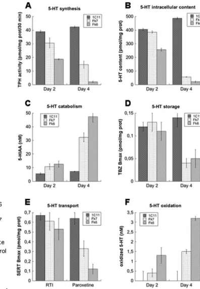
FIGURE 2. Implementation of serotonergic functions in 1C11Fk5-HTcells.
1C11Fk6 and 1C11Fk7 cells were induced to differentiate along the seroton-ergic pathway for 2 or 4 days. 5-HT synthesis (TPH activity) (A), intracellular content (B), catabolism (5-HIAA concentration) (C), storage (VMAT tetraben-azine binding) (D), and transport (SERT RTI-55 and paroxetine binding) (E) were assessed in cell lysates. The concentration of 5-HT-derived oxidized spe-cies (F) was measured in supernatants. Control values were determined from 1C115-HTd2 or d4 cells. Each bar represents the mean⫾ S.E. of triplicate values.
at INRA Institut National de la Recherche Agronomique on June 14, 2018
http://www.jbc.org/
synthesis, and the 5-HT intracellular content after the addition
of Bt2AMP. As shown in Fig. 2, A and B, serotonin synthesis was
detected in 1C11Fk5-HTcells at day 2. However, in
1C11Fk7-and 1C11Fk6-derived progenies, TPH activity reached only 79
and 48%, respectively, of the reference 1C115-HTday 2 level
(Fig. 2A). In parallel, a significant decrease (63% of control) in
5-HT content was recorded in 1C11Fk65-HT-infected cells as
compared with uninfected cells (Fig. 2B). With 1C11Fk75-HT
cells, the 5-HT content was only slightly reduced (95% of con-trol). The impact of prion infection on 5-HT synthesis was even more pronounced at day 4. Although TPH activity and 5-HT content normally increase between day 2 and day 4 of 1C11 serotonergic differentiation (Ref. 15) and Fig. 2, A and B), both
parameters were significantly decreased in 1C11Fk65-HTand
1C11Fk75-HTcells. The TPH enzymatic activity measured at
day 4 in 1C11Fk65-HTcells represented only 5% that recorded in
1C115-HT-uninfected cells. Similarly, the 5-HT content of
1C11Fk65-HTcells was reduced to 4.6% that of the reference
1C115-HTday-4 level. Interestingly, TPH activity and 5-HT
lev-els were less decreased in 1C11Fk7- than in 1C11Fk6-derived cells. Hence, the mean degree of enzymatic perturbation relates
to the mean level of PrPScaccumulation. Although
neurochem-ical analyses cannot be performed at the single cell scale, the
extent of phenotypic alterations (⬎95% of reference level in
1C11Fk65-HTcells) argues against the possibility that a small
proportion of cells within the culture carry the overall in-fectivity and account for 5-HT synthesis impairment. We may instead conclude to a dose-dependent inhibitory effect of prion infection on serotonin synthesis. In contrast to the above find-ings, prion infection had no impact on the activity of unrelated enzymes such as glucose-6-phosphate dehydrogenase, the rate-limiting enzyme in the pentose phosphate pathway, throughout
serotonergic differentiation (supplemental Fig. 1). Hence,
1C11Fk5-HT-infected cells exhibit selective impairment in
sero-tonergic functions. This first set of observations indicates that (i) 1C11Fk cells retain the ability to engage into serotonergic differentiation upon induction, (ii) prion infection hinders the implementation of an optimal TPH activity, (iii) prion-induced alterations are all the more severe since cells accumulate PrPSc
and infectivity (compare Fk6 and Fk7) and, finally, (iv) defects in serotonin synthesis accentuate along the time course of differentiation.
The Overall Serotonin Functions of 1C11Fk5-HT Cells Are Disturbed—We next compared the extent of 5-HT catabolism
in prion-infected 1C11Fk5-HT cells to that of uninfected
1C115-HTcells. At day 4 the concentration of
5-hydroxyindola-cetic acid (5-HIAA), a degradative product of 5-HT, was increased by 6.6-fold in 1C11Fk65-HTcells (Fig. 2C). Again, the
rise in 5-HIAA was less pronounced in 1C11Fk75-HTthan in
1C11Fk65-HT cells (4.5-fold). According to Wolf et al. (25)4,
such an increase in 5-HIAA production may be accounted for by an exacerbated activation of the 5-HT degrading enzyme monoamine oxidase A. It may also relate to a deficiency in 5-HT storage in infected cells. This is indeed substantiated by the reduced abundance of the VMAT in infected cells, as
assessed by [3H]tetrabenazine binding experiments (Bmax in
1C11Fk65-HTcells⫽ 36% of control) (Fig. 2D).
The serotonergic program of 1C115-HTcells is characterized
by the induction at day 4 of a functional SERT, which ensures serotonin re-capture from the extracellular space (21). SERT activity is controlled by post-translational modifications. We have previously reported on pharmacological tools that allow discrimination between functional and nonfunctional forms of the SERT protein (21). Indeed, SSRI antidepressant (e.g. parox-etine) recognition by SERT is restricted to functional mole-cules. On another hand, the cocaine congener RTI-55 allows tracing of total (i.e. immature, functional, and over post-trans-lationally modified) SERT molecules.
We were, thus, able to assess the SERT onset in infected cells.
The total number of SERT molecules in 1C11Fk65-HT and
1C11Fk75-HTcells was measured through125I-labeled RTI-55
binding. It represented nearly 80 and 90%, respectively, of the number measured in uninfected 1C115-HTcells (Fig. 2E). Such a
result indicates that despite infection, most cells have kept the capacity to implement the SERT and to acquire a complete serotonergic phenotype. We next evaluated the ability of the
SERT molecules to bind [3H]paroxetine. With 1C115-HT
con-trol cells, the maximal binding (Bmax) value was identical to the
Bmax for125I-labeled RTI-55, as expected for maximal 5-HT
uptake efficiency (21). With 1C11Fk65-HTcells, the Bmax for
[3H]paroxetine was only 22% that of the125I-labeled RTI-55
Bmax value. Hence, we deduce that in 1C11Fk65-HTcells, only
22% of the SERT molecules have the capacity to take up
sero-tonin. With 1C11Fk75-HT cells, the ratio of paroxetine to
RTI-55 binding equaled 54%, indicating a milder impairment of SERT function. The above results suggest that in prion-infected
1C11Fk5-HTcells from 50% (clone Fk7) to 75% (clone Fk6) of
the total number of SERT molecules have undergone post-translational changes that impair their functionality. Alto-gether, our data indicate that the overall serotonergic functions of 1C115-HTcells (i.e. 5-HT synthesis, catabolism, storage, and
transport) are affected by pathogenic prions.
1C11Fk5-HTSerotonergic Progenies Produce Serotonin-derived Oxidized Species That Inhibit Tryptophan Hydroxylase— Because oxidative stress is a hallmark of TSEs (26, 27), we won-dered whether prion infection would trigger the generation of oxidized bioamine derivatives in 1C11Fk-differentiated
proge-nies. 1C11Fk5-HT cell culture supernatants were analyzed
through HPLC coupled to electrochemical detection. Fractions in phase with redox potential deviation selectively obtained with infected supernatants were collected, dried, and analyzed by fast atom bombardment mass spectrometry and NMR. Chromatographic peaks 1, 7, and 10 were identified as tryptamine 4,5-dione (T-4,5-D), the dimer
7,7⬘-bi-(5-hy-droxytryptamine-4-one) (7,7⬘-D), and
5,6-dihydroxy-tryptamine (5,6-DHT), respectively (Fig. 3). As shown in Fig. 2F, the global concentration of these 5-HT oxidation products
was higher in 1C11Fk65-HTthan in 1C11Fk75-HTsupernatants.
None of these compounds was detected in 1C115-HTcontrol
cells (Fig. 2F). Of note, all three compounds identified in
1C11Fk5-HTsupernatants are detected in central serotonergic
neurons exposed to methamphetamine, where they are endog-enously produced as a consequence of serotonin oxidation (28).
4S. Mouillet-Richard and J.-M. Launay, unpublished observations.
Prion Infection Disturbs Bioaminergic Functions
at INRA Institut National de la Recherche Agronomique on June 14, 2018
http://www.jbc.org/
5,6-DHT and T-4,5-D mediate the neurotoxicity of metham-phetamine toward serotonergic neurons and are classified as serotonin neurotoxins (29). The production of these oxidized
species in 1C11Fk5-HTcells, within the nanomolar range, did
not, however, impact on cell viability according to trypan blue exclusion assay (data not shown).
Because T-4,5-D has been reported to irreversibly inactivate TPH through covalent modification of cysteine residues (30),
we evaluated the ability of 1C11Fk65-HTsupernatants to
inter-fere with TPH activity. In these experiments 1C115-HTcontrol
cells were used as the source of TPH (see “Experimental
Proce-dures”). Upon incubation with 1C11Fk65-HTsupernatant, the
activity of 1C115-HT-derived TPH
was reduced by 87% (Fig. 4A). To get further insight into the mechanisms sustaining the inhibition of TPH
activity, 1C115-HT-derived TPH was
incubated with various protec-tive agents before exposure to
1C11Fk65-HT supernatants. As
shown in Fig. 4A, TPH activity was not protected by DMSO, superox-ide dismutase, or catalase, which scavenge hydroxyl radicals,
super-oxide, and hydrogen peroxide,
respectively. Thus, we may rule out that loss of TPH activity upon
expo-sure to 1C11Fk65-HT supernatants
is directly caused by ROS. In con-trast, GSH or dithiothreitol partially rescued TPH activity from
inhibi-tion by 1C11Fk65-HT supernatants
and cysteine fully protected TPH from inactivation (Fig. 4A). Thus, we conclude that prion-in-fected cells produce reactive compounds distinct from ROS, which alter TPH activity through selective attack on SH groups.
Upon incubation with 1C11Fk65-HTsupernatants, all 12
cys-teine residues of TPH were in fact modified, as determined by titration of free SH groups with DTNB (Fig. 4B). As a whole, these results point to serotonin oxidation products as critical mediators of the damaging effects of prions on neurotransmit-ter-associated functions.
1C11FkNECells Display an Aberrant Noradrenergic
Metabo-lism and Produce Oxidized Derivatives of Catecholamines— Having shown drastic alterations in neurotransmitter homeostasis upon prion infection along the serotonergic differ-entiation pathway, we wondered whether the noradrenergic
progenies of 1C11Fk cells, referred to as 1C11FkNEcells, would
also be affected by the presence of PrPSc(see Fig. 1D). We first
compared the activity of tyrosine hydroxylase, the rate-limiting
enzyme for NE synthesis, in 1C11Fk6NEcells and in 1C11NE
control cells. As depicted in Fig. 5A, tyrosine hydroxylase activ-ity was reduced by 95% in the infected cells. In addition,
1C11Fk6NEcells exhibited dramatic reductions in the levels of
L-DOPA (L-3,4-dihydroxyphenylalanine 13% of control), DA
(4% of control), and NE (3% of control) (Fig. 5B). This reduction in CA content was accompanied by an increase (270% of con-trol) in the level of the NE metabolite
3-methoxy-4-hydroxy-phenylglycol (MHPG) (Fig. 5C). At last, in 1C11Fk6NEcells the
binding capacities of the vesicular transporter (VMAT) were below detectable levels, indicating defects in CA storage (Fig.
5D). 1C11Fk6NE supernatants contained various
cate-cholamine oxidation products, such as 6-hydroxydopamine, and tetrahydroisoquinolines (e.g. salsolinol) (Fig. 5C and data not shown). The noradrenergic progenies of the #7 clone also exhibited altered CA-associated functions. However, in
1C11Fk7NEcells the impact of prion infection was less drastic
than in 1C11Fk6NEcells (Fig. 5). Thus, we may conclude to the
dose-dependent effect of PrPScon CA metabolism, similar to
FIGURE 3. Production of oxidized bioamines in 1C11Fk-derived serotonergic progenies. Chromatogram profiles obtained with supernatants from 1C11Fk65-HT(A) and 1C11Fk75-HT(B) cells. C, peaks were identified as: 1, T-4,5-D; 7, 7,7⬘-D); 10, 5,6-DHT.
FIGURE 4. Impact of 1C11Fk5-HT-derived 5-HT oxidation products on TPH
activity. A, TPH was isolated from 1C115-HTday 4 cells and used to calibrate the control 100% activity. 1C115-HT-derived TPH was preincubated or not with the indicated protectant and exposed to 1C11Fk65-HTsupernatant. Remaining TPH activity is reported as % of the reference level. DTT, dithiothreitol; SOD, superox-ide dismutase. B, free cysteines were titrated by DTNB in 1C115-HT-derived TPH, exposed or not to 1C11Fk65-HTsupernatant (sup). All values are the means⫾ S.E. of n⫽ 4 experiments performed in duplicate.
at INRA Institut National de la Recherche Agronomique on June 14, 2018
http://www.jbc.org/
the changes evidenced along the serotonergic differentiation pathway.
The above results emphasize that, although infected cells have kept the ability to express all biochemical hallmarks of serotonergic or catecholaminergic differentiation, the presence
of PrPScdeviates the overall neurotransmitter metabolism
spe-cific to each pathway. In both cases bioamine synthesis and storage are decreased, and catabolism is enhanced. They point to bioamine-derived oxidized species as a selective conse-quence of prion accumulation in serotonergic or noradrenergic neuronal cells and a proximal cause of neurotransmitter dysfunction.
DISCUSSION
The present work introduces the 1C11 progenitor, which
naturally expresses PrPC, as a novel prion-susceptible cell line.
The 1C11 clone efficiently replicates Chandler, 22L, and Fukuoka-1 prions. The two clones selected here from a
Fukuoka-1-infected culture not only generate PrPScbut also
produce infectivity, as demonstrated by animal assay (Fig. 1C). The incubation time of the disease in inoculated mice was
inversely related to the level of PrPScin the infected Fk clones,
thus highlighting a correlation between PrPScproduction and
infectivity.
The 1C11 cell line used in our study exhibits the properties of a neuroectodermal stem cell. It expresses neuronal precursor cell markers and lacks neuronal functions (15). Noteworthy, prion infection of mouse fetal brain-derived progenitors was recently achieved (31, 32). That 1C11 precursor cells and neu-ronal stem cells sustain prion replication raises the possibility
that prion accumulation may interfere with adult neurogenesis in TSE-affected animals or patients. Actually, recent data
indi-cate that PrPCpositively regulates embryonic and adult
neuro-genesis (33). In view of the notion that prion pathoneuro-genesis
involves some corruption of PrPCnormal function in neuronal
cells (34), that PrPSccould affect neuronal stem cell
differenti-ation in TSEs deserves considerdifferenti-ation.
Our data show that PrPScaccumulation has no detectable
effect on the phenotype of 1C11 undifferentiated cells. Prion replication does not alter the choice of fate of 1C11 cells nor does it hinder the implementation of serotonergic or noradren-ergic functions. On another hand, prion infection has drastic consequences on the overall bioamine metabolism of differen-tiated 1C11Fk progenies. Our study takes advantage of the homogeneous and synchronous onset of neurotransmitter
syn-thesis, catabolism, storage, and uptake in 1C115-HTor 1C11NE
cells (15) and relies on pharmacological approaches to accu-rately quantify the overall phenotypic functions in infected
cells. In both prion-infected 1C11Fk5-HTand 1C11FkNEcells
we detect major impairments in bioamine synthesis, storage, uptake, and increases in serotonin or catecholamine catabo-lism. The prion-induced changes are comparable whether cells differentiate along the serotonergic or the noradrenergic path-way, adding to the significance of our observations. Besides, our quantitative analysis highlights a dose-dependent effect of pri-ons on serotonergic or noradrenergic functipri-ons since the phe-notypic alterations are all the more pronounced because the
cells accumulate PrPScand infectivity (Fk6 versus Fk7). It is of
note that infection of 1C11 cells with Chandler prions pro-moted similar biochemical changes in differentiated cells
(sup-plementalFig. 2). Although unprecedented in in vitro prion
infection studies, our overall observations are in agreement with the perturbations of monoaminergic functions reported in various experimental models of prion diseases (see Ref. 35 for review and references therein). Our conclusions also fit in with clinical (e.g. depression, (36)) or functional (e.g. reduced SERT availability (37)) evidence for dysfunction of the 5-HT system in TSE-affected patients.
Here, one major finding is the detection of serotonin and catecholamine oxidation products in the supernatants of infected cells. Serotonin and catecholamines are highly prone to oxidation and are normally protected against oxidative attack by storage into synaptic vesicles. The formation of oxi-dized derivatives in the differentiated progenies of 1C11Fk-in-fected cells likely involves alterations in 5-HT or CA storage. This idea is indeed substantiated by our observation that VMAT availability is reduced. Moreover, Wrona et al. (28) reported that exposure of cytoplasmic 5-HT to hydroxyl radi-cals yields 5,6-DHT and 4,5-dihydroxytryptamine, itself
con-verting into T-4,5-D. The 7,7⬘-D compound then arises from
dimerization of T-4,5-D (28). Similarly, defects in intracellular DA compartmentalization led to the generation of 6-hydroxy-dopamine and quinones and are thought to contribute to the pathogenesis of Parkinson disease (38). Because we detect these 5-HT- or CA-derived oxidized compounds in the supernatants of 1C11Fk5-HT- or 1C11FkNE-infected cells, they are likely
released through SERT- or NE transporter-mediated efflux (39).
FIGURE 5. Evaluation of noradrenergic functions in 1C11FkNE cells.
1C11Fk6 and 1C11Fk7 cells were induced to differentiate along the noradren-ergic pathway for 8 days. Cell lysates were collected to assess CA synthesis (tyrosine hydroxylase activity) (A), intracellular content (B), catabolism (3-me-thoxy-4-hydroxyphenylglycol (MHPG) concentration) (C), and storage (VMAT tetrabenazine binding) (D). The concentration of CA-derived oxidized species (C) was measured in supernatants. Control values were determined from 1C11NEd8 cells. Each bar represents the mean⫾ S.E. of triplicate values. DOPA,L-3,4-dihydroxyphenylalanine.
Prion Infection Disturbs Bioaminergic Functions
at INRA Institut National de la Recherche Agronomique on June 14, 2018
http://www.jbc.org/
The formation of oxidized derivatives of 5-HT and CA
clearly mirrors elevated ROS levels in infected 1C11Fk5-HTor
1C11FkNEcells. That prion infection alters the cellular redox
state in 1C11Fk cells is in keeping with various in vitro and in
vivoobservations (26, 27, 40). An enhanced ROS level could
simply stem from a deviation of the signal transduction
path-ways normally coupled with PrPC, which involve the
ROS-gen-erating enzyme NADPH oxidase (41, 42). Such a scenario would fit in with the current view that prion injury is primarily
caused by distortions of PrPCnormal function.
What are the molecular mechanisms responsible for the alterations in bioamine metabolism in 1C11Fk progenies? The generation of 5-HT and CA oxidation products appears to greatly contribute to neurotransmitter dysfunction. Our data
indeed demonstrate that 1C11Fk65-HT-derived oxidized
spe-cies have the potential to cancel TPH activity through modifi-cation of SH groups (Fig. 4). A modifimodifi-cation of critical cysteine residues by CA-derived quinones may also account for tyrosine
hydroxylase inactivation in 1C11FkNE cells (43). Because,
T-4,5-D alters SERT function through reaction with sulfhydryl groups (44), we suspect oxidative attack on the SERT cysteines to be involved in transporter dysfunction as well. Actually, we observed that more than 50% of the nonfunctional SERT
mol-ecules in 1C11Fk65-HTcells could revert to a functional state
upon treatment with an SH reducing agent.4The same
mecha-nism of SH oxidation may finally apply to the vesicular mono-amine transporter, which contains multiple cysteine residues critical for tetrabenazine recognition (45). Thus, generation of 5-HT and CA oxidation products capable to react with SH groups may deeply contribute to neurotransmitter dysfunction. The production of such bioamine-derived oxidized species may also account for increased 5-HT or CA catabolism. 5,6-DHT
concentrations below 1Mcan induce a rapid depletion of
5-HT associated with an increase in the level of 5-HIAA (46). The serotonin or catecholamine oxidation products act in a dose-dependent manner (46). At high concentrations the neu-rotoxins 5,7-DHT and 6-hydroxydopamine selectively damage serotonergic or dopaminergic neurons, respectively (29). Here, with 1C11Fk-derived progenies, these oxidized species exert toxicity at the level of neurotransmitter-associated functions only. The low concentration of these oxidation products in
1C11Fk5-HTand 1C11FkNEcells (Fig. 2F and 5C) may explain
why the cells remain viable. Nonetheless, within the context of prion diseases latencies, chronic accumulation of bioamine-de-rived neurotoxins in infected neurons could progressively impair neuronal cell function and, in the long-term, lead to neuronal cell demise in TSEs.
Collectively, our data draw a link between prion infection, cellular redox imbalance, and dysfunctions in neurotransmitter metabolism. We may assume that the disruption of cell
home-ostasis involves prion-induced alterations in PrPCnormal
sig-naling activity (42, 47). Because PrPSc-induced biochemical
alterations coincide with the acquisition of neurotransmitter-related functions, the 1C11 cell line may be a valuable model to investigate into the time-sequence of cellular insults, which contribute to the TSE-associated neurodegeneration. Another challenge will be the identification of therapeutic agents with
the capacity to antagonize PrPScdetrimental effects while
pre-serving PrPCnormal function.
Acknowledgments—We are grateful to Drs. R. Carp for providing 22L-, ME7-, and 22A-infected mouse brains, J. Tateishi and S. Kat-amine for providing Fukuoka-1-infected brains, and C. Weissmann for providing tga20 mice. We are greatly indebted to V. Mutel for fruitful discussion and to M. Bu¨eller (Hoffmann-La Roche) for out-standing experimental support. We thank Danielle Casanova for technical assistance.
REFERENCES
1. Prusiner, S. B. (1998) Proc. Natl. Acad. Sci. U. S. A. 95, 13363–13383 2. Bueler, H., Aguzzi, A., Sailer, A., Greiner, R. A., Autenried, P., Aguet, M.,
and Weissmann, C. (1993) Cell 73, 1339 –1347
3. Brandner, S., Raeber, A., Sailer, A., Blattler, T., Fischer, M., Weissmann, C., and Aguzzi, A. (1996) Proc. Natl. Acad. Sci. U. S. A. 93, 13148 –13151 4. Mallucci, G., Dickinson, A., Linehan, J., Klohn, P. C., Brandner, S., and
Collinge, J. (2003) Science 302, 871– 874
5. Chesebro, B., Trifilo, M., Race, R., Meade-White, K., Teng, C., LaCasse, R., Raymond, L., Favara, C., Baron, G., Priola, S., Caughey, B., Masliah, E., and Oldstone, M. (2005) Science 308, 1435–1439
6. Vilette, D. (2007) Vet. Res. 39, 10
7. Klohn, P. C., Stoltze, L., Flechsig, E., Enari, M., and Weissmann, C. (2003)
Proc. Natl. Acad. Sci. U. S. A. 100,11666 –11671
8. Kocisko, D. A., Morrey, J. D., Race, R. E., Chen, J., and Caughey, B. (2004)
J. Gen. Virol. 85,2479 –2483
9. Feraudet, C., Morel, N., Simon, S., Volland, H., Frobert, Y., Creminon, C., Vilette, D., Lehmann, S., and Grassi, J. (2005) J. Biol. Chem. 280, 11247–11258
10. Rubenstein, R., Deng, H., Scalici, C. L., and Papini, M. C. (1991) J. Gen.
Virol. 72,1279 –1285
11. Schatzl, H. M., Laszlo, L., Holtzman, D. M., Tatzelt, J., DeArmond, S. J., Weiner, R. I., Mobley, W. C., and Prusiner, S. B. (1997) J. Virol. 71, 8821– 8831
12. Milhavet, O., McMahon, H. E., Rachidi, W., Nishida, N., Katamine, S., Mange, A., Arlotto, M., Casanova, D., Riondel, J., Favier, A., and Lehmann, S. (2000) Proc. Natl. Acad. Sci. U. S. A. 97, 13937–13942
13. Cronier, S., Laude, H., and Peyrin, J. M. (2004) Proc. Natl. Acad. Sci.
U. S. A. 101,12271–12276
14. Mouillet-Richard, S., Ermonval, M., Chebassier, C., Laplanche, J. L., Leh-mann, S., Launay, J. M., and KellerLeh-mann, O. (2000) Science 289, 1925–1928
15. Mouillet-Richard, S., Mutel, V., Loric, S., Tournois, C., Launay, J. M., and Kellermann, O. (2000) J. Biol. Chem. 275, 9186 –9192
16. Nishida, N., Harris, D. A., Vilette, D., Laude, H., Frobert, Y., Grassi, J., Casanova, D., Milhavet, O., and Lehmann, S. (2000) J. Virol. 74, 320 –325 17. Vilette, D., Andreoletti, O., Archer, F., Madelaine, M. F., Vilotte, J. L., Lehmann, S., and Laude, H. (2001) Proc. Natl. Acad. Sci. U. S. A. 98, 4055– 4059
18. Levin, R. A., Pollard, H. B., and Kuhn, D. M. (1984) Anal. Biochem. 143, 205–208
19. Costa Rosa, L. F., Curi, R., Murphy, C., and Newsholme, P. (1995) Biochem.
J. 310,709 –714
20. Lin, P. Y. T., Bulawa, M. C., Wong, P., Lin, L., Scott, J., and Blank, C. L. (1984) J. Liq. Chromatogr. 7, 509 –538
21. Launay, J. M., Schneider, B., Loric, S., Da Prada, M., and Kellermann, O. (2006) FASEB J. 20, 1843–1854
22. Riddles, P. W., Blakeley, R. L., and Zerner, B. (1983) Methods Enzymol. 91, 49 – 60
23. Tateishi, J., Sato, Y., Nagara, H., and Boellaard, J. W. (1984) Acta
Neuro-pathol.(Berl.) 64, 85– 88
24. Race, R. E., Fadness, L. H., and Chesebro, B. (1987) J. Gen. Virol. 68, 1391–1399
25. Wolf, W. A., Youdim, M. B., and Kuhn, D. M. (1985) Eur. J. Pharmacol.
at INRA Institut National de la Recherche Agronomique on June 14, 2018
http://www.jbc.org/
109,381–387
26. Milhavet, O., and Lehmann, S. (2002) Brain Res. Brain Res. Rev. 38, 328 –339
27. Yun, S. W., Gerlach, M., Riederer, P., and Klein, M. A. (2006) Exp. Neurol. 201,90 –98
28. Wrona, M. Z., Yang, Z., McAdams, M., O’Connor-Coates, S., and Dry-hurst, G. (1995) J. Neurochem. 64, 1390 –1400
29. Baumgarten, H. G., and Lachenmayer, L. (2004) Neurotox. Res. 6, 589 – 614
30. Wrona, M. Z., and Dryhurst, G. (2001) Chem. Res. Toxicol. 14, 1184 –1192 31. Giri, R. K., Young, R., Pitstick, R., DeArmond, S. J., Prusiner, S. B., and
Carlson, G. A. (2006) Proc. Natl. Acad. Sci. U. S. A. 103, 3875–3880 32. Milhavet, O., Casanova, D., Chevallier, N., McKay, R. D., and Lehmann, S.
(2006) Stem Cells 10, 2284 –2291
33. Steele, A. D., Emsley, J. G., Ozdinler, P. H., Lindquist, S., and Macklis, J. D. (2006) Proc. Natl. Acad. Sci. U. S. A. 103, 3416 –3421
34. Mallucci, G., and Collinge, J. (2005) Nat. Rev. Neurosci. 6, 23–34 35. Ledoux, J. M. (2005) Med. Hypotheses 64, 910 –918
36. Zeidler, M., Johnstone, E. C., Bamber, R. W., Dickens, C. M., Fisher, C. J., Francis, A. F., Goldbeck, R., Higgo, R., Johnson-Sabine, E. C., Lodge, G. J., McGarry, P., Mitchell, S., Tarlo, L., Turner, M., Ryley, P., and Will, R. G. (1997) Lancet 350, 908 –910
37. Kloppel, S., Pirker, W., Brucke, T., Kovacs, G. G., and Almer, G. (2002)
J. Neural Transm. 109,1105–1110
38. Larsen, K. E., Fon, E. A., Hastings, T. G., Edwards, R. H., and Sulzer, D. (2002) J. Neurosci. 22, 8951– 8960
39. Fleckenstein, A. E., Gibb, J. W., and Hanson, G. R. (2000) Eur. J.
Pharma-col. 406,1–13
40. Wong, B. S., Brown, D. R., Pan, T., Whiteman, M., Liu, T., Bu, X., Li, R., Gambetti, P., Olesik, J., Rubenstein, R., and Sy, M. S. (2001) J. Neurochem. 79,689 – 698
41. Schneider, B., Mutel, V., Pietri, M., Ermonval, M., Mouillet-Richard, S., and Kellermann, O. (2003) Proc. Natl. Acad. Sci. U. S. A. 100, 13326 –13331
42. Pietri, M., Caprini, A., Mouillet-Richard, S., Pradines, E., Ermonval, M., Grassi, J., Kellermann, O., and Schneider, B. (2006) J. Biol. Chem. 281, 28470 –28479
43. Kuhn, D. M., Arthur, R. E., Jr., Thomas, D. M., and Elferink, L. A. (1999)
J. Neurochem. 73,1309 –1317
44. Chen, J. C., Crino, P. B., Schnepper, P. W., To, A. C., and Volicer, L. (1989)
J. Pharmacol. Exp. Ther. 250,141–148
45. Thiriot, D. S., and Ruoho, A. E. (2001) J. Biol. Chem. 276, 27304 –27315 46. Wolf, W. A., and Bobik, A. (1988) J. Neurochem. 50, 534 –542
47. Mouillet-Richard, S., Pietri, M., Schneider, B., Vidal, C., Mutel, V., Launay, J. M., and Kellermann, O. (2005) J. Biol. Chem. 280, 4592– 4601
Prion Infection Disturbs Bioaminergic Functions
at INRA Institut National de la Recherche Agronomique on June 14, 2018
http://www.jbc.org/
Odile Kellermann
Schneider, Cécile Féraudet, Jacques Grassi, Jean-Marie Launay, Sylvain Lehmann and
Sophie Mouillet-Richard, Noriyuki Nishida, Elodie Pradines, Hubert Laude, Benoît
Catecholamine-derived Neurotoxins in Neuronal Cells
doi: 10.1074/jbc.M802433200 originally published online July 9, 2008 2008, 283:23782-23790.
J. Biol. Chem.
10.1074/jbc.M802433200
Access the most updated version of this article at doi: Alerts:
When a correction for this article is posted
•
When this article is cited
•
to choose from all of JBC's e-mail alerts
Click here
Supplemental material:
http://www.jbc.org/content/suppl/2008/07/11/M802433200.DC1 http://www.jbc.org/content/283/35/23782.full.html#ref-list-1This article cites 47 references, 23 of which can be accessed free at
at INRA Institut National de la Recherche Agronomique on June 14, 2018
http://www.jbc.org/