1
Prmt5 promotes vascular morphogenesis independently of its methyltransferase 2
activity. 3
4
Aurélie Quillien1*, Manon Boulet1,2, Séverine Ethuin1, Laurence Vandel1,2*
5 6 7
1Centre de Biologie du Développement (CBD), Centre de Biologie Intégrative (CBI),
8
Université de Toulouse, CNRS, UPS, France 9
2Current affiliation : Université Clermont Auvergne, CNRS, Inserm, GReD, F-3000
Clermont-10 Ferrand, France 11 12 13 *Corresponding Authors 14
Aurélie Quillien (aurelie.quillien@gmail.com) 15
Laurence Vandel (laurence.vandel@uca.fr) 16 17 18 19 20 21
ABSTRACT 22
23
During development, the vertebrate vasculature undergoes major growth and remodeling. 24
While the transcriptional cascade underlying blood vessel formation starts to be better 25
characterized, little is known concerning the role and mode of action of epigenetic enzymes 26
during this process. Here, we explored the role of the Protein Arginine Methyl Transferase 27
Prmt5 during blood vessel formation and hematopoiesis in zebrafish. Through the generation 28
of a prmt5 mutant, we highlighted a key role of Prmt5 in both hematopoiesis and blood vessel 29
formation. Notably, we showed that Prmt5 promotes vascular morphogenesis through the 30
transcriptional control of ETS transcription factors and adhesion proteins in endothelial cells. 31
Interestingly, we found that Prmt5 methyltransferase activity is not required to regulate gene 32
expression, and the comparison of chromatin architecture impact on reporter genes expression 33
leads us to propose that Prmt5 rather regulates transcription by acting as a scaffold protein 34
that facilitates chromatin looping in these cells. 35
36
Key words 37
Prmt5; zebrafish; angiogenesis; hematopoiesis; endothelial cells; chromatin looping 38 39 40 41 42 43 44 45 46 47
INTRODUCTION 48
Blood vessel formation is an essential developmental process required for the survival of all 49
vertebrates and much effort has been devoted to understand the molecular pathways and to 50
identify key molecules that regulate different aspects of this process. Interestingly, the vascular 51
anatomy and the mechanisms involved in vessel formation are highly conserved among 52
vertebrates (for a review, (Isogai et al., 2001)). Hence, in the past two decades, zebrafish has 53
been proven to be a useful model to study vascular morphogenesis and blood cell formation 54
in vivo (Beis and Stainier, 2006; Lawson and Weinstein, 2002a; Thisse and Zon, 2002). 55
In vertebrates, blood cell formation is tightly associated with the development of the vascular 56
system. Hematopoietic Stem Cells (HSC), which give rise to the different blood cell lineages, 57
emerge directly from the ventral part of the dorsal aorta, an area referred to as the hemogenic 58
endothelium. Notably, the ETS transcription factor ETV2 functions as a master regulator for 59
the formation of endothelial and hematopoietic cell lineages through the induction of both blood 60
cells and vasculature transcriptional programs, in mouse and in zebrafish (Liu et al., 2015b; 61
Wong et al., 2009). In endothelial cells, ETV2 regulates the expression of other ETS 62
transcription factors, VEGF (Vascular Endothelial Growth factor) signaling receptors and 63
effectors, Rho-GTPases and adhesion molecules (Liu et al., 2015b; Wong et al., 2009). 64
Besides, adhesion molecules have been shown to be crucial players in vascular 65
morphogenesis as Vascular Endothelial cadherin (VE-cad/ cdh5) and endothelial cell-selective 66
adhesion molecule (Esama) are essential for junction remodeling and blood vessel elongation 67
in zebrafish (Sauteur et al., 2017; Sauteur et al., 2014). Indeed, loss of function of both cdh5 68
and esama leads to the formation of disconnected vessels and delayed lumen formation. 69
Likewise, knock down of the scaffold protein Amolt2, which associates to VE-cadherin, also 70
leads to sprout elongation defects and narrowed aortic lumen (Hultin et al., 2014). While the 71
transcriptional cascade underlying blood vessel formation starts to be better characterized, 72
little is known concerning the role and mode of action of epigenetic enzymes during this 73
process. Even though chromatin-modifying enzymes have been described as central in 74
cardiovascular disease and development (Rosa-Garrido et al., 2018; Shailesh et al., 2018), 75
only few examples illustrate in detail the role of epigenetic enzymes during blood vessel 76
development. For instance, the chromatin-remodeling enzyme BRG1 affects early vascular 77
development as well as hematopoiesis in mice (Griffin et al., 2008), and the histone 78
acetyltransferase P300 has been proposed to be recruited at the promoter of specific 79
endothelial genes by the ETS transcription factor ERG (ETS Related Gene) to control their 80
expression both in vivo in zebrafish and in HUVEC (Human Umbilical Vein Endothelial Cell) 81
(Fish et al., 2017; Kalna et al., 2019). 82
Given the common origin of blood and endothelial cells, and their partially shared 83
transcriptional programs, it is plausible that known chromatin-modifying enzymes affecting 84
hematopoiesis could also control blood vessel formation. Along this line, the epigenetic 85
enzyme Prmt5 (Protein Arginine Methyltransferase 5) has been identified as a key player in 86
blood cell formation (Liu et al., 2015a) but its impact on endothelial development has not been 87
investigated to date. Prmt5 catalyzes the symmetric di-methylation of arginine residues on a 88
variety of proteins including histones and therefore acts on many cellular processes such as 89
genome organization, transcription, differentiation, cell cycle regulation or spliceosome 90
assembly, among others (Blanc and Richard, 2017; Karkhanis et al., 2011; Stopa et al., 2015). 91
Prmt5 is mainly known to repress transcription through the methylation of arginine residues on 92
histones H3 and H4 and has been shown to regulate several differentiation processes such as 93
myogenesis, oligodendrocyte and germ cell differentiation or hematopoiesis (Batut et al., 2011; 94
Liu et al., 2015a; Shailesh et al., 2018; Zhu et al., 2019). In mice, prmt5 knock out prevents 95
pluripotent cells to form from the inner cell mass and is embryonic lethal (Tee et al., 2010). 96
Conditional loss of prmt5 in mice leads to severe anemia and pancytopenia and Prmt5 97
maintains Hematopoietic Stem Cells (HSCs) and ensures proper blood cell progenitor 98
expansion (Liu et al., 2015a). Loss of prmt5 leads to oxidative DNA damages, increased cell 99
apoptosis due to p53 dysregulation and as a consequence, to HSC exhaustion. In this context, 100
Prmt5 protects HSCs from DNA damages by allowing the splicing of genes involved in DNA 101
repair (Tan et al., 2019). 102
Here, we explored the role of the Protein Arginine MethylTransferase Prmt5 during blood 103
vessel formation and in hematopoiesis in zebrafish. Through the generation of a prmt5 mutant, 104
we highlight the key role of this gene during vascular morphogenesis via the control of 105
expression of several ETS transcription factors and adhesion molecules. Moreover, we show 106
that Prmt5 methyltransferase activity is not required for blood vessel formation and our results 107
suggest that Prmt5 helps to shape correct chromatin conformation in endothelial cells. 108
109
RESULTS 110
Prmt5 is required for HSC maintenance and lymphoid progenitor expansion 111
To characterize prmt5 function, we generated a prmt5 mutant by targeting the second exon of 112
prmt5 with the CRISPR/Cas9 system. A deletion of 23 nucleotides was obtained, leading to a 113
premature stop codon before the catalytic domain of Prmt5 (Fig. 1A). As a consequence, 114
Prmt5, which was expressed ubiquitously in the trunk at 24 hours post fertilization (hpf), was 115
no longer detected in the mutant (Fig. 1B, C). Similarly, Prmt5 expression was severely 116
reduced in prmt5 morpholino-injected embryos as compared to control morphants (Fig. S1 A, 117
B) (Batut et al., 2011). In order to test whether Prmt5 regulates hematopoiesis in zebrafish, 118
we took advantage of the transgenic line Tg(gata2b:Gal4;UAS:lifeactGFP) that labels 119
Hematopoietic Stem Cells (HSCs) (Butko et al., 2015). HSCs emerge from the ventral wall of 120
the dorsal aorta (DA, Fig. 1D, D’), before migrating into the Caudal Hematopoietic Tissue 121
(CHT) (Fig. 1D) where Hematopoietic Stem and Progenitor Cells (HSPCs) proliferate and 122
undergo maturation (Butko et al., 2015). Reminiscent of the data published in mice (Liu et al., 123
2015a), the loss of prmt5 led to an increased number of gata2b+ HSCs in 36 hpf mutant 124
embryos as compared to wild type ones (Fig. 1E-G). In addition, we found that the relative 125
expression of scla, runx1 or cmyb, which are specifically expressed in emerging HSCs, was 126
increased in prmt5 mutant embryos as compared to wild type embryos (Fig. 1H). These results 127
suggest that Prmt5 regulates the number of emerging HSCs from the dorsal aorta. We next 128
investigated whether blood cell formation was impaired in prmt5 zebrafish mutant as described 129
in mouse (Liu et al., 2015a). HSPCs give rise to different blood cell progenitors, such as 130
lymphoid progenitors which colonize the thymus leading to T lymphopoiesis (Fig. 1D)(Ma et 131
al., 2013). As gata2b+ lymphoid progenitors deriving from gata2b+ HSCs can be detected in 132
the thymus of transgenic zebrafish larvae from day 3 (Butko et al., 2015), we investigated 133
whether Prmt5 could act on theses progenitors. Indeed, we found that at 5 days, the number 134
of gata2b+ lymphoid progenitors in the thymus was significantly reduced in prmt5 mutant and 135
in morphant embryos as compared to wild type embryos (Fig. 1I-K, Fig. S1 C, D, G), 136
suggesting that Prmt5 is required for lymphoid progenitor expansion. Altogether, these data 137
indicate an important and conserved role of Prmt5 during hematopoiesis in zebrafish as in 138
mouse. 139
Prmt5 is required for vascular morphogenesis 140
As Prmt5 regulates zebrafish hematopoiesis, we next asked whether Prmt5 could also play a 141
role during blood vessel formation, either during angiogenesis or vasculogenesis. First, we 142
analyzed the expression and localization of Prmt5 by immunostaining in Tg(fli1a:eGFP) 143
transgenic embryos, in which endothelial cells can be visualized with egfp (Lawson and 144
Weinstein, 2002b). We found that Prmt5 was clearly expressed in early endothelial cells at 14 145
somite stage (Fig. 2A-A’’). At 24 hpf, Prmt5 was expressed in endothelial cells of the dorsal 146
aorta (DA) and of the cardinal vein (CV) (Fig. 2B, B’, D). Prmt5 was also detected in 147
Intersegmental Vessels (ISVs) sprouting from the DA, in either the tip cell (leading the sprout) 148
or the stalk cell (Fig. 2C, C’, D). We then analyzed whether blood vessel formation was affected 149
in transgenic Tg(fli1a:eGFP) prmt5 mutants at 28 hpf. We found that the dorsal aorta diameter 150
of mutant embryos was reduced as compared to the control (Fig. 2D, E, F close-ups), 151
suggesting that lumen formation was perturbed. To confirm this result, we made use of the 152
Notch reporter line Tg(TP1bglob:VenusPEST)s940 in which only the dorsal aorta cells express
the transgene while the cardinal vein endothelial cells do not (Ninov et al., 2012; Quillien et al., 154
2014). In this transgenic context the area occupied by the dorsal aorta in prmt5 morphant 155
embryos was significantly reduced as compared to control embryos (Fig. 2G-I). Prmt5 mutant 156
embryos also showed a defect of sprouting ISV to reach the most dorsal part of the trunk and 157
to connect with other ISVs and form the Dorsal Longitudinal AnastomoticVessel (DLAV) (Fig. 158
2D, E, F). This defect was associated with a significant reduction of ISV length (Fig. 2E, F, K) 159
but with no impact on the number of endothelial cells (Fig. 2J). The observed size reduction of 160
ISVs is thus most likely the result of an elongation issue rather than a proliferation defect. Of 161
note, prmt5 morphants reproduced the phenotype observed in prmt5 mutants i.e. a reduced 162
ISV length at 28 hpf (Fig. S2 A-D). 163
To get a better insight into the impact of Prmt5 on the dynamics of vascular system formation, 164
we performed time-lapse analyses in control and prmt5 morphant embryos. Time-lapse 165
confocal movies were carried out from 28 hpf to 38 hpf to follow the elongation of ISVs to the 166
formation of an effective lumen. As compared to control morphants, prmt5 morphants showed 167
an impaired formation of ISV lumen and DLAV. Indeed, in prmt5 morphants tip cells failed to 168
stay connected to the stalk cells and to contact other tip cells to allow the formation of the 169
DLAV (Fig. 3A-B). Moreover, supernumerary connections were detected in the context of 170
prmt5-loss of function (Fig. 3B). Altogether, these data suggest a central role for Prmt5 in 171
vascular morphogenesis. 172
The master gene regulator ETV2, ETS transcription factors and adhesion proteins have been 173
shown to be involved in blood vessel formation (Craig et al., 2015; Hultin et al., 2014; Pham et 174
al., 2007; Sauteur et al., 2017; Sauteur et al., 2014). Analyzing single cell RNA-sequencing 175
data from Wagner et al. (Wagner et al., 2018), allowed us to determine that prmt5 is expressed 176
in endothelial cells at 10 hpf (like etv2 and fli1a) and that its expression decreases in later 177
stages, when the expression of fli1b, cdh5, agtr2, esama, and amotl2a starts to increase (Fig. 178
S3). To test whether Prmt5 could regulate the expression of these genes, we performed RT-179
qPCR experiments on mutant embryos and on their wild type counterparts. While we found 180
that etv2 expression was not affected, the expression of ETS transcription factors (fli1a, fli1b) 181
and adhesion proteins (cdh5, agtr2, esama and amotl2a), all putative ETV2 target genes (Liu 182
et al., 2015b; Wong et al., 2009), was significantly reduced in prmt5 mutant (Fig. 3C). Of note, 183
we also detected a reduction of fli1a and cdh5 expression in prmt5 mutant by in situ 184
hybridization (Fig. S4). As etv2 expression was unaffected by the loss of prmt5 but its targets 185
were down-regulated, it is tempting to speculate that Prmt5 could modulate ETV2 activity at 186
post-translational level. 187
Prmt5 methyltransferase activity is not required for vascular morphogenesis 188
That Prmt5 modulates gene expression by methylating a variety of proteins including histones 189
but also transcription (co)factors led us to test whether Prmt5 methyltransferase activity was 190
required for vascular morphogenesis and lymphoid progenitor formation. To this end, prmt5 191
mutant or morphant embryos were injected with wild type human prmt5 mRNA (hprmt5WT) or 192
with a catalytic mutant form of this mRNA (hprmt5MUT) (Pal et al., 2003). In mice, the 193
expansion of lymphoid progenitor relies on Prmt5 methyltransferase activity (Liu et al., 2015a). 194
Consistent with this, hprmt5WT but not prmt5MUT mRNA, was able to restore normal lymphoid 195
progenitor expansion in prmt5 morphant embryos (Fig. S1 C-G). This underscores the 196
conserved requirement of PRMT5 methyltransferase activity for lymphoid progenitor formation 197
in human and zebrafish. We then tested whether the same was true for ISV elongation and the 198
expression of etv2 target genes. Surprisingly, we found that both mRNAs were able to restore 199
ISV elongation, albeit to a slightly different extend, as indicated by the average ISV length in 200
injected mutant embryos as compared to non-injected mutants (Fig. 4A-E). Indeed, we 201
observed that the average length of ISVs in hprmt5WT-injected mutants was even longer than 202
intersegmental vessels of wild type embryos, while the average length in hprmt5MUT injected 203
mutants was significantly superior to non-injected mutants but shorter than control embryos 204
(Fig. 4 E). Interestingly, no difference could be seen in the cell number per ISV in the different 205
contexts (Fig. 4F) thus ruling out the possibility that Prmt5 regulates cell proliferation at the 206
ISV. Finally, RT-qPCR experiments revealed that both hprmt5WT and hprmt5MUT mRNAs 207
were able to restore the expression of etv2 target genes, except for fli1a whose expression 208
was only rescued by hprmt5WT (Fig. 4G). In sum, these results indicate that Prmt5 209
methyltranferase activity is largely dispensable for its function in blood vessel formation. 210
Prmt5 might help to shape correct chromatin conformation in endothelial cells 211
As Prmt5 methyltransferase activity seems to be not required for gene expression regulation 212
in vascular morphogenesis, we speculated that Prmt5 could act as a scaffold protein in 213
complexes mediating transcription and chromatin looping. Indeed, Prmt5 has been proposed 214
to promote enhancer-promoter looping at the PPARg2 locus and more broadly to facilitate 215
chromatin connection in adipocytes, via the recruitments of Mediator subunit MED1 and 216
SWI/SNF chromatin remodeling complex subunit Brg1 ATPase (LeBlanc et al., 2016). Thus, 217
we decided to inspect the chromatin architecture of the flanking region of identified Prmt5-218
regulated genes using ATAC-seq data from zebrafish endothelial cells that we previously 219
generated (Quillien et al., 2017). Doing so, we found that putative enhancers are on average 220
distant of 16 kb from the transcriptional start site (TSS) (Table S1, Figure S5), indicating that 221
their expression could rely on proper chromatin looping. To further characterize these specific 222
cis regulatory regions, we turned into the mouse model and analyzed the ChIP-seq data of 223
Etv2 and Prmt5-dependent H4R3 di-methylation to determine whether Prmt5 target genes 224
identified in our study were conserved in mouse (Girardot et al., 2014; Liu et al., 2015a). We 225
found that Etv2 is recruited to the cis regulatory element of amotl2, cdh5 and fli1 (Table 1) and 226
that its binding was associated with the presence of H4R3me2 for some of them, suggesting 227
that ETV2 and Prmt5 can be recruited on the same regions in mouse. In order to gain further 228
insight into the potential role of Prmt5 in supporting proper chromatin conformation in 229
endothelial cells, we analyzed and compared the expression of Gal4 reporter genes in an 230
endogenous (Fig. 5A) and in an artificial chromatin context (Fig. 5E). The first construction 231
used consists in the transgenic line TgBAC(cdh5:GAL4FF);Tg(UAS:GFP) that contains the 232
sequence of an optimized version of Gal4VP16 (GAL4FF) inserted at the TSS of cdh5 gene 233
between cdh5 promoter region (P) and a putative enhancer (E) distant of ~20kb as defined by 234
the presence of two ATAC-seq positive regions (Table S1, Fig. 5A, Fig. S5) (Bussmann and 235
Schulte-Merker, 2011; Quillien et al., 2017). Therefore, in double transgenic individuals, the 236
level of GFP fluorescence intensity correlates with endogenous cdh5 expression. In addition, 237
we generated a transgenic line where the cdh5 promoter and putative enhancer were cloned 238
next to each other, both upstream of the Gal4VP16 coding sequence (Fig. 5E). In double 239
transgenic embryos Tg(cdh5:Gal4VP16); Tg(UAS:KAEDE), the fluorescence intensity of the 240
protein KAEDE is an artificial read out of cdh5 transcription for which chromatin looping is not 241
required. Comparing the level of fluorescence intensity in 242
TgBAC(cdh5:GAL4FF);Tg(UAS:GFP) transgenic line in control condition and in the context of 243
prmt5 knock down, we observed a strong reduction of GFP fluorescence intensity in prmt5 244
morphants (Fig. 5B-D), indicating that Prmt5 is required for cdh5 expression in an endogenous 245
context. In double transgenic embryos Tg(cdh5:Gal4VP16); Tg(UAS:KAEDE), the fluorescent 246
protein KAEDE was expressed in blood vessels (Fig. 5F), validating that the putative enhancer 247
and the promotor region of cdh5 are sufficient to drive gene expression in endothelial cells. 248
However, in this artificial context, prmt5 morpholino injection had no effect on the level of 249
KAEDE fluorescence intensity as compared to control morphants (Fig. 5F-H). This result 250
suggests that in this particular context i.e. when chromatin looping between enhancer and 251
promoter was not needed, Prmt5 was not required either for gene expression. This finding 252
supports the idea that Prmt5 plays a role in the formation of the correct 3D environment for 253
endothelial genes expression. Finally, rescue experiments were performed by injecting either 254
wild type or a catalytic mutant of human prmt5 mRNA to determine whether Prmt5 255
methyltransferase activity was required for the transcriptional control of cdh5 expression in the 256
endogenous context. We found that both wild type and mutant hprmt5 mRNAs restored GFP 257
fluorescence intensity in prmt5 morphants as compared to control embryos (Fig. 5B-D, I-J). 258
Collectively, these data indicate that the transcriptional control of cdh5 is independent of Prmt5 259
methyltransferase activity and could rather rely on a role of Prmt5 as a scaffold protein to 260
provide a proper chromatin conformation context. 261
DISCUSSION 263
Here we have demonstrated a role for Prmt5 in both hematopoiesis and blood vessel formation 264
in zebrafish. Our results suggest that Prmt5 promotes vascular morphogenesis through the 265
transcriptional control of ETS transcription factor and adhesion proteins in endothelial cells. 266
Intriguingly, we have shown that the methyltransferase activity of Prmt5 was not absolutely 267
required to regulate gene expression, leading us to propose a role of scaffold protein for Prmt5 268
to facilitate chromatin looping formation in endothelial cells. 269
We found that, similarly as in mouse (Liu et al., 2015a), Prmt5 plays an important role in 270
zebrafish hematopoiesis by controlling HSCs emergence and HSPCs expansion. We also 271
described for the first time the involvement of Prmt5 in vascular morphogenesis by regulating 272
the expression of known genes that control this process (adhesion proteins or transcription 273
factors). Actually, prmt5 loss of function partially phenocopied loss of function of these genes. 274
Indeed, it was shown that knocking down individual ETS proteins had limited effect on sprout 275
formation, while the combination of morpholinos against both fli1a, fli1b, and ets1 led to a 276
decreased number of vessel sprouts at 24 hpf but to a normal trunk vasculature at 48 hpf 277
(Pham et al., 2007). Moreover, amolt2a knock down in zebrafish led to a reduced diameter of 278
the DA in a similar way as we found in the context of prmt5 loss of function (Hultin et al., 2014). 279
Furthermore, disconnected stalk and tip cells and delayed formation of the DLAV formation 280
that we observed in prmt5 mutant phenocopies loss of function of both cdh5 and esama 281
published in previous studies (Sauteur et al., 2017; Sauteur et al., 2014). However, the loss of 282
function of cdh5 had no effect on HSCs emergence or HSPCs expansion (Anderson et al., 283
2015), suggesting that Prmt5 might act on different set of genes in endothelial cells and in 284
emerging HSCs. In agreement with this hypothesis, Tan et al. have proposed that Prmt5 is 285
playing a critical role in HSC quiescence through the splicing of genes involved in DNA repair 286
(Tan et al., 2019). Of note, this study showed that Prmt5 methyltransferase activity was 287
required for controlling HSC quiescence, in agreement with our findings in the present work. 288
In contrast, our data suggest that the methyltransferase activity of Prmt5 is dispensable in 289
endothelial cells, reinforcing the idea that Prmt5 regulates transcription by different 290
mechanisms in these two processes (Fig. 6). 291
Prmt5 has been shown to facilitate ATP-dependent chromatin remodeling to promote gene 292
expression in skeletal muscles and during adipocyte differentiation (Dacwag et al., 2009; 293
LeBlanc et al., 2012; LeBlanc et al., 2016; Pal et al., 2003). Here, we propose that Prmt5 could 294
also be essential for proper chromatin looping in endothelial cells. Our data suggest that Prmt5 295
influences gene expression only in an endogenous context where chromatin looping is 296
required (e.g. chd5 and TgBAC(cdh5:GAL4FF)), while it is dispensable for gene expression 297
when enhancer and promotor regions are artificially associated (e.g. Tg(cdh5:Gal4VP16)) or 298
close by (e.g. fli1a). This implies that Prmt5 could interact with Brg1 ATPase of SWI/SNF 299
chromatin remodeling complex and with the Mediator complex in endothelial cells as it does in 300
muscle cells and adipocytes. Consistent with this hypothesis, brg1 mutant mouse embryos 301
display an anemia coupled to vascular defects in the yolk sac, characterized by thin vessels 302
and supernumerary sprouts (Griffin et al., 2008), which is reminiscent to our present findings 303
in zebrafish prmt5 mutant. Interestingly, it has been proposed that the mediator complex 304
regulates endothelial cell differentiation (Napoli et al., 2019). Moreover, our analyses of the 305
published single cell expression data (Wagner et al., 2018) indicate that, similarly to prmt5, the 306
expression of smarc4a/brg1 and med12 in zebrafish endothelial cells is detected as early as 307
10 hpf and decreases in subsequent stages. It is thus tempting to speculate that Prmt5, Brg1 308
and the Mediator could act together to regulate chromatin organization in endothelial cells (Fig. 309
6). 310
ChIP-seq data available in mouse revealed that some flanking regions of orthologues of 311
identified Prmt5 target genes are bound by ETV2 and present histone marks associated with 312
the recruitment of Prmt5. In zebrafish, both prmt5 and etv2 genes are expressed at early stage 313
in endothelial cells, and Etv2 binding motif is enriched in cis-regulatory regions identified by 314
ATAC-seq experiment (Quillien et al., 2017). In addition, zebrafish mutant for prmt5 from our 315
study and a mutant for the master regulator etv2 shared similarities in their phenotypes 316
displaying abnormal vasculature at 48 hpf characterized by a lack of lumen formation, a lack 317
of vessel extension and aberrant connections (Craig et al., 2015; Pham et al., 2007). Here, we 318
proposed that Etv2 could be involved in the recruitments of Prmt5 to cis regulatory regions of 319
endothelial genes. Another crucial player of blood vessel formation is the transcription factor 320
Npas4l, which is expressed during late gastrulation and regulates etv2 expression (Marass et 321
al., 2019). Npas4l ChIP-seq data and ATAC-seq data from npas4l mutant also revealed the 322
binding of this transcription factor to a certain number of cis-regulatory regions of Prmt5 target 323
genes identified in the present work. In light of these findings, we speculate that Npas4l could 324
contribute to the recruitment of Prmt5 to endothelial genes (AQ and LV, unpublished data). 325
Even though technically highly challenging at the present time, ChIP-seq against Prmt5 or any 326
known Prmt5 substrates in endothelial cells in zebrafish combined with the corresponding 327
RNA-seq/ATAC-seq experiments in wild type or mutant condition for Prmt5 could help to 328
validate our model and identify all Prmt5 putative target genes. 329
The presence of Prmt5 and Brg1 at promotor regions of the PPARg2 locus or of myogenin was 330
associated with dimethylated H3R8 (histone 3 arginine 8) (Dacwag et al., 2009; LeBlanc et al., 331
2012). Interestingly, prmt5 knock down led to a reduction of both histone methylation and 332
chromatin looping formation (Dacwag et al., 2009; LeBlanc et al., 2012; LeBlanc et al., 2016). 333
In vitro, the addition of Prmt5 to Brg1-immunopurified complexes enhanced histone 334
methylation, while the addition of a catalytic dead version of Prmt5 did not (Pal et al., 2003). 335
Altogether these data suggest that wild type Prmt5, when recruited to target gene promoter 336
regions, acts most likely by dimethylating histone proteins. However, these studies did not 337
assess the ability of Prmt5 to facilitate chromatin looping independently (or not) of its 338
methyltransferase activity. Our data suggest that chromatin looping favored by Prmt5 does not 339
necessarily require its methyltransferase activity. Indeed, rescue experiments demonstrated 340
that Prmt5 was able to restore gene expression independently of its enzymatic activity, with 341
the exception of fli1a expression. Since fli1a putative enhancer is located only at 700 pb from 342
the promoter region, chromatin looping might not be required for fli1a expression and Prmt5 343
might essentially act here through its methyltransferase activity. Hence, depending on the 344
context and the target genes considered, Prmt5 could modulate gene expression in endothelial 345
cell through promotion of chromatin interaction and/or via histones/proteins modification. 346
Finally, we can consider that other proteins of the PRMT family could also regulate endothelial 347
gene expression, as some PRMT members are also expressed in zebrafish endothelial cells 348
(AQ and LV, unpublished data). For instance, ChIP-seq data in chicken erythrocytes suggest 349
that both Prmt5 and Prmt1 are recruited to the same cis-regulatory regions with Prmt1 350
permitting the recruitments of CBP/p300 to acetylate histones (Beacon et al., 2020). Hence, 351
analyses of the role(s) of other PRMT family members in endothelial cells would help to better 352
understand the cross-talks between these enzymes. Besides their function during normal 353
development, it has been shown in a zebrafish xenotransplantation model that Etv2 and Fli1b 354
are required for tumor angiogenesis, suggesting that inhibition of these ETS factors may 355
present a novel strategy to inhibit tumor angiogenesis and reduce tumor growth (Baltrunaite et 356
al., 2017). We found that Prmt5 activates ETV2 target gene expression, and Prmt5 has been 357
proposed as a therapeutic target in many diseases, including cancer (Shailesh et al., 2018). 358
Several Prmt5 inhibitors have been discovered in the past decade and some have been tested 359
in clinical trials for the treatment of tumors (reviewed in (Wang et al., 2018)). However, the vast 360
majority, if not all, compounds discovered and validated so far inhibit Prmt5 enzymatic activity 361
(Lin and Luengo, 2019). Yet, we show here that Prmt5 acts at least in part, independently of 362
its methyltransferase activity to regulate vascular morphogenesis. Hence, our data shed light 363
on a mechanism of action of Prmt5 that will be insensitive to the afore mentioned enzymatic 364
inhibitors and thus calls forth the design of alternative drugs i.e. specific inhibitors of the 365
interaction between Prmt5 and Etv2 in this context. In conclusion, our study highlights different 366
modes of regulation of gene expression by Prmt5 in endothelial cells and strengthens the 367
importance of its enzymatic-independent function in chromatin looping. This non-canonical 368
function of Prmt5 may have a more pervasive role than previously thought in physiological 369
conditions i.e. during development but also in pathological situations such as in tumor 370
angiogenesis and this aspect certainly deserves more attention in the future. 371
372
MATERIALS AND METHODS 373
Zebrafish care and maintenance 374
Embryos were raised and staged according to standard protocols and the Recommended 375
Guidelines for Zebrafish Husbandry Conditions (Alestrom et al., 2019; Kimmel et al., 1995). 376
The establishment and characterization of Tg(gata2b:Gal4;UAS:lifeactGFP), Tg(fli1a:eGFP), 377
Tg(TP1bglob:VenusPEST)s940, TgBAC(cdh5:GAL4FF);Tg(UAS:GFP), Tg(UAS:KAEDE) 378
have been described elsewhere (Bussmann and Schulte-Merker, 2011; Butko et al., 2015; 379
Hatta et al., 2006; Lawson and Weinstein, 2002b; Ninov et al., 2012). Lines generated in this 380
study are described below. Embryos were fixed overnight at 4°C in BT-FIX, after which they 381
were immediately processed or dehydrated and stored at −20°C until use. 382
Ethics statement 383
Fish were handled in a facility certified by the French Ministry of Agriculture (approval number 384
A3155510). The project has received an agreement number APAFIS#7124-20161 385
00517263944 v3. Anesthesia and euthanasia procedures were performed in Tricaine 386
Methanesulfonate (MS222) solutions as recommended for zebrafish (0.16 mg/ml for 387
anesthesia, 0.30 mg/ml for euthanasia). All efforts were made to minimize the number of 388
animals used and their suffering, in accordance with the guidelines from the European directive 389
on the protection of animals used for scientific purposes (2010/63/UE) and the guiding 390
principles from the French Decret 2013–118. 391
Plasmid construction 392
To construct the transgene Tg(cdh5:GAL4VP16), we cloned the putative cdh5 promoter 393
(cdh5P) and enhancer (cdh5E) elements into pme_mcs and p5E_GGWDest+ (Addgene 394
#49319) (Kirchmaier et al., 2013; Kwan et al., 2007) using XhoI, EcoRI and BsaI to give 395
pme_cdh5P and p5E_cdh5E, respectively. The Gal4VP16 sequence from pme_Gal4VP16 396
(Kwan et al., 2007) was then introduced downstream of cdh5P into pme_cdh5P using BamH1 397
and SpeI. A multisite LR recombination reaction (Gateway LR Clonase II Enzyme mix, 398
Invitrogen) was then performed using p5E_cdh5E, pme_cdh5P:Gal4VP16, with pminTol-R4-399
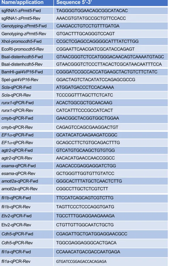
R2pA to give pminTol- cdh5E-cdh5P: Gal4VP16. Oligonucleotide sequences are listed in 400
Table S2. 401
Generation of prmt5-/- mutants by CRISPR/cas9 402
The guide RNA (gRNA) was designed using CHOPCHOP CRISPR Design website (Montague 403
et al., 2014). The designed oligos were annealed and ligated into the gRNA plasmid pDR274 404
after digestion of the plasmid with BsaI (NEB). The gRNA was prepared in vitro using the 405
MEGAshortscript T7 transcription kit (Ambion) after linearizing the plasmid with DraI (NEB) 406
(Talbot and Amacher, 2014) before being purified using illustra MicroSpin G-50 Columns (GE 407
Healthcare). 1 nL of a solution containing 10µM EnGen Cas9 NLS (NEB) and 100 ng/µl of 408
gRNA was injected at the one-cell stage. WT, heterozygous, and homozygous prmt5 animals 409
were identified by PCR. Oligonucleotide sequences are listed in Table S2. 410
Microinjections 411
The Tg(cdh5:GAL4VP16);Tg(UAS:KAEDE) line was generated using pminTol- cdh5E-cdh5P: 412
Gal4VP16 by Tol2 transposition as described previously (Covassin et al., 2009). Control and 413
prmt5 morpholino oligonucleotides (MOs) were described previously (Batut et al., 2011). 414
Embryos from in-crosses of the indicated heterozygous carriers or wild-type adults were 415
injected at the one cell stage with 6 ng of MO. pBluescript II KS+ hPRMT5 WT and pBluescript 416
II KS+ hPRMT5 Mutant (Pal et al., 2003) were linearized by EcoRI (NEB) and transcribed by 417
T7 (Promega). 200 pg hprmt5WT mRNA, or hprmt5 MUT mRNA were injected at one cell 418
stage. 419
RNA extraction, Reverse transcription and real-time PCR 420
Embryos were dissected at the indicated stage after addition of Tricaine Methanesulfonate. 421
Genomic DNA was extracted from dissected embryo heads to identify their genotype and the 422
corresponding dissected tails were conserved in TRIzol Reagent at -20°C. After identification 423
of wild type and mutant embryos, total RNAs from at least 6 identified tails were extracted 424
following manufacturer’s instructions (Invitrogen). Total RNAs were converted into cDNA using 425
Prime Script cDNA Synthesis Kit (Takara) with Oligo(dT) and random hexamer primers for 426
15 min at 37 °C according to manufacturer’s instructions. cDNAs were then diluted 20-fold and 427
quantified by qPCR using SsoFast Evagreen Supermix (Bio-rad) and specific primers. Data 428
were acquired on CFX96 Real-Time PCR detection System (Bio-rad). Samples were analyzed 429
in triplicates and the expression level was calculated relative to zebrafish housekeeping gene 430
EF1α. Oligonucleotide sequences are listed in Table S2. 431
Live imaging 432
For the transgenic lines TgBAC(cdh5:GAL4FF);Tg(UAS:GFP) and 433
Tg(cdh5:GAL4VP16);Tg(UAS:KAEDE), embryos were placed in 1.5% low melt agarose with 434
Tricaine on a glass-bottomed culture dish filled with egg water. Images were acquired using 435
the confocal microscope TCS SP8 (Leica Microsystems) with an L 25 × /0.95 W FLUOSTAR 436
VIZIR objective (zoom X1.25) using the scanner resonant mode. Confocal stacks were 437
acquired every 10 min from 28 to 38 hpf to generate movies. 438
Immunostaining and in situ hybridization 439
After fixation or rehydratation, embryos were washed twice with Phosphate Buffered Saline/1% 440
Triton X-100 (PBST), permeabilized with PBST/0.5% Trypsin for 30 sec and washed twice 441
again with PBST. After blocking with PBST/10% Fetal Calf Serum (FCS)/1% bovine serum 442
albumin (BSA) (hereafter termed ‘blocking solution’) for at least 1 h, embryos were incubated 443
with antibodies directed against either GFP (Torrey Pine, Biolabs), or Prmt5 (Upstate #07405), 444
in blocking solution overnight at 4 °C followed by 5 washing steps with PBST. Embryos were 445
then incubated with the appropriate Alexa Fluor-conjugated secondary antibodies (Molecular 446
Probes) for at least 2 h at room temperature and washed three times. Nuclei were then stained 447
with TO-PRO3 (Molecular Probes) and washed twice with PBST. Embryos were dissected, 448
flat-mounted in glycerol and images were recorded on a confocal microscope as above. 449
Fluorescent in situ hybridization was carried out as previously described (Quillien et al., 2014). 450
Image processing and measurements 451
Confocal images and stacks were either analyzed with ImageJ software or LAS X. Nuclei of 452
ISV cells and gata2b+ cells were counted using the Multipoint tool of ImageJ. ISV lengths were 453
measured by drawing a line between the base and the tip of ISV on ImageJ. Contours of the 454
Dorsal Aorta were drawn using the Freehand Selection Tool with a digital pen and the area 455
was then measured. Fluorescence intensity corresponded to the measurement of average 456
gray value for each entire image. 457
Statistical analysis 458
Statistical comparisons of datasets were performed using GraphPad Prism software. For each 459
dataset, we tested the assumption of normality with D’Agostino-Pearson tests and accordingly, 460
unpaired t-test, Mann-Whitney test, One-way ANOVA, two-way ANOVA or Kruskal-Wallis test 461
were used to compare dataset; means (± SEM) are indicated as horizontal bars on dot plots. 462
The test used as well as the number of independent experiments performed and the minimal 463
number of biological replicates are indicated in each figure legend. 464
Bioinformatic analysis 465
Published single cell data from total embryos at 10hpf, 14hpf, 18hpf and 24hpf (Wagner et al., 466
2018) were analyzed using the R package Seurat (Butler et al., 2018; Stuart et al., 2019). After 467
data clustering, clusters of endothelial cells from each stage were identified by the expression 468
of several endothelial specific genes (etv2, fli1a, …). Then, we examined the level of 469
expression and the percentage of cells expressing our gene of interest at each developmental 470
stage. ATAC-seq data (Marass et al., 2019; Quillien et al., 2017) and Chip-seq data (Girardot 471
et al., 2014; Liu et al., 2015a) were inspected using the Galaxy platform (Afgan et al., 2018). 472
ACKNOWLEDGEMENTS 474
We would like to thank the Blader lab for helpful discussions and support; Dr. Waltzer, Dr. Cau, 475
Dr. Male and Dr. Navajas Acedo for critical reading of the manuscript; A. Laire for excellent 476
zebrafish care; B. Ronsin and S. Bosch from the Toulouse Rio Imaging (TRI) platform; M. 477
Aguirrebengoa from the BigA Core Facility, CBI Toulouse; Dr. Herbomel for the Zebrafish 478
transgenic line Tg(gata2b:Gal4;UAS:lifeactGFP). This work was supported by a grant from the 479
Fondation de l’Association pour la Recherche contre le Cancer (Fondation ARC) and from the 480
Association Française contre les Myopathies (AFM) to LV. AQ was supported by a fellowship 481
from the Fondation ARC. 482
483
COMPETING INTEREST 484
The authors declare no competing interests. 485
486
REFERENCES 487
Afgan, E., Baker, D., Batut, B., van den Beek, M., Bouvier, D., Cech, M., Chilton, J., Clements, 488
D., Coraor, N., Gruning, B.A., et al. (2018). The Galaxy platform for accessible, reproducible 489
and collaborative biomedical analyses: 2018 update. Nucleic Acids Res 46, W537-W544. 490
Alestrom, P., D'Angelo, L., Midtlyng, P.J., Schorderet, D.F., Schulte-Merker, S., Sohm, F., and 491
Warner, S. (2019). Zebrafish: Housing and husbandry recommendations. Lab Anim, 492
23677219869037. 493
Anderson, H., Patch, T.C., Reddy, P.N., Hagedorn, E.J., Kim, P.G., Soltis, K.A., Chen, M.J., 494
Tamplin, O.J., Frye, M., MacLean, G.A., et al. (2015). Hematopoietic stem cells develop in the 495
absence of endothelial cadherin 5 expression. Blood 126, 2811-2820. 496
Baltrunaite, K., Craig, M.P., Palencia Desai, S., Chaturvedi, P., Pandey, R.N., Hegde, R.S., 497
and Sumanas, S. (2017). ETS transcription factors Etv2 and Fli1b are required for tumor 498
angiogenesis. Angiogenesis 20, 307-323. 499
Batut, J., Duboe, C., and Vandel, L. (2011). The methyltransferases PRMT4/CARM1 and 500
PRMT5 control differentially myogenesis in zebrafish. PLoS One 6, e25427. 501
Beacon, T.H., Xu, W., and Davie, J.R. (2020). Genomic landscape of transcriptionally active 502
histone arginine methylation marks, H3R2me2s and H4R3me2a, relative to nucleosome 503
depleted regions. Gene 742, 144593. 504
Beis, D., and Stainier, D.Y. (2006). In vivo cell biology: following the zebrafish trend. Trends 505
Cell Biol 16, 105-112. 506
Blanc, R.S., and Richard, S. (2017). Arginine Methylation: The Coming of Age. Mol Cell 65, 8-507
24. 508
Bussmann, J., and Schulte-Merker, S. (2011). Rapid BAC selection for tol2-mediated 509
transgenesis in zebrafish. Development 138, 4327-4332. 510
Butko, E., Distel, M., Pouget, C., Weijts, B., Kobayashi, I., Ng, K., Mosimann, C., Poulain, F.E., 511
McPherson, A., Ni, C.W., et al. (2015). Gata2b is a restricted early regulator of hemogenic 512
endothelium in the zebrafish embryo. Development 142, 1050-1061. 513
Butler, A., Hoffman, P., Smibert, P., Papalexi, E., and Satija, R. (2018). Integrating single-cell 514
transcriptomic data across different conditions, technologies, and species. Nat Biotechnol 36, 515
411-420. 516
Covassin, L.D., Siekmann, A.F., Kacergis, M.C., Laver, E., Moore, J.C., Villefranc, J.A., 517
Weinstein, B.M., and Lawson, N.D. (2009). A genetic screen for vascular mutants in zebrafish 518
reveals dynamic roles for Vegf/Plcg1 signaling during artery development. Dev Biol 329, 212-519
226. 520
Craig, M.P., Grajevskaja, V., Liao, H.K., Balciuniene, J., Ekker, S.C., Park, J.S., Essner, J.J., 521
Balciunas, D., and Sumanas, S. (2015). Etv2 and fli1b function together as key regulators of 522
vasculogenesis and angiogenesis. Arterioscler Thromb Vasc Biol 35, 865-876. 523
Dacwag, C.S., Bedford, M.T., Sif, S., and Imbalzano, A.N. (2009). Distinct protein arginine 524
methyltransferases promote ATP-dependent chromatin remodeling function at different stages 525
of skeletal muscle differentiation. Mol Cell Biol 29, 1909-1921. 526
Fish, J.E., Cantu Gutierrez, M., Dang, L.T., Khyzha, N., Chen, Z., Veitch, S., Cheng, H.S., 527
Khor, M., Antounians, L., Njock, M.S., et al. (2017). Dynamic regulation of VEGF-inducible 528
genes by an ERK/ERG/p300 transcriptional network. Development 144, 2428-2444. 529
Girardot, M., Hirasawa, R., Kacem, S., Fritsch, L., Pontis, J., Kota, S.K., Filipponi, D., Fabbrizio, 530
E., Sardet, C., Lohmann, F., et al. (2014). PRMT5-mediated histone H4 arginine-3 symmetrical 531
dimethylation marks chromatin at G + C-rich regions of the mouse genome. Nucleic Acids Res 532
42, 235-248. 533
Griffin, C.T., Brennan, J., and Magnuson, T. (2008). The chromatin-remodeling enzyme BRG1 534
plays an essential role in primitive erythropoiesis and vascular development. Development 535
135, 493-500. 536
Hatta, K., Tsujii, H., and Omura, T. (2006). Cell tracking using a photoconvertible fluorescent 537
protein. Nat Protoc 1, 960-967. 538
Hultin, S., Zheng, Y., Mojallal, M., Vertuani, S., Gentili, C., Balland, M., Milloud, R., Belting, 539
H.G., Affolter, M., Helker, C.S., et al. (2014). AmotL2 links VE-cadherin to contractile actin 540
fibres necessary for aortic lumen expansion. Nat Commun 5, 3743. 541
Isogai, S., Horiguchi, M., and Weinstein, B.M. (2001). The vascular anatomy of the developing 542
zebrafish: an atlas of embryonic and early larval development. Dev Biol 230, 278-301. 543
Kalna, V., Yang, Y., Peghaire, C.R., Frudd, K., Hannah, R., Shah, A.V., Osuna Almagro, L., 544
Boyle, J.J., Gottgens, B., Ferrer, J., et al. (2019). The Transcription Factor ERG Regulates 545
Super-Enhancers Associated With an Endothelial-Specific Gene Expression Program. Circ 546
Res 124, 1337-1349. 547
Karkhanis, V., Hu, Y.J., Baiocchi, R.A., Imbalzano, A.N., and Sif, S. (2011). Versatility of 548
PRMT5-induced methylation in growth control and development. Trends Biochem Sci 36, 633-549
641. 550
Kimmel, C.B., Ballard, W.W., Kimmel, S.R., Ullmann, B., and Schilling, T.F. (1995). Stages of 551
embryonic development of the zebrafish. Dev Dyn 203, 253-310. 552
Kirchmaier, S., Lust, K., and Wittbrodt, J. (2013). Golden GATEway cloning--a combinatorial 553
approach to generate fusion and recombination constructs. PLoS One 8, e76117. 554
Kwan, K.M., Fujimoto, E., Grabher, C., Mangum, B.D., Hardy, M.E., Campbell, D.S., Parant, 555
J.M., Yost, H.J., Kanki, J.P., and Chien, C.B. (2007). The Tol2kit: a multisite gateway-based 556
construction kit for Tol2 transposon transgenesis constructs. Dev Dyn 236, 3088-3099. 557
Lawson, N.D., and Weinstein, B.M. (2002a). Arteries and veins: making a difference with 558
zebrafish. Nat Rev Genet 3, 674-682. 559
Lawson, N.D., and Weinstein, B.M. (2002b). In vivo imaging of embryonic vascular 560
development using transgenic zebrafish. Dev Biol 248, 307-318. 561
LeBlanc, S.E., Konda, S., Wu, Q., Hu, Y.J., Oslowski, C.M., Sif, S., and Imbalzano, A.N. 562
(2012). Protein arginine methyltransferase 5 (Prmt5) promotes gene expression of peroxisome 563
proliferator-activated receptor gamma2 (PPARgamma2) and its target genes during 564
adipogenesis. Mol Endocrinol 26, 583-597. 565
LeBlanc, S.E., Wu, Q., Lamba, P., Sif, S., and Imbalzano, A.N. (2016). Promoter-enhancer 566
looping at the PPARgamma2 locus during adipogenic differentiation requires the Prmt5 567
methyltransferase. Nucleic Acids Res 44, 5133-5147. 568
Lin, H., and Luengo, J.I. (2019). Nucleoside protein arginine methyltransferase 5 (PRMT5) 569
inhibitors. Bioorg Med Chem Lett 29, 1264-1269. 570
Liu, F., Cheng, G., Hamard, P.J., Greenblatt, S., Wang, L., Man, N., Perna, F., Xu, H., Tadi, 571
M., Luciani, L., et al. (2015a). Arginine methyltransferase PRMT5 is essential for sustaining 572
normal adult hematopoiesis. J Clin Invest 125, 3532-3544. 573
Liu, F., Li, D., Yu, Y.Y., Kang, I., Cha, M.J., Kim, J.Y., Park, C., Watson, D.K., Wang, T., and 574
Choi, K. (2015b). Induction of hematopoietic and endothelial cell program orchestrated by ETS 575
transcription factor ER71/ETV2. EMBO Rep 16, 654-669. 576
Ma, D., Wei, Y., and Liu, F. (2013). Regulatory mechanisms of thymus and T cell development. 577
Dev Comp Immunol 39, 91-102. 578
Marass, M., Beisaw, A., Gerri, C., Luzzani, F., Fukuda, N., Gunther, S., Kuenne, C., 579
Reischauer, S., and Stainier, D.Y.R. (2019). Genome-wide strategies reveal target genes of 580
Npas4l associated with vascular development in zebrafish. Development 146. 581
Montague, T.G., Cruz, J.M., Gagnon, J.A., Church, G.M., and Valen, E. (2014). CHOPCHOP: 582
a CRISPR/Cas9 and TALEN web tool for genome editing. Nucleic Acids Res 42, W401-407. 583
Napoli, C., Schiano, C., and Soricelli, A. (2019). Increasing evidence of pathogenic role of the 584
Mediator (MED) complex in the development of cardiovascular diseases. Biochimie 165, 1-8. 585
Ninov, N., Borius, M., and Stainier, D.Y. (2012). Different levels of Notch signaling regulate 586
quiescence, renewal and differentiation in pancreatic endocrine progenitors. Development 587
139, 1557-1567. 588
Pal, S., Yun, R., Datta, A., Lacomis, L., Erdjument-Bromage, H., Kumar, J., Tempst, P., and 589
Sif, S. (2003). mSin3A/histone deacetylase 2- and PRMT5-containing Brg1 complex is 590
involved in transcriptional repression of the Myc target gene cad. Mol Cell Biol 23, 7475-7487. 591
Pham, V.N., Lawson, N.D., Mugford, J.W., Dye, L., Castranova, D., Lo, B., and Weinstein, 592
B.M. (2007). Combinatorial function of ETS transcription factors in the developing vasculature. 593
Dev Biol 303, 772-783. 594
Quillien, A., Abdalla, M., Yu, J., Ou, J., Zhu, L.J., and Lawson, N.D. (2017). Robust 595
Identification of Developmentally Active Endothelial Enhancers in Zebrafish Using FANS-596
Assisted ATAC-Seq. Cell Rep 20, 709-720. 597
Quillien, A., Moore, J.C., Shin, M., Siekmann, A.F., Smith, T., Pan, L., Moens, C.B., Parsons, 598
M.J., and Lawson, N.D. (2014). Distinct Notch signaling outputs pattern the developing arterial 599
system. Development 141, 1544-1552. 600
Rosa-Garrido, M., Chapski, D.J., and Vondriska, T.M. (2018). Epigenomes in Cardiovascular 601
Disease. Circ Res 122, 1586-1607. 602
Sauteur, L., Affolter, M., and Belting, H.G. (2017). Distinct and redundant functions of Esama 603
and VE-cadherin during vascular morphogenesis. Development 144, 1554-1565. 604
Sauteur, L., Krudewig, A., Herwig, L., Ehrenfeuchter, N., Lenard, A., Affolter, M., and Belting, 605
H.G. (2014). Cdh5/VE-cadherin promotes endothelial cell interface elongation via cortical actin 606
polymerization during angiogenic sprouting. Cell Rep 9, 504-513. 607
Shailesh, H., Zakaria, Z.Z., Baiocchi, R., and Sif, S. (2018). Protein arginine methyltransferase 608
5 (PRMT5) dysregulation in cancer. Oncotarget 9, 36705-36718. 609
Stopa, N., Krebs, J.E., and Shechter, D. (2015). The PRMT5 arginine methyltransferase: many 610
roles in development, cancer and beyond. Cell Mol Life Sci 72, 2041-2059. 611
Stuart, T., Butler, A., Hoffman, P., Hafemeister, C., Papalexi, E., Mauck, W.M., 3rd, Hao, Y., 612
Stoeckius, M., Smibert, P., and Satija, R. (2019). Comprehensive Integration of Single-Cell 613
Data. Cell 177, 1888-1902 e1821. 614
Talbot, J.C., and Amacher, S.L. (2014). A streamlined CRISPR pipeline to reliably generate 615
zebrafish frameshifting alleles. Zebrafish 11, 583-585. 616
Tan, D.Q., Li, Y., Yang, C., Li, J., Tan, S.H., Chin, D.W.L., Nakamura-Ishizu, A., Yang, H., and 617
Suda, T. (2019). PRMT5 Modulates Splicing for Genome Integrity and Preserves Proteostasis 618
of Hematopoietic Stem Cells. Cell Rep 26, 2316-2328 e2316. 619
Tee, W.W., Pardo, M., Theunissen, T.W., Yu, L., Choudhary, J.S., Hajkova, P., and Surani, 620
M.A. (2010). Prmt5 is essential for early mouse development and acts in the cytoplasm to 621
maintain ES cell pluripotency. Genes Dev 24, 2772-2777. 622
Thisse, C., and Zon, L.I. (2002). Organogenesis--heart and blood formation from the zebrafish 623
point of view. Science 295, 457-462. 624
Wagner, D.E., Weinreb, C., Collins, Z.M., Briggs, J.A., Megason, S.G., and Klein, A.M. (2018). 625
Single-cell mapping of gene expression landscapes and lineage in the zebrafish embryo. 626
Science 360, 981-987. 627
Wang, Y., Hu, W., and Yuan, Y. (2018). Protein Arginine Methyltransferase 5 (PRMT5) as an 628
Anticancer Target and Its Inhibitor Discovery. J Med Chem 61, 9429-9441. 629
Wong, K.S., Proulx, K., Rost, M.S., and Sumanas, S. (2009). Identification of vasculature-630
specific genes by microarray analysis of Etsrp/Etv2 overexpressing zebrafish embryos. Dev 631
Dyn 238, 1836-1850. 632
Zhu, J., Zhang, D., Liu, X., Yu, G., Cai, X., Xu, C., Rong, F., Ouyang, G., Wang, J., and Xiao, 633
W. (2019). Zebrafish prmt5 arginine methyltransferase is essential for germ cell development. 634 Development 146. 635 636 FIGURE LEGENDS 637
Figure 1: Loss of prmt5 affect HSCs and HSPCs production. A- Schematic representation 638
of the sequence targeted by CRISPR/Cas9 leading to a 23 nucleotides deletion, and of wild 639
type and truncated Prmt5 proteins. The catalytic domain “CAT” appears in magenta. B-C- 640
Confocal sections of immunostaining with anti-Prmt5 antibody of wild type and prmt5 mutant 641
embryos at 24 hpf. Scale bar 100 µm. D- Schematic representation of vascular (green) and 642
hematopoietic (red) systems in a zebrafish larva. Circle and bracket indicate the Thymus (T) 643
and the Caudal Hematopoietic Tissue (CHT), respectively. D’- Close-up of the trunk 644
vasculature where HSCs emerge from the ventral wall of the dorsal aorta (DA), bud and 645
migrate. Red line represents the diameter of the dorsal aorta. Cardinal Vein (CV). E-F’- 646
Confocal section of transgenic Tg(gata2b:Gal4; UAS:lifeactGFP) embryos at 36 hpf showing 647
gata2b+ cells in red and TO-PRO-3 in black. Blue arrows indicate HSCs labelled in red in wild 648
type (E, E’) and in prmt5 mutant (F, F’) embryos. Bar scale 100 µm. G- Average number of 649
HSCs enumerated per confocal stack in wild type and in prmt5 mutant embryos at 36 hpf. Data 650
are from 3 independent experiments with at least 6 individuals per experiment and a Mann-651
Whitney test was performed. H- Relative mRNA expressions determined by RT-qPCR in 36 652
hpf wild type and prmt5 mutant embryos, from 3 independent experiments with at least 6 653
animals per condition. Two-way ANOVA was performed. I-J- Confocal sections of wild type (I) 654
and prmt5 mutant (J) thymus from transgenic Tg(gata2b:Gal4; UAS:lifeactGFP) embryos at 5 655
days. Thymus are delimited by a white circle. Bar scale 100 µm. K- Average number of HSPCs 656
enumerated per confocal stack in wild type and prmt5 mutant embryos at 5 days from 3 657
independent experiments with at least 5 individuals per analysis. T-test was performed. * 658
P<0.05, ** P<0.01, ***P<0.001. 659
Figure 2: Loss of prmt5 impairs blood vessel formation. A-C’- Confocal projections of 660
transgenic Tg(fli1a:GFP)y1 embryos with endothelial cells (in green) after immunostaining
661
against Prmt5 (in magenta). A-A’’- Dorsal view of the lateral plate mesoderm at 14 somite- 662
stage. Yellow rectangle delimits the close up of Prmt5+ endothelial cells (A’-A’’). Prmt5+ cells 663
appear in magenta (A-A”) and endothelial cells in green (A-A’). Anterior is on top. Scale bars 664
100 µm (A) and 25 µm (A’). B-B’- Confocal projections focusing on endothelial cells (in green) 665
from the dorsal aorta (DA) and the cardinal vein (CV) at 24 hpf. Red and blue arrows point to 666
Prmt5+ cells (in magenta) from the DA and the CV, respectively. Red and blue lines represent 667
DA and CV diameters, respectively. Scale bar 50 µm. C-C’- Confocal projections focusing on 668
sprouting ISVs (in green) at 24 hpf. Light blue and yellow arrows point to tip and stalk cell, 669
respectively. D- Schematic representation of the trunk vasculature with ISVs sprouting from 670
the DA. The tip cell leads the cell migration and the stalk cell maintains the connection with the 671
DA. E-F- Confocal projections of transgenic Tg(fli1a:GFP)y1 wild type (E) and prmt5 mutant (F)
672
embryos at 28 hpf. Red rectangles delimit where DA close ups were made. White rectangles 673
delimit the higher magnification (x2) of the DA with red lines indicating the dorsal aorta 674
diameters. White arrows indicate the connection point between two ISVs to form the Dorsal 675
Longitudinal Anastomotic Vessel (DLAV). Scale bar 100 µm. G-H- Confocal projections of 676
control morphant (G) and prmt5 morphant (H) transgenic Tg(TP1bglob:VenusPEST)s940
677
embryos labelling cells from the DA at 28 hpf. Yellow lines delimit the measured area occupied 678
by the DA. Scale bar 25 µm I- Average area occupied by the DA in µm2 in control and prmt5
679
morpholino injected embryos from 2 independent experiments with at least 8 animals per 680
condition. T-test was performed. J-K- Average number of endothelial cells per intersegmental 681
vessel (J) and average ISV length in µm (K) in control and in prmt5 mutant embryos from 3 682
independent experiments with at least 3 animals per condition. T-test and Mann Whitney test 683
were performed, respectively. ** P<0.01, ***P<0.001. 684
Figure 3: Prmt5 is required for vascular morphogenesis. A-B- Still images from movies of 685
control (A) and prmt5 morphant (B) Tg(fli1a:GFP)y1 transgenic embryos from 28 to 38 hpf. Red
686
asterisks label missing connections between tip and stalk cells as well as missing connections 687
between tip cells that should lead to DLAV formation. Red arrows point to connecting ISVs 688
leading to DLAV formation. White arrows indicate supernumerary sprouts. Yellow asterisks 689
label the lumen of ISVs. Scale bar 50 µm. C- Relative mRNA expressions of the indicated 690
transcripts were determined by RT-qPCR in 28 hpf wild type and prmt5 mutant embryos, from 691
3 independent experiments with at least 6 animals per condition. Two-way ANOVA was 692
performed. * P<0.05, ***P<0.001. 693
Figure 4: Prmt5 methyltransferase activity is dispensable for vascular morphogenesis. 694
A-D- Confocal projections of transgenic Tg(fli1a:GFP)y1 embryos at 28 hpf. Wild type embryo
695
is on the top left panel (A), prmt5 mutant embryos were not injected (B) or injected with either 696
hprmt5WT mRNA (C) or the mutant form hprmt5MUT mRNA (D). Scale bar 100 µm. E-F- 697
Average ISVs length in µm (E) and average number of endothelial cells per ISVs (F) for wild 698
type, prmt5 mutant embryos not injected or injected with hprmt5WT mRNA, or hprmt5 MUT 699
mRNA, from 3 independent experiments with at least 3 animals per condition. Kruskal-Wallis 700
test (E) and One-way ANOVA (F) were performed. ** P<0.01, *** P<0.001. G- Relative mRNA 701
expressions were determined by RT-qPCR on 28 hpf wild type and prmt5 mutantembryos 702
injected by either hprmt5WT or hprmt5MUT mRNAs, from 2 independent experiments with at 703
least 6 animals per condition. Two-way ANOVA was performed. * P<0.05. 704
Figure 5: Prmt5 promotes chromatin looping. A- Schematic representation of the transgene 705
TgBAC(cdh5:GAL4FF) containing two putative cis-regulatory elements, a promotor region (P) 706
and an enhancer (E), separated by ~20kb with the GAL4FF reporter gene inserted at the TSS 707
of cdh5. B, C, I, J- Confocal projections of transgenic TgBAC(cdh5:GAL4FF);Tg(UAS:GFP) 708
embryos at 28 hpf. Control morphant is on the top left panel (B), prmt5 morphant embryos 709
were not injected (C) or injected by either hprmt5WT mRNA (I) or the catalytic mutant form 710
hprmt5MUT (J) mRNA. The fluorescent intensity is colored-coded, from the Low intensity (L) 711
in black to High intensity (H) in white (intensity scale as in panel B). Scale bar 100 µm. D- 712
Average GFP fluorescence intensity per confocal projection for control, prmt5 morphant 713
embryos injected by hprmt5WT mRNA, or hprmt5 MUT mRNA or not injected, from 3 714
independent experiments with at least 3 animals per condition. One-way ANOVA was 715
performed. *P<0.05, ***P<0.001. E- Schematic representation of the transgene 716
Tg(cdh5:GAL4VP16) containing the two putative cis-regulatory elements next to each other (E 717
and P), upstream of GAL4VP16 reporter gene. F-G- Confocal projection of transgenic 718
Tg(cdh5:GAL4VP16);Tg(UAS:KAEDE) embryos at 26 hpf injected with either a control 719
morpholino (F) or a prmt5 morpholino (G). The fluorescence intensity is color- coded, from the 720
Low intensity (L) in black to High intensity (H) in white (intensity scale in panel B). H- Average 721
KAEDE fluorescence intensity for control and for prmt5 morphant embryos, from 3 independent 722
experiments with at least 5 animals per condition. T-test was performed. 723
Figure 6: A- Schematic representation of two distinct roles of Prmt5 during the formation of 724
hematopoietic lineage development and blood vessels, relying or not on its methyltransferase 725
activity, respectively. B- Proposed model to depict the function of Prmt5 in zebrafish endothelial 726
cells. The transcription factor ETV2 recruited to promoters and enhancers of endothelial 727
specific genes, could favor the recruitment of a complex including Prmt5, Brg1 and the 728
mediator complex to help the formation of chromatin loping and thus facilitate the transcription 729
of specific endothelial genes. Dashed lines indicate potential interactions or plausible 730
recruitments of Brg1 and/or the mediator complex. 731
Figure S1: A-B- Confocal sections of Prmt5 immunostaining in control and prmt5 morphant 732
embryos at 24 hpf. Scale bar 100 µm. C-F- Confocal sections of thymus from transgenic 733
Tg(gata2b:Gal4; UAS:lifeactGFP) embryos at 3 days. Transgenic embryos were injected by 734
control morpholino (C) or prmt5 morpholino only (D) or in combination hprmt5WT mRNA (E) 735
or the catalytic mutant form hprmt5MUT mRNA (F). Thymus is delimited by a white circle. Bar 736
scale 100 µm. G- Average number of HSPCs enumerated per confocal stack in injected 737
embryos at 3 days from 2 independent experiments with at least 3 individuals per analysis. T-738
test was performed. ***P<0.001. 739
Figure S2: A-B- Confocal projections of transgenic Tg(fli1a:GFP)y1 embryos injected by either
740
control morpholino (A) or prmt5 morpholino (B). Scale bar 100 µm. C-D- Average number of 741
endothelial cells per ISV (C) and average ISV length in µm (D), in control and prmt5 morphant 742
embryos, from 3 independent experiments with at least 4 animals per condition. T-test and 743
Mann-Whitney test were performed. *** P<0.001. 744
Figure S3: Expression heatmap for prmt5, etv2 and identified Prmt5 target genes, for 745
endothelial cells at 10hpf, 14hpf, 18hpf and 24hpf. The expression level is colored-coded from 746
absence of expression (in green) to highest level of expression (in white). 747
Figure S4: A-D- Confocal projections of wild type (A, C) and prmt5 mutant embryos (B, D) 748
after fluorescent in situ hybridization against fli1a (A-B) and cdh5 (C-D). Scale bar 100 µm. E-749
F- Percentage of embryos (y axis) presenting a high or a low level of expression of fli1a (E) or 750
cdh5 (F), according to their genotype (x axis), from 3 independent experiments with at least 4 751
animals per condition. 752
Figure S5: Chromatin profile visualization of endothelial cells from the UCSC Genome 753
Browser. ATAC-seq peaks as determined by Quillien et al. (Quillien et al., 2017) flanking 754
indicated genes (cdh5, esama, agtr2, fli1a, fli1b, amotl2a). Promoter regions (P) and 755
numerated putative enhancers (corresponding numerated peaks are found in Table S1) are 756
highlighted in light orange and light purple, respectively. 757
Gene
symbol Peak chr Peak start Peak stop
ETV2 CHIP Peak in mouse embryo
H4R3me2s CHIP Peak in MEF
mouse cells
amotl2 chr9 102,611,688 102,612,810 yes yes
amotl2 chr9 102,624,400 102,624,850 yes no cdh5 chr8 106,619,166 106,620,059 yes yes cdh5 chr8 106,623,076 106,623,670 yes yes cdh5 chr8 106,625,171 106,625,699 yes yes fli1 chr9 32,389,460 32,390,252 yes no fli1 chr9 32,378,074 32,378,681 yes no fli1 chr9 32,363,839 32,364,366 yes yes
fli1 chr9 32,348,223 32,349,647 yes yes
agtr2 chrX _ _ no yes
Table 1: Chromatin profile of mouse orthologous genes of prmt5 identified target genes.
List of Etv2 and H4R3me2s peaks identified by CHIP-seq in mouse embryos and in MEF mouse cells, respectively (Liu et al., 2015a; Girardot et al., 2014).