HAL Id: hal-01604283
https://hal.archives-ouvertes.fr/hal-01604283
Submitted on 27 May 2020
HAL is a multi-disciplinary open access
archive for the deposit and dissemination of
sci-entific research documents, whether they are
pub-lished or not. The documents may come from
teaching and research institutions in France or
abroad, or from public or private research centers.
L’archive ouverte pluridisciplinaire HAL, est
destinée au dépôt et à la diffusion de documents
scientifiques de niveau recherche, publiés ou non,
émanant des établissements d’enseignement et de
recherche français ou étrangers, des laboratoires
publics ou privés.
Distributed under a Creative Commons Attribution| 4.0 International License
Fibrinogen deficiency in a dog - a case report
Franck Jolivet, Armelle Diquélou, Catherine Trumel, Simon Privat, Olivier
Dossin
To cite this version:
Franck Jolivet, Armelle Diquélou, Catherine Trumel, Simon Privat, Olivier Dossin.
Fibrinogen
deficiency in a dog - a case report.
BMC Veterinary Research, BioMed Central, 2017, 13 (1),
C A S E R E P O R T
Open Access
Fibrinogen deficiency in a dog - a case
report
Franck Jolivet
1, Armelle Diquélou
1,2, Catherine Trumel
1, Simon Privat
1and Olivier Dossin
1,2*Abstract
Background: Among coagulation disorders, primary fibrinogen deficiency is very rare in dogs. It is divided into hypofibrinogenemia, afibrinogenemia and dysfibrinogenemia. Afibrinogenemia has been described in three dogs. There are, however, no published case reports of primary hypofibrinogenemia in dogs.
Case presentation: A 1.5 year-old male German Pointer dog was evaluated for a locked-jaw syndrome associated with eye protrusion which appeared after a minor head trauma. Three months before the trauma, a persistent increase in coagulation times was detected by the referring veterinarian after a strong suspicion of snake envenomation. Apart for the primary complaint, physical examination was normal. A complete hemostatic profile revealed a moderately increased prothrombin time, activated partial thromboplastin times and a dramatically decreased fibrinogen concentration (0.34 g/L, reference interval [1.3–4.8 g/L]). Platelet count, plasma D-dimers and antithrombin, were all within the reference intervals and not consistent with a disseminated intravascular coagulation. Other possible causes of hypofibrinogenemia such as chronic hemorrhage and liver failure were excluded by laboratory work-up and imaging studies. Finally, antifibrinogen circulating anticoagulants were excluded using a dilution of citrated plasma from the pooled plasma of healthy dogs. These results supported a diagnosis of congenital fibrinogen deficiency and secondary retrobulbar hematoma and/or cellulitis. The dog’s condition improved rapidly after symptomatic treatment with corticosteroids and antibiotics. At the 1 year follow-up, the dog was clinically normal but a persistent hypofibrinogenemia (≤ 0.8 g/L) remained.
Conclusions: Various clinical presentations may occur in canine primary hypofibrinogenemia which should be included in the list of coagulation disorders. Diagnosis should include fibrinogen determination by coagulometric and non-coagulometric methods to differentiate from dysfibrinogenemia. There is no specific treatment but care should be taken to prevent bleeding and trauma. Emergency management of bleeding episodes with cryoprecipitate is the treatment of choice.
Keywords: Fibrinogen deficiency, Hypofibrinogenemia, Dysfibrinogenemia, Bleeding disorders, Dog Background
Congenital disorders of coagulation are rare in veterin-ary medicine, and consist mainly of hemophilia A and B [1]. Other clotting factor deficiencies have however been described and can be a diagnostic challenge. Among these deficiencies, hypofibrinogenemia has seldom been described in dogs.
Fibrinogen, the soluble precursor of fibrin, is a key coagulation factor involved in both primary and secondary
hemostasis, promoting platelet aggregation and clot formation. Fibrinogen is the most abundant clotting fac-tor, with usual blood concentrations ranging from 1.3 g/L to 4.8 g/L in adult dogs (reference interval (RI) from the veterinary clinical pathology laboratory of the Veterinary Teaching Hospital of the University of Toulouse (VTH UT)). In human and veterinary medicine, three congenital fibrinogen abnormalities have been described: afibri-nogenemia (absence of fibrinogen), hypofibrinogen-emia (quantitative deficiency) and dysfibrinogenhypofibrinogen-emia (qualitative deficiency) [2–6].
Humans with congenital hypofibrinogenemia are usually asymptomatic but may bleed when the hemostatic * Correspondence:o.dossin@envt.fr
1Department of Clinical Sciences, ENVT, University of Toulouse, 31076
Toulouse, France
2IRSD, INSERM 1220, INSERM, INRA, ENVT, UPS, University of Toulouse, 31024
Toulouse, France
© The Author(s). 2017 Open Access This article is distributed under the terms of the Creative Commons Attribution 4.0 International License (http://creativecommons.org/licenses/by/4.0/), which permits unrestricted use, distribution, and reproduction in any medium, provided you give appropriate credit to the original author(s) and the source, provide a link to the Creative Commons license, and indicate if changes were made. The Creative Commons Public Domain Dedication waiver (http://creativecommons.org/publicdomain/zero/1.0/) applies to the data made available in this article, unless otherwise stated.
system is strongly solicited, for example in such situations as trauma or surgery [2].
This report describes a 1.5 year-old German Pointer dog with a protrusion of the right third eyelid and a locked-jaw syndrome associated with extreme pain on mouth-opening due to a congenital hypofibrinogenemia.
Case presentation
A 1.5 year-old male German Pointer dog weighing 27 kg was referred to the VTH UT for a protrusion of the right third eyelid, and a locked-jaw syndrome associated with extreme pain when the dog tried to open his mouth. These signs had appeared 10 days earlier, after the dog was hit on the head by a ball. The dog had remained stable since then. History revealed a strong suspicion of snake envenomation 3 months earlier. At that time, the dog was presented in shock to a veterinarian with marks on the head suggestive of a snake bite. The laboratory workup revealed markedly increased prothrombin time (PT) and activated partial thromboplastin time (aPTT). The veterinarian started a symptomatic treatment [fluid-therapy (Ringer Lactate), corticosteroids (prednisolone, 1 mg/kg, IV, q 12 h), antibiotics (marbofloxacin, 4 mg/kg, IV, q 12 h) and K1 vitamin] and clinical signs improved within a few days. However, the PT and aPTT remained moderately increased after this suspected envenomation. One month later, a gingival wound appeared, requiring a surgical suture to stop the bleeding. On this occasion, PT and APPT were still markedly increased (PT >35 s, RI [12–19 s] and aPTT >200 s, RI [75–105 s]).
Upon admission at the VTH UT, physical examination showed an exophthalmia of the right eye associated with protrusion of third eyelid. The dog was reluctant to be touched on the head and opening the mouth elicited severe pain. Apart from this, clinical examination was unremarkable and a thorough ocular examination did not reveal any other abnormality. The ocular ultrasonog-raphy was consistent with a retrobulbar cellulitis with no image suggesting the presence of a foreign body. Because of the history of abnormal coagulation, the retrobulbar fine needle aspiration was postponed until the laboratory results were available.
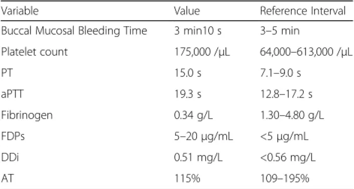
An extensive laboratory profile, including complete hemostatic profile1, was performed at the veterinary clinical pathology laboratory of the VTH UT. Severe hypofibrinogenemia (0.34 g/L, RI [1.30–4.80 g/L] assessed by Clauss’coagulometric method), increased PT (15.0 s, RI [7.10–9.00 s]) and aPTT (19.3 s, RI [12.8–17.2 s]) were observed. All other results including buccal mucosal bleeding time, complete blood count (CBC), D-dimers (DDi), and antithrombin (AT) were within reference inter-vals (Table 1). The fibrinogen/fibrin degradation products (FDPs) value was however in the grey zone (between 5 and 20μg/mL, RI [< 5 μg/mL]).
The hypofibrinogenemia was observed on repeated coagulometric measurements as well as by physical measurement using the Millar heat-precipitation method based on the ability of fibrinogen to precipitate at 56 °C [7]. This method confirmed the severe hypofibrinogen-emia with an almost undetectable fibrinogen precipita-tion band.
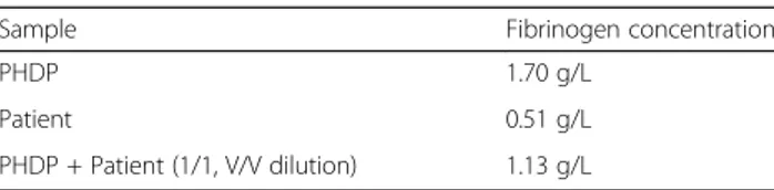
A screening for possible causes of hypofibrinogenemia was performed. The normal liver profile, including a normal bile acid test, excluded liver failure (Table 2). The absence of clinical signs of disease conditions that are associated with disseminated intra-vascular coagula-tion (DIC) together with the absence of any other laboratory signs of hemostasis system activation (normal platelet count, DDi and AT) was not consistent with DIC. Chronic bleeding was ruled out by clinical, diag-nostic imaging and CBC findings. To rule out the hypothesis of circulating anticoagulant interfering with the fibrinogen determination, a 1/1 dilution of the dog’s citrated plasma with pooled healthy dog plasma (PHDP) was performed. PHDP was prepared by pooling identi-cally prepared plasma samples from 9 cliniidenti-cally healthy dogs. The fibrinogen concentration assessed by coagulo-metric method in the mixture was consistent with the expected calculated result (Table 3). This finding ruled Table 1 Complete hemostasis profile
Variable Value Reference Interval Buccal Mucosal Bleeding Time 3 min10 s 3–5 min
Platelet count 175,000 /μL 64,000–613,000 /μL PT 15.0 s 7.1–9.0 s aPTT 19.3 s 12.8–17.2 s Fibrinogen 0.34 g/L 1.30–4.80 g/L FDPs 5–20 μg/mL <5μg/mL DDi 0.51 mg/L <0.56 mg/L AT 115% 109–195%
Table 2 Complete blood chemistry profile
Variable Value Reference Interval Creatinine 75μmol/L 44–133 μmol/L Cholesterol 4.7 mmol/L 3.3–9.3 mmol/L Triglycerides 0.4 mmol/L 0.2–1.3 mmol/L Total Protein 76.1 g/L 48.0–66.0 g/L Albumin 35.3 g/L 23.0–39.0 g/L Alanine Aminotransferase 45 U/L 0.3–50 U/L Alkaline Phosphatase 195 U/L 20–155 U/L Gamma-glutamyltransferase 5 U/L 0.5–25 U/L Total Bilirubin 1.8μmol/L 1.7–12.0 μmol/L Pre-Prandial Bile Acids 2.0μmol/L 0–10.0 μmol/L Post-Prandial Bile Acids 2.0μmol/L 0–20.0 μmol/L
out the presence of circulating anticoagulant with fibrinogen inhibitory properties. A congenital hypofibri-nogenemia was hence diagnosed.
Retrobulbar fine needle aspiration was not per-formed due to the high risk of puncture-induced hemorrhage. In the hypothesis of a retrobulbar cellu-litis, the following treatment was administered: antibi-otics (gentamicin 6.6 mg/kg, IV, q 24 h for 6 days, and amoxicillin/clavulanic acid 20 mg/kg, IV, q 8 h for 6 days, then 12.5 mg/kg, PO, q 12 h for 6 weeks) and corticosteroid therapy (prednisolone 0.3 mg/kg, IV, q 24 h for 6 days then 0.3 mg/kg, PO, q 24 h for 3 weeks, and finally 0.3 mg/kg, PO, every other day, for 2 weeks).
After 2 days of treatment, the protrusion of the third eyelid and the pain associated with mouth opening decreased and resolved after 2 weeks. One month later, the dog was completely normal. Two months later, the dog was stable but fibrinogen remained persistently below the reference interval (Table 4) and the FDP value was negative (< 5 μg/mL). Six months after the initial presentation, no clinical relapse was observed but plasma fibrinogen value remained low.
There is no specific treatment for congenital hypofibri-nogenemia, other than plasma transfusion during acute bleeding crisis. It was therefore strongly recommended to avoid strenuous exercise or hunting with the dog to prevent trauma.
Discussion
This case describes fibrinogen deficiency, an extremely rare congenital bleeding disorder in dogs [1, 8]. Very little data is published on the subject in veterinary medi-cine, with only three case reports on dogs with afibrino-genemia: a Bernese mountain dog [3], a Chihuahua [4], and a Bichon Frise [5]. In an international registry of animal hemostatic disorders, Dodds mentioned a case of dysfibrinogenemia in an inbred family of Borzois and a
case of hypofibrinogenemia in a family of Saint Bernard dogs [6] but to our knowledge these cases have not been reported in detail in a peer-reviewed journal.
In human medicine, congenital fibrinogen deficiency represents 7% of rare congenital bleeding disorders, hemophilia-excluded [2, 9]. Three categories of fibrino-gen deficiency have been described: afibrinofibrino-genemia when fibrinogen is not detectable; hypofibrinogenemia when the value is below the lower limit of the reference interval; and dysfibrinogenemia when the fibrinogen molecule is abnormal and dysfunctional. The only way to differentiate hypofibrinogenemia from dysfibrinogen-emia in humans is a low fibrinogendysfibrinogen-emia assessed by coagulometry with a concurrent normal or subnormal fibrinogen concentration by an immunologic method. Unfortunately, such a method is not routinely avail-able in veterinary medicine [10, 11]. Therefore dysfi-brinogenemia was not completely excluded in our dog. However, because the fibrinogen concentration was similarly decreased with both coagulometric method and heat precipitation method, we concluded it was hypofibrinogenemia.
The clinical presentation of ocular signs in this case is un-usual. Ocular ultrasonography was suggestive of a retro-bulbar cellulitis with effusion. To confirm this diagnosis, cytological and bacteriological analysis of a fine needle as-piration of retrobulbar space is needed. In this case, the first hypothesis was a hematoma because the ocular problems started after the dog was hit on the head and the patient had a background history of sustained increase of coagula-tion times and abnormal bleeding following a benign gin-gival wound. However, the nature of the retrobulbar ultrasonographic abnormality could not be confirmed by fine needle aspiration due to the risk of bleeding. Given the extreme pain associated with jaw opening, a primary hematoma with secondary inflammation/infection was a possible hypothesis. The dog was therefore treated with antibiotic and anti-inflammatory drugs. The rapid clinical improvement after onset of the treatment in a dog, whose signs had been stable for the previous 10 days was consist-ent with this hypothesis.
When hypofibrinogenemia is detected, the first clinical step is to exclude all possibility of a preanalytic error and to confirm the fibrinogen value using another method such as Millar’s method. If the decreased concentration is confirmed, all other causes of decreased fibrinogen pro-duction or increased fibrinogen consumption need to be explored before considering a diagnosis of congenital hypofibrinogenemia. Liver function must be assessed because fibrinogen is produced by the liver. In our case, normal clinical examination and bile acid test excluded decreased hepatic fibrinogen synthesis. The second step is to rule out increased and sustained fibrinogen consumption by activation of the coagulation cascade, Table 3 Fibrinogen concentration after plasma dilution with
PHDP
Sample Fibrinogen concentration
PHDP 1.70 g/L
Patient 0.51 g/L
PHDP + Patient (1/1, V/V dilution) 1.13 g/L
Theoretical expected result for the dilution: 1.105 g/L PHDP Pooled Healthy Dogs Plasma
Table 4 Follow-up of fibrinogen concentrations Test time Fibrinogen concentration RI Admission 0.34 g/L
1 month later 0.70 g/L 1.30–4.80 g/L 2 months later 0.51 g/L
namely DIC or chronic bleeding, by laboratory work-up and diagnostic imaging. No systemic disease, bleeding or inflammatory site, other than the ocular bulging, was sus-pected in our dog. No signs of chronic blood loss were detected on CBC, and normal platelet count, AT and DDi concentrations were inconsistent with DIC.
In our patient, the only hemostatic abnormality other than hypofibrinogenemia was FDPs in the grey zone. FDPs reflect the action of the fibrinolytic enzyme, plas-min, on fibrin and fibrinogen whereas DDi appear only when fibrin is degraded by plasmin [12]. Decreased FDPs clearance due to a failing liver or kidney was excluded by the laboratory results. The FDPs value results could indicate primary hyperfibrinolysis (ie spon-taneous activation of plasmin not triggered by coagula-tion activacoagula-tion). However, primary hyperfibrinolysis had not previously been described in dogs and was consid-ered very unlikely in our case for other reasons including the absence of primary diseases causing hyperfibrinolysis in humans, such as liver cirrhosis [13] or bronchiectasis [14]. Moreover, if the severe hypofibrinogenemia in this dog had been caused by hyperfibrinogenolysis, FDPs would have been very high and not only slightly increased. Finally, the dog’s fibrinogen concentration remained low at the 1 and 2 month follow-ups which was not compatible with a primary hyperfibrinogenolysis that is not a persistent condition. Therefore, we hypoth-esized that the slight increase of the FDPs could be the consequence of the suspected retrobulbar hematoma.
Furthermore, the unusual context of this case’s clinical scenario made it difficult to determine whether the disorder was congenital or acquired. The onset of the condition, discovered by the veterinarian in association with a strong suspicion of snake bite, was initially con-fusing. It concerned an adult dog, with no previous coagulation profile evaluation or surgical history, making it impossible to determine if the coagulation disorders were present prior to this episode. According to the owner, the dog had been perfectly healthy which is consistent with the few cases of hypofibrinogenemia described in the veterinary literature [1, 8].
Because the PT and aPTT values were consistently above the reference intervals after the presumed envenomation, the hypothesis of circulating anticoagu-lants, such as antibodies that would have developed after envenomation and would be directed against fibrinogen, or would impair the coagulation reactions involved in the transformation of fibrinogen into fibrin, had to be investi-gated. The dilution method with a PHDP is a simple method used to explore circulating anticoagulants. It is easily performed in-house when coagulation times are sufficiently increased (at least 1.5 times the upper limit of RI), only requiring the measurement of PT or aPTT in PHDP, the citrated plasma of the affected
animal and a 1/1 (vol/vol) mixture of the two. If PT or aPTT are not normalized in the mixture, it strongly sug-gests a circulating anticoagulant/antibody against a coagu-lation factor [15]. In our case, we chose to measure the fibrinogen concentration ([Fibrinogen]) using a coagulo-metric method rather than coagulation times. Because the fibrinogen concentration in the mixture was close to [PHDP Fibrinogen]/2 + [affected dog’s Fibrinogen]/2, we ruled out the presence of a circulating anticoagulant in the plasma of the affected dog. A possible limitation to this approach would be that serial dilutions might have been more sensitive to detect a circulating antibody if the optimal concentrations had not been obtained at the 1/1 dilution. However, in the only case report of circulating antibody against fibrinogen described in the dog [5], the fibrinogen concentration of the 1/1 mixture was more than 50% below the expected theoretical concentra-tion. A circulating anticoagulant was therefore consid-ered unlikely in our case. Once all the hypotheses had been excluded, the final diagnosis was consistent with a congenital fibrinogen deficiency.
Testing the parents and the siblings to assess coagula-tion abnormalities would have been interesting to inves-tigate the disorder in the whole family and document a possible genetic/hereditary background. Unfortunately, this was not possible in our case.
Cryoprecipitate is the treatment of choice for fibrino-gen deficiencies because it supplies high concentrations of fibrinogen in a low volume. Fresh frozen plasma is an alternative product when cryoprecipitate is unavailable. For patients with active hemorrhage and decreased PCV, a blood transfusion is also indicated. The target fibrino-gen value is 1.0 g/L [16]. The half-life of fibrinofibrino-gen is 4.2 days and relatively long compared to most coagula-tion factors [17]. Therefore, fewer transfusions are often sufficient to temporarily support haemostasis in such patients.
Conclusions
This case is the first complete report of congenital hypofibrinogenemia in a dog. Moreover, it is an ori-ginal presentation of bleeding disorders suggesting that clinicians should consider bleeding disorders in differential diagnosis of locked jaw syndrome or eye protrusion.
Endnotes
1
Sta-Compact, Diagnostica Stago, France.
Abbreviations
aPTT:Activated partial thromboplastin time; AT: Antithrombin; CBC: Complete blood count; DDi: D-dimers; DIC: Disseminated intra-vascular coagulation; FDPs: Fibrinogen/fibrin degradation products; PCV: Packed cell volume; PHDP: Pooled healthy dog plasma; PT: Prothrombin time; RI: Reference interval; VTH UT: Veterinary Teaching Hospital of the University of Toulouse
Acknowledgements
We would like to acknowledge Royal Canin for supporting the position of F. Jolivet as an ECVIM Internal Medicine resident, Michelle Ingelbach for final reviewing and editing of the manuscript and the primary veterinarian for referring the case.
Funding Not applicable.
Availability of data and materials
The data generated, and/or used during the work-up of this case cannot be made publicly available in the interests of retaining patient confidentiality. Authors’ contributions
FJ and OD dealt with the case, drafted the manuscript and FJ, AD, CT, SP and OD have read, and participated in reviewing the manuscript and approved the final manuscript.
Authors’ information
Franck Jolivet, DVM; Armelle Diquélou, DVM, PhD; Catherine Trumel, DVM, PhD, Dipl.ECVCP; Simon Privat, DVM; Olivier Dossin, DVM, PhD, Dipl. ECVIM-CA. Competing interests
The authors declare that they have no competing interests. Consent for publication
The owner of the dog gave his consent for publication. Ethics approval and consent to participate Not applicable.
Publisher’s Note
Springer Nature remains neutral with regard to jurisdictional claims in published maps and institutional affiliations.
Received: 10 March 2017 Accepted: 12 June 2017
References
1. Barr JW, McMichael M. Inherited disorders of hemostasis in dogs and cats. Top Companion Anim Med. 2012;27:53–8.
2. Moen JL, Lord ST. Afibrinogenemias and Dysfibrinogenemias. In: Colman RW, Marder VJ, Clowes AW, et al., editors. Hemostasis and Thrombosis. Basic Principles and Clinical Practice. 5th ed. Philadelphia: Lippincott Williams and Wilkins; 2006. p. 939–52.
3. Kammermann B, Gmür J, StünzI H. Afibrinogenemia in dogs. Zentralbl Veterinarmed A. 1971;18:192–205.
4. Chambers G. Treatment of afibrinogenemia in a Chihuahua. J Am Anim Hosp Assoc. 2013;49:70–4.
5. Wilkerson MJ, Johnson GS, Stockham S, Riley L. Afibrinogenemia and a circulating antibody against fibrinogen in a Bichon Frise dog. Vet Clin Pathol. 2005;34:148–55.
6. Doods WJ. Spontaneous (naturally occurring) disease models: hereditary. In: Doods WJ. Second International Registry of Animal Models of Thrombosis and Hemorrhagic Diseases. Washington DC: National Academy Press; 1981. pp. R3-R25.
7. Millar HR, Simpson JG, Stalker AL. An evaluation of the heat precipitation method for plasma fibrinogen estimation. J Clin Pathol. 1971;24:827–30. 8. Brooks M. A review of canine inherited bleeding disorders: biochemical and
molecular strategies for disease characterization and carrier detection. J Hered. 1999;90:112–8.
9. Peyvandi F. Epidemiology and treatment of congenital fibrinogen deficiency. Thromb Res. 2012;130(Suppl 2):S7–11.
10. Münster AM, Olsen AK, Bladbjerg EM. Usefulness of human coagulation and fibrinolysis assays in domestic pigs. Comp Med. 2002;52:39–43.
11. Ravanat C, Freund M, Dol F, Cadroy Y, Roussi J, Incardona F, et al. Cross-reactivity of human molecular markers for detection of prethrombotic states in various animal species. Blood Coagul Fibrinolysis. 1995;6:446–55.
12. Marder VJ, Francis CW. Physiologic regulation of fibrinolysis. In: Colman RW, Marder VJ, Clowes AW, et al., editors. Hemostasis and Thrombosis. Basic
Principles and Clinical Practice. 5th ed. Philadelphia: Lippincott Williams and Wilkins; 2006. p. 427–36.
13. Nair GB, Ljin M, Muslimani A. A cirrhotic patient with spontaneous intramuscular hematoma due to primary hyperfibrinolysis. Clin Adv Hematol Oncol. 2011;9:249–52.
14. Wei D, Huang G, Su J, Cheng Y. Bronchiectasis combined with primary hyperfibrinolysis : report of two cases. Nan Fang Yi Ke Da Xue Xue Bao. 2012;32:1528–9.
15. Ortel TL. Antiphospholipid syndrome: laboratory testing and diagnostic strategies. Am J Hematol. 2012;87:S75–81.
16. O’Shaughnessy DF, Atterbury C, Bolton Maggs P, Murphy M, Thomas D, Yates S, et al. Guidelines for the use of fresh-frozen plasma, cryoprecipitate and cryosupernatant. Br J Haematol. 2004;126:11–28.
17. Madden RE, Gould RG. The turnover rate of fibrinogen in the dog. J Biol Chem. 1952;196:641–50.
• We accept pre-submission inquiries
• Our selector tool helps you to find the most relevant journal • We provide round the clock customer support
• Convenient online submission • Thorough peer review
• Inclusion in PubMed and all major indexing services • Maximum visibility for your research
Submit your manuscript at www.biomedcentral.com/submit