DOI 10.1007/s00221-007-1077-y
R E S E A R C H A R T I C L E
Neural activation associated with corrective saccades during tasks
with
Wxation, pursuit and saccades
Sven Haller · David Fasler · Sabine Ohlendorf · Ernst W. Radue · Mark W. Greenlee
Received: 29 May 2006 / Accepted: 21 July 2007 / Published online: 24 August 2007 © Springer-Verlag 2007
Abstract Corrective saccades are small eye movements that redirect gaze whenever the actual eye position diVers from the desired eye position. In contrast to various forms of saccades including pro-saccades, recentering-saccades or memory guided saccades, corrective saccades have been widely neglected so far. The fMRI correlates of corrective saccades were studied that spontaneously occurred during Wxation, pursuit or saccadic tasks. Eyetracking was per-formed during the fMRI data acquisition with a Wber-optic device. Using a combined block and event-related design, we isolated the cortical activations associated with visually guided Wxation, pursuit or saccadic tasks and compared these to the activation associated with the occurrence of corrective saccades. Neuronal activations in anterior infe-rior cingulate, bilateral middle and infeinfe-rior frontal gyri, bilateral insula and cerebellum are most likely speciWcally associated with corrective saccades. Additionally, overlap-ping activations with the established pro-saccade and, to a
lesser extent, pursuit network were present. The presented results imply that corrective saccades represent a potential systematic confound in eye-movement studies, in particular because the frequency of spontaneously occurring correc-tive saccades signiWcantly diVered between Wxation, pursuit and pro-saccades.
Keywords Pro-saccades · Corrective saccades · Pursuit eye movement · fMRI · BOLD
Abbreviations
ACG Anterior cingulate cortex
BOLD Blood oxygenation level dependent CEF Cingulate eye Weld
DLPFC Dorsolateral prefrontal cortex FEF Frontal eye Welds
fMRI Functional magnetic resonance imaging PEF Parietal eye Welds
PCG Posterior cingulate cortex SEF Supplementary eye Weld
S Experimental condition pro-saccades
P Experimental condition pursuit eye movement CS Corrective saccades during pro-saccades
CP Corrective saccades during pursuit eye movement CF Corrective saccades during Wxation
Introduction
The human and non-human primate visual system is char-acterized by the fovea, which contains a tightly packed mosaic of cone photoreceptors. The fovea being the center of highest visual acuity, is directed by conjugate eye move-ments to targets of interest, thereby achieving the best pos-sible visual performance (Dodge 1903). There are two S. Haller (&) · D. Fasler · E. W. Radue
Institute of Radiology, Department of Neuroradiology, University Hospital Basel, CH 4031 Basel, Switzerland e-mail: shaller@uhbs.ch
S. Haller
Institute of Radiology, Department of Diagnostic Radiology, University Hospital Basel, CH 4031 Basel, Switzerland
S. Ohlendorf
Department of Neurology,
University Hospital Freiburg, 79106 Freiburg, Germany
M. W. Greenlee
Institute for Experimental Psychology,
fundamental modes of conjugate eye movement. Saccades rapidly direct eye position from one target to the next. Sac-cades occur involuntarily, e.g., not only during reading (Huey 1900) or while scrutinizing a human face (Yarbus
1967) but also may be evoked voluntarily, when attention is directed to an object in a cluttered scene. Pursuit eye move-ment allows for the continuous tracking of a steadily mov-ing target, and can only occur in the presence of a movmov-ing visual target (Pola and Wyatt 1991; Bennett and Barnes
2006). Fixation refers to a constant eye position with respect to a given target and may be considered as the base-line condition for the study of the oculomotor system. Neu-ronal activations associated with these fundamental types of eye movement diVer signiWcantly (Pierrot-Deseilligny et al. 2004).
Corrective saccades are small eye movements that may occur during saccades, pursuit and Wxation. Corrective saccades are necessary whenever the actual eye position diVers from the desired eye position. In order to perform corrective saccades, a neuronal circuitry Wrst needs to detect the diVerence between actual and desired eye posi-tion and then compute an appropriate corrective eye move-ment. This process presumably diVers depending on the on-going eye movement mode executed by the observer. During Wxation, the visual target is stationary and it is suYcient to compute the diVerence between actual and desired eye position. During pro-saccades, the visual tar-get is also stationary but the mismatch between actual and desired eye position must be taken into account when com-puting the next pro-saccade. During pursuit, the motion of the target must be anticipated and the corrective saccades, often referred to as catch-up saccades during pursuit, must be adapted to the speed and direction of the moving target accordingly.
Previous investigations assessed various forms of sac-cades including intentional sacsac-cades (internally triggered saccades towards a target already present), predictive sac-cades (target not yet present), memory guided sacsac-cades (target no longer visible), anti-saccades (saccade in the opposite direction of a visual target) or reXexive (visually guided) saccades often called ‘pro-saccades’ (for a review, Pierrot-Deseilligny et al. 2004). Corrective saccades, in contrast, have been widely neglected so far.
In the present investigation, we simultaneously acquired fMRI data (Ogawa et al. 1990; Belliveau et al. 1992; Fris-ton et al. 1995) to assess neuronal activation, and used an MRI compatible eye tracker to assess eye movements (Kimmig et al. 1999). We tested the hypothesis that a spe-ciWc neuronal circuitry mediates the control of corrective saccades. We expect that the pattern of activation evoked by corrective saccades will partially overlap yet will diVer from the networks subserving pro-saccades, pursuit and Wxation.
Materials and methods Subjects
The study was approved by the local ethics committee. Fourteen subjects (7 females, mean age 28.9 years, §11.8 years standard deviation) gave their written informed consent prior to inclusion in the study and were non-smokers, had normal vision and no history of medical, neurological or psychiatric disorders.
Stimuli and task
A block-design fMRI paradigm was implemented with ditions Wxation (F), saccades (S) and pursuit (P). Each con-dition lasted 20 s. One cycle consisted of a trial sequence: Wxation, saccades, Wxation, pursuit (FSFP). Each run con-sisted of Wve cycles and each subject performed two runs. The stimuli were back-projected onto a translucent screen mounted to the table of the MR imager and mirrors were attached to the head coil. The video projector (PLUS Vision, Tokyo, Japan) with a resolution of 1,024 £ 768 pixels was placed in the control room. The horizontal angle subtended by the entire screen was approx-imately 23°. The imaging room was darkened during the experiment. The subject’s head was immobilized, and noise protection was provided.
In the Wxation condition (F), a stationary dot of 0.5° diameter was presented in the center of the visual Weld. In the saccade condition (S), saccades were performed on aver-age every 500 ms jittered § 100 ms. Saccades were ran-domly performed to targets presented to the far right (equivalent to 11.5°), to the intermediate right (5.75°), to the intermediate left or to the far left. In the pursuit condition (P), the dot performed a smooth, sinusoidal movement to the far left to the far right, starting at the center with a frequency of 0.3 Hz. The speed of the pursuit target was maximal at the central location with a peak velocity of 21.7 per second. Data acquisition
MR imaging was performed with a 1.5-Tesla scanner (Sonata; Siemens Medical Systems, Erlangen, Germany), and transverse functional T2*-weighted MR images were obtained with an echo-planar single-shot pulse sequence. The matrix size was 64 £ 64 (Weld of view, 192 £ 192 mm). Twenty-Wve sections were acquired (4.0-mm section thickness, 1-(4.0-mm gap), which covered the whole brain. The resulting resolution was 3 £ 3 £ 5-mm voxels. Repetition time was 2.5 s, Xip angle was 90°, and echo time was 50 ms. After functional MR imaging, high-spatial-resolution data were acquired (1-mm iso-voxel T1-weighted magnetization-prepared rapid gradient echo;
matrix, 256 £ 256; 176 sections; 1,900 ms repetition time and 3.68 ms echo time) for cortex normalization and cortex surface reconstruction. The head coil used was a standard SIEMENS single-channel birdcage, because this head coil had suYcient space to mount the eye tracker.
Horizontal eye position was recorded using a Wber-optic, MR compatible, IRlight limbus reXection eye tracker (Cambridge Research Systems, http://www.crsltd.com) at a sample rate of 500 Hz. We could resolve saccades that had an amplitude of >0.8° in all subjects.
Data analysis
Anatomical and functional images were analyzed using BrainVoyager QX (Brain Innovation, http://www.brain-voyager.com). Anatomical scans were segmented for iden-tiWcation of the white–gray matter boundary used for cortical surface reconstruction and cortex-based statistics. Concerning the functional time series, the Wrst three of 160 volumes were discarded from further analysis to avoid non¡steady state saturation eVects. Pre-processing con-sisted of three-dimensional motion correction, slice scan time correction, Gaussian spatial Wltering (full width half maximum 4 mm), high-pass temporal Wltering (three cycles per time course) and transformation into standard space (Talairach and Tournoux 1988). Because we assume that corrective saccades depend on the on-going eye movement mode, as discussed in the “Introduction”, we did not gener-ate a single corrective saccades predictor/regressor. Instead, we consider it more appropriate to separate corrective sac-cades occurring during Wxation, pro-sacsac-cades and pursuit into CF, CS and CP, respectively. Consequently, we must take into account smaller expected individual eVects for CF, CS and CP compared to a general linear model with a single corrective saccades predictor/regressor. Therefore, we performed Wxed-eVects GLM analyses. The presented results thus refer to the investigated sample. This GLM included Wve predictors/regressors. Three event-related pre-dictors CF, CS and CP were deWned for each run of each subject by the individual onset of corrective saccades dur-ing Wxation, pro-saccades and pursuit, respectively, ana-lyzed from the eye-tracker recordings in MATLAB (The MathWorks, http://www.mathworks.com). Additionally, we deWned block-design predictors that represent the eye-movement tasks Wxation (F), pro-saccades (S) and pursuit (P). In order not to over-determine the GLM, we deWned only two block-design predictors P and S based on the known sequence of experimental conditions. Condition F was not modeled separately because there are only three conditions F, S and P. Consequently, condition F is imma-nent in the model as non-S and non-P. F might be regarded as baseline condition. All predictors/regressors were con-volved with a hemodynamic reference function consisting
of two superimposed gamma functions (Boynton et al.
1996). Statistical thresholding was corrected for multiple comparisons based on the false discovery rate (FDR) (Genovese et al. 2002) at a false-positive probability of q(FDR) < 0.05. This corresponded to t > 2.27 for pro-sac-cades, t > 2.63 for pursuit, t > 2.75 for pro-saccades versus pursuit, t > 2.71 for CS and t > 3.11 for CF. The spatial extent threshold was 200 mm3.
Statistical analysis of the eye traces was performed using PRISM (GraphPad, http://www.graphpad.com). Individual frequencies of corrective saccades were determined. Because the frequency of CF, CS and CP of the 14 subjects exhibited a non-normal distribution according to the Kol-mogorov–Smirnov Test (P < 0.01), we used non-paramet-ric, repeated-measures Friedman test to test for diVerences between conditions with respect to saccade frequencies. Post-hoc pair-wise comparison was performed using Dunn’s Multiple Comparison Test.
Blinks, drifts and other artifacts were detected and elimi-nated from the oculomotor data. We only classiWed sac-cades as corrective sacsac-cades if their amplitude fell within 0.8° and 8°. Usually these occurred as single events and led to central Wxation of the target stimulus.
To validate the presented analysis and to control for the possible confounding eVects of the activation evoked by the primary (pro-saccades, pursuit) task, we reanalyzed the data but varied the onset of corrective saccades thereby cre-ating new event-related predictors pseudo-CF, pseudo-CS and pseudo-CP. This was achieved by randomly assigning individual event Wles to ‘wrong’ subjects, for example, events (corrective saccades) of subject 1 were assigned to subject 2, etc. This very simple approach has the advantage that when considering the GLM group analysis, the event-related properties such as frequency, temporal distribution, between-subject variation of events, etc. are identical to the ‘correct’ analysis. The block-design regressors S and P were unchanged. We analyzed four randomly chosen vari-ants of the numerous possibilities.
Results Behavioral data
The frequency of corrective saccades during Wxation (CF) was 0.152 § 0.155/s (mean of individual frequency § standard deviation), 0.552 § 0.337 during pro-saccades (CS) and 1.416 § 0.309 during pursuit (CP). These diVer-ences are signiWcant (P < 0.0001). Post-hoc comparison revealed signiWcant diVerences between all pairs: CF versus CS P < 0.05, CF versus CP P < 0.001 and CS versus CP P < 0.05. The majority of corrective saccades during pur-suit were catch-up saccades. During Wxation, the majority
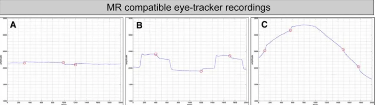
of corrective saccades were a sequence of corrective sac-cade away from the center of gaze, followed by a re-center-ing saccade that is also known as square-wave jerk. Figure1 illustrates an example of eye-tracker recording and corrective saccades during Wxation (A), pro-saccades (B) and pursuit (C).
Pro-saccades and pursuit
Activations associated with pro-saccades were present in a distributed network including bilateral frontal eye Welds
(FEF), supplementary eye Welds (SEF), parietal eye Welds (PEF) and primary visual area (Fig.2a, Table1). Pursuit eye movement was associated with a distributed network, which also included FEF, SEF and PEF and the cerebellum (Fig.2a, Table1).
The direct comparison of pro-saccades and pursuit (Fig.2b, Table2) revealed stronger activations during pro-saccades compared to pursuit within the known pro-sac-cade-associated network including bilateral FEF, SEF and PEF. Stronger activations for pursuit compared to pro-sac-cades were present in the known pursuit-associated
net-Fig. 1 Depiction of an example of eye-tracker recordings of one vol-unteer during Wxation (a), pro-saccades (b) and pursuit (c). Corrective saccades occur during all conditions and are marked with red circles. Note that pro-saccade targets were randomly presented to left or right,
half or full amplitude. The example b displays two consecutive pro-saccades in the same direction with half amplitude. X-axis indicates time (in ms), Y-axis indicates raw eye position (arbitrary units)
Fig. 2 Pro-saccades (S, green) and pursuit eye movement (P, blue) evoke activations in a distributed and largely overlapping network including FEF, SEF, PEF and the human homologue of MT/MST, which is in general more prominently activated for S except MT/MST. The direct statistical comparison (Table1) conWrms this observation and reveals additional activations for S compared to P in bilateral middle frontal gyri, left inferior frontal gyrus and left anterior insula.
Conversely, P compared to S (blue) evoked stronger activations in bilateral cerebellum. Activations are superimposed on the inXated MNI reference brain in Talairach space; activations are based on a Wxed-eVects GLM corrected for multiple comparisons at q(FDR) < 0.05. Essential anatomical regions are labelled: FEF frontal eye Welds, PEF posterior eye Welds, SEF supplementary eye Welds,
Table 1 Activation clusters for the analysis of S, P and the direct comparison of S versus P based on general linear models corrected for multiple comparisons at q(FDR) < 0.05
A pro-saccades and pursuit
Size t max TAL X TAL Y TAL Z Side Anatomic region BA
Pro-saccades
188,899 25.3 0.89 § 26 ¡64 § 14 11 § 22 Left/right Superior parietal lobule including PEF 7
Precuneus 7
Inferior parietal lobule 40
Cuneus 17 18 19
Middle occipital gyrus 18 19
Inferior occipital gyrus 18
Lingual gyrus 17 18 19
Superior temporal gyrus 22
Middle temporal gyrus 21
Inferior temporal gyrus 37
Cerebellum NA
25.3 ¡8 ¡70 5 Left Lingual gyrus 18
20.8 11 ¡77 5 Right Lingual gyrus 18
15.1 ¡28 ¡56 48 Left Superior parietal lobule 7
11.8 ¡42 ¡67 2 Left Middle temporal gyrus 37
11.7 21 ¡58 51 Right Superior parietal lobule 7
11.5 42 ¡61 3 Right Middle temporal gyrus 37
11.5 ¡26 ¡75 21 Left Cuneus 18
10.3 23 ¡79 23 Right Cuneus 18
38,716 12.9 ¡0.41 § 30 ¡6.7 § 6.6 47 § 9.6 Left/right Superior, middle frontal gyrus including FEF 6 9 46 Medial frontal gyrus including SMA, SEF 6
Anterior cingulate 24 32
Inferior frontal gyrus 44
12.9 26 ¡10 52 Right Middle frontal gyrus 6
11.8 ¡28 ¡9 47 Left Middle frontal gyrus 6
10.0 ¡2 ¡4 54 Left/right Medial frontal gyrus 6
790 3.1 ¡4.2 § 5.8 15 § 4.6 3.1 § 3 Left/right Caput of caudate nucleus NA
273 3.2 ¡38 § 2.1 9.9 § 1.7 22 § 1.7 Left Middle frontal gyrus 9 46
Pursuit
73,364 17.5 2.1 § 13 ¡69 § 12 5.5 § 13 Left/right Cuneus 17 18 19
Posterior cingulate gyrus 23 29 30 31
Lingual gyrus 17 18 19
Parahippocampal gyrus 27 30
Cerebellum NA
17.5 ¡9 ¡71 6 Left Lingual gyrus 18
17.2 10 ¡68 8 Right Lingual gyrus 18
2,500 6.8 ¡42 § 3.8 ¡66 § 3.9 3.5 § 4 Left Middle temporal gyrus 37
Middle occipital gyrus 19
1,234 5.7 41 § 2.3 ¡62 § 3.2 3.5 § 4.1 Right Middle temporal gyrus 37
Middle occipital gyrus 19
249 3.7 37 § 1.9 ¡11 § 1.6 49 § 2 Right Precentral gyrus 4
work including bilateral cuneus, precuneus, posterior cingulate and lingual gyrus. Additional activations were present in bilateral posterior cerebellum. These patterns of activation are in close agreement with published results (Petit et al. 1997; Kimmig et al. 1999).
Corrective saccades during Wxation, pro-saccades and pursuit
Activations associated with corrective saccades are illus-trated in Fig.3 and Table2.
Table 1 continued
Size of activation cluster in mm3, max t value, center of gravity in Talairach space § standard deviation, anatomic region and Brodmann Area
(BA). Local maxima are provided for large activation clusters if applicable in italics A pro-saccades and pursuit
Size t max TAL X TAL Y TAL Z Side Anatomic region BA
Pro-saccades
B pro-saccades versus pursuit
Size t max TAL X TAL Y TAL Z Side Anatomic region BA
Pro-saccades > pursuit
18,501 5.4 16 § 17 ¡54 § 11 44 § 5.8 Right Inferior parietal lobule 40
Left/right Superior parietal lobule including PEF 7
Left/right Precuneus 7
5.4 30 ¡50 44 Right Superior parietal lobule 7
7,359 5.6 ¡31 § 6.3 ¡49 § 10 39 § 7 Left Inferior parietal lobule 40
Superior parietal lobule including PEF 7 7,348 5.9 ¡0.74 § 22 ¡6.6 § 6.7 51 § 4.8 Left/right Medial frontal gyrus including SEF 6
Left/right Anterior cingulate 24 32
Left Superior, middle frontal gyrus including FEF 6 9
5.9 26 ¡10 53 Right Middle frontal gyrus 6
4.6 ¡2 ¡3 54 Left/right Medial frontal gyrus 6
4.2 ¡24 ¡13 53 Left Middle frontal gyrus 6
2,659 5.1 46 § 5.1 ¡42 § 3.5 12 § 4.2 Right Superior temporal gyrus 22
Middle temporal gyrus 22
1,216 4.0 ¡49 § 2.7 ¡44 § 5 9.4 § 3 Left Superior temporal gyrus 22
Middle temporal gyrus 21
623 3.4 ¡29 § 3.6 ¡78 § 5.4 ¡5.3 § 2.2 Left Lingual gyrus 18 19
614 3.6 38 § 4.6 ¡2.7 § 2.3 34 § 3.6 Right Precentral gyrus 6
535 4.2 34 § 2.4 ¡45 § 3.5 ¡18 § 1.6 Right Fusiforme gyrus 20
479 4.6 ¡23 § 1.9 ¡79 § 2.9 ¡23 § 2.3 Left Cerebellum NA
290 3.8 ¡37 § 2.1 39 § 1.7 24 § 1.9 Left Middle frontal gyrus 10
244 3.2 35 § 2.2 ¡67 § 2.1 15 § 1.7 Right Middle temporal gyrus 39
234 3.3 16 § 2.4 33 § 2 30 § 1.7 Right Medial frontal gyrus 9
219 3.3 ¡40 § 2.5 8.7 § 1.5 31 § 1.8 Left Middle frontal gyrus 9
208 3.6 42 § 1.9 ¡31 § 1.7 0.62 § 1.5 Right Superior temporal gyrus 41
Pursuit > pro-saccades
24,577 ¡7.0 ¡0.87 § 8.8 ¡68 § 10 9.7 § 9.4 Left/right Cuneus 17 18 19 31
Left/right Precuneus 31
Left/right Posterior Cingulate 23 30
Left/right Lingual gyrus 18 19
Left/right Cerebellum NA
¡6.9 0 ¡78 28 Left/right Cuneus 19
¡6.8 ¡8 ¡62 8 Left Cuneus 31
Table 2 Activation clusters for CS, CF and CP (otherwise as in Table1) Corrective saccades
Size t max TAL X TAL Y TAL Z Side Anatomic region BA
A corrective saccades during pro-saccades—CS
27,041 6.2 5.5 § 11 ¡71 § 9.3 ¡0.38 § 12 Left/right Cuneus 17 18 Lingual gyrus 17 18 19 Parahippocampal gyrus 30 Cerebellum NA 6.2 3 ¡76 14 Right Cuneus 18 5.3 ¡9 ¡67 ¡15 Left Cerebellum NA 4.9 3 ¡77 27 Right Cuneus 18 4.9 28 ¡74 ¡15 Right Cerebellum NA 4.8 9 ¡70 ¡11 Right Cerebellum NA 15,782 5.0 36 § 17 ¡47 § 12 32 § 19 Right Precuneus 7
Superior parietal lobule including PEF 7
Inferior parietal lobule 40
Superior temporal gyrus 22
Middle temporal gyrus 21 22
5.0 36 ¡42 40 Right Inferior parietal lobule 40
4.8 51 ¡36 22 Right Superior temporal gyrus 22
4.6 50 ¡42 3 Right Middle temporal gyrus 21
10,933 4.7 41 § 7.8 16 § 14 19 § 17 Right Superior, middle frontal gyrus including FEF 6 8 9 10
Precentral gyrus 4 6
Inferior frontal gyrus 44 45
Insula 13
Superior temporal gyrus 22
4.7 45 5 36 Right Middle frontal gyrus 9
4.2 33 19 6 Right Anterior insula 13
4.1 33 27 37 Right Middle frontal gyrus 9
3.9 28 44 ¡4 Right Middle frontal gyrus 10
8,774 4.8 ¡47 § 7.9 ¡47 § 8 26 § 14 Left Superior parietal lobule including PEF 7
Inferior parietal lobule 40
Superior temporal gyrus 22
Middle temporal gyrus 21
4.8 ¡51 ¡40 35 Left Inferior parietal lobule 40
4.3 ¡37 ¡53 39 Left Inferior parietal lobule 40
4.1 ¡52 ¡40 21 Left Superior temporal gyrus 22
6,337 4.9 0.53 § 3.8 0.85 § 9.9 50 § 8.4 Left/right Medial frontal gyrus including SEF 6
Cingulate gyrus 24 32
2,011 4.9 ¡46 § 4 7.5 § 3.8 2.4 § 3.4 Left Inferior frontal gyrus 44 45
Insula 13
Superior temporal gyrus 22
1,676 4.2 ¡29 § 3.9 ¡62 § 5.9 ¡20 § 2.9 Left Cerebellum 0
752 3.6 ¡36 § 3 48 § 3 2.3 § 2.3 Left Middle frontal gyrus 10
586 3.6 30 § 2.1 44 § 1.9 13 § 4.5 Right Middle frontal gyrus 10
495 4.0 37 § 2.2 ¡5 § 2.8 ¡7.5 § 2.1 Right Insula 13
403 3.3 ¡39 § 3.4 1.5 § 2.3 34 § 2.8 Left Middle frontal gyrus 9
Table 2 continued
Size of activation cluster in mm3, max t value, center of gravity in Talairach space § standard deviation, anatomic region and Brodmann Area
(BA). Local maxima are provided for large activation clusters if applicable in italics Corrective saccades
Size t max TAL X TAL Y TAL Z Side Anatomic region BA
A corrective saccades during pro-saccades—CS
357 3.4 ¡34 § 2.6 ¡11 § 2 49 § 2.3 Left Precentral gyrus 4
Left Middle frontal gyrus 6
338 3.9 ¡59 § 1.8 ¡20 § 2.1 14 § 2.2 Left Transverse temporal gyrus 41
Superior Temporal gyrus 42
300 3.4 ¡41 § 2.2 ¡30 § 1.8 12 § 1.8 Left Transverse temporal gyrus 41
278 3.7 ¡30 § 1.7 ¡27 § 2.1 13 § 2.4 Left Insula 13
Transverse temporal gyrus 41
269 3.3 1.8 § 1.5 29 § 2.6 29 § 2.1 Right Cingulate gyrus 32
B corrective saccades during Wxation—CF
5,542 5.9 0.28 § 7.3 ¡75 § 6.5 4.7 § 3.9 Left/right Lingual gyrus 17 18
1,310 4.4 13 § 3.7 ¡56 § 3 3.2 § 4.5 Right Lingual gyrus 19
1,289 5.3 ¡48 § 4.4 ¡54 § 4.9 11 § 2.7 Left Middle temporal gyrus 39
702 4.7 ¡48 § 3.7 10 § 3.3 ¡1 § 2.1 Left Insula 13
476 4.1 ¡51 § 1.5 ¡38 § 2.5 27 § 4.8 Left Inferior parietal lobule 40
250 3.6 ¡44 § 1.4 5.5 § 3.4 37 § 3.9 Left Middle frontal gyrus 8 9
213 4.0 3 § 1.4 ¡9.2 § 1.5 53 § 2.3 Right Medial frontal gyrus 6
203 4.0 ¡21 § 1.9 ¡11 § 1.5 59 § 1.7 Left Middle frontal gyrus including FEF 6 C corrective saccades during pursuit—CP
No suprathreshold activations
Fig. 3 illustrates activations associated with corrective saccades dur-ing pro-saccades (CS, red) and Wxation (CF, orange-yellow). The acti-vations of pro-saccades are superimposed for means comparison in green. CS evoked activations in the prosaccades network and addi-tional activations in anterior inferior cingulate and bilateral middle and inferior frontal gyri and bilateral insula. CF yielded activations
domi-nantly within the pursuit network and additionally in left insula. No su-pra-threshold activations were present for corrective saccades during pursuit (CP). Essential anatomic regions are labelled: MFG middle frontal gyrus, IFG inferior frontal gyrus, STG superior temporal gyrus,
MTG middle temporal gyrus, aACG anterior aspect of anterior
Corrective saccades occurring during pro-saccades (CS) were associated with a distributed network of activations, which largely overlaps with the pro-saccade network. Acti-vations in additional areas were present in anterior–inferior cingulate and bilateral middle and inferior frontal gyri and bilateral anterior insula (Table2).
Corrective saccades during Wxation (CF) yielded activa-tions within the pro-saccades network (notably bilateral lin-gual gyrus, left middle frontal gyrus including FEF and left middle temporal gyrus). Additional activations were found in left insula.
Corrections saccades during pursuit (CP) did not evoke any supra-threshold activations. The reasons for this lack of CP activation are discussed below.
Discussion
The combination of functional magnetic resonance imag-ing (fMRI) and MR compatible eye trackimag-ing was imple-mented to assess the neuronal circuitry mediating the execution of corrective saccades. More speciWcally, neu-ronal activations associated with corrective saccades were related to the modiWcation of the neuronal activations associated with pro-saccades and pursuit eye movement. In accordance to our hypothesis, we demonstrated speciWc neuronal activations associated with corrective saccades in addition to common neuronal activations evoked by pro-saccades and, to a lesser extent, by pursuit eye move-ments.
Pro-saccades and pursuit eye movement
In a Wrst step, we assessed neuronal activations associated with the execution of pro-saccades (S) and pursuit eye movement (P) in a block-design, which served as the basis for the later comparison with corrective saccades. In line with previous investigations, S and P yielded activations in largely overlapping networks including SEF, FEF and PEF (Berman et al. 1999; Petit and Haxby 1999) and the occip-ito-temporal junction (Petit and Haxby 1999), which is con-sidered as human homologue of monkey MT/MST (Zeki et al. 1991; Watson et al. 1993).
S compared to P evoked stronger activations in several areas deWned in the analysis of S, notably SEF, FEF and PEF, in line with a previous study (Petit and Haxby 1999). In another study, these areas had a trend to greater activa-tions during S compared to P, which was signiWcant only in FEF (Berman et al. 1999). In contrast to these investiga-tions, we compared the magnitude of activations in the S and P conditions using statistical parametric mapping. This revealed additional activations for S versus P in bilateral middle frontal gyri, left inferior frontal gyrus and left
ante-rior insula. It was shown recently that these areas are active in visually and memory guided saccade tasks (Brown et al.
2004; Ozyurt et al. 2006).
The inverse comparison P versus S yielded stronger acti-vations in cuneus, precuneus (Berman et al. 1999; Petit and Haxby 1999), lingual gyrus and posterior cingulate (Ber-man et al. 1999). Correspondingly, the direct comparison of P versus S yielded additional activations in bilateral cere-bellum. This region has been shown to be involved in the control of closed-loop, oculo-motor control of pursuit (O’Driscoll et al. 2000).
Corrective saccades
In contrast to previous investigations (Berman et al. 1999; Petit and Haxby 1999), we assessed eye movements during the fMRI experiments using an MR-compatible eye tracker (Kimmig et al. 1999). This allows for an event-related anal-ysis of neuronal activations associated with corrective sac-cades.
The frequency of spontaneously occurring corrective saccades diVered signiWcantly depending on the performed oculomotor task. The frequency of spontaneous corrective saccades increased from Wxation across pro-saccades to pursuit eye movement.
The most pronounced activations associated with correc-tive saccades were present during pro-saccades (CS). The resulting activation network largely overlapped with the acti-vation network deWned for pro-saccades, yet revealed addi-tional activations in anterior inferior cingulate and bilateral middle and inferior frontal gyri and bilateral–anterior insula. This implies a speciWc role of these above-mentioned areas in the execution of corrective saccades. These areas have not been reported in previous investigations, which assessed var-ious aspects of the neural control of saccades including inten-tional saccades, predictive saccades, memory guided saccades, anti-saccades or pro-saccades (for a review, Pier-rot-Deseilligny et al. 2004) and were accordingly not active in the pro-saccades condition of the presented study. It should be mentioned however that some previous studies have focused on the established neural circuitry that controls the oculomotor system (e.g., SEF, FEF and PEF) and did not investigate the whole brain. In particular, middle and inferior frontal gyri were not covered in the investigated volume in some studies (Berman et al. 1999; Kimmig et al. 2001).
Activations in the posterior aspect of the anterior cingu-late gyrus (pACG), which has also been called the ‘cingu-late eye Weld’ (Pierrot-Deseilligny et al. 2004), and activations in posterior cingulate gyrus (PCG) are known to be associated with saccade control (Paus et al. 1993; Nobre et al. 1997; Gaymard and Pierrot-Deseilligny 1999; Pierrot-Deseilligny et al. 2004). In agreement with these investiga-tions, pACG and PCG were active during S and CS in the
present study. The anterior–inferior aspect of the anterior cingulate cortex (aACG), in contrast, was active only dur-ing CS. The activations in aACG and bilateral inferior fron-tal gyri/bilateral anterior insula are very similar to those reported in an fMRI study, which investigated recentering-saccades (Raemaekers et al. 2005). Recentering-saccades direct the gaze back to the center of the display, following e.g., pro-saccades and anti-saccades. We suggest that these recentering-saccades share fundamental similarities with corrective saccades because both forms of saccades redirect the gaze. During the central Wxation periods between two consecutive pro-saccades, i.e., for approximately 50% of the time in the S condition, corrective saccades actually redirect gaze to the central visual target and hence are very similar to the above-mentioned recentering-saccades.
Corrective saccades during Wxation (CF) evoked similar yet less pronounced activations. This can be attributed to the lower frequency of corrective saccades during Wxations as compared to during pro-saccades. Activations were predom-inantly evident within the pro-saccade network. Additional activation was present in left insula, in agreement with CS.
Corrective saccades during pursuit (CP) yielded no signiWcant activations. The fact that CP yielded no supra-threshold activation in contrast to CS and CF appears contra-intuitive at Wrst glance because corrective saccades occurred almost three times more frequently during pursuit (P) than during pro-saccades (S). If the occurrence of cor-rective saccades increases, the linear dependence between the block-design regressor, e.g., P and the event-related regressor CP increases and it becomes progressively less possible to diVerentiate neuronal activations associated with P and CP. Note that the hemodynamic response func-tion (HRF) (Friston et al. 1998) introduces an additional temporal Wlter, that causes similar regressors for P and CP given the high number of events. Under the assumption of this eVect in particular in CP, we suggest that part of the activations in the established pro-saccade network during P may be related to corrective saccades rather than to pursuit per se. This consideration is supported by the observations that the amplitude of the BOLD response, in particular in FEF, increases almost linearly with increasing frequency of the pro-saccades while the amplitude of pro-saccades is irrelevant (Kimmig et al. 2001). If this observation can be transferred to corrective saccades, this implies that only the frequency of corrective saccades is of importance while their (small) amplitude is irrelevant. This suggests that the high number of spontaneously occurring corrective sac-cades during P should evoke pronounced activations, for e.g., in FEF. In line with this argumentation, it was sug-gested that FEF activation might be related to corrective saccades, which has been shown to occur during erroneous anti-saccades in a randomized pro- and anti-saccade task (Cornelissen et al. 2002).
The presented results imply that corrective saccades represent a potential systematic confound for the interpreta-tion of the neuronal correlates of oculomotor control, in particular if eye movements are not measured and corrective saccades are not included in the GLM analysis. This concern is in particular supported by the fact that the spontaneous occurrence of corrective saccades depends on the oculomotor task.
Limitations
The major limitation of the present investigation resides in the nature of corrective saccades, which occur spontane-ously and by deWnition only together with on-going eye movements. This complicates the separation of neuronal activations related to corrective saccades and the neuronal activations associated with, e.g., pursuit per se. The invol-untary occurrence renders impossible standard fMRI ON– OFF designs and/or systematic parametric variation in task diYculty, which would represent one possibility to over-come this limitation (Amaro and Barker 2006). We there-fore modeled the basic eye movement condition as non-explanatory regressors. Ideally, variance in BOLD response associated with the primary eye movement condition should be represented in the corresponding regressor. The remaining variance in BOLD response, which is associated with the behavioral occurrence of corrective saccades, pre-sumably reXects neuronal activations due to corrective sac-cades. The presented results regarding the neuronal correlates of corrective saccades should therefore be considered with caution given the intrinsic diYculty of separating the eVects of corrective saccades from those of the on-going oculomotor task. Notwithstanding, we argue that corrective saccades are a fundamental component of eye movement control, which implies that some methodo-logical constraints must be taken into account. Under the assumption that the occurrence of corrective saccades depends on the diYculty of the on-going eye-movement task, one possible amendment for future studies would reside in the titration of the eye movement task diYculty to evoke the frequency of corrective saccades, which is opti-mal for separation of eye movement-related and corrective saccades-related neuronal activations. For example, the occurrence of corrective saccades during smooth pursuit is lower for slower targets and for sinusoidal rather than con-stant velocity movement (Berman et al. 1999).
In addition to the frequency, the amplitude of corrective saccades might be of interest. In principle, it is possible to estimate the amplitude of eye movements with the used MR eye-tracker. The MR eye-tracker sensor is attached to the head coil. Consequently, head motion causes a shift of the center of gaze relative to this camera. We compared the measured eye position to the known stimulus position. We
do not report the corrective saccades amplitude results because in some volunteers, even (inevitable) small head motion evoked substantial and non-linear mis-estimation of the eye-positions in relation to the relatively small ampli-tude of corrective saccades. It has, however, been demon-strated that the amplitude of pro-saccades is irrelevant for the BOLD activations while the frequency of pro-saccades almost linearly correlates with the BOLD response (Kim-mig et al. 2001). Although this observation was made for pro-saccades and not for corrective saccades, this Wnding at least suggests that the amplitude of corrective saccades is less relevant than the frequency. The frequency could be determined with high accuracy with the MR eye-tracker.
Given these methodological constraints of the analysis of BOLD activations associated with corrective saccades, as discussed above, we cannot rule out that hypometria of the primary saccades and not necessarily the performance of corrective saccades per se might at least contribute to the observed activations of CS. One possibility to validate the presented results would be a parametric analysis of other ocular motor parameters such as saccade gain or saccade dysmetria, which might support a speciWc association of the reported activations with the performance of corrective sac-cades. At least in our experimental setup, the amplitude of the corrective saccades is however small in relation to the accuracy of the estimated eye position, as discussed above. This impedes a reliable parametric analysis of our data, but might be an option in a future setup. As discussed above, the temporal estimation of the onset of corrective saccades, in contrast, was possible at high accuracy.
We chose another approach to validate the presented results and reanalyzed the data with randomly varied onsets of corrective saccades, creating pseudo-corrective saccades event-related regressors. The block-design regressors for pro-saccades and pursuit were unchanged. As expected, the resulting activation patterns for the pseudo-corrective sac-cades diVered between diVerent variants of this random analysis, while the block-design regressors remained largely constant. This supports the assumption that the reported results are speciWcally related to the onset of cor-rective saccades, and illustrates the power of GLM analyses to separate the underlying task (here e.g., pro-saccades) from additional events (here e.g., corrective saccades dur-ing pro-saccades).
Perspectives
The present investigation provides functional MRI results concerning the neural control of corrective saccades. We demonstrate the feasibility of assessing eye movements and corrective saccades during fMRI and illustrate that correc-tive saccades represent a putacorrec-tive systematic confound in functional imaging of eye movements. At this stage, we
could not directly compare BOLD activations of corrective saccades arising in the CS and CP conditions, owing to diVerences in the spontaneous frequency of the events. We found substantial and signiWcant diVerence in the frequency of corrective saccades between these conditions, which would represent a systematic confound in the direct com-parison. Consequently, the interpretation of these diVerence analyses is problematic. It is diYcult to control the fre-quency of corrective saccades, as these are involuntary events. As discussed above, it might be possible to inXu-ence the frequency of corrective saccades by modifying the diYculty of the eye-movement task. Corrective saccades can be directly compared in future studies only if it is possi-ble to achieve similar frequencies of corrective saccades in the diVerent conditions. Additionally, we did not diVerenti-ate corrective saccades into sub-types. For example, correc-tive saccades during pursuit are also called catch-up saccades and it might physiologically be relevant to dis-criminate catch-up saccades in ON direction (i.e., the direc-tion of pursuit) and OFF direcdirec-tion (i.e., in the opposite direction of pursuit). Likewise, during pro-saccades, it might be relevant to discriminate corrective saccades towards the center versus towards the periphery. Further-more single corrective saccades might be diVerentiated from multiple corrective saccades. Subdivision of correc-tive saccades on the one hand reduces the number of events per condition and on the other hand requires additional pre-dictors/regressors in the GLM analysis. Both modiWcations likely reduce the level of signiWcance of the fMRI activa-tions. SpeciWc assessment of these sub-types of corrective saccades must be performed in future, speciWcally tailored investigations.
Conclusion
We demonstrated a neuronal network that is most likely speciWcally associated with corrective saccades and par-tially overlaps with the established pro-saccade and pursuit eye movement networks. The presented results suggest that corrective saccades represent a potential systematic con-found in eye-movement studies in particular because the frequency of spontaneously occurring corrective saccades depends on the underlying oculomotor task.
References
Amaro E Jr, Barker GJ (2006) Study design in fMRI: basic principles. Brain Cogn 60:220–232
Belliveau JW, Kwong KK, Kennedy DN, Baker JR, Stern CE, Benson R, Chesler DA, WeisskoV RM, Cohen MS, Tootell RB et al (1992) Magnetic resonance imaging mapping of brain function. Human visual cortex. Invest Radiol 27(Suppl 2):S59–S65
Bennett SJ, Barnes GR (2006) Combined smooth and saccadic ocular pursuit during the transient occlusion of a moving visual object. Exp Brain Res 168:313–321
Berman RA, Colby CL, Genovese CR, Voyvodic JT, Luna B, Thul-born KR, Sweeney JA (1999) Cortical networks subserving pur-suit and saccadic eye movements in humans: an FMRI study. Hum Brain Mapp 8:209–225
Boynton GM, Engel SA, Glover GH, Heeger DJ (1996) Linear systems analysis of functional magnetic resonance imaging in human V1. J Neurosci 16:4207–4221
Brown MR, DeSouza JF, Goltz HC, Ford K, Menon RS, Goodale MA, Everling S (2004) Comparison of memory- and visually guided saccades using event-related fMRI. J Neurophysiol 91:873–889
Cornelissen FW, Kimmig H, Schira M, Rutschmann RM, Maguire RP, Broerse A, Den Boer JA, Greenlee MW (2002) Event-related fMRI responses in the human frontal eye Welds in a randomized pro- and antisaccade task. Exp Brain Res 145:270–274
Dodge R (1903) Five types of eye movement in the horizontal meridian plane of the Weld of regard. Am J Physiol 8:307–329
Friston KJ, Holmes AP, Worsley KJ, Poline JB, Frith CD, Frackowiak RSJ (1995) Statistical parametric maps in functional imaging: a general linear approach. Hum Brain Mapp:189–210
Friston KJ, Fletcher P, Josephs O, Holmes A, Rugg MD, Turner R (1998) Event-related fMRI: characterizing diVerential responses. Neuroimage 7:30–40
Gaymard B, Pierrot-Deseilligny C (1999) Neurology of saccades and smooth pursuit. Curr Opin Neurol 12:13–19
Genovese CR, Lazar NA, Nichols T (2002) Thresholding of statistical maps in functional neuroimaging using the false discovery rate. Neuroimage 15:870–878
Huey EB (1900) On the psychology and physiology of reading. Am J Psychol 11:283–302
Kimmig H, Greenlee MW, Huethe F, Mergner T (1999) MR-eyetrac-ker: a new method for eye movement recording in functional magnetic resonance imaging. Exp Brain Res 126:443–449 Kimmig H, Greenlee MW, Gondan M, Schira M, Kassubek J, Mergner
T (2001) Relationship between saccadic eye movements and cor-tical activity as measured by fMRI: quantitative and qualitative aspects. Exp Brain Res 141:184–194
Nobre AC, Sebestyen GN, Gitelman DR, Mesulam MM, Frackowiak RS, Frith CD (1997) Functional localization of the system for vi-suospatial attention using positron emission tomography. Brain 120(Pt 3):515–533
O’Driscoll GA, WolV AL, Benkelfat C, Florencio PS, Lal S, Evans AC (2000) Functional neuroanatomy of smooth pursuit and predictive saccades. Neuroreport 11:1335–1340
Ogawa S, Lee TM, Nayak AS, Glynn P (1990) Oxygenation-sensitive contrast in magnetic resonance image of rodent brain at high mag-netic Welds. Magn Reson Med 14:68–78
Ozyurt J, Rutschmann RM, Greenlee MW (2006) Cortical activation during memory guided saccades. Neuroreport 17:1005–1009 Paus T, Petrides M, Evans AC, Meyer E (1993) Role of the human
anterior cingulate cortex in the control of oculomotor, manual, and speech responses: a positron emission tomography study. J Neurophysiol 70:453–469
Petit L, Haxby JV (1999) Functional anatomy of pursuit eye move-ments in humans as revealed by fMRI. J Neurophysiol 82:463– 471
Petit L, Clark VP, Ingeholm J, Haxby JV (1997) Dissociation of sac-cade-related and pursuit-related activation in human frontal eye Welds as revealed by fMRI. J Neurophysiol 77:3386–3390 Pierrot-Deseilligny C, Milea D, Muri RM (2004) Eye movement
con-trol by the cerebral cortex. Curr Opin Neurol 17:17–25
Pola J, Wyatt HJ (1991) Smooth pursuit: response characteristics, stimuli and mechanisms. Macmillan, London
Raemaekers M, Vink M, van den Heuvel MP, Kahn RS, Ramsey NF (2005) Brain activation related to retrosaccades in saccade exper-iments. Neuroreport 16:1043–1047
Talairach J, Tournoux P (1988) Co-planar stereotaxic atlas of the human brain. Thieme, New York
Watson JD, Myers R, Frackowiak RS, Hajnal JV, Woods RP, Mazziotta JC, Shipp S, Zeki S (1993) Area V5 of the human brain: evidence from a combined study using positron emission tomography and magnetic resonance imaging. Cereb Cortex 3:79–94
Yarbus AL (1967) Eye movements during perception of complex objects. Plenum Press, New York
Zeki S, Watson JD, Lueck CJ, Friston KJ, Kennard C, Frackowiak RS (1991) A direct demonstration of functional specialization in human visual cortex. J Neurosci 11:641–649