A new genus and species for the amiiform fishes
previously assigned to Amiopsis from the Early
Cretaceous of Las Hoyas, Cuenca, Spain
HUGO MARTÍN-ABAD
1,2,3*
and FRANCISCO JOSÉ POYATO-ARIZA
11
Unidad de Paleontología, Departamento de Biología, Universidad Autónoma de Madrid, Calle Darwin
2, 28049 Madrid, Spain
2
JURASSICA Museum, Route de Fontenais 21, 2900 Porrentruy, Switzerland
3
Department of Geosciences, University of Fribourg, Chemin du Musée 6, 1700 Fribourg, Switzerland
The Mesozoic actinopterygian fish Amiopsis has been reported from three different localities in the Iberian Peninsula. Amiopsis woodwardi was described from the Berriasian–Valanginian of El Montsec (Lérida, Spain). Isolated verte-bral centra referred to Amiopsis were found in the Barremian of Buenache de la Sierra (Cuenca, Spain). Finally, amiiform material from the Barremian of Las Hoyas (Cuenca, Spain) were reported as Amiopsis cf. A. woodwardi, due to the overall similarity with the species from El Montsec. Here a detailed anatomical description of the material from Las Hoyas is provided for the first time. The unique combination of characters seen in this taxon, some of which are not shared with Amiopsis but with more derived amiids, indicates that it represents a new taxon, which is here named Hispanamia newbreyi gen. nov., sp. nov.
ADDITIONAL KEYWORDS: Amiidae – Amiopsinae – Amiopsis woodwardi – Barremian – El Montsec – Hispanamia newbreyi gen. nov., sp. nov. – Vidalamiinae.
INTRODUCTION
Amiopsis Kner, 1863 is a genus included in the family Amiidae, the only genus of the subfamily Amiopsinae (Grande & Bemis, 1998). There are sev-eral valid species currently referred to it, all from European localities (Martín-Abad & Poyato-Ariza, 2013a; Poyato-Ariza & Martín-Abad, 2013): A. lepidota (Agassiz, 1833) from the Late Jurassic of Solnhofen (Germany), A. damoni (Egerton, 1858) from the Late Jurassic of England, A. woodwardi (Sauvage, 1903) from the Early Cretaceous of El Montsec (Spain), A. dolloi Traquair, 1911 from the Early Cretaceous of Bernissart (Belgium) and A. prisca Kner, 1863, the type species, from the Late Cretaceous of Croatia. The monophyly of this genus is currently supported by only one multistate character state, the presence of three
or more lateral fossae on each side of most abdominal centra (Grande & Bemis, 1998).
Concerning the amiid record of the Iberian Peninsula, isolated remains reported as Amiidae indet. or Amiiformes indet. from Asturias ( Ruiz-Omeñaca et al., 2006), the Cameros Basin in La Rioja (Bermúdez-Rochas & Poyato-Ariza, 2007), Vega de Pas in Cantabria (Bermúdez-Rochas et al., 2007) and Galve (e.g. Ruiz-Omeñaca et al., 2004) might actually belong to Amiopsis but are in need of further analysis ( Martín-Abad & Poyato-Ariza, 2013b). The genus Amiopsis has been recorded from three different localities in Spain. (1) A. woodwardi was described by Sauvage (1903) from the Berriasian–Valanginian laminated limestones of La Pedrera de Rùbies, in El Montsec (Lérida, north-east Spain). This species was later revised by Wenz (1968, 1988; see also Wenz & Poyato-Ariza, 1995), and more recently by Grande & Bemis (1998). (2) Vertebral centra referred to the subfamily Amiopsinae have been recovered from the Barremian of Buenache de la Sierra (Cuenca, Spain; Buscalioni et al., 2008). Isolated amii-form teeth were also found at this site, but they were *Corresponding author. E-mail: hugo.martin.abad@gmail.com
[ Ve r s i o n o f R e c o r d , p u b l i s h e d o n l i n e 5 Ju n e 2 0 1 7 ; http://zoobank.org/ urn:lsid:zoobank.org:pub:213A1503-1053- 459E-8C7D-F83D4C9F47E1]
http://doc.rero.ch
Published in "Zoological Journal of the Linnean Society 181 (3): 604–637, 2017"
which should be cited to refer to this work.
classified as Amiidae indet. (3) Finally, the Barremian laminated limestones of Las Hoyas (Cuenca, Spain) have also yielded specimens traditionally referred to Amiopsis (e.g. Sanz et al., 1988; Poyato-Ariza & Wenz, 1995; Poyato-Ariza, 2005a). Several amiiform taxa from this locality are currently under study; initially, the fish fauna of Las Hoyas was considered to be similar to that of El Montsec, at least at the generic level (Sanz et al., 1988), with three amiiform genera – Caturus, Amiopsis and Vidalamia – known from both. However, a more detailed comparison of both faunas has revealed that they differ more than initially thought with, for instance, the occurrence of several endemic taxa at Las Hoyas, such as the pycnodonts Turbomesodon praeclarus Poyato-Ariza & Wenz, 2004 and Stenamara mia Poyato-Ariza & Wenz, 2000, the gonorhynchiform Gordichthys conquensis Poyato-Ariza, 1994, and with different assemblages of primitive teleosts ( Poyato-Ariza, 1997; see also Poyato-Ariza & Martín-Abad, 2016). The last-named study suggested that the three amiiform species from Las Hoyas belong to new taxa (Poyato-Ariza & Martín-Abad, 2016).
The objective of the present paper is to describe the taxon of amiiform fishes from Las Hoyas that was previously referred to Amiopsis and to test whether it represents the same taxon as that from El Montsec. A complete phylogenetic analysis will be carried out after other amiiform taxa from Las Hoyas are revised.
MATERIAL AND METHODS
MATERIAL EXAMINED
The material of this new taxon is deposited in the Las Hoyas collection at the Museo de las Ciencias de Castilla-La Mancha (MCCM LH) in Cuenca, Spain:
MCCM LH 023, small, complete and articulated skeleton with excellent preservation.
MCCM LH 076a/b, very small skeleton, nearly complete but not totally ossified, with very good preservation.
MCCM LH 085R, small, complete and articulated skeleton with excellent preservation.
MCCM LH 151Pa/b, small, almost complete and mostly articulated skeleton with good preservation.
MCCM LH 374Ra/b, medium-sized, anterior half of skeleton, articulated and excellently preserved. The skull was mechanically and chemically prepared.
MCCM LH 9576a/b, small, complete and articulated skeleton with excellent preservation. It was mechani-cally and chemimechani-cally prepared lomechani-cally.
MCCM LH 9645a/b, complete and articulated, lacking only the tips of fin rays and some teeth. Slab ‘a’ was acid-prepared locally, not transferred to resin. It is excellently preserved, with only some bones of the skull and a few neural arches damaged.
Slab ‘b’ shows only the imprint of some damaged skull bones.
MCCM LH 11286, small, complete and articulated skeleton with excellent preservation. It was mechani-cally and chemimechani-cally prepared lomechani-cally.
MCCM LH 15783a/b, very small skeleton, complete but not totally ossified, with very good preservation.
MCCM LH 16040a/b, complete and articulated, lack-ing only part of the opercular series, the pectoral girdle and some fin ray segments. Slab ‘b’ was mostly covered by matrix, with only part of the abdomen exposed, and was mechanically and acid-prepared locally, not trans-ferred to resin. Preservation is very good, but the ver-tebrae of the exposed surface of the fish were already broken. Slab ‘a’ shows only the originally exposed surface.
MCCM LH 16257a, small, almost complete and articulated skeleton, with excellent preservation. It was acid-prepared locally.
MCCM LH 17274a/b, small, fairly complete and articulated skeleton, with excellent preservation. Both slabs have several cracks, which especially affect the skull and the axial skeleton.
MCCM LH 22600a, the smallest known specimen, complete and articulated skeleton not yet totally ossi-fied, with excellent preservation.
MCCM LH 23062a/b, small, complete and articu-lated skeleton with good preservation. It was mechani-cally and chemimechani-cally prepared lomechani-cally.
MCCM LH 30878a/b, very small, complete and articulated skeleton not yet totally ossified, with good preservation.
MCCM LH 32022a/b, small, nearly complete and articulated skeleton, with good preservation.
MCCM LH 32244a/b, very small, nearly complete and articulated skeleton not yet totally ossified, with good preservation.
TYPEOFPRESERVATIONAND PREPARATION METHODS
Fossil fishes from Las Hoyas usually appear complete and articulated and, even in the case of rare isolated elements, the preservation is exceptional. They are typically preserved as dark reddish to brown coloured bones in a light brown to light grey matrix. Nearly all the known specimens are preserved in lateral view, including their skulls, and are laterally compressed, rarely showing any three-dimensional preservation. Specimens are usually found as slab and counterslab after splitting the thin laminated limestones. Most of the specimens were prepared mechanically, chemically or a combination of both. For chemical preparation, a 2–4% solution of formic acid buffered with calcium phosphate was used; bones were coated with a highly diluted solution of B-72 paraloyd in acetone.
ANATOMICALABBREVIATIONS
ang, angular; ao, antorbital; ar, articular; arp,
ascend-ing ramus of parasphenoid; bop, branchiopercular; br, branchiostegals; cl, cleithrum; co, coronoid; d, dentary;
dplp, posterior dermopalatine; dpt, dermopterotic; dr,
distal radials; dsp, dermosphenotic; ecp, ectoptery-goid; ep, epural; epx, epaxial fin rays; es, extrascapu-lar; fr, frontal; g, guextrascapu-lar; h, hyomandibula; ha, haemal arch; hpx, hypaxial fin rays; hpxa, anteroventralmost hypaxial caudal fin ray; hpxd, dorsalmost hypaxial caudal fin ray; hs, haemal spine; hyp, hypural; ihm, infrahaemal; io, infraornbital bones; iop, interopercu-lar bone; l, lacrimal; le, lateral ethmoid; lloc, caudal extension of lateral line canal; mpt, metapterygoid;
mr, middle radial; mtg, metapterygium; mx, maxilla; n, nasal; op, opercular bone; pa, parietal; pas,
paras-phenoid; pastp, parasphenoid tooth patch; pb, ossified portion of basipterygium; pcl, postcleithrum; pfr, prin-cipal fin rays; phy, parhypural; pmx, premaxilla; pop, preopercular bone; pr, proximal radials; prfr, precur-rent fin rays; pt, posttemporal; ptg, propterygium;
q, quadrate; r, rib; ra, elongate proximal radials; ro,
rostral; sag, supraangular; scl, supracleithrum; smx, supramaxilla; sn, supraneural; sop, subopercular bone;
su, supraorbital bones; sym, symplectic; u, ural
cen-trum; ud, urodermal; una, ural neural arch; vo, vomer.
SYSTEMATIC PALAEONTOLOGY
SUBCLASS ACTINOPTERYGII COPE, 1887 INFRACLASS NEOPTERYGII REGAN, 1923,
SENSU ROSENETAL., 1981
DIVISION HOLOSTEI MÜLLER, 1844, SENSU GRANDE, 2010
SUBDIVISION HALECOMORPHI COPE, 1872, SENSU GRANDE & BEMIS, 1998
ORDER AMIIFORMES HAY, 1929, SENSU GRANDE & BEMIS, 1998
SUPERFAMILY AMIOIDEA BONAPARTE, 1838, SENSU GRANDE & BEMIS, 1998
FAMILY AMIIDAE BONAPARTE, 1838 GENUS HISPANAMIAGEN. NOV.
u r n : l s i d : z o o b a n k . o r g : a c t : A C 5 3 1 9 7 6 - D 3 6 A - 4089-8858-1C13D95DD668
? Amiopsis Kner, 1863: Sanz et al., 1988: 619–620, table 1.
Urocles Jordan, 1919 (Megalurus Agassiz): Poyato-Ariza, 1989: 113, fig. 22B, table 1.
Amiopsis Kner, 1863: Poyato-Ariza, 1989: 113, fig. 22C, table 1.
? Amiopsis Kner, 1863: Gómez-Pallerola, 1990: 52.
Urocles Jordan, 1919 (Megalurus Agassiz) pro parte:
Poyato-Ariza & Wenz, 1990: 303, fig. 5.
Amiopsis Kner, 1863 (Megalurus Agassiz) pro parte:
Poyato-Ariza & Wenz, 1990: 304, figs 2B, 5.
Urocles Jordan, 1919 (Megalurus Agassiz): Sanz et al., 1990: table 1.
Amiopsis Kner, 1863 (Megalurus Agassiz): Sanz et al., 1990: table 1.
Amiopsis Kner, 1863: Poyato-Ariza, 1992: 117, fig. 31. Amiopsis Kner, 1863: Sanz et al., 1994: 186.
Amiopsis Kner, 1863 pro parte: Wenz & Poyato-Ariza, 1994: 204–210.
Urocles Jordan, 1919 (Megalurus Agassiz): Fregenal-Martínez, 1995: 150, table 1.
Amiopsis Kner, 1863 (Megalurus Agassiz): Fregenal-Martínez, 1995: 150, table 1.
Amiopsis Kner, 1863: Poyato-Ariza & Wenz, 1995, 47, fig. IV-11.
Amiopsis Kner, 1863: Ortega et al., 1999, 206. Amiopsis Kner, 1863: Sanz et al., 1999: 158. Amiopsis Kner, 1863: Sanz et al., 2000: 158. Amiopsis Kner, 1863: Sanz et al., 2001: 359. Amiopsis Kner, 1863: Ortega et al., 2003: 434, 447. Amiopsis Kner, 1863: Delclòs et al., 2004: 40.
Amiopsis Kner, 1863: Poyato-Ariza, 2005b: 162, fig. 2C. Amiopsis Kner, 1863: Buscalioni & Fregenal-Martínez,
2010: 311.
Amiopsis Kner, 1863 pro parte: Martín-Abad & Poyato-Ariza, 2013a: fig. 4.
Amiopsis Kner, 1863: Martín-Abad & Poyato-Ariza, 2013b: fig. 2.
Amiopsis Kner, 1863: Poyato-Ariza & Martín-Abad, 2016.
Type species: Hispanamia newbreyi sp. nov. by monotypy. Generic diagnosis: Amiid fish differing from all other amiids by the following combination of derived and primitive characters: suborbital bones absent; jaw teeth labiolingually compressed, with sharply cari-nate caps; three lateral fossae on most of the verte-bral centra; articular bone of the lower jaw formed by the fusion of an anterior and a posterior element; faint ornamentation on the dermal bones of the skull formed by small, irregularly disposed foramina and thin, shallow, parallel grooves; relatively short pari-etal (width-to-length ratio range well exceeding 0.90); ten or fewer ural centra; preopercular long and narrow, crescent-shaped; and posttemporal with lateral edge shorter than anterior edge.
Etymology: Hispania, ancient Latin name of Spain, and Amia (Aμια), ancient Greek name for a type of fish (probably the bonito), and name of the type genus of the family Amiidae.
HISPANAMIANEWBREYISP. NOV. (FIGS 1–11)
u r n : l s i d : z o o b a n k . o r g : a c t : E 5 9 B 8 E 0 F - B 6 C 7 - 42D6-B8EF-FAC356648163
Urocles woodwardi (Sauvage, 1903): Sanz et al., 1988: 619, table 1, pl. 2 fig.2.
Urocles woodwardi (Sauvage, 1903) pro parte: Gómez-Pallerola, 1990: 52, 70.
Amiopsis woodwardi (Sauvage, 1903) pro parte: Wenz & Poyato-Ariza, 1994: 204–210, tables 1, 2.
Amiopsis woodwardi (Sauvage, 1903) pro parte: Grande & Bemis, 1998: 516.
Amiopsis woodwardi (Sauvage, 1903) pro parte: Poyato-Ariza, Buscalioni & Cartanyà, 1999: 511. Amiopsis woodwardi (Sauvage, 1903):
Fregenal-Martínez & Meléndez, 2000: table 1.
Amiopsis woodwardi (Sauvage, 1903) pro parte: Wenz, 2003: 495.
Amiopsis woodwardi (Sauvage, 1903): Escaso, Sanz & Ortega, 2005: 228.
Amiopsis woodwardi (Sauvage, 1903): Poyato-Ariza, 2005a: 286.
Amiopsis woodwardi (Sauvage, 1903) pro parte: Martín-Abad & Poyato-Ariza, 2009: 265.
Amiopsis woodwardi (Sauvage, 1903) pro parte: Martín-Abad & Poyato-Ariza, 2013a: 78–82, figs 4, 6, table 1. Amiopsis cf. A. woodwardi (Sauvage, 1903):
Martín-Abad & Poyato-Ariza, 2013b: fig. 1.
Amiopsis cf. A. woodwardi (Sauvage, 1903): Poyato-Ariza & Martín-Abad, 2016 b: c.pl. 12.2.B.
New taxon #1: Martín-Abad, 2016.
Type locality: Las Hoyas, Cuenca, Spain.
Type stratum: Calizas de la Huérguina Formation (Vilas et al. 1982), Rambla de Las Cruces II Sequence at Las Hoyas (Fregenal-Martínez, 1998). Upper Barremian (Early Cretaceous).
Geographical distribution: The species is endemic to Las Hoyas.
Species diagnosis: As for the genus (monotypic genus). Etymology: newbreyi, after Dr Michael G. Newbrey, palaeontologist and ichthyologist.
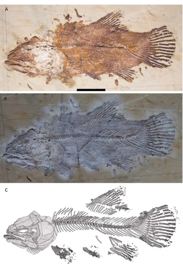
Holotype: MCCM LH 9645a/b, part and counterpart (Fig. 1).
Paratypes: MCCM LH 023, 076a/b, 085R, 151Pa/b, 374Ra/b, 9576a/b, 11286, 15783a/b, 16040a/b (Fig. 2), 16257a, 17274a/b, 22600a, 23062a/b, 30878a/b, 32022a/b, 32244a/b.
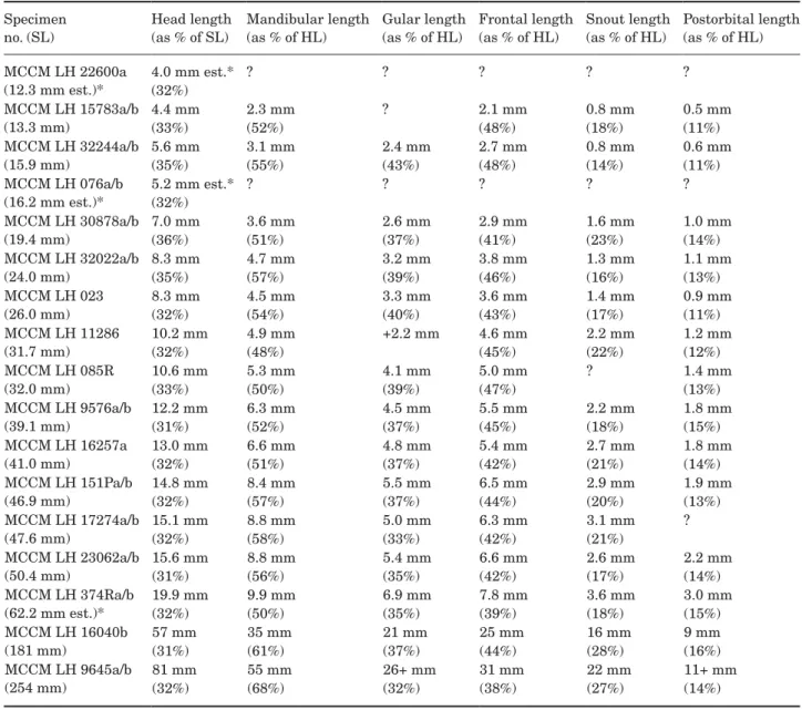
DESCRIPTION
Descriptive counts and measurements are provided in Tables 1–10. The known size of this species ranges from 12.3 mm standard length (SL) and 15.6 mm total length (TL) (specimen MCCM LH 22600a) to 254 mm SL and 308 mm TL (MCCM LH 9645a/b). A large pro-portion of the articulated specimens found so far are juveniles, some of them with a very low degree of ossi-fication, and there are only a few subadult and even fewer large adult specimens. One specimen (MCCM LH 9645a/b) is much larger than the rest; it is almost 50% longer than the second largest specimen.
SKULL
Braincase and ethmoid region: All known specimens of Hispanamia newbreyi gen. nov., sp. nov. have the skull preserved in lateral view. This, together with the good preservation of the cheek bones of the specimens, always with a very complete dermocranium, does not allow any bone of the sphenotic, otic and occipital regions of the braincase to be viewed. Only part of the ethmoid region can be seen on some specimens.
The rostral (ro) is an unpaired V-shaped bone with relatively large paired lateral expansions (wings). Posteriorly, the rostral bone articulates with the nasals. The tip of each lateral arm of the rostral is continuous with the tip of the corresponding antorbital. The ros-tral bone overlies the premaxilla. On the medial ante-rior surface of each wing of the rostral there is a small opening to the rostral canal.
The paired lateral ethmoid (le) lies just ventral to the anterolateral corner of the frontal and the postero-lateral corner of the nasal, and dorsal to the vomer and lacrimal. The lateral ethmoid is roughly hemispherical posteriorly and flatter anteriorly. It is partially over-lapped by the antorbital. The lateral ethmoid articu-lated in life with the palatal complex (usually with the autopalatine), forming the anterior suspension of the palatal complex to the skull (Grande & Bemis, 1998).
Otoliths: As stated above, this region is not clearly visible in any of the known specimens and no otoliths have been observed.
Skull roof: The skull roof is formed by six paired bones: nasal, frontal, dermosphenotic, dermopterotic, parietal and extrascapular. As stated above, all the known specimens are preserved in lateral view, so the skull roof is never fully observable. The skull of the holotype (Fig. 3), nonetheless, is slightly compressed, and both the left and part of the right sides of the skull roof are visible. The skull roof is, however, not as well preserved as the rest of the specimen. Nevertheless, the shape and margins of most of the bones can be confidently traced. The skull of specimen MCCM LH 16040a/b (Fig. 4) is preserved in lateral view.
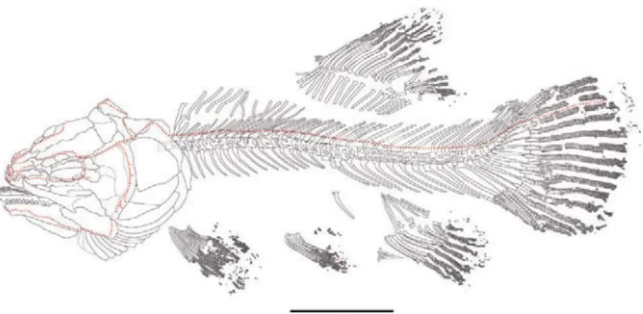
Figure 1. Hispanamia newbreyi gen. nov., sp. nov. (A) specimen MCCM LH 9645a (holotype). (B) specimen in A slightly coated with ammonium chloride. (C) digital drawing of the skeleton as preserved in specimen in A. Scale bar = 5 cm.
The bones of the skull roof are very smooth, lacking a developed ornamentation. Even in the largest specimen the ornamentation consists only of very few, small irreg-ularly disposed foramina and thin, shallow, parallel grooves. This contrasts with the taxa from other amiid subfamilies (Grande & Bemis, 1998), for which the orna-mentation is usually moderately to highly developed. As found in Amia calva, there are no fenestrae, fonta-nelles or pineal openings on the skull roof. The bones are tightly sutured to each other in adult individuals.
Also as in A. calva, there are several pore clusters on the skull roof corresponding to the supraorbital sensory canal. These pores conform a linear pattern that runs from the nasal into the frontal, running near the lateral edge of the latter; whether this canal penetrates into the parietal is hard to confirm. The canal then continues into the dermosphenotic, where it branches towards the infraorbital series to connect with the infraorbital sensory canal. Finally, it continues through the dermopterotic, extrascapular and the lateral edge of the posttemporal.
At least one trough, running on the posterolateral part of the parietal and into the dermopterotic, is observable on the skull roof. This is termed middle pit-line trough (Grande & Bemis, 1998). In life, a row of closely spaced pit-line neuromasts would be situated along the floor of the trough, as found in A. calva (e.g. Jarvik, 1980).
The anteriormost bone of the skull roof is the nasal (n), which lies posterior to the rostral. The nasal is an elongated bone, slightly wider anteriorly than posteri-orly. On the anterolateral margin of the nasal is a notch which forms, together with the antorbital, the posterior edge of the opening for the anterior narines. The nasal
is located anterior to the frontal, but does not actually articulate with it; in life, both bones are separated by connective tissue in A. calva, and probably in H. new-breyi as well. The left and right nasals are sutured to each other medially by means of an interdigitating suture. There are short, shallow, parallel grooves both on the anterior and on the posterior regions of the nasal.
Posterior to the nasal is the frontal (fr). As in A. calva, the frontal is the longest element of the skull roof, occupying almost 40% of the head length in the largest specimen. The frontal is approximately twice as long as wide, with a sub-rectangular general shape. It is wider posteriorly, and tapers abruptly at the level of the orbit. Anteriorly, the frontal has thin laminar projections that partially overlap the supramaxilla and, in turn, are overlapped by the nasal. The ante-rolateral margin is greatly excavated, and articulates with a series of supraorbital bones. The frontal has an irregular rather than straight contour, indicating inter-digitating sutures with the parietal, dermopterotic and dermosphenotic. The frontal has very few relatively shallow, parallel grooves running from the centre of the bone towards the posterior and anterior margins.
Posterior to the frontal and medial to the dermopter-otic is the parietal (pa). The shape of the parietal is highly variable, both among individuals and between the left and right sides of the same individual, depending on the curvature of the margins of articulation with other skull roof bones. In general, it is approximately as wide as long. The right and the left parietals are very similar in size. Medially (and probably anteriorly, but this cannot be con-firmed because all the specimens show articulated skull
Figure 2. Hispanamia newbreyi gen. nov., sp. nov. (A) specimen MCCM LH 16040b. (B) specimen in A slightly coated with ammonium chloride. Scale bar = 5 cm.
roofs) the parietal has well-developed ventral laminar shelves that are overlain by the other-side parietal (and by the frontal anteriorly), forming a very tight articula-tion. The margins are irregularly curved, forming inter-digitating sutures with the other-side parietal medially, with the frontal anteriorly and with the dermopterotic laterally. The posterior margin of the parietal is straight, and articulates with the extrascapular. The presence on
the posterior edge of a foramen to a canal, present on other amiid species including A. calva (Grande & Bemis, 1998: 42), cannot be confirmed.
Lateral to the frontal is the dermosphenotic (dsp). This bone, which is actually part of the infraorbital series, is tightly sutured into the skull roof, as is the common con-dition for amiid fishes, and is thus described in this sec-tion. The dermosphenotic is a small bone, slightly longer
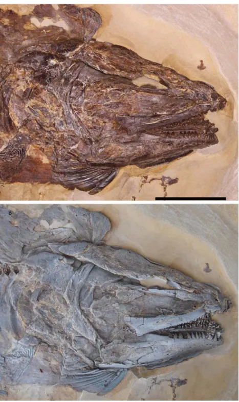
Figure 3. Hispanamia newbreyi gen. nov., sp. nov. (A) skull from specimen MCCM LH 9645a (holotype) slightly coated with ammonium chloride. (B) digital drawing of the skeleton as preserved in specimen in A. Scale bar = 2 cm.
than wide. It exhibits a more or less curved anterolateral edge, which forms part of the margin of the orbit, and an irregular posteromedial edge to articulate with the fron-tal. The posterior margin is irregular as well, and articu-lates with the dermopterotic. The margin of the orbit is more excavated anterior to the dermosphenotic, despite
the presence of supraorbital bones. Posterolaterally, the dermosphenotic articulates loosely with the last infraor-bital. In lateral view, and just posterior to the orbital margin, the dermosphenotic has a pore that might cor-respond to an opening for the infraorbital canal, as in A. calva (Grande & Bemis, 1998: 48).
Figure 4. Hispanamia newbreyi gen. nov., sp. nov. (A) skull from specimen MCCM LH 16040b. (B) specimen in A slightly coated with ammonium chloride. Scale bar = 2 cm.
Posterior to the dermosphenotic and lateral to the posterior part of the frontal is the dermopterotic (dpt). The dermopterotic is longer than wide. It is wider pos-teriorly and tapers anpos-teriorly, although there is individ-ual variability in the width between the posterior and the anterior parts. It has straight posterior and lateral edges, and the posterolateral corner is very slightly expanded and curved. The medial and, especially, the anterior edge are more sinuous, conforming interdigi-tating sutures with the dermosphenotic, frontal and parietal. The dermopterotic is almost twice as long as the parietal and slightly narrower. It bears very few shallow, parallel grooves running along its longitudinal axis. The presence of a ventral laminar process and of an opening for the postotic sensory canal, which is the junction between the lateral line canal of the dermop-terotic and the preopercular canal, typically observable in lateral view in amiid dermopterotics (e.g. A. calva in Grande & Bemis, 1998, figs 17, 24), cannot be confirmed.
The posteriormost bone of the skull roof is the extrascapular (es). The extrascapular is subtriangular in shape, and tapers medially. It is wider than long, with a straight anterior margin, and a posterior mar-gin that tapers abruptly in the midline. The lateral margin is straight to slightly curved, and the medial margin is curved, not acute. The extrascapular has short, shallow, parallel grooves on its posterolateral region, difficult to see on some specimens.
Circumorbital bones: The infraorbital series is com-posed of eight bones. Only the second to the seventh are typically termed infraorbitals, because the first is the antorbital, and the eighth is the dermosphenotic, which is tightly sutured into the skull roof. All the bones of the infraorbital series enclose the infraorbi-tal sensory canal; this canal branches anteriorly at the antorbital and posteriorly at the dermosphenotic, in both cases towards the supraorbital canal. For this reason, sensory canal pores can be seen on all the infraorbital bones if the preservation is good enough.
The anteriormost bone of the infraorbital series is usually referred to as the antorbital (ao). The antorbital is an elongate, slightly curved bone, wider in its middle. The antorbital articulates only loosely with the nasal of the skull roof; it does not contact the frontal posteriorly, but partially overlies the lateral ethmoid and the lacri-mal. Anteriorly, the antorbital extends very close to the rostral bone, yet it does not articulate with it. The rior region of the antorbital has openings for the ante-riormost part of the lateral line sensory system, which continues through the lateral wing of the rostral bone. Posteriorly, the antorbital articulates only loosely with the next element of the infraorbital series, the lacrimal.
The lacrimal (l), usually termed infraorbital 1 as well, is the bone that forms the anterior margin of the orbit. The shape of the lacrimal is variable in amiids (Grande & Bemis, 1998); in H. newbreyi the lacrimal
is a flat bone, subtriangular in shape, located between the maxilla and the skull roof, and reaching the pre-maxilla anteriorly. It is longer than deep. The lacrimal articulates loosely with and is partly overlapped by the antorbital. Posteriorly, it has a notch for the articu-lation with the next infraorbital bone.
There are three infraorbitals (io2–4) forming the ventral margin of the orbit. These infraorbitals at the ventral margin of the orbit are termed subinfraorbitals by Grande & Bemis (1998). Only two of these three bones are visible in specimen MCCM LH16040b. These three infraorbitals are little more than ossified tubes. In adults, they are short bones, yet longer than deep, and subrectangular in shape. The first (io2) of them is slightly more expanded dorsoventrally than the other two. Their contour is irregular rather than straight.
There are two infraorbitals (io5–6) constituting the posterior margin of the orbit. These bones at the poste-rior margin of the orbit are termed postinfraorbitals by Grande & Bemis (1998). The shape and relative size of these infraorbitals are highly variable in amiid fishes (Grande & Bemis, 1998). For example, the infraorbitals 5–6 of H. newbreyi are much less developed than those of A. calva (e.g. Grande & Bemis, 1998). Infraorbital 5 is not complete in the holotype. It is roughly ovoid in shape, higher posteriorly than anteriorly. Its surface is not as smooth as that of other dermal bones; it has short, shal-low grooves on its margins, and is slightly depressed on the centre. The infraorbitary sensory canal runs through the anterior part of the bone and penetrates into the next infraorbital. Infraorbital 6 is not completely preserved on the holotype either, and thus it is difficult to confirm its relative size in comparison to infraorbital 5, which is con-sidered to be of phylogenetic interest (Grande & Bemis, 1998). In specimen MCCM LH 16040a/b, only infraor-bital 5 is preserved, but there is a very small displaced bone that might correspond to infraorbital 6, thus con-firming a smaller size of the uppermost (io6) relative to the lowermost (io5) posterior infraorbitals. Nevertheless, this area is not very well preserved in this specimen, so nothing conclusive can be stated.
There are no suborbitals. These bones, located poste-rior to the last infraorbitals when present, do not exist in any known species of the subfamilies Amiinae and Vidalamiinae either. In contrast, Solnhofenamia elon-gata and several Amiopsis species have one or more suborbitals (Grande & Bemis, 1998).
There are two supraorbital bones (su). They are short, yet longer than high, with irregular rather than straight margins. The supraorbitals are attached loosely to the lateral margin of the anterior part of the frontal, where the latter shows a distinct facet forming the orbital mar-gin. Both supraorbitals are arranged into a single row.
Neither the holotype nor specimen MCCM LH 16040a/b show clear remains of an ossified sclerotic ring. However, smaller specimens (e.g. MCCM LH 151Pa/b,
374Rb, 9576b, 11286) show a sclerotic ring that seems to be faintly ossified into two pieces (Fig. 5A, B).
Upper jaw: The upper jaw consists of a premaxilla, a maxilla and a supramaxilla. The anteriormost ele-ment is the premaxilla (pmx). The oral border of the premaxilla, where the teeth are implanted, is thick, robust and concave, articulating with its counterpart medially. Dorsally and posteriorly the premaxilla has a thin expansion, the nasal process of the premaxilla. This expansion seems to extend back to the anterior edge of the frontal. The premaxilla is overlapped by the nasal,
the antorbital and the rostral bone, with which it is not articulated. In turn, the premaxilla posteriorly covers the anterior part of the vomer, whose teeth are just pos-terior to, and in functional continuity with, those of the premaxilla. Posterolaterally, the premaxilla articulates with the maxilla. The premaxilla bears a single row of teeth that are large and slightly recurved towards the oral cavity. They are sharply pointed and have a cari-nate (keeled) acrodin cap. There are usually eight teeth on each premaxilla, all of them approximately of the same size.
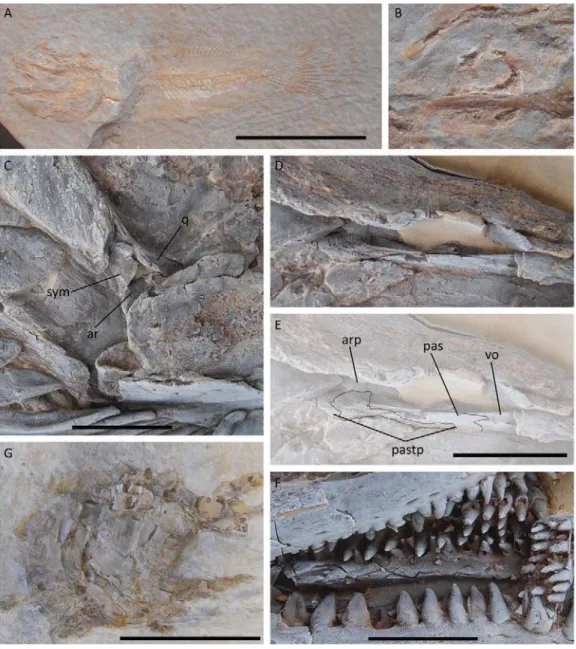
Figure 5. Hispanamia newbreyi gen. nov., sp. nov. (A) specimen MCCM LH 151Pa. Scale bar = 2 cm. (B) close-up of specimen from A showing sclerotic ring ossification. (C) close-up of jaw articulation from specimen MCCM LH 16040b slightly coated with ammonium chloride showing both the quadrate and the symplectic articulating with a single articular element. Scale bar = 5 mm. (D–E) close-up of orbit from specimen MCCM LH 16040b slightly coated with ammonium chloride show-ing the parasphenoid tooth patch. Scale bar = 1 cm. (F) close-up of mouth openshow-ing from specimen MCCM LH 16040b slightly coated with ammonium chloride showing keeled teeth. Scale bar = 5 mm. (G) specimen MCCM LH 9645b (holotype). Scale bar = 5 cm.
The maxilla (mx) seems to articulate only loosely with the rest of the skull, as in A. calva, where it rotates around the axis of a peg-like anterior process which fits into a socket formed by the vomer, premaxilla and preethmoid (Grande & Bemis, 1998). The premaxillary process of the maxilla is thin and ellipsoidal in section. The posterior expansion of the maxilla is much higher and flatter. The maxilla bears a single row of teeth, which extends back to over three-quarters of the length of the maxilla. These teeth are smaller than those of the premaxilla, and become much smaller posteriorly. The largest teeth are slightly curved towards the oral cavity. Each maxilla has around 25 teeth; according to Grande & Bemis (1998), the number of maxillary teeth increases throughout the life of the fish, at least in A. calva, a con-dition that, based on the specimens available, seems to occur also in H. newbreyi. The teeth are sharply pointed, and have keeled acrodin tips. The anterior dorsal margin of the maxilla is slightly excavated; when the mouth is closed, the lacrimal and, at least, the infraorbital 2 fit into this depression. Posteriorly, the dorsal edge of the max-illa is also excavated to hold the supramaxmax-illa; this exca-vation, best observed in specimen MCCM LH 16040a/b, is termed the supramaxillary notch. The supramaxilla is tightly sutured to this excavation. The posterior edge of the maxilla is concave, forming the posterior maxil-lary notch, which seems not to be greatly developed; this notch is not clearly observed in the holotype specimen, because this region of the maxilla is not well preserved. The lateral surface of the maxilla is moderately smooth, with very shallow grooves on its posterior part.
The supramaxilla (smx) articulates with the max-illa immediately posterior to the supramaxmax-illary notch. This bone is thin, elongate and relatively low. It is almost as long as half the length of the maxilla. Its lateral surface is smooth.
Lower jaw: To date, no specimen has been found showing a medial view of a complete lower jaw; only the anteriormost part of the right dentary and coro-noids are visible on specimens preserved on their left side, and vice versa. As a consequence, only the pres-ence of the following paired bones can be confirmed: dentary, at least three coronoids, angular, supraan-gular, articular (apparently formed by two fused ele-ments, as commented later on) and a retroarticular.
On their lateral surface, both the dentary and the angular show relatively large pores corresponding to the mandibular sensory canal. This canal seems to turn upwards on the posterior part of the angular, approximately towards the ventral end of the pre-opercular, suggesting a possible connection through the skin with the sensory canal running through the latter, as is common in all neopterygians. In A. calva this connection exists and is termed the preopercular– mandibular canal (Grande & Bemis, 1998).
The anteriormost bone of the lower jaw is the den-tary (d). The denden-tary is the largest element of the lower jaw. It bears a single row of approximately 15 teeth, which are the largest in the lower jaw. They are slightly recurved towards the oral cavity, and not completely round in cross-section but rather slightly compressed labiolingually. They have an acrodin cap that is strongly carinate or keeled. In lateral view, the dentary is deep-est posteriorly and tapers anteriorly. The anterior part of the dentary is curved medially, to meet its counter-part forming a slightly rounded symphyseal region. The anterior part of the ventral margin of the bone is slightly concave. The dorsal margin is expanded posterior to the last tooth; this expansion, the coronoid process, is sub-triangular in shape, and its posterior margin articulates with the supraangular bone. The posterior margin of the dentary is deeply notched in its articulation with the angular. In lateral view, the dentary is fairly smooth, with very shallow longitudinal grooves. The most pro-nounced structure on the surface of the dentary is the series of pores of the mandibular sensory canal, which runs ventrally on the anterior part of the dentary, then through the lower half of its lateral surface describing a soft curve, and then very close to the ventral surface of the bone posteriorly. In medial view, the dentary has a thin bony shelf where the coronoids are lodged. This bony shelf is actually the upper part of the meckelian groove, which harbours Meckel’s cartilage.
Posterior to the dentary is the angular (ang). The angular is a thick bone, approximately half as long as the dentary, and as high as the middle part of the den-tary. The ornamentation on the lateral surface of the angular is usually more pronounced than that of the dentary, with somewhat deeper grooves. A thin ante-rior projection of the angular sutures with the posteante-rior notch of the dentary; another projection on the ventral surface of the angular also sutures with the dentary, so the articulation is V-shaped. The mandibular sensory canal continues from the dentary through the ventro-lateral part of the angular. Posteriorly, the canal turns upwards and runs very close to the posterior margin of the angular. It exits the bone through its dorsal margin, where a large opening serves as the connection point with the sensory canal of the preopercular through the skin. The posterior margin of the angular is curved and ventrally notched. Its dorsal margin is fairly straight, and articulates with the supraangular.
The supraangular (sag) is smaller than the dentary and the angular. It articulates with the posterior edge of the dorsal expansion of the dentary and the dorsal margin of the angular. It constitutes the highest point of the lower jaw and of the coronoid process, providing the posterior part of the lower jaw with a roughly tri-angular shape. The supratri-angular has a smooth lateral surface, devoid of ornamentation.
There are, at least, three coronoid bones (co) on the inner side of the lower jaw. The coronoids are arranged in line and articulate with each other by means of interdigi-tating anterior and posterior margins. The posteriormost coronoid is the one articulating with the anterior margin of the prearticular, but this bone is not observable in any of the known specimens. The coronoids are short bones constituted by a bony base and numerous teeth; each coronoid bears 20–30 teeth. These teeth are irregularly arranged in three or four series. The teeth are conical and slightly recurved towards the oral cavity. They have a sharply pointed acrodin cap that is keeled. The teeth on the coronoids are smaller than those on the dentary, but are located nearly at the same level on the lower jaw.
Among the bones that usually ossify from Meckel’s cartilage, only the retroarticular and the articular are observable on the known specimens.
The articular (ar) is only observable in specimen MCCM LH 16040a/b, and is slightly broken, so it is dif-ficult to confirm whether it is a single bone or two differ-ent elemdiffer-ents fused together. The presence of a very thin line that could be a suture, and the different directions of the anterior and posterior parts suggest the latter option as more probable. So, it is interpreted that the articular is apparently formed by the fusion of an ante-rior plus a posteante-rior element. The articular forms the so-called double jaw articulation with the quadrate and the symplectic, which is a traditional diagnostic charac-ter for halecomorph fishes; the ancharac-terior part of the artic-ular bone articulates with the condyle of the quadrate, whereas the posterior part of the articular bone articu-lates with the socket-like ventral end of the symplectic (Fig. 5C). Overall, the articular is small and convex; its anterior part is located between the angular and the prearticular (which is actually not visible in any speci-men), and is anterolaterally directed. The posterior part of the articular bone articulates with the medial surface of the posterodorsal corner of the angular. The articular is visible in lateral view through the posterior corner of the articulation between the angular and supraangular. An articular formed by two elements tightly sutured to each other constitutes an important phylogenetic char-acter according to Grande & Bemis (1998).
The retroarticular is the posteriormost bone of the lower jaw. The retroarticular articulates with the poster-oventral corner of the medial surface of the angular. In lateral view, it is visible through the posteroventral notch of the angular. The retroarticular is a small rounded bone that is dorsally concave and has a small dorsal expansion on its medial surface, creating a sort of hemispheric fossa. In A. calva, this fossa serves as the surface of attachment for the large hyomandibula and interoperculomandibu-lar ligaments (Grande & Bemis, 1998).
Palate: The palatal complex of amiid fishes includes one unpaired bone, the parasphenoid, and numerous paired bones. Among them, the autopalatine cannot
be seen in any known specimen, probably because it is placed in a deep plane and laterally overlapped by the large lacrimal. The presence of parasphenoid, vomer, two dermopalatines, ectopteryogid, endopterygoid and metapterygoid can be confirmed. None of these bones can be described in detail, because they are only partially visible; they are never observable in ventral or medial view, where most of their important characteristics are, nor are they usually completely visible in lateral view.
The parasphenoid (pas) is observable in lateral view crossing the orbit of most of the specimens, but this view does not provide much morphological infor-mation. It extends, at least, from almost the anterior margin of the orbit to the hyomandibula. The tooth patch of the parasphenoid extends a long way anteri-orly, almost to the suture with the vomers (Fig. 5D, E). Posteriorly, the tooth patch extends into the ascending rami of the parasphenoid. This patch is formed by hun-dreds of small and pointed teeth.
The vomer (vo) is relatively long. It extends from the premaxilla and exceeds the anterior margin of the orbit. The suture between the parasphenoid and the vomers is interdigitated.
The anterior dermopalatine is a short, low bone located medial to the anterior part of the maxilla. It probably articulates with the vomer, as in other ami-ids, but this cannot be confirmed. It articulates poste-riorly with the posterior dermopalatine by means of an interdigitating suture. It bears a row of very few, large, curved teeth (at least three or four are observable). These teeth are larger than those of the maxilla and coronoids, but smaller than those of the premaxilla and dentary. They have a small acrodin cap that is keeled, but not as strongly as those of the dentary, coronoids and premaxilla. The presence on this bone of more teeth medial to the row of large teeth cannot be confirmed, although in specimen MCCM LH 16040a/b there seems to be at least one smaller tooth medial to the large teeth.
The posterior dermopalatine (dplp) is approximately as long as the anterior one. It is also a low bone, and is located between the anterior dermopalatine and the ectopterygoid, with which it also articulates by means of an interdigitating suture; the large teeth of these three bones form a medial row continuous to that of the maxilla. The teeth of the posterior dermopalatine are as large as, or even slightly larger than, those of the ante-rior dermopalatine, and very similar in shape. They are very few in number (two to three are distinguishable).
The ectopterygoid (ecp) is a long bone, reaching the level of the posterior end of the maxilla. In lateral view, it is a thin, tube-like bone, slightly higher anteriorly, and with a long, low, triangular posterodorsal expan-sion. It bears, at least, a row of teeth (at least four or five are visible) whose size decreases from anterior to posterior, smaller than those of the dermopalatines. As in the anterior dermopalatine, the occurrence of
additional, smaller, medial teeth, present in other ami-ids, cannot be confirmed.
The endopterygoid is a flat, relatively large bone. Its anterior margin, supposedly articulating with the autopalatine and dermopalatine, is largely covered lat-erally by the lacrimal and infraorbitals 2–4. Latlat-erally it articulates with the dorsomedial margin of the ectopterygoid, and extends inwards to the parasphe-noid. Its posterior margin is sutured to the anterior margin of the metapterygoid.
The metapterygoid (mpt) is the posteriormost ele-ment of the palatal complex. The metapterygoid is a chondral bone that ossifies within the palatoquadrate cartilage (in addition to the quadrate and the autopala-tine; Jollie, 1984). This region of the skull, which usually is partially overlaid by the infraorbital series, is best seen on specimen MCCM LH 16040a/b. The metaptery-goid is a relatively large, flat bone. Its dorsal margin is deeply notched, bearing a laterally pointed middle process. In life, this notch is crossed by the trigeminal nerve in A. calva (Grande & Bemis, 1998). The anterior, ventral and posterior margins of the bone form a more or less continuous curve; the anterodorsal and postero-dorsal expansions that form the notch are subtriangu-lar in shape. The metapterygoid is tightly articulated to the endopteryogoid and ectopterygoid anteriorly. Posteriorly, it partially overlaps the hyomandibula, and ventrally it is tightly articulated to the quadrate.
Teeth: The large teeth of the jaws and palatal bones are not completely conical, but rather labiolingually compressed, especially those of the dentary. The teeth are sharply pointed, with an acrodin cap usually pre-served. This cap is keeled in most of the teeth, espe-cially in those of the dentary, coronoids and premaxilla (Fig. 5F). The largest teeth are on the dentary and pre-maxilla. The large teeth have long, hollow shafts as in all other amiids (Grande & Bemis, 1998). There are patches of very numerous little teeth (shagreen) on several pala-tal bones; these tiny teeth are conical and pointed.
Hyoid arch: In addition to the palatal bones dis-cussed above, the hyomandibula, the quadrate and the symplectic also contribute to the suspensorium. As for the metapterygoid, these bones are best seen in speci-men MCCM LH 16040a/b, but also on the counterslab of the holotype, especially the hyomandibula (Fig. 5G). The hyomandibula and the symplectic are part of the hyoid arch, which also includes the anterior and poste-rior ceratohyal, the hypohyal and the basihyal; the last two are not visible in any known specimen.
The hyomandibula (h) is a large bone located poste-rior and dorsal to the metapterygoid. The hyomandibula is not tightly sutured to the metapterygoid, but rather is overlapped by its posterodorsal margin, as found in relatively small specimens of A. calva (Grande & Bemis, 1998). Dorsally, it reaches the level of the ventrolateral surface of the dermopterotic; in the extant A. calva, the
hyomandibula articulates with the autopterotic carti-lage into a groove under the dermopterotic (Grande & Bemis, 1998). The dorsal margin is the longest part of the hyomandibula; the bone tapers in the middle, and then becomes slightly longer again ventrally, giving it the shape of a hatchet. Posteriorly, the hyomandibula has a little expansion near the dorsal margin that extends backwards to the opercular, with which it articulates. Ventrally, the hyomandibula reaches the quadrate. Close to the centre of the bone there is a large foramen plus a groove for passage of the hyomandibular trunk nerves (Grande & Bemis, 1998) that are visible both on the lat-eral and on the medial surfaces of the bone. Around this foramen plus groove, the hyomandibula has a Y-shaped prominent ridge on its lateral surface. In lateral view, the hyomandibula is partly covered by the preopercular.
The quadrate (q) is part of the double jaw articula-tion by contacting ventrally the anterior part of the articular bone of the lower jaw. The quadrate is a later-ally flat bone with a ventral, convex, laterlater-ally directed process, which is the articular surface for the jaw articulation. Overall, the quadrate has a subtriangular shape with rounded corners. Anteriorly, the quadrate is sutured with the metapterygoid; whether it articulates with the ectopterygoid as well, as found in A. calva (Grande & Bemis, 1998), cannot be confirmed, because the anteroventral margin of the quadrate is overlapped by the supraangular. The posteroventral margin of the quadrate has a slightly convex edge, which continues the ventral articular process, and which is in contact with the anteroventral margin of the preopercular. The quadrate does not contact the symplectic, at least as far as can be seen in the available specimens.
The symplectic (sym) cannot be described in much detail. In lateral view it is almost completely overlaid by the preopercular, except for its ventralmost part. This ventral part is rounded and has several thin lobe-like expansions, which form a slightly concave surface for articulation with the posterior part of the articular bone of the lower jaw.
The anterior ceratohyal is visible in specimen MCCM LH 16040a/b, whereas the presence of the posterior cer-atohyal is more difficult to confirm. The anterior cerato-hyal can be observed in several smaller individuals (e.g. MCCM LH 023, 085R, 374Rb, 9576, 16257a, 17274a) as well. The anterior ceratohyal is a long bone with a slightly expanded anterior end and that gets gradually wider posteriorly. Its posterior margin is straight. The branchiostegal rays articulate with its lateral surface.
Opercular series, branchiostegal rays, and gular: The opercular series is composed of the opercular, suboper-cular and interopersuboper-cular bones plus the long preoper-cular, which lies anterior to them. The branchiostegal rays are located ventral to the opercular series, and the large gular plate, which is an unpaired bone, is located anterior to the branchiostegals. The opercular,
subopercular and interopercular bones are not well preserved in lateral view in any of the large specimens, but they are preserved in medial view in the counter-slab of the holotype, and their medial imprint is also visible on specimen MCCM LH 16040a/b. For this rea-son, the ornamentation of these bones, which is usually fairly developed in other amiids, cannot be confirmed. However, the smooth branchiostegals, together with a comparatively faint ornamentation on the rest of the dermal skull bones, suggest that the bones of the oper-cular series were probably faintly ornamented as well. The preopercular (pop) is a long, narrow, crescent-shaped bone. It is relatively constant in width, with only the ventral and especially the dorsal ends being slightly narrower. Anteriorly it overlies the symplectic and the posterior part of the hyomandibula, and is in contact with the posterior margin of the quadrate. Its dorsal end contacts the dermopterotic; through this point, the supratemporal sensory canal and the preopercular canal are connected. The preopercular sensory canal runs along the entire length of this bone, and several lateral line pores can be seen on its posterior edge. Ventrally the preopercular reaches the level of the angular bone of the lower jaw, and the preopercular canal is continuous with the mandibular canal. In medial view (observable in the counterslab of the holotype specimen) the preopercu-lar has a long ridge that serves for the insertion of the anterior edges of the opercular, subopercular and inter-opercular, which lie posterior to it. The preopercular is smooth, except on its dorsalmost part, where it bears sev-eral short but relatively deep, parallel grooves, and a long groove running on the anterior part of its lateral surface.
The opercular (op) is approximately as wide as it is high, the widest part being at the ventral margin of the bone. The dorsal and posterior edges of the opercu-lar bone form a continuous curve, whereas the anterior and ventral margins are straight, forming a slightly acute angle. The articular facet of the opercular, which articulates with the posterior opercular process of the hyomandibula, is small, and is located close to the anterodorsal corner of the bone. Ventral to the opercu-lar is the subopercuopercu-lar.
The subopercular (sop) is as long as the opercular, but much shallower. The anterodorsal process of the subopercular lies anterior to the opercular, because, in contrast to the usual condition in amiids, the opercular does not have an anteroventral notch for it to fit into. The dorsal and ventral edges of the subopercular, which articulate with the opercular and interopercular, respec-tively, are straight and nearly parallel to each other. The anterior edge, which articulates with the preopercular, is only slightly curved, and forms a rounded anteroven-tral corner. The posterior edge is curved, and articulates with the lateral surface of the last branchiostegal ray.
Ventral to the subopercular is the interoper-cular (iop). The interoperinteroper-cular is a smaller bone,
subtriangular in shape. Its anterior edge, which also articulates with the preopercular, is straight. Its pos-terior edge, longer than the anpos-terior, is slightly con-vex, and makes an acute, rounded ventral corner. Its dorsal edge, which articulates with the ventral margin of the subopercular, is rather straight. It articulates ventrally with the last brachiostegal ray.
There are usually 11–12 branchiostegal rays (br), including the last or dorsalmost one, termed branchi-opercular (bop) by Grande & Bemis (1998). In general, the branchiostegal rays are elongated, slightly curved bones that form a series that extends from the ven-tral part of the skull, between the lower jaws, into its posterolateral side, the last one articulating with the subopercular and interopercular bones. The anterior-most branchiostegal rays are smaller, and they become progressively longer and larger towards the end of the series. The last branchiostegal ray is, thus, the long-est and larglong-est one; it is greatly expanded. Anteriorly, the branchiostegal rays are thin, almost tube-like in shape, and they get flatter and more expanded posteri-orly, except the last and largest branchiostegals, which are subtriangular anteriorly. The anterior ends of the branchiostegal rays are not visible in the holotype; in specimen MCCM LH 16040a/b, they seem to articulate with a flat, relatively broad bone which is the anterior and/or posterior ceratohyal. Anterior to the first, ven-tralmost branchiostegal rays is the gular plate.
The gular (g), or gular plate, is a median bone that covers the bucco-pharyngeal cavity of the skull ventrally between the two rami of the lower jaw. It is a very flat and broad bone. The gular plate is not entirely observable in any of the large specimens. However, as far as can be seen, it is ovoid in shape, with curved posterior and lateral margins, and tapers anteriorly. Annular concentric marks can be seen on its ventral and/or dorsal surface. In smaller speci-mens (e.g. MCCM LH 151Pa, 374Rb, 17274a) the gular plate is entirely visible; in the smallest ones, it has a straighter posterior margin (Fig. 6A, B) that becomes more curved as the fish grows.
Branchial arches: The branchial arches complex of amiids, as for all other actinopterygians, is constituted by numerous bones, several of which have attached patches of pointed teeth. Unfortunately, this complex is never accurately preserved in fossils. It is almost completely unknown in H. newbreyi, either because it is missing (not preserved) or, most probably, because it is covered by other skull bones. In specimen MCCM LH 16040a/b there are a few tooth patches that would be attached to bones of the branchial arches. Each patch comprises around three to ten very thin and pointed teeth, approximately the same size as those of the tooth patch of the parasphenoid.
Sensory canals: The main sensory canals are illus-trated in Figure 7. The dermal bones comprising
the sensory canals of the head are among the first to ossify, and they do so as delicate, thin tubes around the canals (Fig. 6C, D). This is clearly visible in some of the smallest specimens, such as MCCM LH 15783a/b (13 mm SL) or MCCM LH 32244a/b (15.5 mm SL).
AXIAL SKELETON
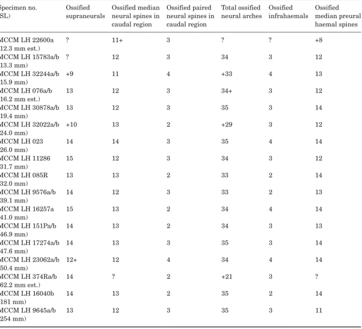
Figure 8 shows the division of the axial skeleton in dif-ferent regions. The numbers of vertebral elements are given in Tables 6 and 7.
Vertebrae: The autocentra are solid, perichordally ossified and amphicoelous. A small opening for the
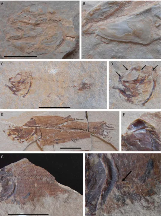
Figure 6. Hispanamia newbreyi gen. nov., sp. nov. (A) skull from specimen MCCM LH 151Pa. Scale bar = 1 cm. (B) close-up of specimen from A showing the gular plate. Note the annular concentric marks. (C) specimen MCCM LH 15783a. Scale bar = 5 mm. (D) close-up of specimen from C showing early ossification of dermal bones comprising sensory canals, as indicated by arrows. (E) specimen MCCM LH 17274a. Scale bar = 1 cm. (F) close-up of specimen from E showing ventral rod-like process, possibly for articulation with the intercalar bone of the braincase. (G) specimen MCCM LH 374Ra. Scale bar = 2 cm. (H) close-up of specimen from G showing a probable scapulocoracoid ossification.
notochordal canal remains in each autocentrum even in the largest specimens. The autocentra are approximately rounded in contour in anterior/posterior view and higher than long. All the autocentra have two or three deep excavations, termed lateral oval fossae, on each side of their lateral surface. Additionally, they can have smaller fossae, either circular or elongated in shape. There are several diplospondylous vertebrae, both the normal and the alternating type. There are no epineurals or epi-pleurals, as found in all other known amiid species. The vertebrae of all large specimens are only observable in lateral view, or slightly tilted, which allows us to confirm their rounded contour, but does not allow the dorsal and ventral views to be described in great detail.
Abdominal region: There are usually 18–21 abdomi-nal autocentra. The anterior few (two or three) abdom-inal autocentra are lower than the rest. The first one or two vertebrae are covered by the skull bones. In addition to them, the abdominal vertebrae bear well-developed ribs (r), which are curved, broader on their
proximal end than they are on their distal end. The distal tips of the ribs are pointed. The abdominal ver-tebrae lack ossified parapophyses. The ribs seem to articulate more laterally and anteriorly on the ante-riormost autocentra, and gradually move to a more ventral and posterior position on the more posterior abdominal autocentra. The last ribs also become grad-ually slightly smaller, the last pair being the smallest, although they are not extremely smaller, and have the same narrow shape as the rest. The last abdominal autocentrum lacks ribs. All the abdominal vertebrae have neural arches. There are no median spines on the abdominal autocentra, since the right and left half neural arches do not fuse to each other distally, but develop into paired spine-like structures, as found in A. calva (Grande & Bemis, 1998). The neural arches have pointed distal tips and proximal ends or bases that are expanded and lie dorsal to the posterior half of the autocentrum, even overlying part of the next auto-centrum, especially the posteriormost ones. The neural
Figure 7. Hispanamia newbreyi gen. nov., sp. nov. Skeletal reconstruction of specimen MCCM LH 9645a (holotype) showing the main sensory canals. Scale bar = 5 cm.
Figure 8. Hispanamia newbreyi gen. nov., sp. nov. Axial skeleton of specimen MCCM LH 9645a (holotype). Scale bar = 5 cm.
arches of the first abdominal vertebrae are shorter and slightly flattened in the sagittal plane. Associated with the first neural arches is a series of supraneu-rals (sn). Supraneusupraneu-rals are long, thin bones flattened in the sagittal plane located slightly dorsal to the first neural arches, sometimes inserting between two con-secutive neural arches, except the first one, which is shorter and articulates with its corresponding neural arch. Some variability in the number of supraneurals is observable, with 13–15, usually 14, supraneurals present, the first one just posterior to the skull and the last ones lying ventral to the anteriormost axonosts of the dorsal fin.
Preural caudal region: The preural caudal region begins with a vertebra bearing haemal arches. There are usually 23–25 preural caudal autocentra. This region contains all of the diplospondylous vertebrae. In summary, diplospondyly is the duplication of the num-ber of autocentra for each body segment or myosep-tum, presumably to increase flexibility in the posterior part of the body (Schaeffer, 1967; see also Wenz, 1977; and Schultze & Arratia, 1986).
The preural caudal region is usually composed of three monospondylous vertebrae anteriorly and becomes diplospondylous only more posteriorly. The first diplospondylous vertebrae are of the normal type; posteriorly, there is a shift to alternating diplospondyly, which affects only three or four vertebrae (vertebrae 28–31 in the holotype specimen). Finally, the last ver-tebra or pair of verver-tebrae of the preural caudal region are of the normal diplospondyly type again. Ventrally, the anteriormost vertebrae of the preural caudal region (the three first in the case of the holotype, and at least two in specimen MCCM LH 16040a/b) bear short haemal arches (ha) that do not fuse distally to form median haemal spines. Instead, these short ante-rior haemal arches articulate with long, spine-like median bones termed infrahaemals (ihm), which are also present in the extant A. calva. The next preural caudal vertebrae bear haemal arches that do fuse distally to form median haemal spines. The infrahae-mals and the haemal spines are approximately the same length, much longer than the abdominal ribs, and both are thin bones with pointed distal tips. The last few haemal spines are involved in the support of the caudal fin forming the caudal endoskeleton. These last haemal spines gradually become flatter distally, acquiring a hypural-like shape. The last one is called the parhypural (phy). Dorsally, the vertebrae of the preural caudal region bear a pair of neural arches that form neural spines distally. The neural arches of the first three or four vertebrae do not fuse distally, and thus they form paired neural spines, like those of the abdominal region. The neural arches of the rest of the preural caudal vertebrae do fuse to each other distally,
forming median neural spines. The paired and the median neural spines of the preural caudal region are approximately the same length; they are also the same length as those of the abdominal region and, as with the latter, they have pointed distal tips.
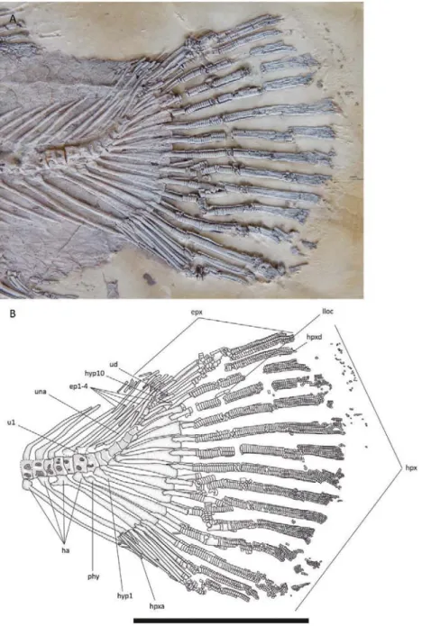
Ural region: The ural caudal region of the vertebral column comprises the vertebrae that bear hypurals. They support fin rays, forming the caudal endoskel-eton (see description below).
APPENDICULARSKELETON
Pectoral girdle and fin: The pectoral girdle of amiids is composed of numerous paired bones. As Grande & Bemis (1998) show, not all the elements of the pectoral girdle ossify in all amiid species. The dorsalmost ele-ment of the pectoral girdle is the posttemporal (pt). The anterior edge of the posttemporal is overlaid by the posterior part of the extrascapular, the posterior-most bone of the skull roof. The posttemporal is trian-gular in shape, with straight margins forming rounded corners (Fig. 3). The anterior margin of the bone, which is disposed perpendicular to the longitudinal axis of the body, is as long as, or slightly longer than, its lat-eral margin, which is parallel to the longitudinal axis of the body. Its medial surface is covered by the ante-riormost dorsal scales, which are inserted between the posteromedial margin of the extrascapular and the anteromedial margin of the posttemporal. The dorsal surface of the posttemporal is irregularly ornamented with shallow grooves. There are also pores of the lat-eral line sensorial system that enters from the extras-capular and runs diagonally through the posttemporal towards its posterolateral corner, where the bone con-nects with the supracleithrum. The posttemporal is not observable in ventral view in any of the large spec-imens; however, one of the juvenile specimens (MCCM LH 17274a) has a posttemporal with a ventral rod-like process possibly for articulation with the intercalar bone of the braincase (Fig. 6E, F), as found in A. calva (Grande & Bemis, 1998: fig. 36).
The supracleithrum (scl) is placed ventral to the posstemporal (Fig. 3). The supracleithrum is a rela-tively large, flat bone mostly covered by the operculum in lateral view. Its posterior margin is convex, with a more or less developed posterodorsal expansion. It is smooth, but it has a couple of large pores for the lateral line sensory system on its dorsal margin. The canal exits the bone posteriorly, connecting with the lateral line body system. Ventrally, the supracleithrum articulates with the cleithrum anteriorly, and with the postcleithrum posteriorly, overlying them.
The postcleithrum (pcl) is much smaller than the supracleithrum and the cleithrum (Fig. 3). It is par-tially covered by the supracleithrum dorsally and by
the cleithrum ventrally. The visible lateral surface of this bone is smooth; it is higher than long, and its pos-terior margin is convexly rounded.
The cleithrum (cl) is, as usual, the largest bone of the pectoral girdle (partially covered by the branchiostegal rays in Fig. 3). This bone is composed of an anterior pointing arm and a dorsal pointing arm. The dorsal arm is shorter than the anterior arm; it overlies the anterior part of the postcleithrum and articulates with the ventral part of the supracleithrum. It is partially covered by the operculum laterally. The end of this arm is a thick, tube-like expansion. The posterior margin of the bone, where both the anterior and the dorsal arms are joined, is convexly rounded, depicting a continu-ous curve. This posterior expansion of the bone is very thin, and it is smooth in lateral view, in contrast to the condition in amiines. The anterior arm is not clearly visible in any of the large specimens. Smaller speci-mens have a very thick dorsal margin on the anterior arm. The anterior end of this arm is slightly expanded and forms an acute angle. The clavicle elements are not visible in any of the known specimens. According to Grande & Bemis (1998), these elements are not nor-mally visible in well-articulated skulls, because they lie between the anterior arm of the cleithrum and bones of the opercular series. They do not articulate tightly with the cleithrum either, and thus they disar-ticulate easily.
There are also several chondral elements in the pec-toral girdle of amiids. The scapulocoracoid complex it is not visible in any of the known specimens, so it seems not to be ossified, as is often the case in other amiid species. The only exception is a juvenile individual (MCCM LH 374Ra/b), which has a small ossification apparently articulating with both the cleithrum and the radial elements of the pectoral fin that might be the scapulocoracoid (Fig. 6G, H); this part of the speci-men is, however, not very well preserved, so nothing conclusive can be really said. Nonetheless, the scapulo-coracoid complex might be covered by the cleithrum in most specimens, since it articulates on the medial sur-face of the latter. According to Grande & Bemis (1998), the scapulocoracoid complex does not ossify in A. calva until very late in ontogeny.
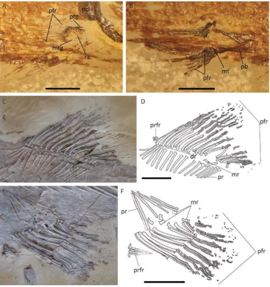
In most specimens, the radial elements are pre-served only as imprints, suggesting a faint ossification. There are seven to nine radial elements in the pectoral fins (Fig. 9A). The first of them is thicker and shorter, and probably corresponds to the propterygium (ptg). The radials (ra) are thin, short, cylindrical bones. The last element is also shorter and thicker, and is not com-pletely parallel to the others, but somehow diagonally disposed, suggesting it might be the posterior end of a metapterygium (mtgo). According to Grande & Bemis (1998), only the posterior part of the metapterygium
ossifies in adult individuals of A. calva. Posteriorly, the ensemble of the radial elements form a convex surface, where the fin rays articulate. The pectoral rays, 13–16 in number, are not in a one-to-one ratio with the radi-als; instead, several rays articulate with each radial. The pectoral fin rays are long, the more posterior ones gradually decreasing in length, so that the pectoral fin has a slightly convexly rounded posterior margin. The pectoral fin rays become branched as the fish grows; the longest rays are branched twice in the largest spec-imen, the holotype.
Pelvic girdle and fin: The only elements of the pel-vic girdle and fin that apparently ossify are the basip-terygium and the fin rays (Fig. 9B). A small ossification, slightly wider anteriorly than posteriorly, occurring in a small specimen (MCCM LH16257a) might represent the metapterygium (mtg). The basipteryigium (pb), whose ossification is also termed pelvic bone, is rela-tively large, flat and hourglass-shaped. The narrower point of the bone is much closer to the posterior end than to the anterior end, both of which get gradually wider. In adults, the anterior margin is twice as wide as the posterior margin. There are six or seven principal rays in the pelvic fin, which are segmented and branched in large individuals. There is also a much smaller, neither segmented nor branched ray anterior to the rest. The pelvic fin has a slightly convex posterior margin. The prepelvic length represents approximately 50% of the standard length of the fish in large individuals. The pel-vic fin is smaller and shorter than the pectoral fin.
UNPAIRED FINS
Dorsal and anal fins: The dorsal fin (Fig. 9C, D) is short and triangular in shape, with a straight to slightly convexly rounded posterior margin. The dor-sal fin is located only slightly closer to the caudal fin than to the head; the predorsal length represents approximately 60% of the standard length of the fish in large individuals, and the dorsal fin base 16%. It has 14–16 principal rays supported by 15–17 ptery-giophores. The pterygiophores are median structures comprising a proximal radial (pr), a middle radial (mr) and a distal radial (dr). The proximal radials are the longest elements, and they are spine-like, round in cross-section, with a broadened distal end. All of the proximal radials are approximately the same length. Apparently, the proximal radial of the first pterygio-phore is slightly curved anteriorly and closer to the next one than are any of the others. The proximal ends of the proximal radials are not in contact with the neural spines of the vertebral column but notably separated from them. The proximal radials are dis-posed diagonally to the longitudinal axis of the body, in anteroventral to posterodorsal sense. The distal end
of each proximal radial articulates with the proximal end of a middle radial. Middle radials are small, tube-like bones with broad tips. As seen in A. calva (Grande & Bemis, 1998), the posterodistal end of each middle radial seems to articulate with the anterodistal end of the next proximal radial. Thus, each proximal radial, except the first one, makes contact with two middle radials. The distal radials are very small bones that articulate with the distal ends of the middle radials and lie between the two halves of their corresponding lepidotrichia. Each pterygiophore supports one fin ray, except the first one or two, which can support more than one due to the existence of a few, usually two or three, precurrent rays at the beginning of the dorsal
fin. These precurrent rays (prfr) are very small, and neither segmented nor branched. The principal rays are both segmented and branched. Except for the first one or two principal fin rays, which are shorter, the principal dorsal fin rays are longer anteriorly and gradually shorter posteriorly.
The anal fin (Fig. 9E, F) is also short, with a slightly convexly rounded margin. Its anterior inser-tion is located anterior to the posterior end of the dorsal fin; the preanal length represents 72–74% of the standard length of the fish in the largest spec-imens. The anal fin is slightly closer to the caudal fin than to the pelvic fins. The anal fin base repre-sents approximately 10% of the standard length.
Figure 9. Hispanamia newbreyi gen. nov., sp. nov. (A) pectoral fin from specimen MCCM LH 16257a. Scale bar = 2 mm. (B) pelvic fin from specimen MCCM LH 16257a. Scale bar = 2 mm. (C) dorsal fin from specimen MCCM LH 9645a (holotype) slightly coated with ammonium chloride. (D) interpretative drawing of specimen in C. Scale bar = 5 cm. (E) anal fin from specimen MCCM LH 9645a (holotype) slightly coated with ammonium chloride. (F) interpretative drawing of specimen in E. Scale bar = 5 cm.