O R I G I N A L A R T I C L E
Mutation in exon 1a of
PLEC, leading to disruption
of plectin isoform 1a, causes autosomal-recessive
skin-only epidermolysis bullosa simplex
Katarzyna B. Gostyńska
1
, Miranda Nijenhuis
1
, Henny Lemmink
2
,
Hendri H. Pas
1
, Anna M.G. Pasmooij
1
, Kristin Kernland Lang
3
,
Maria J. Castañón
4
, Gerhard Wiche
4
and Marcel F. Jonkman
1,
*
1
Department of Dermatology and
2Department of Genetics, University Medical Center Groningen, University of
Groningen, Groningen, The Netherlands,
3Department of Dermatology, University of Bern, Bern, Switzerland and
4Max F. Perutz Laboratories, Department of Biochemistry and Cell Biology, University of Vienna, Vienna, Austria
*To whom correspondence should be addressed. Tel: +31 503612520; Fax: +31 503619247; Email: [email protected]
Abstract
PLEC, the gene encoding the cytolinker protein plectin, has eight tissue-specific isoforms in humans, arising by alternate splicing of thefirst exon. To date, all PLEC mutations that cause epidermolysis bullosa simplex (EBS) were found in exons common to all isoforms. Due to the ubiquitous presence of plectin in mammalian tissues, EBS from recessive plectin mutations is always associated with extracutaneous involvement including muscular dystrophy, pyloric atresia and cardiomyopathy. We studied a consanguineous family with sisters having isolated blistering suggesting EBS. Skin disease started with foot blisters at walking age and became generalized at puberty while sparing mucous membranes. DNA sequencing revealed a homozygous nonsense mutation (c.46C>T; p.Arg16X) in thefirst exon of the plectin variant encoding plectin isoform 1a (P1a). Immunofluorescence antigen mapping, transmission electron microscopy, western blot analysis and qRT-PCR were performed on patient skin and cultured keratinocytes, control myocardium and striated muscle samples. We found hypoplastic hemidesmosomes and intra-epidermal‘pseudo-junctional’ cleavage fitting EBS. Screening for cardiomyopathy and muscle dystrophy showed no abnormalities. We report thefirst cases of autosomal-recessive EBS from P1a deficiency affecting skin, while mucous membranes, heart and muscle are spared. The dominant expression of the P1a isoform in epidermal basal cell layer and cultured keratinocytes suggests that mutations in thefirst exon of isoform 1a cause skin-only EBS without extracutaneous involvement. Our study characterizes yet another of the eight isoforms of plectin and adds a tissue-specific phenotype to the spectrum of ‘plectinopathies’ produced by mutations of unique first exons of this gene.
Introduction
Epidermolysis bullosa (EB [MIM 131900]) is a heterogeneous group of genodermatoses characterized by lifelong skin fragility and extracutaneous complications present from birth (1). Affected individuals suffer from trauma-induced blistering of skin and
mucous membranes with severity ranging from mild to lethal. For EB, up until now there have been mutations identified in 18 different genes affecting different proteins in the epidermis and basement membrane zone (BMZ) (1). Depending on the af-fected protein, the epidermis is rendered fragile by cleavage of
Received: January 16, 2015. Revised and Accepted: February 13, 2015
© The Author 2015. Published by Oxford University Press. All rights reserved. For Permissions, please email: [email protected] doi: 10.1093/hmg/ddv066
Advance Access Publication Date: 24 February 2015 Original Article
the skin at its biological location. The most commonly occurring subtype is epidermolysis bullosa simplex (EBS) where skin cleav-age occurs in the epidermis (2). In basal EBS the split occurs in the basal cell layer as witnessed by immunofluorescence anti-gen mapping or electron microscopy. Basal EBS is commonly as-sociated with four different genes: KRT5, KRT14, BPAG1 and PLEC. In 8% of EBS cases, PLEC is the mutated gene (3). PLEC en-codes the cytolinker protein plectin, which is ubiquitously pre-sent in skin, mucous membranes, gut, muscle and heart tissue. The phenotypes associated with PLEC mutations are mucocuta-neous blistering, pyloric atresia, muscular dystrophy and cardio-myopathy (4,5). Here, we identify a novel mutation causing cutaneous EBS without extracutaneous involvement. This non-sense mutation found in the distinctfirst exon 1a encoding the N terminus of plectin isoform 1a (P1a) only disrupts this one isoform whose tissue dominant distribution in the epidermis results in skin-only EBS.
Results
Our index female patient (V:1, EB 315-01) was 27-years old when referred to our clinic for consultation of her unclassified EB sub-type. She was one of two affected daughters born to consanguin-eous Turkish parents (Fig.1A). Mild blistering began after walking age at 1.5 years and slightly restricted her physical education activities at school. Both hand and toenails were dystrophic from the age of 3 years. Blistering became generalized with prur-itus when in puberty at 14 years and lesions healed with scarring and hyperpigmentation. Mucous membranes were not affected and enamel was normal. She displayed excoriated and verrucous papules and intact blisters (Fig.1B) most pronounced on the acra with residual post-inflammatory hyperpigmentation (Fig.1C–E). Focal plantar hyperkeratosis and severe nail dystrophy on both hand and toenails was seen (Fig.1E). Scalp, axillary and pubic hair was normal. Her one-year younger sister (V:2, EB 315-02)
Figure 1. Clinical picture. (A) Pedigree of family 1 with EBS-plectin 1a. DNA was obtained from individuals V-1 and V-2. The rest of the family members was unavailable for consultation. (B–E) Clinical presentation of patient V:1. Generalized verrucous papules, excoriations and post-inflammatory hyperpigmentation of abdomen, thighs, shins and dorsal surface of arms.
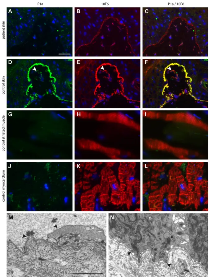
had a similar history and distribution of progressive skin lesions, however displayed less pronounced hyperkeratotic papules and plantar hyperkeratosis possibly due to minor pruritus as com-pared to her sister (Supplementary Material, Fig. S1). Routine histopathology of a papule revealed dyskeratosis with papillar inflammation of the dermis (Supplementary Material, Fig. S2). Immunofluorescence antigen mapping of lesional skin showed a thin granular lining of keratin staining on the blisterfloor revealing a very low intra-epidermal cleavage in basal cells that led us to the diagnosis of EBS. Double immunofluorescence stain-ing of intact patient’s skin with affinity-purified isoform-specific rabbit antibodies to P1a and pan-plectin mouse monoclonal anti-body (mAb) 10F6 recognizing all isoforms of plectin was negative for P1a (Fig.2A), but positive for pan-plectin, producing a linear pattern along the epidermal BMZ, albeit of markedly reduced intensity compared to control skin (Fig.2B). In the latter, staining for P1a revealed the same pattern and intensity of linear fluores-cence along the BMZ as that for pan-plectin (Fig.2D–F). The expression of integrinα6β4 was normal (data not shown). A 1.5-mm punch biopsy, obtained forfixation and transmission elec-tron microscopy, revealed hypoplastic hemidesmosomes and intra-epidermal pseudo-junctional cleavage with the plasma membrane of basal cells visible on the blisterfloor (Fig.2M and N). Together, thesefindings indicated PLEC as a candidate gene. DNA was isolated from peripheral blood and mutation analysis of the coding region of the PLEC gene including exon-intron boundaries of all 8 isoforms revealed a homozygous nonsense mutation present in P1a, c.46C>T leading to a premature termin-ation codon p.Arg16X (Fig.3A and B). The affected sister of the index patient was a carrier of the same homozygous mutation. Further PLEC sequence analysis also revealed a nucleotide transi-tion in the promoter sequence of isoform 1, c.-27C>T. Following mutation analysis, immunofluorescence mapping was extended with isoform-specific antibodies to P1a and anti-pan-plectin mAb 10F6 on control striated muscle and myocardium from nor-mal cadavers. P1a was not expressed by myocytes in striated muscle and myocardium, whereas the shared plectin epitope (10F6) was prominently stained at Z-disks of striated muscle (Fig.2H) and intercalated discs of myocardium (Fig.2K). We also examined stratified epithelia other than skin of human controls, and found expression of P1a along the basement membrane of the oral cavity mucosa and to a limited extent in the conjunctiva (Supplementary Material, Fig. S3).
When cell lysates of cultured skin keratinocytes from both of the affected sisters were subjected to immunoblotting using mAb 10F6 and antibodies to P1a, reduced 10F6 signals (V:1 16.5% and V:2 15.4%) compared to control cells were observed in both cases (Fig.3C,first panel), whereas in neither case were P1a-positive bands detectable (Fig.3C, second panel). Blot stain-ing done with antibodies to desmoplakin as a control, for both patients revealed unchanged signals compared to control cells (Fig.3C, third panel).
We performed quantitative RT-PCR (qRT-PCR) on cultured cells and found dominance of P1a transcription compared to plectin isoform 1c (P1c) in normal human keratinocytes and fibroblasts (Fig.3D). The transcription level of P1a, but not of P1c, was reduced in patient keratinocytes andfibroblasts (Fig.3D). We also checked the transcription level of plectin isoforms in mouse epidermis and keratinocytes to compare with our human data and to line up with literature (6). Absolute quantification un-veiled two important differences in isoform expression between epidermis and primary mouse keratinocytes. While P1c accounts for a substantial fraction (∼80%) of the plectin transcript in epi-dermis, this is no longer the case in primary keratinocytes
(∼20%), conversely, P1a transcript levels are higher (more than 50% of total) in primary keratinocytes than in epidermis (∼20% of total) (Supplementary Material, Fig. S4).
Considering that plectin mutations leading to EBS can mani-fest with extracutaneous complications of muscle dystrophy and/or cardiomyopathy (3,4), the index patient was screened by a neurologist and cardiologist respectively. Upon physical exam-ination no symptoms of muscular dystrophy were found and a muscle biopsy was deemed unnecessary. Cardiologic consult-ation including physical examinconsult-ation, electrocardiogram and an echocardiogram was carried out with no associatedfindings of cardiomyopathy. Moreover, her affected younger sister had no symptoms of muscular or cardiologic pathology. Currently both sisters, now aged 29 and 30 years, exhibit no signs of muscu-lar dystrophy or cardiomyopathy.
Discussion
The spectrum of diseases in humans caused by PLEC mutations, ‘the plectinopathies’, continues to expand and be classified. The cytolinker scaffold protein plectin is present in essentially all mammalian tissue. This cardinal supporting structure is ex-pressed in tissues such as heart, muscle, striated epithelia as well as the blood–brain barrier. At the cellular level, plectin strengthens tissues in regions of mechanical stress, such as link-ing desmin and the dystrophin glycoprotein complex in myo-cytes of the heart. In skin, plectin is a fundamental building block of the hemidesmosome, anchoring basal keratinocytes of the epidermis to the underlying dermis (7). PLEC is a large gene consisting of 32 exons with 8 tissue-specific isoforms which arise by alternative 5′splicing of transcripts (8–10). Molecularly, the plectin protein is built of a centralα-helical coiled-coil rod do-mainflanked by N- and C-terminal globular domains (11). All 8 isoforms share exons 2-32 and differ only by distinctfirst exons (4) (Supplementary Material, Table S1). The various isoforms of plectin continue to be classified according to occurrence and tissue specificity arising from the alternate promoter regions and cis splicing from several differentfirst coding exons (4,7). The most prominently expressed isoform of plectin in mouse and human cultured keratinocytes is P1a, followed by P1c and other isoforms among them P1, plectin isoform 1f (P1f ) and P1b (Supplementary Material, Fig. S4) (see also Ref.12).
That P1a is also dominantly expressed at the dermal–epidermal junction of human epidermis is suggested by (i) the identical pattern and intensity of P1a and pan-plectin staining using double immunofluorescence labeling of normal human skin (Fig.2F), (ii) the severe reduction of the pan-plectin signal in pa-tient skin (Fig.2B), (iii) the∼84% reduction of pan-plectin in pa-tient keratinocyte extracts (Fig.3C,first panel) and (iv) qRT-PCR showing a higher level of P1a than P1c transcripts in cultured human keratinocytes in normal control cell populations (Fig.3D). The dominance of P1c transcripts in mouse epidermis (Supplementary Material, Fig. S4A) is simply explained by the fact that primary keratinocytes lack the suprabasal layers where P1c is predominantly expressed while they are rich in hemidesmosome-like structures where P1a is found.
P1a was expressed also in oral mucosa, and to a limited extent in conjunctiva (Supplementary Material, Fig. S3). Since P1a is ex-pressed in these epithelia in normal human controls one may have expected mucosal lesions in the patients. The fact that they were absent suggests that in these tissues P1a’s loss was compensated by other proteins or isoforms of plectin.
Mutations in the exons common to all 8 isoforms (2-32) of this one gene continue to be reported and classified within the main
Figure 2. Double immunofluorescence microscopy of human skin, muscle and heart tissue using antibodies to P1a (green) and an epitope shared between plectin isoforms (red) and transmission electron microscopy of skin. In skin of patient V:1, P1a (A) is absent, whereas 10F6 staining at the BMZ is reduced (B and C). In control skin, P1a (D) and the shared epitope of all plectin isoforms (10F6) (E) display a strong linear staining that co-localizes (F) at the BMZ. Note that the staining of dermal blood vessels (arrowhead) with antibodies 10F6 is more dominant than that for P1a. Striated muscle of healthy controls shows no expression of P1a (G), whereas Z-disks clearly stain for the shared epitope of plectin (H and I). Myocardium of healthy controls shows no expression of P1a (J), whereas Z-disks and intercalated disks stain strongly for the shared plectin epitope (K and L). Bar in A corresponds to 25μm for all immunofluorescence images. Transmission electron microscopy of ultrathin sections of patient skin (M and N). Lesional skin (M) reveals remnants of keratinocyte membranes (arrowheads) lining the blisterfloor fitting with a ‘pseudo-junctional’ intra-epidermal split level. The intact skin (N) shows hypoplastic hemidesmosomes (arrowheads). Bar in M corresponds to 2μm for both EM images.
phenotypic groups (Table1). New PLEC mutations resulting in EBS are frequently reported and account for 8% of all cases of EBS (3). However, except for one (13), all of them reside within exons that are common to all isoforms (4). Epidermolysis bullosa simplex with pyloric atresia (EBS-PA, [MIM 612138]) is characterized by neonatal blistering, pyloric atresia and rapid clinical decline
(14). Epidermolysis bullosa simplex with muscular dystrophy (EBS-MD,[MIM 226670]) displays congenital blistering and myop-athy onset in later life (15). Additional myasthenic symptoms have been reported in several patients having EBS-MD character-izing yet another subtype, EBS-MD-Mys (16). An autosomal dom-inant (AD) disease is EBS Ogna (EBS-O [MIM 131950]) with mild
Figure 3. DNA sequence analysis, immunoblotting and RT-PCR of P1a in patients. DNA sequencing of PLEC (A) revealed a homozygous mutation in exon 1a encoding the isoform-specific sequence of P1a in patient V:1. A cytosine at position 46 is exchanged for a thymine (c.46C>T). This homozygous nucleotide change results in an arginine at position 16 rendering a stop codon ( p.Arg16X) in isoform 1a but not in any other plectin isoform (B). The isoform-specific exon 1a is highlighted in yellow. (C) Immunoblotting of cell lysates from cultured keratinocytes derived from normal humans (lanes C) and patients (lanes V:1 and V:2). First panel (10F6): A marked reduction (80–84%) of band intensity is seen for both patients using mAbs 10F6 which bind to the rod domain common to all isoforms of plectin, including P1a. Because of the absence of P1a, the 10F6 signal (representing total plectin in skin) is reduced as expected. Arrow, position of plectin (molecular mass∼ 500 kDa) deduced from the position of molecular size markers (not shown). Second panel (P1a): P1a-specific antibodies show no detectable expression of the protein in keratinocytes from patients, whereas P1a was markedly expressed in normal human keratinocytes. Arrow, as infirst panel. Third panel (DP2.15): Antibodies to human desmoplakin I (250 kDa) and II (215 kDa) show normal expression of both proteins in both patients, similar to cultured keratinocytes from unaffected humans. Arrow, position of desmoplakin I. (D) Quantitative RT-PCR of P1a (gray) and P1c (white) mRNA expression in cultured keratinocytes andfibroblasts from human control (CK1-3 and CF1,2, respectively) and corresponding patient (V:1,V:2) specimens. Bars represent expression relative to household gene B2M. Error bars, standard deviations of experiments run in triplicates. Note the predominance of P1a above P1c in control keratinocytes (CK1-3), and the diminished expression in patient cells.
skin fragility (17) that renders the plectin polypeptide vulnerable to proteolysis (6). Of particular interest are the consequences of mutations in the variousfirst exons because it is their tissue spe-cificity which dictates disease manifestation. Comprehensive understanding of isoform expression in different tissues is a work in progress shared by different medical specialties. Recent-ly, Gundesli et al. reported and characterized that mutations in thefirst coding exon of plectin isoform P1f are responsible for limb-girdle muscular dystrophy (LGMD2Q) without any skin in-volvement (13). Andrä et al.first reported and characterized the targeted cellular localization of P1a as the essential plectin isoform present in the hemidesmosome co-localizing with the integrinα6β4 subunit in mice (12). They suggested that this iso-form was the culprit responsible for hereditary EBS in humans. This was later confirmed by Walko et al. who showed that hemi-desmosome failure was caused by decreased levels of isoform P1a in knock-in mice for dominantly inherited epidermolysis bullosa simplex type Ogna (EBS-Ogna) (6). In mice, P1a is the pre-dominant isoform that localizes to the epidermal BMZ (12). The persistent expression of common plectin epitopes in the epider-mal BMZ of our patients suggests that in humans, like in mice, other plectin isoforms are co-expressed in the epidermal BMZ. Compensation of the additional non-mutated isoforms known to be expressed in the epidermis (i.e. P1c) did not preserve the hemidesmosome architecture at adult age and led to skin fragil-ity. However, both studied patients did not develop generalized lesions until puberty implicating that perhaps compensation oc-curred from their non-mutated isoforms during childhood.
The expression of P1a was absent in both normal human striated and cardiac muscle tissues as shown in this study. Due to the fact that the pathogenic mutation did not affect coding exons shared by other isoforms whose role has previously implicated Z-disks of striated muscle and intercalated disks of myocardium, we do not expect development of myopathy or car-diomyopathy. Moreover, late-onset plectin-mediated (cardio) myopathy develops before the age of 30 years (18). The con-sequence at the protein expression level of the homozygous c.–27C > T variant of plectin isoform P1 is not known. In most genes at this position TATA box promoter sequence elements are surrounded by GC-rich sequences for transcription regulation (19). In the case of c.–27C > T no such sequences can be distin-guished. It is possible that c.–27C > T might be a rare polymorph-ism, commonly present in the Turkish population.
In conclusion, we present and characterize thefirst human phenotype of the genodermatosis EBS caused by a homozygous nonsense mutation in exon 1a of the PLEC gene. Ourfindings uphold those of previous ones that have implicated P1a as
the dominant isoform upon which hemidesmosome stability is dependent (20).
Materials and Methods
PatientsThe patients (EB 315-01, V:1 and EB 315-02, V:2), were two sisters diagnosed with EBS based on clinicalfindings, immunofluores-cence microscopy, electron microscopy and molecular analysis. All experiments performed were done with material obtained earlier for diagnostic purposes which did not require extra approval from the institutional ethical committee. Patients gave informed consent for the use of photographs and tissue samples. All non-patient tissue samples were contributed and analyzed anonymously. All experiments were conducted accord-ing to the principles of the Declaration of Helsinki.
Immunofluorescence antigen mapping
Four-mm skin biopsies of fresh blisters and healthy skin under the left arm were taken for immunofluorence antigen mapping from both affected individuals. Immunofluorescence antigen mapping of skin biopsies and cultured human keratinocytes were done as described before (21). The monoclonal antibodies and their origin have been described previously (6,22,23). Mouse monoclonal antibodies 10F6 were directed against an epitope in plectin’s rod domain (24). Isoform-specific antibodies to P1a, raised in rabbit against a synthetic peptide corresponding to a 12-amino acid residue-long sequence (KRTSSEDNLYLA) encoded by human exon 1a, were used after affinity-purification (6,12,25); the sequence of the peptide used as immunogen is conserved between man, mouse and rat. Antibodies were diluted in PBS/ OVA 1% and sections were incubated at room temperature for 45 min. The secondary antibodies used for double staining were green FITC-conjugated F(ab’)2 goat anti-rabbit IgG (1:50; Zymed Laboratories, Carlsbad, CA) and red LRSC-conjugated donkey anti-mouse IgG (1:50; Jackson Immuno Research, Newmarket, UK). Validity of species-specific binding was ascertained by cross experiments. Hoechst 33342 was used to stain nuclei. Pictures of sectors were merged with Adobe Photoshop CS3 software.
Electron microscopy
Two-mm punch biopsies of perilesional and non-lesional skin taken from the upper arm were obtained and prepared as described previously (21).
Table 1. The plectinopathies
Subtype Exons affected Inheritance Organs affected Severity First reported case EBS-MD 9–32 AR Skin, striated muscle, myocardium Severe and progressive Smith et al. (15) EBS-MD-Mys 13,31,32 AR Skin, striated muscle Severe and progressive Selcen et al. (16)
EBS-PA Distal exons AR Skin, pylorus Lethala Charlesworth et al. (29)
EBS-Ogna 31 AD Skin Mild Koss-Harnes et al. (17)
EBS-plectin 1a 1a AR Skin Moderate This article
LGMD2Q 1f AR Striated muscle Severe and progressive Gundesli et al. (13)
Subtypes listed: epidermolysis bullosa simplex-muscular dystrophy (EBS-MD), epidermolysis bullosa simplex-muscular dystrophy with myasthenic syndrome (EBS-MD-Mys), epidermolysis bullosa simplex with pyloric atresia (EBS-PA), epidermolysis bullosa simplex type Ogna (EBS-Ogna), epidermolysis bullosa simplex with pathogenic mutation in thefirst coding exon (exon 1a) of isoform 1a (EBS-plectin 1a), limb-girdle muscular dystrophy (LGMD2Q). Inheritance: Autosomal recessive (AR), Autosomal dominant (AD).
Molecular analysis
Genomic DNA was extracted from peripheral blood lympho-cytes using standard laboratory methods. The PLEC gene en-compassing the isoforms 1, 1a, 1b, 1c, 1d, 1e, 1f and 1g were screened for mutations by sequencing analysis of all exons in-cluding exon–intron boundaries. Primer sequences are available upon request.
Western blotting
SDS–PAGE and western blotting were performed as described previously (26,27). As substrate we used extracts from cultured keratinocytes from our patients and healthy controls. After SDS–PAGE and transfer onto nitrocellulose, the proteins were identified with the same antibodies as used for immunofluores-cence microscopy; in addition antibodies DP 2.15 directed against desmoplakin I/II (Abcam®) were used. To calculate plectin ex-pression total protein content of patient and control extracts were compared by running serial dilutions of all extracts on SDS–PAGE gels. After visualization of the proteins with ‘Blue Sil-ver’ staining (28) the gels were scanned and the intensity of stain-ing of the lanes was used to calculate the relative protein content of the different extracts. Next, serial dilutions were run on one SDS–PAGE gel, and after western blotting plectin was visualized with antibody 10F6. The blot was scanned and the relative plectin expression was calculated from the intensity of staining using Quantiscan software.
Quantitative RT-PCR (qRT-PCR)
For human samples, transcriptor universal cDNA master (Roche, Basel, Switzerland) was used for reverse transcription of a single RNA isolation of each cell population. P1a, P1c and the house-keeping gene beta-2-microglobulin (B2M) were verified by qPCR using FastStart Universal SYBR Master (Rox) reagent and an Applied Biosystems 7900ht (Foster City, CA, USA). Primers used are in Supplementary Material, Table S2.
Quantitative RT-PCR of mouse samples was performed as previously described (Fig. S8 in ref6). Estimated absolute copy numbers of plectin isoforms were calculated from standard curves obtained by serial dilutions of plasmids harboring the dif-ferent exons. Values were normalized to hypoxanthine-guanine phosphoribosyltransferase (HPRT) transcript levels.
Supplementary Material
Supplementary Material is available at HMG online.
Acknowledgements
We wish to thank J. Zuiderveen and G. Meijer performing skillful immunofluorescence stainings, and N. Kloosterhuis for excellent help with quantative RT-PCR.
Conflict of Interest statement. None declared.
Funding
G.W. was supported by grant P20744-B11 from the Austrian Sci-ence Research Fund (FWF). M.J. was supported by a grant from Vlinderkind (Dutch Butterfly Children Foundation).
References
1. Fine, J.D., Bruckner-Tuderman, L., Eady, R.A.J., Bauer, E.A., Bauer, J.W., Has, C., Heagerty, A., Hintner, H., Hovnanian, A., Jonkman, M.F. et al. (2014) Inherited epidermolysis bullosa: updated recommendations on diagnosis and classification. J. Am. Acad. Dermatol., 70, 1103–1126.
2. Bolling, M.C., Lemmink, H.H., Jansen, G.H. and Jonkman, M.F. (2011) Mutations in KRT5 and KRT14 cause epidermolysis bullosa simplex in 75% of the patients. Br. J. Dermatol., 164, 637–644.
3. Bolling, M.C., Jongbloed, J.D., Boven, L.G., Diercks, G.F., Smith, F.J., McLean, W.H. and Jonkman, M.F. (2014) Plectin mutations underlie epidermolysis bullosa simplex in 8% of patients. J. Invest. Dermatol., 134, 273–276.
4. Winter, L. and Wiche, G. (2013) The many faces of plectin and plectinopathies: Pathology and mechanisms. Acta Neuro-pathol., 125, 77–93.
5. Charlesworth, A., Chiaverini, C., Chevrant-Breton, J., DelRio, M., Diociaiuti, A., Dupuis, R.P., El Hachem, M., Le Fiblec, B., Sankari-Ho, A.M., Valhquist, A. et al. (2013) Epidermolysis bul-losa simplex with PLEC mutations: new phenotypes and new mutations. Br. J. Dermatol., 168, 808–814.
6. Walko, G., Vukasinovic, N., Gross, K., Fischer, I., Sibitz, S., Fuchs, P., Reipert, S., Jungwirth, U., Berger, W., Salzer, U. et al. (2011) Targeted proteolysis of plectin isoform 1a ac-counts for hemidesmosome dysfunction in mice mimicking the dominant skin blistering disease EBS-ogna. PLoS Genetics, 7, e1002396.
7. Castañón, M.J., Walko, G., Winter, L. and Wiche, G. (2013) Plec-tin-intermediatefilament partnership in skin, skeletal mus-cle, and peripheral nerve. Histochem. Cell Biol., 140, 33–53. 8. Fuchs, P., Zörer, M., Rezniczek, G.A., Spazierer, D., Oehler, S.,
Castañón, M.J., Hauptmann, R. and Wiche, G. (1999) Unusual 5′ transcript complexity of plectin isoforms: Novel tissue-specific exons modulate actin binding activity. Hum. Mol. Genet., 8, 2461–2472.
9. Rezniczek, G.A., Abrahamsberg, C., Fuchs, P., Spazierer, D. and Wiche, G. (2003) Plectin 5′-transcript diversity: short al-ternative sequences determine stability of gene products, ini-tiation of translation and subcellular localization of isoforms. Hum. Mol. Genet., 12, 3181–3194.
10. Zhang, T., Haws, P. and Wu, Q. (2004) Multiple variablefirst exons: a mechanism for cell- and tissue-specific gene regula-tion. Genome Res., 14, 79–89.
11. Wiche, G., Becker, B., Luber, K., Weitzer, G., Castañón, M.J., Hauptmann, R., Stratowa, C. and Stewart, M. (1991) Cloning and sequencing of rat plectin indicates a 466-kD polypeptide chain with a three-domain structure based on a central alpha-helical coiled coil. J. Cell Biol., 114, 83–99.
12. Andrä, K., Kornacker, I., Jörgl, A., Zörer, M., Spazierer, D., Fuchs, P., Fischer, I. and Wiche, G. (2003) Plectin-isoform-specific rescue of hemidesmosomal defects in plectin (−/−) keratinocytes. J. Invest. Dermatol., 120, 189–197.
13. Gundesli, H., Talim, B., Korkusuz, P., Balci-Hayta, B., Cirak, S., Akarsu, N.A., Topaloglu, H. and Dincer, P. (2010) Mutation in exon 1f of PLEC, leading to disruption of plectin isoform 1f, causes autosomal-recessive limb-girdle muscular dystrophy. Am. J. Hum. Genet., 87, 834–841.
14. Pfendner, E. and Uitto, J. (2005) Plectin gene mutations can cause epidermolysis bullosa with pyloric atresia. J. Invest. Dermatol., 124, 111–115.
15. Smith, F.J., Eady, R.A., Leigh, I.M., McMillan, J.R., Rugg, E.L., Kelsell, D.P., Bryant, S.P., Spurr, N.K., Geddes, J.F., Kirtschig, G.
et al. (1996) Plectin deficiency results in muscular dystrophy with epidermolysis bullosa. Nat. Genet., 13, 450–457.
16. Selcen, D., Juel, V.C., Hobson-Webb, L.D., Smith, E.C., Stickler, D.E., Bite, A.V., Ohno, K. and Engel, A.G. (2011) Myasthenic syndrome caused by plectinopathy. Neurology, 76, 327–336. 17. Koss-Harnes, D., Hoyheim, B., Anton-Lamprecht, I., Gjesti, A.,
Jørgensen, R.S., Jahnsen, F.L., Olaisen, B., Wiche, G. and Gedde-Dahl, T. Jr. (2002) A site-specific plectin mutation causes dominant epidermolysis bullosa simplex ogna: Two identical de novo mutations. J. Invest. Dermatol., 118, 87–93. 18. Bolling, M.C., de Visser, M., Aronica, E., Pfendner, E.G., van
den Berg, M.P., Diercks, G.F., Suurmeijer, A.J. and Jonkman, M.F. (2010) PLEC1 mutations underlie adult-onset dilated car-diomyopathy in epidermolysis bullosa simplex with muscu-lar dystrophy. J. Invest. Dermatol., 130, 1178–1181.
19. Savinkova, L.K., Ponomarenko, M.P., Ponomarenko, P.M., Drachkova, I.A., Lysova, M.V., Arshinova, T.V. and Kolchanov, N.A. (2009) TATA box polymorphisms in human gene promo-ters and associated hereditary pathologies. Biochemistry (Mos-cow), 74, 117–129.
20. Walko, G., Castañón, M.J. and Wiche, G. (2014) Molecular architecture and function of the hemidesmosome. Cell Tissue Res. Dec 9. [Epub ahead of print], PMID: 25487405.
21. Jonkman, M.F., De Jong, M.C., Heeres, K. and Sonnenberg, A. (1992) Expression of integrinα6β4 in junctional epidermolysis bullosa. J. Invest. Dermatol., 99, 489–496.
22. Jonkman, M.F., De Jong, M.C., Heeres, K., Pas, H.H., Van der Meer, J.B., Martinez de Velasco, A.M., Niessen, C.M. and Son-nenberg, A. (1995) 180-kD bullous pemphigoid antigen (BP180) is deficient in generalized atrophic benign epiderm-olysis bullosa. J. Clin. Invest., 95, 1345–1352.
23. Pasmooij, A.M., Pas, H.H., Bolling, M.C. and Jonkman, M.F. (2007) Revertant mosaicism in junctional epidermolysis bul-losa due to multiple correcting second-site mutations in LAMB3. J. Clin. Invest., 117, 1240–1248.
24. Foisner, R., Feldman, B., Sander, L., Seifert, G., Artlieb, U. and Wiche, G. (1994) A panel of monoclonal antibodies to rat plec-tin: distinction by epitope mapping and immunoreactivity with different tissues and cell lines. Acta Histochem.(Jena), 96, 421–438.
25. Rezniczek, G.A., de Pereda, J.M., Reipert, S. and Wiche, G. (1998) Linking integrin alpha6beta4-based cell adhesion to the intermediatefilament cytoskeleton: Direct interaction between the beta4 subunit and plectin at multiple molecular sites. J. Cell Biol., 141, 209–225.
26. Pas, H.H., de Jong, M.C., Jonkman, M.F., Heeres, K., Slijper-Pal, I.J. and Van der Meer, J.B. (1995) Bullous pemphigoid: Serum antibody titre and antigen specificity. Exp. Dermatol., 4, 372–376.
27. Pas, H.H., Kloosterhuis, G.J., Heeres, K., van der Meer, J.B. and Jonkman, M.F. (1997) Bullous pemphigoid and linear IgA dermatosis sera recognize a similar 120-kDa keratinocyte col-lagenous glycoprotein with antigenic cross-reactivity to BP180. J. Invest. Dermatol., 108, 423–429.
28. Westermeier, R. (2006) Sensitive, quantitative, and fast mod-ifications for coomassie blue staining of polyacrylamide gels. Proteomics, 6, Suppl 2, 61–64.
29. Charlesworth, A., Gagnoux-Palacios, L., Bonduelle, M., Ortonne, J.P., Raeve, L.D. and Meneguzzi, G. (2003) Identifica-tion of a lethal form of epidermolysis bullosa simplex asso-ciated with a homozygous genetic mutation in plectin. J. Invest. Dermatol., 121, 1344–1348.