HAL Id: tel-02145207
https://tel.archives-ouvertes.fr/tel-02145207
Submitted on 2 Jun 2019HAL is a multi-disciplinary open access archive for the deposit and dissemination of sci-entific research documents, whether they are pub-lished or not. The documents may come from teaching and research institutions in France or abroad, or from public or private research centers.
L’archive ouverte pluridisciplinaire HAL, est destinée au dépôt et à la diffusion de documents scientifiques de niveau recherche, publiés ou non, émanant des établissements d’enseignement et de recherche français ou étrangers, des laboratoires publics ou privés.
cancer-risk agents using experimental systems
Maria Zhivagui
To cite this version:
Maria Zhivagui. Genome-wide modeling of mutation spectra of human cancer-risk agents using exper-imental systems. Cancer. Université de Lyon, 2017. English. �NNT : 2017LYSE1278�. �tel-02145207�
N°d’ordre NNT : 2017LYSE1278
THESE de DOCTORAT DE L’UNIVERSITE DE LYON
opérée au sein de
l’Université Claude Bernard Lyon 1
Ecole Doctorale
N° accréditationBiologie Moléculaire, Intégrative et Cellulaire de Lyon (BMIC)
Spécialité de doctorat
:Cancérologie
Discipline
:Biologie moléculaire et cellulaire
Soutenue publiquement/à huis clos le 30/11/2017, par :
Maria ZHIVAGUI
Genome-wide modeling of mutation spectra of
human cancer-risk agents using experimental
systems
Devant le jury composé de :M. PHILLIPS David Professeur, King’s College London Président M. SICHEL Francois Professeur, Université de Caen-Normandie Rapporteur M. BESARATINIA Ahmad Professeur, Keck School of Medicine, University of
Southern California Rapporteur M. BAROUKI Robert Professeur, Inserm-Université Paris Descartes Examinateur Mme. FERVERS Beatrice Professeur, Centre Léon Bérard Examinatrice
Mme. BERNET Agnès Professeur, Centre Léon Bérard Examinatrice M. ZAVADIL Jiri Docteur, Centre International de Recherche sur le Cancer Directeur de thèse M. MCKAY James Docteur, Centre International de Recherche sur le Cancer Co-directeur de thèse M. KORENJAK Michael Docteur, Centre International de Recherche sur le Cancer Co-superviseur
UNIVERSITE CLAUDE BERNARD - LYON 1
Président de l’UniversitéPrésident du Conseil Académique Vice-président du Conseil d’Administration
Vice-président du Conseil Formation et Vie Universitaire Vice-président de la Commission Recherche
Directrice Générale des Services
M. le Professeur Frédéric FLEURY
M. le Professeur Hamda BEN HADID M. le Professeur Didier REVEL M. le Professeur Philippe CHEVALIER M. Fabrice VALLÉE
Mme Dominique MARCHAND
COMPOSANTES SANTE
Faculté de Médecine Lyon Est – Claude Bernard
Faculté de Médecine et de Maïeutique Lyon Sud – Charles Mérieux Faculté d’Odontologie
Institut des Sciences Pharmaceutiques et Biologiques Institut des Sciences et Techniques de la Réadaptation
Département de formation et Centre de Recherche en Biologie Humaine
Directeur : M. le Professeur G.RODE Directeur : Mme la Professeure C. BURILLON Directeur : M. le Professeur D. BOURGEOIS Directeur : Mme la Professeure C. VINCIGUERRA Directeur : M. X. PERROT
Directeur : Mme la Professeure A-M. SCHOTT
COMPOSANTES ET DEPARTEMENTS DE SCIENCES ET TECHNOLOGIE
Faculté des Sciences et TechnologiesDépartement Biologie
Département Chimie Biochimie Département GEP
Département Informatique Département Mathématiques Département Mécanique Département Physique
UFR Sciences et Techniques des Activités Physiques et Sportives Observatoire des Sciences de l’Univers de Lyon
Polytech Lyon
Ecole Supérieure de Chimie Physique Electronique Institut Universitaire de Technologie de Lyon 1 Ecole Supérieure du Professorat et de l’Education Institut de Science Financière et d'Assurances
Directeur : M. F. DE MARCHI
Directeur : M. le Professeur F. THEVENARD Directeur : Mme C. FELIX
Directeur : M. Hassan HAMMOURI
Directeur : M. le Professeur S. AKKOUCHE Directeur : M. le Professeur G. TOMANOV Directeur : M. le Professeur H. BEN HADID Directeur : M. le Professeur J-C PLENET Directeur : M. Y.VANPOULLE
Directeur : M. B. GUIDERDONI Directeur : M. le Professeur E.PERRIN Directeur : M. G. PIGNAULT
Directeur : M. le Professeur C. VITON Directeur : M. le Professeur A. MOUGNIOTTE Directeur : M. N. LEBOISNE
LABORATORY
Groupe de Mécanismes Moléculaire et Biomarqueurs (MMB) Centre international de Recherche sur le Cancer (CIRC) 150 Cours Albert Thomas
69372 Lyon cedex 08 France
i
Abstract
Résumé en français
Modélisation à l'échelle du génome des spectres de mutations des agents de risque de cancer humain en employant des systèmes expérimentaux
Les génomes du cancer présentent une mosaïque de types de mutations. Trente signatures mutationnelles ont été identifiées à partir d'un grand nombre de tumeurs humaines primaires. Déchiffrer l’origine de ces signatures mutationnelles pourrait aider à identifier les causes du cancer humain. Environ 40% des signatures décrites sont d’origine inconnue, soulignant la nécessité de modèles expérimentaux contrôlés pour étudier l’origine de ces signatures. Au cours de mon travail de doctorat, j'ai caractérisé et utilisé des modèles in vitro et in vivo d'exposition aux cancérogènes, en particulier, les cellules primaires Hupki MEF, les lignées cellulaires HepaRG et lymphoblastoïdes (LCL) ainsi que les tumeurs des rongeurs. Ensuite, que j’ai caractérisé les signatures mutationnelles au niveau de génome entier de plusieurs composés cancérogènes pour lesquels le spectre de mutations n’était pas connu ou controversé.
Tout d'abord, les conditions de cytotoxicités et genotoxicités pour chaque composé ont été établies et la formation d'adduits d'ADN a été évaluée. Suite au séquençage du gène TP53, un séquençage au niveau génomique a été effectué des clones de MEF immortalisés dérivés de l'exposition à l'acrylamide, au glycidamide et à l'ochratoxine A (OTA).
Le travail suggère une nouvelle signature mutationnelle unique pour l’acrylamide médiée par son métabolite actif, le glycidamide. En fait, le profil de la signature mutationnelle a récapitulé les types de mutations attendus en fonction de l'analyse des adduits d'ADN.
En outre, une analyse intégrée utilisant un modèles cellulaire, les Hupki MEF, et tumoral, les tumeurs rénales des rats exposés à l’OTA, suggère un manque de mutagénicité directe pour l'OTA avec une contribution potentielle d'un mode d'action lié à la production des radicaux libres observée dans la signature mutationnelle d’OTA dans les MEF.
Cette stratégie expérimentale simple et puissante peut faciliter l'interprétation des empreintes de mutations identifiées dans les tumeurs humaines, élucider l'étiologie du cancer et éventuellement soutenir la classification des cancérigènes par le CIRC en fournissant des preuves mécanistes.
Mots clés : Facteurs de risque de cancer, modèles d’expositions in vitro, tissues de tumeurs, séquençage du genome entier, spectres de mutations, signatures mutationnelles.
ii
Résumé en anglais
Genome-wide modeling of mutation spectra of human cancer-risk agents using experimental systems
Cancer genomes harbour a mosaic of mutation patterns from which thirty mutational signatures have been identified, each attributable to a particular known or yet undetermined causal process. Deciphering the origins of these global mutational signatures in full could help identify the causes of human cancer, especially for about 40% of those signatures identified thus far that remain without a known etiological factor. Thus, well-controlled experimental exposure models can be used to assign particular mutational signatures to various mutagenic factors.
During the time frame of my PhD work, I characterized and employed innovative in vitro and in vivo models of carcinogen exposure, namely, primary Hupki MEF cells, HepaRG and lymphoblastoid cell lines as well as rodent tumors. The cytotoxic and genotoxic conditions for each tested exposure compound were established and DNA adduct formation was assessed in select cases. Following a pre-screen by TP53 gene sequencing, genome-wide sequencing of immortalized Hupki MEF clones derived from exposure to acrylamide, glycidamide and ochratoxin A was performed, alongside whole genome sequencing of ochratoxin A induced rat renal tumors.
The results reveal a novel mutational signature of acrylamide mediated by its active metabolite, glycidamide, a pattern that can be explained by the parallel analysis of individual glycidamide-DNA adducts. In addition, an integrative mutation analysis using in vitro and in vivo models suggests a lack of direct mutagenicity for OTA and possible indirect effects ROS-mediated in MEF cells.
The presented robust experimental strategy can facilitate the interpretation of mutation fingerprints identified in human tumors, thereby elucidating cancer etiology, elucidating the relationship between mutagenesis and carcinogenesis and ultimately providing mechanistic evidence for IARC’s carcinogen classification.
Key words: Cancer-risk factors, in vitro exposure models, FFPE tissues, genome-wide sequencing, mutation spectra, mutational signature.
iii
ACKNOWLEDGMENTS
I would like to express my special appreciation and thanks to my thesis supervisor and advisor Dr. Jiri Zavadil. He has been a great mentor and teacher to me. I thank him for allowing me to grow as a research scientist. I would like to add to this special acknowledgement my co-supervisors Dr. Michael Korenjak and Dr. James McKay. I thank them all for their guidance at various levels throughout the different stages of my doctoral thesis, and for sustaining an environment where knowledge and expertise have been shared in frequently enriching and inspiring discourses.
I would also like to show gratitude to Dr. Zdenko Herceg for his wisdom, guidance and for lifting me up in difficult moments. I appreciate his support and thank him for his valuable encouragement.
My further thanks go to the key Molecular Mechanisms and Biomarker’s collaborators who provided expertise and advice for parts of my project, namely to Dr. Dinesh Kumar Barupal for his help with the compounds prioritization method, Dr. Silvia Balbo and Dr. Andrea Carra for their eager contribution to screen for OTA-DNA adducts, Dr. Frederick Beland and Dr. Mona Churchwell for the establishment of GA-DNA adducts in the MEF cell lines, , Dr. Ron Herbert from the US NTP for providing access to NTP bioassays material, Dr. Steve Rozen and Dr. Arnoud Boot for assisting with the whole-genome data analysis and Dr. Ludmil B. Alexandrov for offering great explanations and advices at different stage of my PhD work. I thank the members of my thesis committee for following on my PhD work throughout these three years as well as for their guidance, advices and discussions, namely, Prof. David Phillips and Dr. Virginie Petrilli.
I would like to thank Prof. Agnes Bernet, Prof. Béatrice Fervers, Prof. David Phillips, Prof. Francois Sichel and Prof. Robert Barouki for their willingness and graceful acceptance to serve as jury members.
I would like to thank again Prof. Francois Sichel as well as Prof. Ahmad Besaratinia for reviewing my thesis manuscript, for their time and for their comments.
I would like to also express my thanks to all the members of the IARC MCA section and other IARC colleagues, especially to Dr. Akram Ghantous for his professional guidance.
I thank my parents for their support, and a special thanks goes to my mom. She is a fighter and she has continuously supported us and encouraged us to always aim high and make our dreams come true. I owe her all the respect and the love. Thanks to my dad who worked
iv hard for our education. Thanks to my brothers and sisters for their support and for all the lovely memories we have shared during these distant years. Lastly, thanks to my friends who have been enormously supportive during my PhD work and for being there.
v
List of abbreviations
3-NBA 3-nitrobenzanthrone AA aristolochic acid ACR acrylamide AFB1 aflatoxin B1AHRR Aryl-Hydrocarbon Receptor Repressor
APOBEC Apolipoprotein B mRNA Editing Enzyme, Catalytic Polypeptide-Like
BaP benzo[a]pyrene
BBCE Barrier Bypass-Clonal Expansion
BBN N-butyl-N(4-hydroxybutil)nitrosamine
BEN Balkan Endemic Nephropathy
B-gal β-Galactosidase
bp base pair
BSA Bovine Serum Albumin
C* crisis
CB crisis bypass
CE clonal expansion
CK-19 cytokeratin-19
COSMIC Catalogue of Somatic Mutation In Cancer
Cr(VI) chromium (VI)
CYP40 cytochrome P450
DMBA 7,12-dimethylbenz[a]anthracene
DMSO Dimethylsulfoxide
dR deoxyribose
EBV Epstein Barr Virus
EDTA Ethylenediaminetetraacetic acid
vi
FACS Fluorescence-Activated Cell Sorting
FASAY Functional Analysis of Separated Allele In Yeast
FCS Fetal Bovine Serum
FDR False Discovery Rate
FF Fresh-Frozen
FFPE Formalin-Fixed Paraffin-Embedded
FSC Forward-Scattered Light
fwd forward
GA glycidamide
GDC Genomic Data Commons
JH2Ax Phosphorylated H2Ax
GIV Global Imbalance Variation
GLYPH Glyphosate
H&E Hematoxylin And Eosin
HBV Hepatitis B Virus
HCC Hepatocellular Carcinoma
HKG Housekeeping Genes
HK-2 Human Kidney Cell Line
HMEC Human Mammary Epithelial Cells
HPV Human Papilloma Virus
IARC International Agency of Research on Cancer
ICGC International Cancer Genome Consortium
JIA IARC Junior Investigator Award
IMO IARC Monographs Section
indel Insertion-Deletion
iPS induced Pluripotent Stem cells
LC/MS Liquid Chromatography/Mass Spectrometry
vii
LCL Lymphoblastoid Cell Line
MCA Mechanisms of Carcinogenesis
MEF Mouse Embryonic Fibroblast
MMB Molecular Mechanism and Biomarkers Group
MMR DNA Mismatch Repair
MNNG 1-methyl-3-nitro-1-nitrosoguanidine
MNU N-methyl-N-nitrosourea
MS mass spectrometry
MTT 3-(4,5-Dimethylthiazol-2-Yl)-2,5-Diphenyltetrazolium Bromide
MutSpec Mutation Spectra
N1-GA-Ade N1-(2-carbamoyl-2-hydroxyethyl) Adenine
N3-GA-Ade N3-(2-carbamoyl-2-hydroxyethyl) Adenine
N7-GA-Gua N7-(2-carbamoyl-2-hydroxyethyl) Guanine
NCI National Cancer Institute
N-GLYPH N-nitroso-glyphosate
NGS next-generation sequencing
NHGRI National Human Genome Research Institute
NMF Non-Negative Matrix Factorization
NL neutral loss
NSLC non-small-cell lung cancer
NTP National Toxicology Program
OTA ochratoxin A
PCA Principal Component Analysis
PCAWG Pan-Cancer Analysis of Whole Genomes
PCR Polymerase Chain Reaction
PEG Polyethylene Glycol
PhIP 2-amino-1-methyl-6-phenylimidazo[4,5-b]pyridine
viii
qRT-PCR Quantitative Real Time-Polymerase Chain Reaction
rev reverse
ROS Reactive Oxygen Species
S* senescence
SBI Senescence Bypass/ Immortalization
SBS single base substitution
SD Standard Deviation
SNP single nucleotide polymorphism
Spont Spontaneous
SSC Side-Scattered Light
TBP TATA Box Binding Protein
TCGA The Cancer Genome Atlas
TRC Toronto Research Chemicals
TSG tumor suppressor gene
UTUC upper tract urothelial carcinoma
UV Ultraviolet Light
WES whole-exome sequencing
WGS whole-genome sequencing
ix
Table of Contents
Abstract ... i ACKNOWLEDGMENTS ... iii List of abbreviations ... v Table of Contents ... ix INTRODUCTION ... 11. Cancer prevalence, incidence and mortality: ... 1
2. Cancer biology: ... 3
2.1. Hallmarks of cancer: ... 3
2.2. Cancer genome: ... 4
2.2.1. Epigenetic changes in a cancer genome: ... 4
2.2.2. Somatic mutations in a cancer genome: ... 5
2.2.3. Driver and passenger mutations ... 7
3. Causes of mutations in human cancer: ... 9
3.1. Intrinsic versus extrinsic exposures leading to cancer mutations ... 9
3.2. Approaches to identify the sources of the somatic mutations: ... 10
3.2.1. Single-gene approaches ... 11
3.2.1.1. Reporter gene assays ... 11
3.2.1.2. Single-gene mutation profiles ... 12
3.2.2. Massively parallel sequencing and computational analysis ... 14
4. Cancer genomics repositories: ... 18
4.1. The Catalogue Of Somatic Mutations in Cancer (COSMIC) ... 18
4.2. The Cancer Genome Atlas (TCGA) ... 18
4.3. The International Cancer Genome Consortium (ICGC) ... 19
5. IARC Monographs on the evaluation and classification of carcinogenic risks to humans ... 20
x
5.2. Evaluation and rationale ... 20
5.3. Overall evaluation ... 21
6. The MutSpec project: Molecular Mechanisms and Biomarkers group, IARC ... 23
6.1. The experimental model systems ... 23
6.1.1. Mouse embryonic fibroblast: Hupki MEF cells ... 24
6.1.2. Human cell models ... 27
6.1.2.1. HepaRG cells: human hepatic bipotent progenitor cells ... 27
6.1.2.2. Human lymphoblastoid cell lines: LCL ... 28
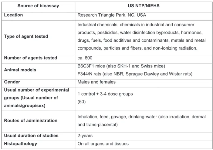
6.1.3. Rodent bioassays: powerful in vivo exposure study systems ... 29
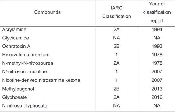
6.2. High priority compounds, background and relative interests ... 30
6.2.1. Acrylamide and glycidamide ... 30
6.2.2. Ochratoxin A ... 32
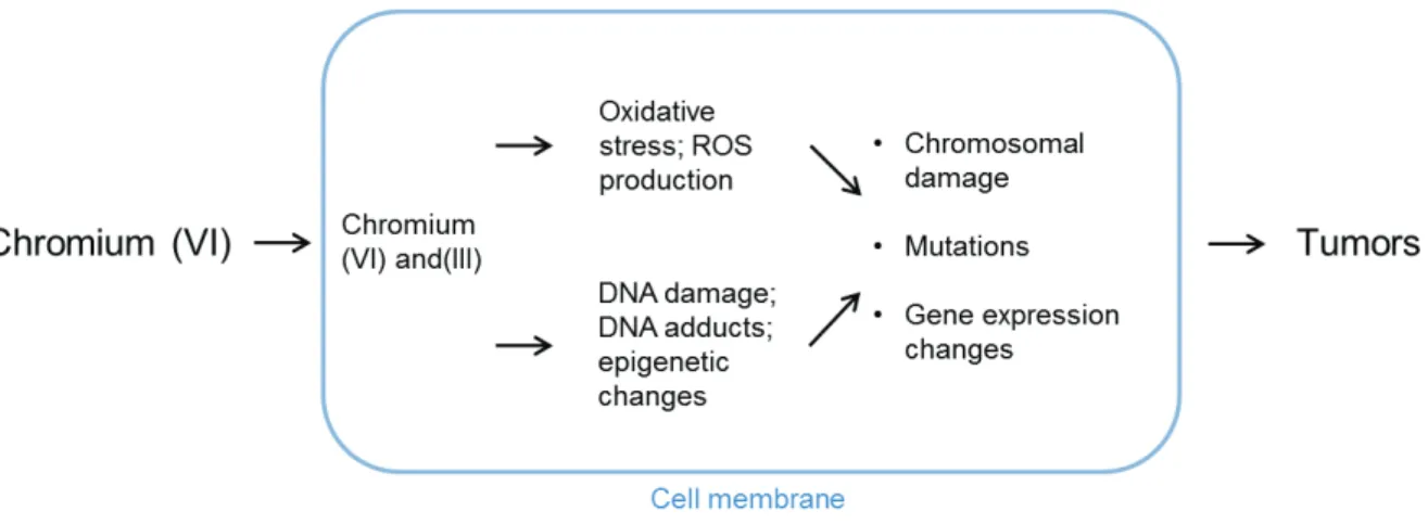
6.2.3. Glyphosate and N-nitroso-glyphosate ... Error! Bookmark not defined. 6.2.4. Hexavalent chromium ... 33
6.2.5. N-Nitroso-N-methylurea ... 35
OBJECTIVES ... 37
MATERIALS AND METHODS ... 39
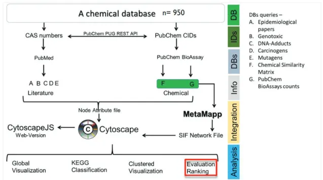
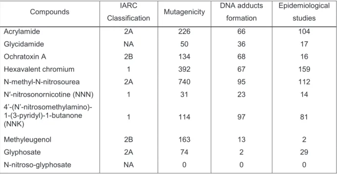
1. Prioritization of compounds for testing ... 39
2. Compounds preparation ... 41
3. Hupki MEFs cell culture, exposure and immortalization ... 42
4. HepaRG cell culture, exposure and clonal expansion ... 42
5. Lymphoblastoid cell lines culture, exposure and clonal expansion .. Error! Bookmark not defined. 6. Cytotoxicity assessment upon compound exposure ... 43
7. Genotoxicity assessment upon compound exposure ... 43
8. DNA adduct analysis ... 44
9. RNA extraction ... 45
xi
11. TP53 genotyping of primary and immortalized cells ... 45
12. DNA extraction from cultured cells ... 46
13. Animal bioassay FFPE sample processing ... 46
14. DNA extraction from animal tissues ... 47
15. Library preparation for WGS ... 48
16. Library preparation for WES ... 49
17. Bioinformatics pipeline and processing of NGS data ... 49
18. Statistical analysis ... 50
RESULTS ... 52
Objective 1: Development of mammalian cell models for exposure assays ... 52
1. Hupki MEF cells ... 52
2. HepaRG cell model ... 53
2.1. Clonal Expansion assay: Single-cell subcloning ... 54
2.2. Barrier-Bypass Clonal Expansion assay: Clonal outgrowth ... 55
2.2.1. Hepatocyte-like cells isolation ... 55
2.2.2. Exposure of progenitor bipotent cells ... 57
3. The LCL ... Error! Bookmark not defined. Objective 2: Identification of cytotoxic and genotoxic effects of high priority compounds ... 59
1. Identification of the cytotoxic effect of high priority chemical agents ... 59
2. DNA damage-dependent γH2Ax response to exposure to high priority compounds60 Objective 3: Characterization of the mutational signatures specific to mutagens ... 62
Paper 1: Summary of findings regarding the dietary compounds acrylamide and glycidamide ... 62
Paper 2: Summary of findings regarding the mycotoxin compound ochratoxin A ... 103
1. DNA adduct analysis ... 103
2. Hupki MEFs immortalization andTP53 mutations ... 105
3. FFPE tissues processing ... 105
xii
5. Mutational signature analysis... 106
6. Distribution of somatic mutations on the genome ... 108
DISCUSSION ... 111
1. Establishing mammalian in vitro models for exposure assays ... 111
2. Considerations for applying NGS to analyze FFPE tissues ... 115
3. NGS and mutational signature identification using Hupki MEF system ... 117
3.1. Acrylamide and its metabolite glycidamide ... 117
3.2. Ochratoxin A ... 118 3.3. Other compounds ... 120 CONCLUSION ... 121 BIBLIOGRAPHY ... 122 APPENDICES ... 136 Appendix A ... 136 Appendix B ... 137
Appendix C: DNA adduct analysis protocol ... 138
1. DNA extraction for adductomics analysis ... 138
2. DNA enzymatic digestion ... 138
3. dG quantitation method ... 139
4. Hydrophobic reversed phase fraction collection ... 140
5. LC/MS3 Adductomic Analysis ... 141
6. Adductomic Data Analysis ... 143
Appendix D: Published review (Zhivagui et al., 2016) ... 145
Appendix E ... 152
1
INTRODUCTION
1. Cancer prevalence, incidence and mortality:
Cancer is one of the leading causes of mortality worldwide, causing one of six deaths globally (Ferlay et al., 2015). As stated by the World Health Organization (WHO), 70% of new cancer cases will arise in the next two decades. Public health concerns have grown immensely trying to understand the biology and the burden of cancer on society.
According to the GLOBOCAN project (Global Cancer Reports 2014; Ferlay et al., 2015), prevalence estimates for 2012 indicate that for all cancers combined (excluding non-melanoma skin cancer) there were 32.6 million people (older than 15 years) alive who had been diagnosed with cancer in the previous five years. 48% of the 5-year prevalent cancer cases occurred in the less developed world, and 52% occurred in the high-income countries of North America and Western Europe, together with Japan, the Republic of Korea, Australia, and New Zealand. Figure 1 represents the 5-year prevalence of new cancer cases.
Figure 1: Estimated numbers of prevalence cases (5-year), in both sexes, from all cancers excluding non-melanoma skin cancer, worlwide in 2012. Data source: GLOBOCAN 2012; Graph production: IARC, World Health Organization (http://gco.iarc.fr/today).
Despite the higher incidence rate of cancer in the developed world largely due to tobacco smoking, high overall calorie intake coupled with the sedentary lifestyle in the rich populations, the level of mortality in the less developed world is remarkably higher than in the rich countries, accounting for 64.9% and 35.1%, respectively. In fact, regions such as Africa,
2 Asia, and Central and South America represent about 70% of the cancer deaths worldwide (Ferlay et al., 2015). This increased death rate is caused by multiple challenges facing the less developed countries. Attempts to control cancer development are less effective in the less developed countries given the remarkable disparities in resources compared to the rich countries. Different factors contribute to a vicious cycle wherein the poor world is trapped including poverty and low education level, limited government funds for health care expenditure and lack of trained professionals and managing cancer (see Figure A.1, meaning Appendix A figure A.1). Escaping from this cycle would require improvements in health care as well as in the socioeconomic status of the countries (Internal Network for Cancer Treatment and Prevention, INCTR). The most common causes of cancer deaths in the world are lung, breast, colorectal, prostate, stomach, and liver cancers (Figure A.2). Worldwide distribution of particular cancer types indicates marked differences between populations, mostly attributed to discrepancies in risk-factors exposure. The substantial burden of cancer on societies in low- and high-income countries is a major driving force for continued research to better understand the causes of cancer, and hence the development of therapeutic and preventive measures (Ferlay et al., 2015).
3
2. Cancer biology:
Cancer is a generic term reflecting neoplasms that can affect different organs and tissues of the body. The complexity of this disease has been extensively studied in the past decades generating a rich knowledge on the dynamic changes that drive a normal cell to become malignant (Hanahan and Weinberg, 2000). One defining feature of cancer is the abnormal growth of cells beyond their boundaries, the ability to invade adjacent tissues and the blood circulation leading to the dispersal of the cells into different organs, a process termed metastasis. Tumorigenesis is defined by a number of molecular and cellular hallmarks driving cell transformation (Hanahan and Weinberg, 2000, 2011). This process follows the Darwinian evolution by which a cell is subjected to a succession of genetic or epigenetic changes that confer a growth advantage and lead to the progressive conversion of a normal cell into a malignant mass (Stratton et al., 2009).
2.1. Hallmarks
of
cancer:
Throughout cancer development, cells accumulate hallmark characteristics enabling their transformation into a malignant entity with the ability to proliferate indefinitely. The hallmarks of cancer development have been described and revised in (Hanahan and Weinberg, 2000, 2011), resulting in a total of ten biological capabilities, such as sustained proliferative signaling, resistance to cell death, replicative immortality, invasion and metastasis or genome instability. The ten hallmarks (summarized in Figure 2) are acquired differently and at various times across different cancer types and individuals. Among these hallmarks, the ability to invade the blood circulation and adjacent tissues, leading to the dispersal of cancer cells to different organs, a process termed metastasis, is the main cause of cancer deaths worldwide. Induction of genome instability, for example, is brought about by mutations affecting pathways that monitor genomic integrity, such as TP53 (“the guardian of the genome”), which results in the accumulation of random mutations and structural rearrangements that can subsequently orchestrate other hallmark capabilities.
4
Figure 2: The hallmark of cancer: Hallmarks of cancer development. Taken from (Hanahan and Weinberg, 2000) and (Hanahan and Weinberg, 2011).
2.2. Cancer
genome:
In spite of the more recent emergence of epigenetic changes during tumorigenesis, cancer is primarily considered a genetic disease, causing complex abnormalities in the genomes of cancer cells (Nowell, 2002; Vogelstein and Kinzler, 1993). In analogy to Darwinian evolution, cells continuously acquire stochastic, heritable genomic alterations, which through natural selection can give rise to the phenotypic diversity and heterogeneity of tumors. Some of the acquired mutations can be deleterious and others can provide a growth advantage to the cells, which ultimately allows cancer cells to survive, proliferate, invade and metastasize (Stratton et al., 2009).
2.2.1. Epigenetic changes in a cancer genome:
In recent years, evidence has emerged linking epigenetic changes to environmental factors and human malignancies (Feil and Fraga, 2012). Cancer genomes frequently undergo epigenetic changes, which follow the Darwinian natural selection process and favor the growth of cells with characteristically altered chromatin structure and deregulated gene expression (Stratton, 2013). These changes are brought about by epigenetic modifier genes, such as DNA methyltransferases/demethylases, histone modifiers or ATP-dependent
5 chromatin remodelers that are frequently mutated in human cancer (Feinberg et al., 2016). The results of epigenetic deregulation can range from the misexpression of individual oncogenes or tumor suppressor genes to large-scale chromatin structure alterations and genomic instability.
Well-established cancer-risk agents and lifestyle factors have been studied in terms of epigenome deregulation, improving the understanding of their long-lasting effects on cancer outcome. Tobacco smoking, diet, infections, inflammation and age are known to affect epigenetic states and can play a role in the early onset of cancers through different mechanisms. Smoking, which is the strongest exposure factor causing lung cancer, harbors an epigenetic signature characterized by consistent methylation changes in the Aryl-hydrocarbon receptor repressor (AHRR) gene. Age is the strongest demographic risk factor for cancer and, interestingly, DNA methylation profiles of chronological age established an “epigenetic clock” that can be affected by different external and endogenous factors (Horvath, 2013).
Progress in epigenetic research can open the door to a new era where epigenetic biomarkers can serve as surrogate for diagnostics and risk stratification of cancer in tissues and can provide evidence on the interactive role of epigenetic deregulation in the roadmap between environmental exposures and cancer (Herceg et al. 2017).
2.2.2. Somatic mutations in a cancer genome:
Throughout the lifetime of a cancer patient, mutations are accumulating in the genome. These acquired mutations are termed somatic mutations, differentiating them from germline mutations which are inherited changes linked to familial predisposition (Stratton et al., 2009). Somatic mutations in cancer cells can encompass different structural classes of DNA sequence changes (Figure 3). They include:
1. Point mutations:
a. Single base substitutions (SBS) of one base to another. Depending on the base change and position, these can have varying effects. Silent or synonymous mutations do not alter the protein sequence. Alternatively, they can lead to a truncated or inactive protein when the SBS introduces a stop codon (missense mutation) or induces an amino-acid change (non-synonymous mutation), respectively. Finally, mutations can fall in gene regulatory regions disrupting the transcriptional activity of the gene. For example, the first cancer-causing gene change was discovered in 1982 when
6 researchers identified a G>T substitution in codon 12 of the HRAS gene causing a glycine to valine substitution (Reddy et al., 1982; Tabin et al., 1982). b. Small insertions and deletions (Indels) that result from loss or gain of nucleotide base pairs can produce abnormal protein sequences, thus affecting their function (Jego et al., 1993).
2. Chromosomal rearrangements, in which DNA segments break off and re-attach at a different genomic location, within the same chromosome or on a different chromosome, termed intra- and interchromosomal rearrangements, respectively (Figure 3). This can lead to gene disruption, the fusion of two genes or the translocation of a gene adjacent to regulatory elements, resulting in abnormal gene expression. Translocations are mostly operative in leukemias, lymphomas and sarcomas (Nowell et al., 1960; Rowley, 1973). More recently, rearranged cancer fusion genes were discovered in half of prostate cancer patients (Tomlins et al., 2005) as well as in non-small-cell lung cancer (NSCLC) cases (Soda et al., 2007).
3. Copy number variations:
a. Copy number increases, from two copies in a diploid genome, to several hundreds of copies. These are referred to as gene amplifications, which are a common mechanism for the activation of oncogenes (Alitalo, 1984), by increasing mRNA levels and thus gene expression.
b. Copy number reductions resulting from large deletions. This may induce the complete absence of a DNA segment, resulting in the loss of an associated gene, and most commonly observed as a mutational mechanism for TSG (Harris et al., 1991).
4. Insertion of new DNA sequence, originating from exogenous sources, notably viruses such as human papilloma virus (HPV), Epstein Barr virus (EBV), hepatitis B virus (HBV). These viruses have been unambiguously implicated in the development of different types of cancer (Talbot and Crawford, 2004).
7
Figure 3: Visualisation of the different types of genomic alterations present in cancer genome. Circos plot are used to depict chromosomes, point mutations, copy number and rearrangements from the outer circle to the inner circle. Each alteration is represented relative to its position on chromosomes. Adopted from (Stratton et al., 2009).
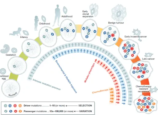
2.2.3. Driver and passenger mutations
Mutations accumulate progressively during the lifespan of an individual. Nonetheless, not all of these mutations result in tumor development. To reflect this concept, mutations are classified according to their consequences on cancer development and referred to as driver or passenger mutations (Figure 4). A driver mutation is a mutation that is implicated in oncogenesis; it has been positively selected in the microenvironment of the tumor tissue by conferring a growth advantage to the tumor. Such mutations are carried along in the clonal growth of a cancer and can help maintain and promote its growth. A passenger mutation is a mutation that does not contribute to cancer development and that has not been selected for during the evolution of the cancer (Vogelstein et al., 2013). Passenger mutations do not have functional consequences on tumor growth. By exploiting the functional contribution of driver genes, such as oncogenes and TSG, to tumor development it is possible to define and distinguish the clustered nature of driver mutations, which occur in a small number of genes, from passenger mutations, which are randomly distributed throughout the genome. Nevertheless, this task remains challenging as some mutation processes target specific genomic regions, generating clusters of passenger mutations that can be mistaken for driver alterations (Stratton et al., 2009). In addition, identification of cancer driver genes hinges in part on mutation analysis of the most commonly mutated genes within a particular type of cancer (Cancer Genome Atlas Research Network, 2008). This suggests that there are more
8 driver genes still to be identified, including drivers infrequently mutated across cancers or driver gene rearrangements that demand advanced genomic annotations for identification (Greenman et al., 2007). In order to identify a driver gene that is mutated in more than 5% of tumors of the same type with sufficient confidence, sequencing of hundreds of cases will be required.
Figure 4: Cellular lineage of cancer cell. Coloured symbols represent the progressive accumulation of somatic mutations between the fertilised egg and a fully malignant cancer cell. Embryogenesis represents a sensitive stage where embryos are prone to intrinsic mutation processes. After birth and during childhood, more mutations start accumulating due to environmental or lifestyle exposure. Chronic exposure to some cancer-risk agents can promote a mutator phenotype leading to an over-proliferation of cells and finally provoking cancer. Moreover, therapeutic approaches to eradicate cancer can cause the development of mosaic cells resistant to chemotherapy and thus the recurrence of the disease. Adopted from (Stratton, 2013).
9
3. Causes of mutations in human cancer:
Regarded mostly as a genetic disease, research has been heavily focused on the identification of cancer driver genes, as they represent attractive targets for therapeutic drugs. With the announcement of the reference human genome sequence in 2002 and the development of NGS, human cancer genomes have been sequenced at an unprecedented rate, uncovering the identity of genes operative in cancer development and accounting for more than 1% of all human genes (Futreal et al., 2004). Interestingly, while examining the cancer data for cancer genes in a myriad of sequence changes, researchers noticed that the mutational patterns differed by frequency and mutations type across the cancer types. This was suggestive that each cancer type can be a consequence of distinct mutagenic processes.
3.1. Intrinsic versus extrinsic exposures leading to cancer mutations
Cancer genomes represent a historical archive of the different mutagens that acted on the organism and were ultimately responsible for the development of cancer. Mutations can be prompted by endogenous factors (intrinsic mutagens), such as the inherent genetic instability and defects in the DNA repair machinery, or exogenous factors (extrinsic mutagens), such as dietary compounds, smoking, viruses, occupational and environmental carcinogens (Nowell, 2002; Vogelstein and Kinzler, 1993).Lately, the identification of the source of mutations found in human cancer genomes has become a subject of controversy.
It is widely accepted that mutations can be introduced upon exposure to carcinogens or via inherited predisposition to cancer. However, differences in incidence rate across cancer types have stimulated a discussion on whether stochastic DNA changes, due to variations in stem cell divisions in different organs, could explain this observation (Tomasetti and Vogelstein, 2015; Tomasetti et al., 2017a, 2017b). Quantitative correlation analysis of the lifetime risk of developing a certain cancer with the number of stem cells divisions within the same organ implied that replication-related mutations are required to drive neoplastic development (Tomasetti and Vogelstein, 2015).
An extended analysis based on a novel mathematical model and DNA sequencing and epidemiological data from 69 countries, representing 4.8 billion people, replicated the previous findings (Tomasetti et al., 2017b). This model computed 29% of driver gene mutations to be linked to environmental factors, whereas 66% were attributed to replicative errors.
10 However, it is difficult to completely separate replication-related mutations from exogenous factors. Consumption of very hot beverages for example, which has been linked to esophageal carcinogenesis, induces severe damage to the cellular lining of the esophagus, which in turn will trigger stem cells located in the deep layers to divide in order to replace the damaged cells. Therefore, stem cells divisions, caused by an exogenous factor, can introduce replication-related mutations (López-Lázaro). Moreover, the endogenous production of reactive oxygen species (ROS), which can trigger replicative mutations, has been considerably associated with several environmental compounds that act indirectly on the DNA, such as pesticides, mycotoxins and heavy metals (Frenkel, 1992).
Numerous epidemiological studies emphasize the contribution of the environment to the cancer burden observed in particular populations (Wild et al., 2015). For instance, hepatocellular carcinoma shows a high incidence rate in regions with a high risk of exposure to aflatoxin B1 (AFB1) compared to other regions where AFB1 exposure is minimal (Wild et
al., 1990). In addition, comparison of cancer incidence of Japanese subjects residing in Hawaii versus those living in Japan, especially from Okinawa, revealed a dramatic decrease in cancers of the mouth, pharynx and esophagus in all Japanese migrants, suggesting that they have escaped exposure to an environmental cancer risk factor peculiar to Okinawa region (Stemmermann et al., 1991). Furthermore, Wu et al. (Wu et al., 2016) provided extensive discussion on the causes of cancer mutations being attributed more significantly to environmental factors by employing different approaches. This resulted in an estimated 70-90% contribution of extrinsic factors to cancer development, with the rest being due to intrinsic factors.
Uncovering the causes of cancer allows cancer prevention measures, seeking to reduce or remove the exposure factors (Brennan and Wild, 2015; Colditz et al., 2012). Current estimates indicate that the majority of the global cancer risk could be preventable (Ferlay et al., 2015). Taking the ongoing discussion regarding the contribution of replication errors into account, a smaller proportion of cancers would be amenable to a reduction of environmental exposures, while the majority would require cancer prevention measures based on early detection and intervention.
3.2. Approaches to identify the sources of the somatic mutations:
Human cancer genomes harbour complex mutation patterns reflecting tumor heterogeneity that can stem from exposures to multiple carcinogenic agents (Greenman et al., 2007). Some mutagenic carcinogens leave specific SBS mutation imprints on the DNA, exemplified by tobacco smoke carcinogens, ultraviolet light (UV) and AFB1, causing characteristic
11 mutation patterns as seen in lung (G>T), skin (C>T) and liver (G>T) cancers, respectively (Hollstein et al., 1991; Pleasance et al., 2010a, 2010b). Further refined classification of these SBS mutations can be applied by taking the nucleotide bases flanking the mutated base on 5’ and 3’ into consideration. Thus, it became possible to discriminate between the mutation patterns of G>T transitions observed in lung and liver cancers, and the analysis of human cancer mutation spectra now offers the possibility to study cancer etiology (Hollstein et al., 2017).
3.2.1. Single-gene approaches
Previously, mutagenicity and genotoxicity evaluation of compounds relied on simple assays employing prokaryotic systems, such as the Ames test, and assays that are laborious, such as the comet and micronucleus assays. However, these assays do not provide insights regarding the specific base changes and the sequence context (Zhivagui et al., 2016).
Using single-gene sequencing experimental models as well as primary human tumors provides an alternative to study the mutagenic processes associated with specific carcinogenic exposures. The experimental systems used for this purpose depend either on a phenotypic selection method (e.g. bacterial reporter genes) or on genes that are frequently mutated in human cancers.
3.2.1.1. Reporter gene assays
Commonly utilized in vitro reporter genes rely on endogenous genes, e.g. HPRT, DHFR and TK, to convert certain media supplements to toxic metabolites implying the occurrence of genetic changes in the encoding genes. In contrast, animal in vivo model systems include the genomic integration of a transgene consisting of a reporter gene (such as lacI, lacZ, gpt, gpa, hprt, aprt, supF and cII genes) and a viral shuttle vector. After exposure, the transgene is packaged into phage particles ensuring the efficient delivery of the target gene into a bacterial host. Mutation detection is examined using chromogenic or viability selection (Boverhof et al., 2011). Reporter gene assay allowed the assessment of the mutagenicity of a number of carcinogens, for instance, the heterocyclic amine 2-amino-1-methyl-6-phenylimidazo[4,5-b]pyridine (PhIP), a common dietary carcinogen in cooked meat, which was associated with an increased rate of G>T transversions. Other examples include the dietary carcinogen acrylamide (A>T and G>C), AFB1 (G>T) and the chemotherapeutic agent 8-methoxypsoralen (T>A) (Zhivagui et al., 2016).
12
3.2.1.2. Single-gene mutation profiles
Sequencing of cancer genes facilitates the identification of driver mutations in a cancer type. This approach provided the first evidence regarding the molecular mechanisms by which environmental carcinogens leave characteristic imprints on the DNA. Examples of the most frequently mutated genes in human tumors include TP53, KRAS and BRAF genes. Single cancer gene sequencing in skin and lung tumors identified mutation patterns characteristic of exposures to UV-light and benzo[a]pyrene (BaP), respectively (Brash, 2015; Brash et al., 1991; Pfeifer et al., 2002). UV-light induces C>T transitions at dipyrimidines in skin cancers, and tobacco smoke prompts G>T transversions in lung tumors (Figure 5) (Olivier et al., 2010). These tumor-associated mutation patterns are in agreement with results from controlled experimental exposure studies of UV-light and BaP (Denissenko et al., 1996; Miller, 1982). Indeed, sequencing of the TP53 gene in cancer patients with different exposures history (exposed vs. non-exposed) strengthened the link between environmental factors and cancer (Hollstein et al., 1991). Lung tumors from smokers display a predominant G>T mutation pattern which is not evident in lung tumors from non-smokers, and the G>T imprint correlates with the level of tobacco consumption (Pfeifer et al., 2002).
Human exposure to the plant carcinogen aristolochic acid (AA) has been linked to the endemic Balkan nephropathy and upper tract urothelial carcinoma (UTUC) (Grollman et al., 2007). Indeed, TP53 mutation screening of these cancers identified a pronounced A>T mutation fingerprint associated with the unique mutation pattern of AA observed in the laboratory (Hollstein et al., 2013; Nedelko et al., 2009).
Finally, the TP53 gene exhibits a unique mutation profile characterized by predominant G>T transversions in hepatocellular carcinoma (HCC) cases from regions where AFB1 exposure is prevalent (Bressac et al., 1991; Montesano et al., 1997; Wogan, 1992). Liver cancers from other populations where AFB1 exposure is minimal and other risk factors prevail exhibit distinct TP53 mutation fingerprints (Montesano et al., 1997).
13
Figure 5: Data extracted from IARC TP53 database (http://p53.iarc.fr/TP53SomaticMutations.aspx). Pie charts representation of the proportion of TP53 SBS changes observed in human skin, lung, kidney and liver cancers linked to the external factors, UV, smoking, AA and AFB1 respectively.
Different in vivo and in vitro experimental systems contributed to the extraction of TP53 mutation patterns, representing “rudimentary signatures” of mutagens and their association to specific cancer types. These models include a genetically engineered mouse system, harboring the human TP53 gene, from which embryonic fibroblasts were derived. Hupki (Human TP53 knock-in) mice were exposed to UVB inducing characteristic TP53 gene mutations similar to those predominantly observed in human skin cancer (C>T) (Luo et al., 2001a, 2001b). Moreover, Hupki mouse embryonic fibroblasts (Hupki MEFs) were exposed to a number of carcinogens elucidating analogy between the Sanger sequenced TP53 genes from the in vitro assay and human tumors associated with the same exposure (Zhivagui et al., 2016).
Yeast systems were also exploited for TP53 mutagenesis using a strain transfected with an expression vector harboring human TP53 cDNA that had been UV-irradiated in vitro. The results revealed CC>TT transversions which is in line with the observations from human skin cancer data (Inga et al., 1998).
14 Finally, normal human fibroblasts were treated with known carcinogens, such as BaP, AFB1 and acetaldehyde and mutation patterns of TP53 were evaluated by functional analysis of separated allele in yeast (FASAY) (Paget et al., 2012).
Notably, experimental identification of carcinogen-specific mutation patterns demonstrates effectiveness in convergence of the mutation data with the epidemiological studies for the establishment of causal associations between environmental exposures and human cancers (Hollstein et al., 2013; Zhivagui et al., 2016).
Despite their significant contribution to understanding the sources of somatic mutations in cancer, single gene sequencing studies harbor major limitations: first, TP53 mutation, which confers a selective growth advantage, may not always occur or be selected for during cell transformation; second, many samples from a specific cancer type are needed to accumulate enough alterations to extract a specific mutation profile (Hollstein et al., 2017; Zhivagui et al., 2016). Fortunately, advances in NGS and bioinformatics analysis can help address these challenges and allow efficient testing of hypotheses regarding putative cancer-risk factors.
3.2.2.
Massively parallel sequencing and computational analysisMassively parallel sequencing has revolutionized many aspects of biology, including mutation research, due to high speed sequencing capacities and the reduction in the overall sequencing cost. NGS enables the extraction of mutation patterns from individual tumor samples, overcoming the need to pool many individuals for single-gene mutation profiling. The mutation spectra observed in cancer genomes are the consequence of exposure to multiple risk factors during the individuals’ lifetime. In order to establish the contribution of individual exposures to the final mutation spectra, a simple mathematical model based on non-negative matrix factorization (NMF) can be used to deconvolute the spectra into mutational signatures characteristic for cancer-risk factors (Figure 6). The NMF algorithm was first used by Alexandrov and colleagues to achieve an elegant reconstruction of the original sources of mutations, using mutation data from 7042 cancer patients in 30 different cancer types to extract 21 distinct mutational signatures (Alexandrov et al., 2013a) that were later expanded to 30 and are available on the COSMIC website (source: http://cancer.sanger.ac.uk/cosmic/signatures) (Alexandrov et al., 2013b).
15
Figure 6: An example of using the NMF tool to tease apart different mutational signatures from tumor sequencing data. In the first case with low exposure risk, the only observed mutational signatures are C and B, whereas in case 2 with high exposure risk a new mutational signature A is extracted, and by the mean of epidemiological studies and patient exposure history the mutagenic factor can be identified. The pie charts represent the relative contribution of each signature to the overall mutation load in each tumor. These two cases illustrate the difference in cancer etiology (Hollstein et al., 2017).
Currently, modeling of mutation spectra and mutational signatures in human cancers addresses the SBS data only. There are six possible types of SBS: C:G>A:T, C:G>G:C, C:T>T:A, T:A>A:T, T:A>C:G and T:A>G:C. SBS are conventionally reported as the pyrimidine of the mutated Watson-Crick base pair. Thereby, a C:G>A:T substitution will refer to both C>A and G>T substitutions. The profile includes the trinucleotide sequence context of each mutated base (with the mutated base in the centre) on the x-axis, generating 96 possible SBS mutation types. The y-axis represents the proportion of each mutation with respect to the overall SBS counts (see Figure 6). Moreover, an additional feature can be attributed to a mutational signature taking in consideration the proportion of the mutations generated on the transcribed and the non-transcribed DNA strand, referred to as transcription strand bias. A ratio between the mutation counts on the non-transcribed (numerator) and the transcribed strand (denominator) greater than 1 denotes the activation of transcription-related DNA repair machinery, reducing the number of mutations in the denominator. Reversely, a replication-related strand bias can occur when the mutation counts are relatively increased on the transcribed strand compared to the non-transcribed strand.
16 Differences in mutation patterns in different cancer types are immediately evident. Small cell lung carcinoma, for example, displays a predominant C:G>A:T pattern with transcription strand bias (signature 4), related to tobacco smoke carcinogens. Upper tract urothelial carcinoma shows a unique mutational signature characterized by T:A>A:T in 5’-CAG-3’ sequence context (signature 22), ascribed to AA exposure, whereas melanoma harbors a mutational signature characterized by predominant C:G>T:A at dipyrimidine nucleotide (signature 7) attributed to UV-light exposure (Alexandrov et al., 2013b). Importantly, mutational signatures with a strong link to a specific cancer type and its main etiological factor replicate observations from the single-gene TP53 sequencing approach.
As tumor mutation spectra are a composite of superimposed mutational signatures left by various mutagenic insults, seven of the thirty known mutational signatures, for example, were identified in liver cancers. The known risk factors attributed to liver cancer occurrence are HBV and HCV, alcohol consumption and AFB1 exposure. Among the identified signatures, signature 16, characterized by T:A>G:C transitions at 5’-NAT-3’ sites is observed exclusively in 90% of the liver tumors. Its etiology is nevertheless still unknown. Signature 24 was also uncovered in AFB1-exposed liver cancer spectra, characterized by a transcription asymmetry of C:G>A:T transversions (Schulze et al., 2015). This was elegantly confirmed using an integrated experimental analysis across human cell lines, animals and primary HCC tumors (Huang et al., 2017).
Surprisingly, endogenous mutagenic processes constitute almost half of the identified mutational signatures. Signature 1, attributed to the spontaneous deamination of 5’-methylcytosine, is seen in almost all tumors and is pronounced in some, such as in acute myeloid leukaemia. Together with signature 5, it has been linked to the clock-like cellular processes reflecting the chronological age of patients at diagnosis (Alexandrov et al., 2015). Furthermore, a number of signatures have been attributed to the disruption of processes regulating DNA homeostasis (Helleday et al., 2014), such as malfunction of DNA repair polymerases eta and epsilon (signatures 9 and 10, respectively), defective DNA mismatch repair (MMR) (signatures 6, 15 and 20), and BRCA1/2 mutations indicating a failure of DNA double strand repair by homologous recombination (signature 3). Notably, in more than half of the cancer types analysed to date, APOBEC (apolipoprotein B mRNA editing enzyme, catalytic polypeptide-like) deaminase mutational signatures (signatures 2 and 13) have been identified. APOBEC signatures are supported by extensive work in experimental model systems (Burns et al., 2013; Chan et al., 2015; Harris et al., 2002; Kazanov et al., 2015). These enzymes are implicated in virus restriction and suppression of retrotransposition (Smith et al., 2012). Signature 2 was found in cervical and in head and neck cancers, both of
17 which are related to HPV infection, implying the recurrent activation of APOBEC upon viral infection (Rebhandl et al., 2015; Warren et al., 2015).
Among the 30 distinct mutational signatures thus far identified, 40% remain with unknown etiology reflecting the need for controlled experimental mutation studies (Zhivagui et al., 2016).
18
4. Cancer genomics repositories:
Hand-in-hand with the remarkable technological advances in sequencing, a complete catalogue of all somatically acquired variants in cancer genomes was established in coordination with different data repositories, which keep the mutation data freely accessible and mineable. In addition to the somatic mutations, these repositories frequently contain additional omics data, such as gene expression data from RNA-seq, proteomics and epigenetic profiles, of the same cancer cases, and have been correlated with basic clinical features.
There are three large-scale data repositories that exist today:
x The Catalogue Of Somatic Mutations In Cancer (COSMIC) which is maintained by the Sanger Institute UK, using manually curated data from the scientific literature.
x The Cancer Genome Atlas (TCGA) which collaborates with the Pan-Cancer analysis project and is funded by the NIH.
x The International Cancer Genome Consortium (ICGC) which coordinates the generation of comprehensive catalogues of genomic abnormalities internationally.
4.1. The Catalogue Of Somatic Mutations in Cancer (COSMIC)
The Catalogue Of Somatic Mutations in Cancer (COSMIC) is easily accessible through its website (http://www.sanger.ac.uk/cosmic) (Forbes et al., 2011, 2017). COSMIC is the broadest database initiative of cancer mutation recurrence exploring targets and trends in the genome of human cancers worldwide. Release v78 (September 2016) includes 1,235,846 tumors samples, and 28,366 tumors genome-wide sequenced. Using 23,489 scientific publications for manual curation (accounting for 60% of COSMIC content), this high-resolution resource focuses on 186 key genes across all cancers. Molecular profiling of this large number of tumors allowed annotation of over 4 million coding mutations and one million copy number variants (Forbes et al., 2017). Around 30% of COSMIC content has been selected from consortia sources such as TCGA and ICGC.
4.2. The Cancer Genome Atlas (TCGA)
The Cancer Genome Atlas (TCGA) is accessible on http://cancergenome.nih.gov/. It is managed by the National Cancer Institute (NCI) and the National Human Genome Research Institute (NHGRI). The primary aim of TCGA is to assimilate and interpret molecular profiles from DNA, RNA and protein sequencing as well as epigenetic patterns from clinical cases of different types of cancer. Data annotation is not confined to point mutations only, but also to
19 the characterization of copy number variations, DNA methylation, mRNA and miRNA expression and sequence and transcript splice variations (Weinstein et al., 2013). Cancer samples are chosen on the basis of poor prognosis and public health impact and on the availability of human tumor-matched normal tissue samples. In 2017, a launch of a new data portal, the Genomic Data Commons (GDC), will take place, which includes 29 different human tumor sites.
4.3. The International Cancer Genome Consortium (ICGC)
The International Cancer Genome Consortium (ICGC) is publically accessible on http://icgc.org/. The aim of the ICGC platform is to attain a comprehensive elucidation of cancer genome abnormalities by harnessing genomic, transcriptomic and epigenomic changes in 50 different tumor types denoting clinical and public health importance throughout the world (2010). To date, ICGC analyzed over 25,000 cancer genomes using different omics approaches and identified around 46 million somatic mutations in 21 different tumor types. The Pan-Cancer Analysis of Whole Genomes (PCAWG) collaboration was established, covering about 700 researchers from around the world. PCAWG encompasses whole genome data of 2,834 donors with matched tumor/normal samples using ICGC data repositories. It aims for meaningful cross-tumor comparisons and standardized bioinformatic analyses using gold-standard, benchmarked, version-controlled algorithms (Campbell et al., 2017).
20
5. IARC Monographs on the evaluation and classification of
carcinogenic risks to humans
The International Agency of research on Cancer (IARC) is the specialized cancer agency of the World Health Organization (WHO). It seeks to prompt broad collaboration in cancer research to uncover the causes of cancer so that prevention measures can be adopted in order to reduce the burden of this disease and related suffering. The IARC Monographs are expert evaluations of a compendium of carcinogenic chemicals and their causal effect on the human population.
5.1. Objective and scope
The objective of the IARC Monographs program is to review public scientific reports for evidence linking a wide range of agents to human cancer occurrence. The Monograph represents the first step in carcinogen risk assessment through examination of published data, positive or negative, in order to evaluate whether or not a compound could induce cancer in humans. The term ‘agent’, frequently used, refers to a broad range of agent categories including chemicals, complex mixtures, occupational and environmental exposures, as well as biological organisms.
5.2. Evaluation and rationale
Evaluation of cancer risk agents depends on the strength of evidence available in the literature related to carcinogenesis in human and animals as well as to mechanistic data. The classification of a compound doesn’t revolve around the carcinogenic potency but around the strength of evidence (sufficient or insufficient). This classification can be changed when new evidence becomes available.
Establishing a causal association between exposure to a studied agent and human cancer relies on the available epidemiological studies that allow the categorization of the compound into one of the following: a) sufficient evidence of carcinogenicity; b) limited evidence of carcinogenicity; c) inadequate evidence of carcinogenicity; and d) evidence suggesting lack of carcinogenicity.
Similarly, the carcinogenicity of a compound in experimental animals is classified based on conventional animal bioassays (mostly rodents), including those that employ genetically-modified animals.
Mechanistic and other relevant data have the power to affect the evaluation and the classification of an agent by weighting data on preneoplastic lesions, tumour pathology,
21 genetic and related effects, structure-activity relationships, metabolism and toxicokinetics, physicochemical parameters and analogous biological agents. The agent is then categorized based on the strength of evidence as “weak”, “moderate” or “strong”. The Working Group can identify mechanistic data that are likely to operate in humans and consider whether multiple mechanisms contribute to carcinogenesis, whether different mechanisms function at different dose ranges, whether distinct mechanisms drive tumorigenesis in humans compared to animals and whether a unique mechanism is activated only in a susceptible group. The evidence is strengthened when experiments are performed in different models with consistency in the results and biological plausibility.
5.3. Overall
evaluation
Based on the strength of evidence inferred from the human epidemiology data, experimental animal studies and mechanistic and other relevant data, the agent is subsequently classified into one of the following categories (see also Table 1):
x Group 1: The agent is carcinogenic to humans. This group comprises 120 agents. x Group 2: The agent is probably (2A) or possibly (2B) carcinogenic to humans. This
group contains 81 and 299 agents, respectively.
x Group 3: The agent is not classifiable as to its carcinogenicity to humans. Group 3 embraces 502 agents.
x Group 4: The agent is probably not carcinogenic to humans. This group consists of 1 agent, caprolactam.
22
Table 1: A summary of evaluation instructions to classify an agent in one of the groups assigned by IARC monographs based on the strength of evidence in humans and experimental models. ESLC: evidence suggesting lack of carcinogenicity. Table source: IARC monographs.
Mechanistic data can be pivotal when the human data are not conclusive and can, therefore, result in the change of classification of a compound. Table C.1 is complementary to Table 1, depicting the impact of mechanistic data on cancer-risk agent classification.
23
6. The MutSpec project: Molecular Mechanisms and Biomarkers
group, IARC
With respect to IARC’s core activities, elucidating the mechanisms of environmental exposures through genetic and epigenetic alterations can provide evidence base for the etiology of cancer, strengthen the data on carcinogen evaluation and classification and may ultimately influence prevention measures.
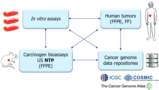
As part of the Mechanisms of Carcinogenesis (MCA) section, a main focus of the Molecular Mechanisms and Biomarkers (MMB) group at IARC, led by Dr. Jiri Zavadil, is to decipher the origins of the molecular changes that shape human cancer genomes. Such changes can arise from environmental exposures or endogenous processes that leave fingerprints on the DNA. In 2014, the “MutSpec” project, short for Mutation Spectra, was launched in coordination with the IARC Monographs section (IMO) and other IARC groups, in order to experimentally generate mutational signatures specific to cancer-risk agents and to elucidate the enigmatic signatures observed in human tumors. For this purpose, a list of high priority compounds has been generated, reflecting MMB group interests as well as recommendations of the Advisory Group regarding compounds of interest for carcinogen classification by the IARC Monographs section (Straif et al., 2014). The “MutSpec” project seeks to identify carcinogen mutation spectra and signatures in well-controlled experimental settings, using robust mammalian in vitro exposure assays and tumor tissue from animal bioassays.
6.1. The
experimental
model
systems
In vivo exposure bioassays as well as in vitro exposure assays are two roads that can lead to a controlled assessment of the genotoxicity, mutagenicity and carcinogenicity of a compound. Ideally, such exposure studies would use model systems that enable the testing of a large number of compounds within a reasonable timeframe. Cellular models suitable for mutation spectra analysis should include a bottleneck step followed by clonal expansion and mimic key steps of carcinogenesis (initiation via exposures, promotion and progression). There are two approaches to be considered for in vitro systems: 1) Bypass of a biological barrier, like crisis or senescence, and emergence of an immortalized clonal population, referred to as Barrier-Bypass Clonal Expansion (BBCE); 2) Cells to which a selective biological bypass step is not applicable require single-cell subcloning after exposure, referred to as Clonal Expansion (CE). Moreover, these models should be able to recapitulate key aspects of human biology (e.g. metabolism, DNA repair pathways) (Zhivagui et al., 2016).
24
6.1.1. Mouse embryonic fibroblast: Hupki MEF cells
Several model systems used for the inquiry of mutational signatures by the means of massively parallel sequencing meet some but not all of the above mentioned criteria (Zhivagui et al., 2016).
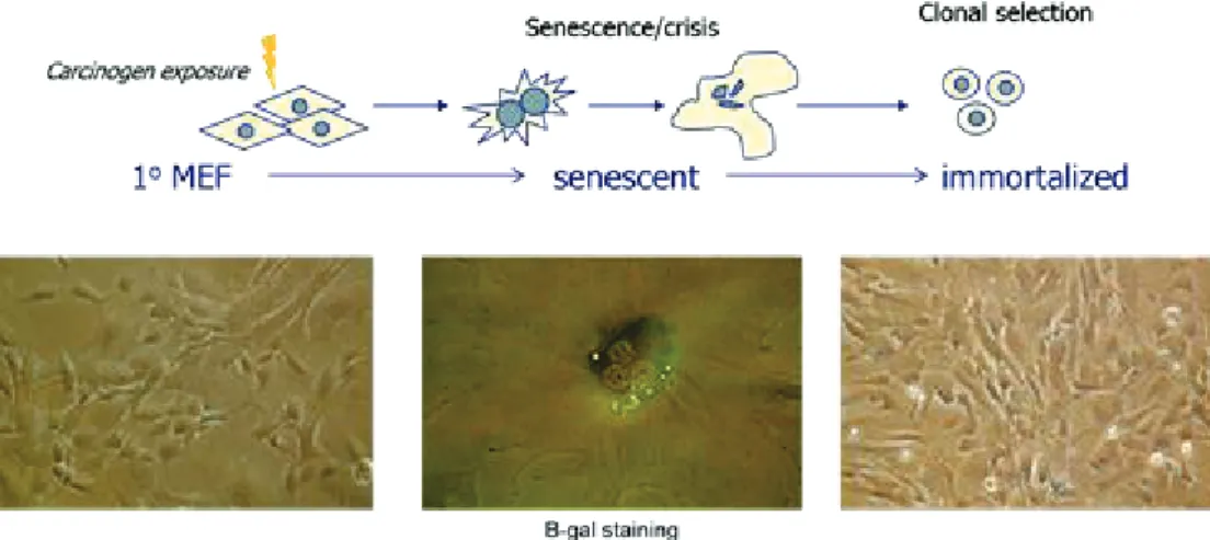
Hupki MEFs were first established for single-gene studies using Hupki mice (Liu et al., 2004). Using this cell system, exposures to UV light, AA, benzo[a]pyrene (B[a]P) and 3-nitrobenzanthrone (3-NBA) were carried out (vom Brocke et al., 2008; Feldmeyer et al., 2006; Liu et al., 2004, 2005). It is characterized by a biological barrier (senescence), which cells can bypass in a clonal manner (see Figure 7). Sanger sequencing of the TP53 gene recapitulated human cancer TP53 mutation profiles associated with the same exposures (Besaratinia and Pfeifer, 2010; Brocke et al., 2006; Kucab et al., 2010), namely in skin, kidney and lung tumors.
Figure 7: Hupki MEF exposure. MEFs are exposed as primary cells to carcinogens. The cells are propagated in culture until they reach senescence, manifested in modified cellular morphology (e.g. increase in cytoplasmic size) due to the inability of the cells to undergo a full cell cycle, hence, the formation of multi-nucleated cells. Senescence can be detected biochemically using beta-galactosidase staining. Mouse cells have the ability the bypass senescence generating immortalized cell lines representing a number of clones or subclones.
More recently, Hupki MEF cell lines derived from exposure to UV-light class C, AA, B[a]P and methylnitronitrosoguanidine (MMNG) were subjected to whole-exome sequencing. In agreement with the TP53 sequencing studies, extracted SBS-mutational signatures recapitulated the mutation profiles observed in human cancer linked to same exposures, (melanoma, UTUC, lung and brain cancer, respectively) (Olivier et al., 2014) (Figure 8). The immortalized cell lines represent relatively homogenous populations of one predominant
25 clone and less represented subclones, which allows reliable identification of enriched SBS patterns upon sequencing at reasonable coverage (Zhivagui et al., 2016). These findings were validated at the whole-genome scale allowing investigations beyond SBS mutations and towards structural variations, large insertions and deletions and copy number alterations (Nik-Zainal et al., 2015).
Nevertheless, using mouse cell lines has caveats to recapitulate exposures in human beings due to limitations in the differences in genetic background, species-specific repair machineries and metabolic restrictions (Zhivagui et al., 2016). The addition of human S9 fraction, comprising active metabolic enzymes such as CYP450 and transferases, can boost metabolism of pro-carcinogens and thus circumvent the latter limitation. Interestingly, immortalization of primary mouse cells requires only one barrier bypass event such as disruption of the p19/ARF/p53 axis, making it an easier and faster system compared with the human cells necessitating disruptions of several critical genetic pathways (Hahn and Weinberg, 2002).
26
Figure 8: Carcinogens’ mutational fingerprints in human primary tumors recapitulated in the Hupki MEF experimental system. (a) The upper panels show the mutational signature identified in smoking-related cancer patients (COSMIC signature 4 and 29). Lower panel: Hupki MEF cells treated with B[a]P under well controlled settings. (b) The upper panel represents the mutational signature identified in UTUC patients (COSMIC signature 22) correlating with AA exposure in Hupki MEFs (lower panel). (c) Mutational signature from skin cancer patients, attributed to UV-light (upper panel) (COSMIC signature 7) recapitulated by Hupki MEFs exposed to UV-light (lower panel).