HAL Id: hal-01468144
https://hal.sorbonne-universite.fr/hal-01468144
Submitted on 15 Feb 2017
HAL is a multi-disciplinary open access archive for the deposit and dissemination of sci-entific research documents, whether they are pub-lished or not. The documents may come from teaching and research institutions in France or abroad, or from public or private research centers.
L’archive ouverte pluridisciplinaire HAL, est destinée au dépôt et à la diffusion de documents scientifiques de niveau recherche, publiés ou non, émanant des établissements d’enseignement et de recherche français ou étrangers, des laboratoires publics ou privés.
cervical dystonia?
Loïc Carment, Marc A. Maier, Sophie Sangla, Vincent Guiraud, Serge Mesure,
Marie Vidailhet, Påvel G Lindberg, Jean-Pierre Bleton
To cite this version:
Loïc Carment, Marc A. Maier, Sophie Sangla, Vincent Guiraud, Serge Mesure, et al.. Does dystonic muscle activity affect sense of effort in cervical dystonia?. PLoS ONE, Public Library of Science, 2017, 12 (2), pp.e0172019. �10.1371/journal.pone.0172019�. �hal-01468144�
RESEARCH ARTICLE
Does dystonic muscle activity affect sense of
effort in cervical dystonia?
Loïc Carment1*, Marc A. Maier1,2, Sophie Sangla3, Vincent Guiraud4,5, Serge Mesure6,
Marie Vidailhet7,8, Påvel G Lindberg1,9☯‡, Jean-Pierre Bleton3☯‡
1 FR3636, CNRS / Universite´ Paris Descartes, Sorbonne Paris Cite´, Paris, France, 2 Universite´ Paris Diderot, Sorbonne Paris Cite´, Paris, France, 3 Service de Neurologie, Fondation OPH de Rothschild, Paris, France, 4 Universite´ Paris Descartes, Sorbonne Paris Cite´, INSERM U894, Paris, France, 5 Service de Neurologie et Unite´ Neurovasculaire, Hoˆpital Sainte-Anne, Paris, France, 6 UMR 7287, CNRS Aix Marseille Universite´, Institut des Sciences du Mouvement, Marseille, France, 7 Sorbonne Universite´s, UPMC Univ Paris 06, UMR S 1127, Paris, France, 8 AP-HP, Hoˆpital de la Pitie´ Salpêtrière, De´partement de Neurologie, Paris, France, 9 Centre de Psychiatrie et Neurosciences, Inserm U894, Paris, France
☯These authors contributed equally to this work. ‡ These authors are senior authors on this work.
Abstract
Background
Focal dystonia has been associated with deficient processing of sense of effort cues. How-ever, corresponding studies are lacking in cervical dystonia (CD). We hypothesized that dystonic muscle activity would perturb neck force control based on sense of effort cues.
Methods
Neck extension force control was investigated in 18 CD patients with different clinical fea-tures (7 with and 11 without retrocollis) and in 19 control subjects. Subjects performed force-matching and force-maintaining tasks at 5% and 20% of maximum voluntary contraction (MVC). Three task conditions were tested: i) with visual force feedback, ii) without visual feedback (requiring use of sense of effort), iii) without visual feedback, but with neck exten-sor muscle vibration (modifying muscle afferent cues). Trapezius muscle activity was recorded using electromyography (EMG).
Results
CD patients did not differ in task performance from healthy subjects when using visual feed-back (ANOVA, p>0.7). In contrast, when relying on sense of effort cues (without visual feed-back, 5% MVC), force control was impaired in patients without retrocollis (p = 0.006), but not in patients with retrocollis (p>0.2). Compared to controls, muscle vibration without visual feedback significantly affected performance in patients with retrocollis (p<0.001), but not in patients without retrocollis. Extensor EMG during rest, included as covariate in ANOVA, explained these group differences.
a1111111111 a1111111111 a1111111111 a1111111111 a1111111111 OPEN ACCESS
Citation: Carment L, Maier MA, Sangla S, Guiraud
V, Mesure S, Vidailhet M, et al. (2017) Does dystonic muscle activity affect sense of effort in cervical dystonia? PLoS ONE 12(2): e0172019. doi:10.1371/journal.pone.0172019
Editor: Yuqing Li, University of Florida, UNITED
STATES
Received: November 21, 2016 Accepted: January 30, 2017 Published: February 13, 2017
Copyright:© 2017 Carment et al. This is an open access article distributed under the terms of the
Creative Commons Attribution License, which permits unrestricted use, distribution, and reproduction in any medium, provided the original author and source are credited.
Data Availability Statement: All relevant data are
within the paper.
Funding: This work was supported by the
Rothschild Foundation Hospital and the Association Franc¸aise pour la Recherche et l’Evaluation en Kine´sithe´rapie. The funders had no role in study design, data collection and analysis, decision to publish, or preparation of the manuscript.
Competing interests: LC reports grants from
Universite´ Pierre et Marie Curie, Paris VI, outside the submitted work. SS reports personal fees from
Conclusion
This study shows that muscle afferent feedback biases sense of effort cues when controlling neck forces in patients with CD. The bias acts on peripheral or central sense of effort cues depending on whether the task involves dystonic muscles. This may explain why patients with retrocollis more accurately matched isometric neck extension forces. This highlights the need to consider clinical features (pattern of dystonic muscles) when evaluating sensori-motor integration in CD.
Introduction
Cervical dystonia (CD) is clinically characterized by involuntary neck muscle contraction lead-ing to abnormal movement or posture [1]. Integration of multimodal sensory information is necessary to accurately execute voluntary movements. This integration seems to be deficient in CD [2]. Similarly, controlling forces using sense of effort cues are affected in focal dystonia [2,3]. Sense of effort contributes to the control of body position, forces and movements and includes central cues (efferent copy: derived from the motor command) as well as peripheral cues (from muscle afferents) [4]. It has been suggested that involuntary neck muscle contrac-tions may perturb the peripheral contribution to the sense of effort in CD [1]. However, despite potential interest for targeted rehabilitation approaches, studies investigating the rela-tion between clinical features of CD, neck control and sense of effort are lacking.
Kinematic studies have shown that in CD neck extension amplitude and velocity are reduced toward the non-dystonic side (anti-dystonic) in voluntary movements compared to movements toward the dystonic side (pro-dystonic) [5,6]. These results were explained by more efficient muscle activation patterns (muscle synergies) during pro-dystonic movements [5]. During anti-dystonic movements, the overflow phenomenon, i.e., a lack of muscle selectiv-ity often described in focal dystonia [7], was thought to impede appropriately timed relaxation of dystonic muscles [8]. Thus, involuntary muscle activity reduces specificity of task-related sense of effort cues.
Furthermore, even though anti-dystonic movements are impaired in all planes, movement control in the sagittal plane, i.e. in flexion-extension, may be more severely affected [5,6].
Together these previous studies suggest that control of neck extension movements and forces will differ depending on clinical features, i.e. on whether task-related muscles are dys-tonic or not. This leads to the prediction that neck control involving dysdys-tonic muscles will be less affected than neck control involving non-dystonic muscles in patients with CD. Particu-larly, in CD flexion-extension afferent feedback will be differentially affected by the presence or absence of retrocollis. The underlying rationale is that the reliability of sensorimotor infor-mation processing [9] depends in part on muscle afferent feedback.
We hypothesized that multisensory integration (of visual and sense of effort cues) during voluntary isometric neck force control would be differentially affected in CD patients with varying clinical features (i.e., presence or absence of retrocollis).
Methods
Participants
Eighteen patients with primary focal CD were recruited and categorized according to clinical features, i.e. presence (CD_R+, N = 7) or absence (CD_R-, N = 11) of a retrocollis. Patients
Allergan, personal fees from Ipsen, personal fees from Merz-Pharma, outside the submitted work. VG has nothing to disclose. SM reports grants from Fondation Paul Bennetot, outside the submitted work. MV reports grants from INSERM, grants from APHP, grants from APTES, grants from France Parkinson, grants from AMADYS, personal fees from Movement Disorders Society, outside the submitted work; and Member of the advisory board for Merz and Medtronic. PGL reports grants from Gloria and Jacques Gosweiler Foundation, outside the submitted work and owns shares in Aggero MedTech AB, a company commercializing a measurement instrument for spasticity. In addition, PGL and MAM have patented a method for measurement of manual dexterity (EP2659835A1). JPB reports personal fees from MERZ-PHARMA, personal fees from IPSEN, grants from AMADYS, grants from AFREK, outside the submitted work. This does not alter the authors’ adherence to PLOS ONE policies on sharing data and materials.
with tardive/drug-induced dystonia were excluded. None of the patients received botulinum toxin injections for at least 3 months prior to this study. Nineteen healthy (age- and gender-matched) control subjects were also recruited (Table 1: demographic and clinical details). The study received ethical approval from the regional ethics committee (Ile de France VIII) and all subjects provided written informed consent.
Clinical assessments
Dystonic symptoms were assessed with the Toronto Western Spasmodic Torticollis Rating Scale (TWSTRS) [11]. Maximum voluntary neck extension force (MVC) was recorded using a dynamometer (Biometrics, France) applied to the back of the head.
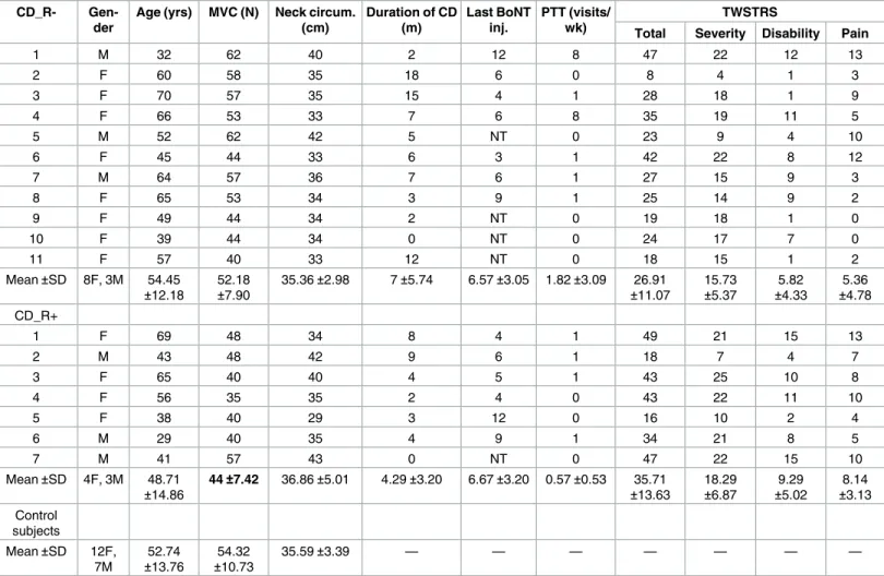
Table 1. Demographic and clinical data. Clinical data for patients with a retrocollis (CD_R+) and without a retrocollis (CD_R-). The only significant differ-ence between groups was lower neck extension MVC in CD_R+ patients compared to the control group (p<0.019). Scores of the clinical scales did not differ between CD_R+ and CD_R- patients. There was no difference in the demographic data between groups.
CD_R- Gen-der
Age (yrs) MVC (N) Neck circum. (cm) Duration of CD (m) Last BoNT inj. PTT (visits/ wk) TWSTRS
Total Severity Disability Pain
1 M 32 62 40 2 12 8 47 22 12 13 2 F 60 58 35 18 6 0 8 4 1 3 3 F 70 57 35 15 4 1 28 18 1 9 4 F 66 53 33 7 6 8 35 19 11 5 5 M 52 62 42 5 NT 0 23 9 4 10 6 F 45 44 33 6 3 1 42 22 8 12 7 M 64 57 36 7 6 1 27 15 9 3 8 F 65 53 34 3 9 1 25 14 9 2 9 F 49 44 34 2 NT 0 19 18 1 0 10 F 39 44 34 0 NT 0 24 17 7 0 11 F 57 40 33 12 NT 0 18 15 1 2 Mean±SD 8F, 3M 54.45 ±12.18 52.18 ±7.90 35.36±2.98 7±5.74 6.57±3.05 1.82±3.09 26.91 ±11.07 15.73 ±5.37 5.82 ±4.33 5.36 ±4.78 CD_R+ 1 F 69 48 34 8 4 1 49 21 15 13 2 M 43 48 42 9 6 1 18 7 4 7 3 F 65 40 40 4 5 1 43 25 10 8 4 F 56 35 35 2 4 0 43 22 11 10 5 F 38 40 29 3 12 0 16 10 2 4 6 M 29 40 35 4 9 1 34 21 8 5 7 M 41 57 43 0 NT 0 47 22 15 10 Mean±SD 4F, 3M 48.71 ±14.86 44±7.42 36.86±5.01 4.29±3.20 6.67±3.20 0.57±0.53 35.71 ±13.63 18.29 ±6.87 9.29 ±5.02 8.14 ±3.13 Control subjects Mean±SD 12F, 7M 52.74 ±13.76 54.32 ±10.73 35.59±3.39 — — — — — — —
Abbreviations: TWSTRS = Toronto Western Spasmodic Torticollis Rating Scale; Yrs = years; N = Newton; circum = circumference; m = months; BoNT inj. = time (in months) of last botulinum neurotoxin injection; NT = non treated, PTT = Physical therapy treatment according to standardized physical therapy program including retraining of neck movements and posture [10], wk = week.
doi:10.1371/journal.pone.0172019.t001
Neck extension tasks
Two tasks were developed (using Spike2/CED1401,http://ced.co.uk) to investigate multi-modal sensory processing during isometric force control.
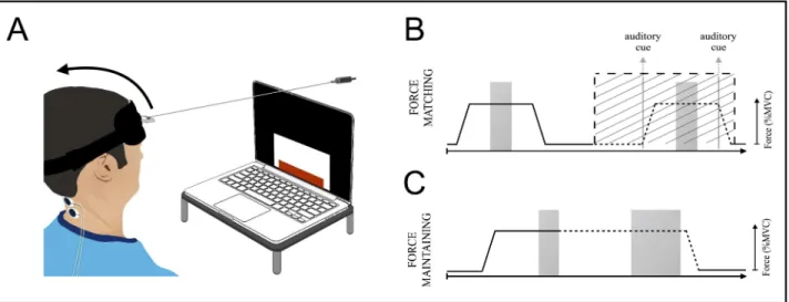
1. A force-matching task (Fig 1A and 1B) was used to assess performance of voluntary neck force modulation at 5% and 20% MVC under three different task conditions:
a. Condition_Vis: subjects had to match their neck extension force, displayed in real-time on a screen (i.e.with visual feedback, Vis), to a visually indicated target level. In each trial, force had to be increased to target force level, then maintained (for 3s), and finally released.
b. Condition_noVis: subjects had to reproduce the previous trial without visual feedback (noVis). This required the use of sense of effort cues to match performance between trials.
c. Condition_noVis+Vib: this condition was similar to condition b, but with muscle vibra-tion (+Vib) to modify muscle afferent feedback [12] (70Hz vibration on the left and right trapezius muscles; Vibrasens1VB115,www.technoconcept.fr).
2. A force-maintaining task was used to assess the ability to maintain steady neck extension force at 5% and 20% MVC (Fig 1C) [13]. This task was performed in the same three task conditions as theForce-matching task.
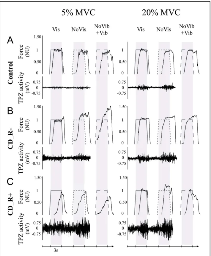
Left and right trapezius activity was recorded using surface electromyography (EMG)
(www.grasstechnologies.com;Fig 2A–2C). EMG from five subjects was not exploitable, due
to either poor signal to noise ratio (N = 2) or technical failure (N = 3).
Fig 1. Setup and visuomotor tasks. (A) Setup for visuomotor tasks. Subjects were seated in front of a computer screen. A headband was attached to the force sensor by a non-extensible wire. The task consisted of a series (trials) of visually displayed target forces (height of white rectangle) to be matched as closely as possible using visual feedback of the exerted neck force (height of red rectangle). (B) Force matching task: subjects matched the neck extension force to an indicated target level (5% or 20% MVC) with visual force feedback (condition_Vis) and reproduced the same force level without visual feedback (condition_NoVis). In conditions without vision, subjects were given an auditory cue indicating force onset, offset or hold. Five trials/condition were presented in a pseudo-randomized order. Force exerted during the stable part of the hold phase, indicated by grey shading, was analyzed. (C) Force-maintaining task: subjects maintained their extension force at target level with visual feedback (condition_Vis). The visual feedback was then removed for six seconds
(condition_NoVis) and vibration was applied (condition_NoVis+Vib).
Fig 2. Comparison of raw data for a control subject, a CD_R- and a CD_R+ patient. (A) Control subject: raw data recorded during the force matching task at 5% MVC and 20% MVC: Neck extension force was first down-sampled (100Hz) and normalized for each subject to the target force level (NU: normalized units). Lower trace: EMG activity of right trapezius (TPZ). Example trials show force and EMG traces during condition_Vis, condition_NoVis, and condition_ NoVis+Vib. Note: EMG was not recorded during vibration. (B) CD_R- patient: corresponding examples. (C) CD_R+ patient: corresponding examples.
doi:10.1371/journal.pone.0172019.g002
Data analysis and statistics
Raw data was analyzed using MatlabV8.6 (The MathWorks, Inc., Natick, MA, USA) and statis-tics performed under Statistica10 (StatSoft, Inc., USA). Forces and EMGs were averaged across all trials in each task condition. Group differences were analyzed using a general linear model repeated measures ANOVA with one GROUP factor (CD_R-/CD_R+/Controls) and two within-group factors: FORCE (5%/20% MVC) and CONDITION (a/b/c). We used Fisher LSD to test for post-hoc differences. The level of significance was set to p<0.05 and adjusted in order to correct for multiple comparisons with the Benjamini and Hochberg method [14]. All reported p values <0.05 met corrected levels of significance.
Results
Isometric force-matching and force-maintaining
The two tasks were completed successfully by all subjects. Patient groups did not differ in total TWSTRS (Mann-Whitney U Test p = 0.23).Fig 2A–2Cshows examples of the isometric force-matching task.
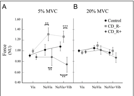
In the force-matching task (Fig 3A and 3B) the ANOVA of force showed a significant GROUP difference (F = 3.63, p = 0.04). This GROUP difference interacted with FORCE (F = 4.96, p = 0.01); no other interaction was found. Post-hoc testing revealed that neck exten-sion forces were similar in patients and control subjects when using visual cues (condition_Vis, p>0.7) and GROUP differences were specific to 5% MVC-level (p<0.001 between CD groups).
Fig 3. Group performance in the visuomotor force-matching task. Mean force (mean±SD) for the three groups during condition_Vis, condition_NoVis and condition_NoVis+Vib at 5% MVC (A) and at 20% MVC (B). (A) No significant difference in mean force between groups in condition_Vis. Significant differences at 5% MVC during condition_NoVis between CD_R- and control subjects (p = 0.006), and also between CD_R- and CD_R+ patients (p = 0.002). Note that the variability of mean force (SD) increased about 5-fold in all three groups. Significant differences during condition_NoVis+Vib between CD_R+ and control subjects (p = 0.006) and also between CD_R+ and CD_R- patients (p<0.001). (B) No difference in mean force at 20% MVC.*= p<0.05;**= p<0.01;***= p<0.001.
However, performance of CD patients differed in conditions requiring sense of effort cues: in condition_noVis (5% MVC), force was significantly increased in CD_R- patients (1.31±0.47N) compared to control subjects (1.06±0.43N, p = 0.006), but not in CD_R+ patients (0.89±0.32N, p = 0.27). Thus, CD_R- patients applied significantly higher forces than CD_R+ patients (p = 0.002). Control subjects showed no significant force difference betweencondition_Vis (0.92±0.09N) and condition_noVis (1.07±0.43N, p>0.05).
During muscle vibration (condition_noVis+Vib, 5% MVC), CD_R+ patients showed signif-icantly reduced mean force (0.74±0.33N) compared to CD_R- patients (1.26±0.35N, p<0.001) and to control subjects (1.08±0.37N, p = 0.006). Hence, compared to control subjects, CD_R-patients tended to overshoot, whereas CD_R+ CD_R-patients undershot target forces.
The above ANOVA was repeated with baseline EMG activity (during rest) as covariate. This cancelled the statistical main (GROUP, p = 0.3) and post-hoc differences between CD patients and control subjects.
In the force-maintaining task, the performance of CD_R- and CD_R+ patients were quali-tatively similar to those seen during force-matching. Similar between group differences were found: (i)condition_Vis: no significant difference (p>0.2); (ii) condition_noVis: force CD_R-> force controls (p = 0.003); (iii)condition_noVis +Vib: force CD_R- > force CD_R+, (p = 0.001).
Electromyography
EMG activity during MVC was similar between groups (F = 0.69, p = 0.51). However, during force-matching, the ANOVA of EMG activity showed significant effects of GROUP (F = 10.52, p<0.001) and FORCE (F = 92.63, p<0.001), but not of CONDITION (F = 0.27, p = 0.60). Post-hoc analyses showed increased EMG activity in CD patients compared to control subjects (p<0.01). EMG activity was higher in CD_R+ than in CD_R- patients (p = 0.04).
Discussion
We have shown that clinical features of CD (presence/absence of a retrocollis) differentially affect multisensory integration of visual and sense of effort signals during voluntary neck force control. We have also shown that the type of force control deficit depends on the characteris-tics and sources of sensory information (with/without vision; with/without perturbed muscle afferents). Our results are in line with the optimal multisensory integration theory, [9] which postulates that to control movement, subjects (through mechanisms of gating/weighting) rely on the most reliable information available among multi-modal sensory feedback.
With visual force feedback, performance of CD patients was similar to that of control sub-jects. CD patients presumably used the most reliable sensory modality (vision) and gated less reliable feedback. This is consistent with previous findings [15,16] and with the theory of opti-mal use of multisensory cues [9].
Without visual feedback, subjects were required to match forces (from the previous trial) by using sense of effort cues exclusively. The variability around the average force increased for all subjects. However, force control was impaired only in CD patients without retrocollis, since CD_R- patients overshot, whereas CD_R+ patients and control subjects showed no change. These results suggest that CD_R- and CD_R+ patients optimized their use of sense of effort cues differently. Presumably, CD_R+ patients favoured peripheral cues since voluntary activa-tion of dystonic task-related muscles helped keeping agonist afferent feedback reliable. CD_R-patients may have chosen central cues since non-agonist dystonic muscles may have produced sensory afferent crosstalk, rendering the efferent copy more reliable. Baseline EMG explained
group differences suggesting that spontaneous (dystonic) neck muscle activity at rest can account for modified sense of effort.
Modifying peripheral sensory cues (by vibration of neck extensor muscles) clearly affected CD_R+, but not CD_R- patients. The fact that muscle vibration acts directly on peripheral cues corroborates our assumption that CD_R- patients relied more on central sense of effort cues (not or less affected by vibration), and that CD_R+ patients relied more on peripheral sense of effort cues (strongly affected by vibration). Moreover, target forces were overshot by CD_R- patients and undershot by CD_R+ patients. This is consistent with vibration acting on dystonic agonist muscles in CD_R+ and on non-dystonic agonists in CD_R- patients. Dys-tonic muscles are more sensitive to vibration than non-dysDys-tonic muscles [17] and provide an over-proportional muscle afferent feedback in CD_R+ patients. This could explain why CD_R+ patients overestimated forces (and undershot the target given their over-proportional feedback). In contrast CD_R- patients, less affected by muscle vibration, underestimated forces (and overshot the target), as they did in the condition ‘without visual feedback’.
Lastly, we confirm that in focal dystonia, modulating voluntary force according to sense of effort cues is affected [2]. CD patients showed deficits at 5% but not at 20% MVC, consistent with dystonia affecting motor control specifically at low forces [2,18]. This is consistent with dystonic muscle activity being proportionally greater relative to voluntary EMG activity at low force levels. Hence, dystonic muscle activity presumably disrupts peripheral cues to a greater extent at low force levels (more sensory cross-talk). The involuntary movements contributing to this deficit in force control may result from deficient cortical and subcortical gating mecha-nisms: [19–21] thalamocortical connectivity patterns vary in dystonic patients with different symptoms, providing a mechanistic rationale for dystonic phenotype [22,25]. A cerebellar component cannot be excluded either [25].
Further studies in CD are needed to investigate whether our findings can be generalized to movements other than flexion-extension, such as rotational neck movements. Although this study is limited by a relatively small sample size and lack of antagonist EMG, our findings sug-gest that modifying sense of effort through training or neuromodulation may be a useful thera-peutic approach in CD [19,23,24].
In conclusion, we found impaired voluntary neck force control in CD patients, when suc-cessful task completion required the use of sense of effort cues. Our results showed that this impaired control may be explained by altered muscle afferent feedback related to dystonic muscle contractions, which in turn may hamper optimal use of multi-modal sensory informa-tion [9], including sense of effort. In particular, our data suggest that peripheral sense of effort cues alter sensorimotor integration in CD [25]. Our findings highlight the need to consider clinical features when investigating sensorimotor control in CD.
Acknowledgments
We thank the Centre de Recherche Clinique of the Saint Anne Hospital and the Unite´ de Recherche Clinique of the Rothschild Foundation Hospital for their help. This work was sup-ported by the Rothschild Foundation Hospital and the Association Franc¸aise pour la Recher-che et l’Evaluation en Kine´sithe´rapie. The funders had no role in study design, data collection and analysis, decision to publish, or preparation of the manuscript.
Author Contributions
Conceptualization: PGL JPB MAM MV. Formal analysis: MAM LC PGL.
Funding acquisition: JPB PGL. Investigation: JPB LC.
Methodology: PGL JPB MAM LC. Resources: SS VG SM.
Supervision: PGL JPB.
Writing – original draft: LC MAM PGL.
Writing – review & editing: LC MAM SS VG SM MV PGL JPB.
References
1. Albanese A, Bhatia K, Bressman SB, Delong MR, Fahn S, Fung VSC, et al. Phenomenology and classi-fication of dystonia: a consensus update. Mov Disord. 2013 Jun 15; 28(7):863–73. doi:10.1002/mds. 25475PMID:23649720
2. Avanzino L, Tinazzi M, Ionta S, Fiorio M. Sensory-motor integration in focal dystonia. Neuropsycholo-gia. 2015 Dec; 79(Pt B):288–300. doi:10.1016/j.neuropsychologia.2015.07.008PMID:26164472
3. Bleton J-P, Teremetz M, Vidailhet M, Mesure S, Maier MA, Lindberg PG. Impaired force control in writ-er’s cramp showing a bilateral deficit in sensorimotor integration. Mov Disord. 2014 Jan; 29(1):130–4. doi:10.1002/mds.25690PMID:24123136
4. Proske U, Gandevia SC. The proprioceptive senses: their roles in signaling body shape, body position and movement, and muscle force. Physiol Rev. 2012 Oct; 92(4):1651–97. doi:10.1152/physrev.00048. 2011PMID:23073629
5. Boccagni C, Carpaneto J, Micera S, Bagnato S, Galardi G. Motion analysis in cervical dystonia. Neurol Sci. 2008 Dec; 29(6):375–81.
6. Gregori B, Agostino R, Bologna M, Dinapoli L, Colosimo C, Accornero N, et al. Fast voluntary neck movements in patients with cervical dystonia: a kinematic study before and after therapy with botulinum toxin type A. Clin Neurophysiol. 2008 Feb; 119(2):273–80.
7. Obermann M, Vollrath C, de Greiff A, Gizewski ER, Diener H-C, Hallett M, et al. Sensory disinhibition on passive movement in cervical dystonia. Mov Disord. 2010 Nov 15; 25(15):2627–33. doi:10.1002/mds. 23321PMID:20725914
8. Buccolieri A, Avanzino L, Marinelli L, Trompetto C, Marchese R, Abbruzzese G. Muscle relaxation is impaired in dystonia: a reaction time study. Mov Disord. 2004 Jun; 19(6):681–7. doi:10.1002/mds. 10711PMID:15197708
9. Ronsse R, Miall RC, Swinnen SP. Multisensory integration in dynamical behaviors: maximum likelihood estimation across bimanual skill learning. J Neurosci. 2009 Jul 1; 29(26):8419–28. doi:10.1523/ JNEUROSCI.5734-08.2009PMID:19571132
10. van den Dool J, Visser B, Koelman JHTM, Engelbert RHH, Tijssen MAJ. Cervical dystonia: effective-ness of a standardized physical therapy program; study design and protocol of a single blind random-ized controlled trial. BMC Neurol. 2013 Jul 15; 13:85. doi:10.1186/1471-2377-13-85PMID:23855591
11. Boyce MJ, Canning CG, Mahant N, Morris J, Latimer J, Fung VSC. The Toronto Western Spasmodic Torticollis Rating Scale: reliability in neurologists and physiotherapists. Parkinsonism Relat Disord. 2012 Jun; 18(5):635–7. doi:10.1016/j.parkreldis.2012.02.007PMID:22405838
12. Roll JP, Vedel JP, Ribot E. Alteration of proprioceptive messages induced by tendon vibration in man: a microneurographic study. Exp Brain Res. 1989; 76(1):213–22. PMID:2753103
13. Rothwell JC, Traub MM, Day BL, Obeso JA, Thomas PK, Marsden CD. Manual motor performance in a deafferented man. Brain. 1982 Sep; 105 (Pt 3):515–42.
14. Benjamini Y, Hochberg Y. Controlling the False Discovery Rate: A Practical and Powerful Approach to Multiple Testing. J R Stat Soc. 1995; 57(1):289–300.
15. Lekhel H, Popov K, Anastasopoulos D, Bronstein A, Bhatia K, Marsden CD, et al. Postural responses to vibration of neck muscles in patients with idiopathic torticollis. Brain. 1997 Apr; 120 (Pt 4):583–91. 16. Sadnicka A, Patani B, Saifee TA, Kassavetis P, Paree´s I, Korlipara P, et al. Normal motor adaptation in
cervical dystonia: a fundamental cerebellar computation is intact. Cerebellum. 2014 Oct; 13(5):558–67. doi:10.1007/s12311-014-0569-0PMID:24872202
17. Kaji R, Rothwell JC, Katayama M, Ikeda T, Kubori T, Kohara N, et al. Tonic vibration reflex and muscle afferent block in writer’s cramp. Ann Neurol. 1995 Aug; 38(2):155–62. doi:10.1002/ana.410380206
PMID:7654062
18. Beck S, Schubert M, Richardson SP, Hallett M. Surround inhibition depends on the force exerted and is abnormal in focal hand dystonia. J Appl Physiol. 2009 Nov; 107(5):1513–8. doi:10.1152/japplphysiol. 91580.2008PMID:19713426
19. Avanzino L, Fiorio M. Proprioceptive dysfunction in focal dystonia: from experimental evidence to reha-bilitation strategies. Front Hum Neurosci. 2014; 8:1000. doi:10.3389/fnhum.2014.01000PMID:
25538612
20. Abbruzzese G, Berardelli A. Sensorimotor integration in movement disorders. Mov Disord. 2003 Mar; 18(3):231–40. doi:10.1002/mds.10327PMID:12621626
21. Quartarone A, Hallett M. Emerging concepts in the physiological basis of dystonia. Mov Disord. 2013 Jun 15; 28(7):958–67. doi:10.1002/mds.25532PMID:23893452
22. Vo A, Sako W, Niethammer M, Carbon M, Bressman SB, UluğAM, et al. Thalamocortical Connectivity Correlates with Phenotypic Variability in Dystonia. Cereb Cortex. 2015 Sep; 25(9):3086–94. doi:10. 1093/cercor/bhu104PMID:24860017
23. Rosenkranz K, Butler K, Williamon A, Rothwell JC. Regaining motor control in musician’s dystonia by restoring sensorimotor organization. J Neurosci. 2009 Nov 18; 29(46):14627–36. doi:10.1523/ JNEUROSCI.2094-09.2009PMID:19923295
24. Zittel S, Helmich RC, Demiralay C, Mu¨nchau A, Ba¨umer T. Normalization of sensorimotor integration by repetitive transcranial magnetic stimulation in cervical dystonia. J Neurol. 2015 May 28;1–7.
25. Shaikh AG, Zee DS, Crawford JD, Jinnah HA. Cervical dystonia: a neural integrator disorder. Brain. 2016 Oct; 139(Pt 10):2590–9. doi:10.1093/brain/aww141PMID:27324878