JON 2675 D. T. Winkler P. Lyrer A. Probst D. Devys T. Haufschild S. Haller N. Willi M. J. Mihatsch A. J. Steck M. Tolnay
Hereditary Systemic Angiopathy (HSA)
with cerebral calcifications, retinopathy,
progressive nephropathy,
and hepatopathy
Received: 8 January 2007
Received in revised form: 14 May 2007 Accepted: 6 June 2007
Published online: 22 January 2008
D. T. Winkler, MD, PhD · P. Lyrer, MD · A. J. Steck, MD
Dept. of Neurology University Hospital Basel Petersgraben 4 4031 Basel, Switzerland D. T. Winkler, MD, PhD · A. Probst, MD · M. J. Mihatsch, MD · M. Tolnay, MD Dept. of Neuropathology Institute of Pathology University Hospital Basel Petersgraben 4
4031 Basel, Switzerland D. Devys, MD, PhD
Institut de Génétique et de Biologie Moléculaire et Cellulaire CNRS/INSERM/ULP B.P. 10142 67404 Illkirch Cedex CU de Strasbourg, France T. Haufschild, MD University Eye Clinic University Hospital Basel Petersgraben 4
4031 Basel, Switzerland
S. Haller, MD
Dept. of Neuroradiology Institute of Radiology University Hospital Basel Petersgraben 4
4031 Basel, Switzerland N. Willi, MD
Cantonal Institute of Pathology Mühlemattstrasse 22
4410 Liestal, Switzerland M. Tolnay (쾷)
Institute of Pathology University Hospital Basel Schönbeinstrasse 40 PO Box 4031 Basel, Switzerland Tel.: +41-61/265-2896 Fax: +41-61/265-3194 E-Mail: mtolnay@uhbs.ch
■ Abstract Several hereditary conditions affecting cerebral, reti-nal and systemic microvessels have recently been described. They in-clude CADASIL, CRV, and HERNS. We here report on a variant form of a hereditary systemic angiopathy (HSA) affecting two generations of a Caucasian family. Clinical symptoms of HSA appear in the mid-forties and are characterized by visual impairment, migraine-like headache, skin rash, epileptic seizures, progressive motor paresis
and cognitive decline. Late symp-toms include hepatic and renal failure. Retinal capillary microan-eurysms and arteriolar tortuosity are associated with marked optic disc atrophy. Radiological hall-marks consist of multiple cerebral calcifications and tumor-like sub-cortical white matter lesions. Brain, peripheral nerve, muscle, kidney and colon biopsies have revealed a multi organ small vessel involve-ment with partly altered endothe-lium, perivascular inflammation and thrombotic microangiopathy. No curative therapeutic options are known for hereditary cerebral vasculopathies. The use of cyclo-phosphamide, azathioprine and methotrexate was of no benefit in our cases of HSA. Early diagnosis of hereditary systemic angiopathies is important in order to prevent patients from repetitive invasive diagnostic measures and to avoid the use of inappropriate and poten-tially harmful drugs.
■ Key words angiopathy ·
vasculopathy · cerebral calcification · small vessel disease · HERNS · CRV · HVR · CADASIL
077_088_Winkler_JON_2675.indd 77
Abbreviations
HSA hereditary systemic angiopathy
ADAMTS a disintegrin-like and metalloproteinase with thrombospondin motifs
CADASIL cerebral autosomal dominant arteriopathy with subcortical infarcts and leukoen-cephalopathy
CARASIL cerebral autosomal recessive arteriopathy with subcortical infarcts and leukoen-cephalopathy
CRV cerebroretinal vasculopathy
HERNS hereditary endotheliopathy with retinopa-thy, nephroparetinopa-thy, and stroke
HIHRTL hereditary infantile hemiparesis, retinal arteriolar tortuosity, and leukoencepha-lopathy
MELAS mitochondrial myopathy, encephalopathy, lactic acidosis and stroke-like episodes vWF von Willebrand factor
Introduction
Several hereditary small vessel diseases have recently been described. CADASIL is the prototypical example of a hereditary systemic small vessel disease and remains the only one which has been genetically characterized [9]. Other conditions with small vessel pathology in-clude CARASIL [29], HERNS [7], CRV [5, 28], HVR [17, 23, 25], HIHRTL [27] and a recently described syndrome with leukoencephalopathy, cerebral calcifications, and cysts [10, 14]. HERNS, CRV and HRV have been linked to chromosome 3p21.1–3p21.3 [17] and may therefore have a common genetic origin. Interestingly, migraine-like headaches have been reported in most hereditary small vessel diseases, but little is known about the mech-anisms underlying this association.
We here report on a three generations family in which affected members developed a variant form of a hereditary systemic angiopathy (HSA) with partly oc-cluding sclerosis of brain microvessels, perivascular
in-flammation, cerebral calcifications and pseudotumoral brain lesions. The vessel pathology was compatible with a thrombotic microangiopathy and endotheliopathy, which was likely to be responsible for the elevation of vWF factor. The HSA we describe shares clinical and pathological features with other hereditary small ves-sel diseases such as HERNS, CRV and HVR but exhibits additional traits such as extensive cerebral calcification, perivascular inflammation and progressive hepatic and renal failure which sets it apart from previously reported families with hereditary small vessel disease.
Clinical, radiological, histopathological and
genetic findings
■ Patients
Data have been collected from five members of a three generations Caucasian family. Three subjects of two generations have been analyzed in details (Fig. 1). They presented clinical evidence of a severe progressive CNS-disorder that was recognized as a hereditary syndrome. Clinical, radiological and histological data are summa-rized in Table 1. Informed consent was obtained from the subjects or next of kin for DNA samples collected and neuropathological studies performed.
■ Subject I/2 (see pedigree)
Only limited clinical data on this subject were obtained. The patient committed suicide at the age of 28 after a short history of mood-disorder, which led to psychiatric exploration. The patient’s husband lived healthy up to the age of 70, at which he died from leukemia.
■ Patient II/1
K.B. 1924–1982, male. This toolmaker noted first minor visual disturbances at the age of 41. Ophthalmologic
II/1
III/1 III/2
II/2 II/3
III/3 III/4 III/5
II/4 II/5
I/2 I/1
Fig. 1 Pedigree of the family. Black symbols indi-cate documented HSA with clinical, brain imaging and/or bioptic evidence of HSA in the 2nd generation
and actual diagnostic work-up in the 3rd generation.
Grey symbols indicate subjects with suspected HSA.
077_088_Winkler_JON_2675.indd 78
investigation at the age of 52 documented a central scotoma on the left and marked atrophy of optic discs. Fluorescein angiography revealed multiple capillary an-eurysms and increased tortuosity of the vessels in the posterior pole of both eyes. At the age of 54, mild mem-ory deficits, diffuse weakness of both legs and impaired balance were noted. Neurological examination at the age of 56 showed left central facial palsy, mild weakness of the left arm, brisk tendon reflexes on the left (3+) and stooped posture as he walked. Multiple cerebral calcifi-cations were seen on CT scan and were attributed to a progressive encephalopathy of unknown vascular ori-gin. Cerebro spinal fluid cell count and protein level were normal. One year later, the patient presented confused and required permanent supervision at home. Mean-while bilateral loss of vision with pale optic discs had occurred. Left side muscle tone was increased and his gait was slow and uncoordinated. He died at the age of 58. Autopsy was not performed.
■ Patient II/4
L.H. 1929–1978, female. Chronic progressive headache at the beginning of her 5th decade of life was followed by mental decline, emotional lability and major depressive episodes. She was admitted to a psychiatric clinic because of stupor at the age of 48. First generalized epileptic sei-zures occurred one month later. Aphasia, apraxia, partial optic atrophy in the right eye and motor hemiparesis on her right side were noted in our neurological clinic. A fronto-parietal solid tumor-like lesion was biopsied and glucocorticosteroids were given to reduce the cerebral edema. This was followed by an almost complete recov-ery of the hemiparesis and a clinically stable period of a few months. Thereafter, seizure incidence increased and
her clinical condition deteriorated with severe aphasia, partial gaze palsy to the right, right central facial palsy and worsening of her right spastic hemiparesis. Labora-tory findings furthermore revealed a progressive chole-static hepatopathy. The patient died nine months after her first admission to the hospital from a hypovolemic shock after an episode of melena. No autopsy was per-formed.
■ Subject II/5
Limited data were available on this subject. According to family members, this woman was hospitalized in a psy-chiatric institution due to a mental disorder. Thereafter, contact with her relatives was lost.
■ Patient III/4
P.H. 1960–2005, male. This salesman suffered from chronic migraine-like headaches since his late 30’s. Episodic oral aphtous lesions and chronic severe diar-rhea occurred. The presence of anti-gliadin antibodies suggested celiac disease, which was supported by the findings of a duodenal biopsy. Strict diet nevertheless remained without clinical improvement. Mild muscular weakness was known since childhood. At the age of 39, the patient presented with a progressive weakness of his left leg with a paresis of the left peroneal and tibial muscles. Electromyography showed myopathic changes and an ischemic forearm exercise test revealed slightly increased lactate levels. One year later, the patient com-plained about numbness in the territory of the oph-thalmic and maxillary branches of the right trigeminal nerve. Brain MRI revealed multiple bihemispheric white
Patient II/1 II/4 III/4
Age at onset ± 40 ± 40 ± 35
Initial symptoms visual impairment migraine, psychiatric chronic diarrhea,
disturbances migraine
Age at death 58 48 44
Migraine Ø +++ ++
Epileptic seizures Ø +++ +++
Progressive motor paresis ++ ++ ++
Cognitive decline and +++ +++ +
psychiatric disturbances Cerebral calcifications +++ + +++ Subcortical infarcts + + + Retinopathy ++ + ++ Myopathy Ø Ø ++ Hepatopathy – ++ +++ Nephropathy – + ++ Enteropathy Ø – ++
– = absent; Ø = not reported; + = present; ++ = moderate finding; +++ prominent sign Table 1 Overview on clinical and radiological
find-ings in our HSA patients
077_088_Winkler_JON_2675.indd 79
matter lesions. CSF was normal. Soon thereafter, the pa-tient experienced a sudden loss of vision of the right eye. Ophthalmologic examination stated a pale left papilla and marked reduction of blood flow in the inferotempo-ral region as a result of vascular occlusion. Fluorescein angiography revealed reduced perfusion in the tempo-ral half of the macula (Fig. 2). Shunt vessels and slight leakage were seen in the late phase. Few months later, a severe attack of headaches was followed by a sensory-motor left hemiparesis. Cranial CT showed a tumor-like fronto-temporal mass on the right side and multiple small subcortical calcifications. Parathormone levels were normal. Stereotactic brain biopsy revealed a severe fibrosis of small vessels with perivascular inflammation. Biventricular cardiopathy with decreased cardiac output progressive hepatopathy, intestinal malabsorption with persistent, therapy-refractory diarrhea and an intermit-tent pleural effusion were diagnosed. A renal biopsy was taken due to chronic renal failure. Liver enzymes (alanine aminotransferase, aspartate aminotransfer-ase, gamma-glutamyltransferaminotransfer-ase, alkaline phosphatase) and plasma creatinine were progressively increasing. Negative titers were measured for rheumatoid factors, antineutrophil cytoplasmic antibodies, anti-DNS-an-tibodies, anti-extractable nuclear antigens antibodies (Sm, RNP, SSA, SSB, Scl-70, Jo-1, histones, centromere), anti-cardiolipin-IgM and -IgG. Nevertheless, a connec-tive tissue disease with multi-organ involvement was suspected. The patient underwent immunosuppressive treatment with steroids and cyclophosphamide, which after two months was replaced by azathioprine. Because of continuous deterioration, immunoglobulins were given intravenously, followed by methotrexate adminis-tration over 8 months.
At the age of 42, serial partial motor epileptic seizures and secondarily generalized seizures occurred. Treat-ment with phenytoin and levetiracetam was started. Both, total protein (819mg/l) and albumin (548mg/l) levels were elevated in CSF and 2 specific bands were
detectable, but no pleocytosis. CSF PCR was negative for JC-virus, BK-polyoma virus, CMV, HSV and toxoplasma gondii.
Analysis of the coagulation system revealed a mas-sive increase of von Willebrand factor (vWF, 500 %), factor VIII (200 %) and slightly elevated D-dimers. The activity of the vWF-cleaving protease ADAMTS 13 was within normal range. There were no protease inhibitory antibodies detected. Under low molecular heparin treat-ment, clinical course became transiently stabilized for al-most one year. Neuropsychological examination showed moderate cognitive deficits. Episodes of depression and agitation were reported. Then, general condition dete-riorated, mainly due to the progressive protein-losing enteropathy. Hepatic insufficiency was associated with ascites and portal hypertension. Due to a persistent subi-leus, the patient was fed parenterally for several months before he died at the age of 44 from bronchopneumonia. A brain autopsy was performed.
■ Neuroimaging studies Patient II/1
Axial non-enhanced (Fig. 3 A) and enhanced CCT sec-tions at the age of 56 showed bilateral, multiple and nod-ular calcifications. Approximately 50 calcifications were found in the frontal, parietal and temporal lobes and in the basal ganglia, pons and cerebellum. Their size varied from a few millimetres up to 2 cm.
Patient II/4
EMI scan at the age of 48 revealed an oval-shaped con-trast enhancing lesion in the left frontoparietal region. The lesion was surrounded by an extensive perifocal edema causing slight midline shifting to the right.
Fig. 2 Fluorescein angiography of subject III/4 at the age of 42. Right eye with marked reduction of flow in the inferotemporal region as a result of a vascular occlusion three years earlier (A). Shunt ves-sels surrounding the increased avascular zone of the fovea. Left eye with increased central avascular zone. Both sides lack signs of a vasculitis (B).
077_088_Winkler_JON_2675.indd 80
Patient III/4
Axial non-enhanced (Fig. 3 B) and enhanced (Fig. 3 C) CCT sections showed bilateral, multiple and nodular calcfications at the age of 42, similar to the findings in subject II/1. There was marked edema in the right fron-tal lobe with mild mass-effect.
The size of the edematous lesions exhibited a charac-teristic fluctuating time-course, as illustrated by serial MR imaging. The first axial TSE T2w image at the age of 39 (Fig. 4 A) revealed mild temporo-occipital intracranial edema but no relevant mass-effect. One year later, there was a massive progression of edema with now marked mass-effect and midline-shift towards the left (Fig. 4 B). In the centre of the edematous region, there was a rela-tively hypodense area surrounded by ring-enhancement in CE T1w images.
Three years after the first scan, at the age of 42 (Fig. 4 C), the right temporo-occipital edema and the mass-effect had almost completely resolved. However, there was a new frontal right edema with moderate mass-effect. Again, there was in its centre an irregularly shaped, rim enhancing lesion in the CE T1w scan (Fig. 4 D).
■ Histopathological studies
Biopsies were taken from the brain, skin and liver of pa-tients II/4 and III/4 and from kidney, peripheral nerve, skeletal muscle, duodenum and colon of patient III/4. Brain autopsy was performed from patient III/4.
■ Brain biopsies (II/4 and III/4) and brain autopsy (III/4) Brain biopsies taken from the white matter of the left (II/4, at the age of 48) and right (III/4, at the age of 39 and 42) frontal lobes revealed multiple, often conflu-ent, foci of coagulation necrosis (Fig. 5 A). In other parts of the biopsy there were subnecrotic areas of subtotal ischemia with reactive astrocytic gliosis. Some small
calcifications were present within necrotic foci. A quite prominent feature was the presence of perivascular in-flammation, consisting of B- and T-lymphocytes, his-tiocytes and plasma cells (Fig. 5 B). Features of a vascu-litis, however, were not observed. Occasionally sparse lymphocytic inflammation was present in non-necrotic brain tissue in addition to edema, reactive astrogliosis and microvessel proliferation (Fig. 5 C). Necrosis and ischemic brain tissue changes were associated with a variety of vasculopathic changes involving mainly small arteries and veins. Fibrinoid necrosis of vessel walls with extravasation of fibrinoid material into adjacent brain tissue and occasional thrombosis of small vessels were found in both necrotic and non-necrotic tissue (Fig. 5 D). A hallmark feature consisted of markedly thickened, fibrotic and sclerotic small vessel walls with an often severe narrowing of the lumen (Fig. 5 E, F). There was no PAS-positive granular material in the vessel wall. In many microvessels endothelial cells appeared enlarged with hyperchromatic nuclei and prominent nucleoli (Fig. 5 G). Material reprocessed for electron microscopy did not allow for detailed analysis. There was a promi-nent thickening of the intimal layer in small arteries and a thickening of the capillary basement membrane. In none of the vessels there was deposition of granular osmiophilic material (GOMs). Brain autopsy of patient III/4 revealed multiple, partly cystic and largely calci-fied, old necrotic lesions in the white matter of the right frontoparietal (8 × 4.5 × 3 cm) (Fig. 5 H, I), right temporo-parietal (2 × 1.5 × 1 cm) and right and left parietooccipi-tal lobes (2 × 1 × 1 cm). Similar, but smaller lesions (about 5 mm in diameter) were also present in the cerebellar white matter while no such changes could be found in the brainstem. Autopsy further confirmed the small ves-sel pathology (Fig. 5 I) already obtained by earlier brain biopsies, however, perivascular inflammatory changes were absent.
Fig. 3 An axial non-enhanced CT scan of patient II/1 (A) shows multiple nodular calcifications spread in both hemispheres. Similar findings are seen in a native axial CT scan of patient III/4 presenting multiple nodular cerebral calcification foci (B). Note the right frontal hypodense white matter lesion consistent with focal edema causing a mild mass effect to the left hemisphere. This tumor-like lesion is lacking focal en-hancement of contrast media (C).
077_088_Winkler_JON_2675.indd 81
■ Skeletal muscle and peripheral nerve biopsies (III/4) A striking feature in a biopsy taken from the left pero-neal muscle was the presence of many muscle fascicles that showed a centrofascicular atrophy associated with hypertrophy of the adjacent perifascicular muscle fi-bres (Fig. 6 A). Since there were many, mainly arterial, small vessels with a thickened fibrotic wall (Fig. 6 B) that resulted in a severe narrowing of the lumen, we inter-preted centrofascicular changes to be most likely due to ischemia. Inflammatory changes were not observed, and there were no ragged red fibres. In a sural nerve biopsy there was loss of myelinated nerve fibres and axonal breakdown with signs of segmentation myelin sheat el-lipsoides (Fig. 6 C). As in the brain and skeletal muscle, there were small vessels with an obliterated lumen due to fibrous thickening of the vessel wall (Fig. 6 D).
■ Colon and duodenal biopsy (III/4)
A colon biopsy performed at the age of 42 showed dis-organisation of the crypts and fibrosis of the lamina propria. In the mucosa and submucosa there were many small vessels with hyalin thickening of their walls. The lumen was either dilated, narrowed or even totally obliterated (Fig. 7 A). Duodenal biopsy at the age of 35 showed discrete mononuclear inflammatory infiltrates in the lamina propria and slight atrophy of villi. The ves-sels, however, were reported to be normal.
■ Renal biopsy (III/4)
The small renal biopsy contained cortex and medulla. The latter was unchanged. In the cortex there were six glomeruli which showed severe collapse lesions. The small arteries exhibited severe intimal fibrosis with a Fig. 4 Serial MRIs of patient III/4, illustrating the
characteristic disease progression in phases (A–D). At the age of 39, a hyperintense signal-alteration right temporo-occipital adjacent to the right lateral ven-tricle is seen in an axial TSE T2w image (A). Note, that the lesion, which is compatible with perifocal edema, spares the grey matter and lacks relevant mass-effect at this time point. An equivalent slice at the age of 40 reveals massive progression of the right temporo-oc-cipital edema with mass-effect and midline-shift to the left hand side (B). Again, the cortex is relatively spared. The further course at the age of 42 reveals almost complete resolution of the temporo-occipital right edema (C). However, a new frontal right lesion with extensive perifocal edema has developed. In an axial SE T1w scan post Gadolinium (D), this subcor-tical lesion exhibits irregularly shaped rim enhance-ment. The surrounding hypointense signal alteration, corresponding to the T2w hyperintense edema, is again sparing the cortical layers.
077_088_Winkler_JON_2675.indd 82
significant narrowing of the vessel lumens (Fig. 7 B). The arterioles exhibited severe narrowing of the vascular lumen due to prominent protein deposits in the vessel walls, partly in the subendothelium, partly in the media (Fig. 7 C). In the tubulo-interstitial space a striped pat-tern of fibrosis with tubular atrophy without significant inflammation was present. Immunofluorescent micros-copy revealed medium severe unspecific deposits of IgM, C3c and C5b-9 in the arterioles and minor unspecific de-posits of IgM, IgA, C3c, C4, C5b-9 and C1q in glomeruli. Electron microscopy showed a massive thickening of the
intimal layer in small arteries due to matrix accumula-tion and minor myofibroblast proliferaaccumula-tion with a severe narrowing of the vessel lumen. In glomerular capillaries, prominent thickening of the peripheral basement mem-branes – especially of the lamina rara interna – was pres-ent without any osmiophilic deposits (Fig. 7 D). GOMs were found to be absent in all vessels investigated. On the basis of the light, immunohistological as well as the electron microscopic findings, the diagnosis of throm-botic microangiopathy was made.
Fig. 5 Brain biopsies from patient II/4 (A–C) and III/4 (D–G) and brain autopsy from patient III/4 (H, I). Coagulation necrosis is apparent in the white mat-ter (A). Some vessels are surrounded by prominent perivascular inflammation (B). Edema, astrocytic gli-osis and proliferation of microvessels with thickened vessel walls (C). Fibrinoid necrosis of vessel walls. A microthrombus is observed in the small vessel to the right (D). (D). White matter vessels with mark-edly thickened, fibrotic and sclerotic vessel wall (E, F) are sometimes associated with adventitial fibrosis (F). Activated endothelial cells in microvessels are characterised by enlarged endothelial cells with hy-perchromatic nuclei and prominent nucleoli (G). Old, partly cystic and calcified necrosis in the right fronto-parietal white matter (H). White matter vessel with obliterated lumen in the vicinity of a mostly calcified necrosis (I). H&E stain. A, B, I x150; C, D, x200; E, F x400; G x630.
077_088_Winkler_JON_2675.indd 83
■ Liver biopsies (II/4 and III/4)
Liver biopsies of both patients showed foci of hepatic fibrosis and a dilatation of periportal sinusoids. There were few, most likely reactive inflammatory cells con-sisting of B- and T-lymphocytes and some plasma cells. These changes were interpreted to be compatible with an obstruction of vascular outflow.
■ Skin biopsies (II/4 and III/4)
A skin biopsy taken from the left elbow (II/4) revealed epidermal parakeratotic changes, mild unspecific in-flammation and some ectatic small vessels exhibiting enlarged endothelial cells. A skin biopsy taken from pa-tient III/4 revealed subepidermal lymphocytic and neu-trophilic infiltration, which was considered compatible Fig. 6 Peroneal muscle (A, B) and sural nerve
bi-opsy (C, D) from patient III/4. A striking feature of the peroneal muscle is the presence of centrofascicular atrophy while there is hypertrophy of adjacent peri-fascicular muscle fibres (A). A small endomysial artery shows a markedly thickened and sclerotic vessel wall (B). Loss of myelinated fibres is observed in the sural nerve biopsy. Breakdown and segmentation into el-lipsoids of the myelin sheath (arrows) indicates ongo-ing axonal degeneration (C). Abnormally thickened vessel wall with obliteration of the lumen is seen in a small epineural artery (D). A, D: H&E stain; B: Mas-son-Trichrom stain; C: Holmes-Luxol stain. A x100; B x280; D x400; C x200.
Fig. 7 Colon (A) and renal biopsy (B–D) of patient III/4. Small vessels of the colon mucosa show hyalin thickening of their walls (A). Some vessels appear dilated (asteriks), others of reduced diameter (black dot) or totally obliterated (arrow). A small artery of the renal cortex exhibits a severe intimal fibrosis with significant narrowing of its lumen (B). Prearteriole with thickening of its vessel wall (left) and an arte-riole that shows a severe narrowing of its lumen due to protein deposition in the subendothelium and the media (arrows) (C). By electron microscopy glomeru-lar capilglomeru-laries exhibit thickening of the peripheral basement membranes (D). A: H&E stain; B, C: PAS stain. A x400; B, C x630; D x7300.
077_088_Winkler_JON_2675.indd 84
with herpetiform dermatitis of Duhring. Skin vessels were found to be unchanged.
■ Genetic studies
Chromosomal analysis of patient III/4 showed a normal 46 XY karyotype without signs of major structural de-fects.
Genetic analysis of mitochondrial DNA was per-formed on tissue samples obtained from a muscle biopsy in patient III/4 as published previously [11]. In brief, de-letion or duplication of the mitochondrial genome were excluded by Southern blotting. No point mutations have been found at the positions 3243 (MELAS), 3271 (ME-LAS), 8344 (MERRF) and 8356 (MELAS/MERRF) by a combined PCR and RT-PCR approach.
Screening for polyglutamine proteins was performed as described [26] and did not reveal elevated levels of polyglutamine containing proteins, when supernatant from homogenized lymphoblastoid cell lines of patient III/4 was analyzed by Western blotting using the poly-glutamine sensitive antibody 1C2.
Discussion
We report on a Caucasian family affected by a vari-ant form of a hereditary systemic angiopathy (HSA). Multiple cerebral calcifications on brain CT and MRI scans were found to be associated with a sclerosing and necrotizing vasculopathy of small and medium-sized cerebral vessels. Clinical hallmarks were (a) chronic migraine and (b) retinopathy, early in the course of the disease, followed by (c) motor paresis, (d) severe intractable seizures, (e) cognitive decline, (f) chronic liver sclerosis with progressive hepatic dysfunction and (g) nephrosclerosis with renal dysfunction and chronic anemia. Polyneuropathy, myopathy, cardiopathy and gastrointestinal symptoms with chronic diarrhea and malabsorption were prominent in one patient (see Ta-ble 1).
In all subjects, diagnosis of HSA was delayed and a broad work up for differential diagnosis was performed. Diagnostic difficulties originated from the stepwise pro-gressive nature of the disease as well as from the unfa-miliar radiological and pathological findings. Initial brain imaging studies suggested recurrent edematous inflammatory or tumoral brain lesions rather than a vas-culopathy. This led to multiple brain biopsies in two of the subjects. Several cerebral lesions were of large size and altered their shape rapidly. Some lesions diminished their size spontaneously, others in response to steroid treatment. Differential diagnosis was broad and ranged from tuberous sclerosis, early in the course of the dis-ease, to rheumatological disorders, when multiple organs
became involved, up to rare hereditary conditions (see Table 2) [19].
In our patients, progressive occlusion of arterial retinal vessels leads to ischemic retinopathy with sub-sequent optic disc atrophy and formation of capillary aneurysms and shunts. These findings are reminiscent of the retinal pathology reported in subjects suffering from HRV [25]. Interestingly, in contrast to HSA, disease penetrance seems to be very low in HRV since only 6,9 % of all HRV family members present retinopathy, while retinopathy is present in all our affected HSA patients. Vascular retinopathy was also reported in a French fam-ily [6] and in CRV [28], where it may occur together with focal cerebral calcifications [15, 16]. Compared to HSA, these syndromes are less systemic in nature, although hepatic involvement has been suspected in two CRV pa-tients [22, 28]. A combination of prominent retinopathy and systemic vasculopathy (HERNS) has been reported in a Chinese family [7]. In contrast to HSA, neither ce-rebral calcifications nor recurrent seizures have been reported in HERNS patients. Furthermore, the clinical symptoms in HERNS patients neither include promi-nent liver and gastrointestinal dysfunction nor my-opathy or cardimy-opathy [7, 21]. An autosomal recessive cerebroretinal vasculopathy with cerebral calcifications has recently been reported in a German sister pair [15, 16, 20]. This disorder, however, differs from HSA by the juvenile onset, the presence of microcephaly and the ab-sence of systemic disease manifestations.
■ Pathology and mechanisms of disease progression The histopathological findings in brain tissue of HSA patients share similarities with brain lesions reported in CRV [5, 28] and in HERNS [7]. In all these syndromes there is coagulation necrosis reminiscent of delayed radiation-induced brain tissue damage. Moreover, the vasculopathic changes are quite comparable, both at the level of light and transmission electron microscopy (e. g., thickened and/or multilayered basement mem-branes) [7], while perivascular inflammation seems to be an inconsistent finding among the different syn-dromes. Brain autopsy in one of our patients revealed large confluent cerebral white matter calcifications. Histologically, calcifications have not been reported in HERNS [7]. In contrast, a true calcinosis characterised by calcified arteriolar vessel walls and “encrusted” neu-ral cells, has been described in a French family by Ram-baud et al. [18].
The finding of activated endothelial cells (e. g., en-larged endothelial cells with hyperchromatic nuclei and prominent nucleoli) in many microvessels of the brain, as well as the presence of thrombotic microangiopathy in the renal biopsy suggests that an endothelial damage might be an important factor causing HSA. One
conse-077_088_Winkler_JON_2675.indd 85
quence might be capillary and arteriolar dysfunction, which in turn leads to slow progressive systemic multi-organ failure. In line with the hypothesis of primary en-dothelial damage, we measured increased levels of von Willebrand factor (vWF) and factor VIII, while ADAMTS 13 protease dysfunction could be excluded as a causative factor for vWF elevation.
Interestingly, HSA seems to progress in stages as it happens in other thrombotic microangiopathies, like the thrombocytopenic purpura [13]. Hypothetically, a critical vascular injury may lead to an activation of the coagulation cascade with subsequent microthrombosis and systemic microinfarctions.
Genetic knowledge about vasculopathies is limited and only a few hereditary cerebrovascular syndromes such as CADASIL [8] and hereditary hemorrhagic
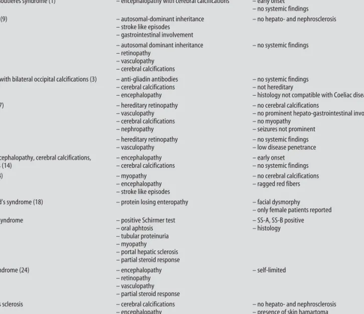
tele-angiectasia [12] have been genetically identified. Due to common clinical features, a common genetic origin has been considered for CRV, HRV and HERNS, and recently all these syndromes have been linked to the same locus on chromosome 3p21 [17]. Interestingly, the Aicardi-Goutieres syndrome, a further disease with progressive leukoencephalopathy, has been located to 3p21 [2], a re-gion to which also the polyglutamine disease spinocere-bellar ataxia 7 has been linked. We screened for polyglu-tamine proteins in one patient. Although we did not find polyglutamine proteins in our screen, we cannot exclude the possibility of a poly-repeat phenomenon other than glutamine-repeats as the causative defect in HSA. Table 2 Differential diagnosis of HSA
Disease Similarities with the presented HSA family Differences from the presented HSA family Aicardi-Goutieres syndrome (1) – encephalopathy with cerebral calcifications – early onset
– no systemic findings
CADASIL (9) – autosomal-dominant inheritance – no hepato- and nephrosclerosis
– stroke like episodes
– gastrointestinal involvement
CRV (28) – autosomal dominant inheritance – no systemic findings
– retinopathy
– vasculopathy
– cerebral calcifications
Epilepsy with bilateral occipital calcifications (3) – anti-gliadin antibodies – no systemic findings
– cerebral calcifications – not hereditary
– encephalopathy – histology not compatible with Coeliac disease HERNS (7) – hereditary retinopathy – no cerebral calcifications
– vasculopathy – no prominent hepato-gastrointestinal involvement
– cerebral calcifications – no myopathy
– nephropathy – seizures not prominent
HRV (25) – hereditary retinopathy – no systemic findings
– vasculopathy – low disease penetrance
Leukoencephalopathy, cerebral calcifications, – encephalopathy – early onset and cysts (14) – cerebral calcifications – no systemic findings
MELAS (4) – myopathy – no cerebral calcifications
– encephalopathy – ragged red fibers
– stroke like episodes
Rambaud’s syndrome (18) – protein losing enteropathy – facial dysmorphy
– only female patients reported
Sjögren syndrome – positive Schirmer test – SS-A, SS-B positive
– oral aphtosis – histology
– tubular proteinuria
– myopathy
– portal hepatic sclerosis
– partial steroid response
Susac syndrome (24) – encephalopathy – self-limited
– retinopathy
– vasculopathy
– partial steroid response
Tuberous sclerosis – cerebral calcifications – no hepato- and nephrosclerosis
– encephalopathy – presence of skin hamartoma
– seizures – no retinopathy
– autosomal-dominant inheritance
077_088_Winkler_JON_2675.indd 86
■ Treatment of HSA and cerebral vasculopathies So far no treatment for cerebral vasculopathies is known. Nevertheless, in all our patients, long-term use of low dose steroids proved helpful to control brain edema sur-rounding cerebral lesions. Acute cerebral mass lesions responded to steroid pulse therapy.
Assuming a role for microthrombi in the pathogen-esis of HSA, the use of antiplatelet agents can be tried. Low molecular heparin therapy led to stabilization of the course of the disease over about one year in one of our patients. Successful use of aspirin and pentoxifylline has previously been reported in a patient suffering from CRV [28].
In contrast, cyclophosphamide, azathioprin and methotrexate worsened the pre-existing hepatic dys-function without beneficial effects on overall disease progression in our patients. The use of immunosup-pressants also was not favorable in CRV [28] and should be discouraged due to its side-effects. Early recognition of HSA and other hereditary vasculopathies is thus of particular importance to protect patients from harmful and inappropriate drug treatment.
In summary, diagnosis of HSA can be suspected in
the presence of a clinical constellation with migraine-like headache, motor paresis, recurrent epileptic sei-zures, in combination with cerebral calcifications visible in CT-scans, retinal aneurysms in fluorescein angiogra-phy and a biopsy of an affected organ revealing angio-pathic changes including activated endothelial cells and microthrombi. It is important to note that HSA is not responsive to immunosuppressants that have the poten-tial to provoke additional damage to liver and kidneys. Administration of low molecular heparin can be tried to minimize the formation of microthrombi. When ce-rebral mass lesions provoke midline shift, steroid ap-plication has been shown to reduce the edema. Future work will help to elucidate the pathogenic mechanisms responsible for the endothelial damage in HSA.
■ Acknowledgement We wish to thank Prof. Dr. G. Spagnoli, Univer-sity of Basel, for establishing lymphoblast cell culture lines. We thank Dr. L.J. Trecjokas, PhD, MD, consultant neurologist, Santa Monica, CA, USA, for his help by providing clinical and radiological data. We fur-ther acknowledge Prof. Dr. E.W. Radue, Dept. of Neuroradiology, Prof. Dr. A. Tyndall, Dept. of Rheumatology, Prof. Dr. G. Marbet, Dept. of Hematology, University of Basel, for discussion. We also thank Prof. Dr. B. Lämmle, University of Bern, for measuring ADAMTS 13 prote-ase activity.
References
1. Aicardi J, Goutieres F (1984) A pro-gressive familial encephalopathy in infancy with calcifications of the basal ganglia and chronic cerebrospinal fluid lymphocytosis. Ann Neurol 15(1):49–54
2. Crow YJ, Jackson AP, Roberts E, van Beusekom E, Barth P, Corry P, Ferrie CD, Hamel BC, Jayatunga R, Karbani G, Kalmanchey R, Kelemen A, King M, Kumar R, Livingstone J, Massey R, McWilliam R, Meager A, Rittey C, Stephenson JB, Tolmie JL, Verrips A, Voit T, van Bokhoven H, Brunner HG, Woods CG (2000) Aicardi-Goutieres syndrome displays genetic hetero-geneity with one locus (AGS1) on chromosome 3p21. Am J Hum Genet 67(1):213–221
3. Gobbi G, Bouquet F, Greco L, Lam-bertini A, Tassinari CA, Ventura A, Zaniboni MG (1992) Coeliac disease, epilepsy, and cerebral calcifications. The Italian Working Group on Coeliac Disease and Epilepsy. Lancet 340(8817):439–443
4. Goto Y, Nonaka I, Horai S (1990) A mutation in the tRNA(Leu)(UUR) gene associated with the MELAS subgroup of mitochondrial encephalomyopa-thies. Nature 348(6302):651–653
5. Grand MG, Kaine J, Fulling K, Atkinson J, Dowton SB, Farber M, Craver J, Rice K (1988) Cerebroretinal vasculopathy. A new hereditary syndrome. Ophthal-mology 95(5):649–659
6. Gutmann DH, Fischbeck KH, Sergott RC (1989) Hereditary retinal vasculop-athy with cerebral white matter lesions. Am J Med Genet 34(2):217–220 7. Jen J, Cohen AH, Yue Q, Stout JT,
Vinters HV, Nelson S, Baloh RW (1997) Hereditary endotheliopathy with retinopathy, nephropathy, and stroke (HERNS). Neurology 49(5):1322–1330 8. Joutel A, Bousser MG, Biousse V,
La-bauge P, Chabriat H, Nibbio A, Maci-azek J, Meyer B, Bach MA, Weissenbach J, et al. (1993) A gene for familial hemi-plegic migraine maps to chromosome 19. Nat Genet 5(1):40–45
9. Joutel A, Corpechot C, Ducros A, Vahedi K, Chabriat H, Mouton P, Ala-mowitch S, Domenga V, Cecillion M, Marechal E, Maciazek J, Vayssiere C, Cruaud C, Cabanis EA, Ruchoux MM, Weissenbach J, Bach JF, Bousser MG, Tournier-Lasserve E (1996) Notch3 mutations in CADASIL, a hereditary adult-onset condition causing stroke and dementia. Nature 383(6602): 707–710
10. Labrune P, Lacroix C, Goutieres F, de Laveaucoupet J, Chevalier P, Zerah M, Husson B, Landrieu P (1996) Extensive brain calcifications, leukodystrophy, and formation of parenchymal cysts: a new progressive disorder due to dif-fuse cerebral microangiopathy. Neurol-ogy 46(5):1297–1301
11. Magistris MR, Kohler A, Pizzolato G, Morris MA, Baroffio A, Bernheim L, Bader CR (1998) Needle muscle biopsy in the investigation of neuromuscular disorders. Muscle Nerve 21(2):194–200 12. McAllister KA, Grogg KM, Johnson
DW, Gallione CJ, Baldwin MA, Jack-son CE, Helmbold EA, Markel DS, McKinnon WC, Murrell J, et al. (1994) Endoglin, a TGF-beta binding protein of endothelial cells, is the gene for he-reditary haemorrhagic telangiectasia type 1. Nat Genet 8(4):345–351 13. Moake JL (2002) Thrombotic
micro angiopathies. N Engl J Med 347(8):589–600
14. Nagae-Poetscher LM, Bibat G, Philip-part M, Rosemberg S, Fatemi A, Lac-erda MT, Costa MO, Kok F, Costa Leite C, Horska A, Barker PB, Naidu S (2004) Leukoencephalopathy, cerebral calci-fications, and cysts: new observations. Neurology 62(7):1206–1209
077_088_Winkler_JON_2675.indd 87
15. Niedermayer I, Graf N, Schmidbauer J, Reiche W (2000) Cerebroretinal vas-culopathy mimicking a brain tumor. Neurology 54(9):1878–1879
16. Niedermayer I, Reiche W, Graf N, Mes-tres P, Feiden W (2000) Cerebroretinal vasculopathy and leukoencephalopa-thy mimicking a brain tumor. Report of two early-onset cases with Fanconi’s anemia-like phenotypes suggesting an autosomal-recessive inheritance pat-tern. Clin Neuropathol 19(6):285–295 17. Ophoff RA, DeYoung J, Service SK,
Joosse M, Caffo NA, Sandkuijl LA, Ter-windt GM, Haan J, van den Maagden-berg AM, Jen J, Baloh RW, Barilla-LaBarca ML, Saccone NL, Atkinson JP, Ferrari MD, Freimer NB, Frants RR (2001) Hereditary vascular reti-nopathy, cerebroretinal vasculopathy, and hereditary endotheliopathy with retinopathy, nephropathy, and stroke map to a single locus on chromo-some 3p21.1–p21.3. Am J Hum Genet 69(2):447–453
18. Rambaud JC, Galian A, Touchard G, Morel-Maroger L, Mikol J, Van Ef-fenterre G, Leclerc JP, Le Charpentier Y, Haut J, Matuchansky C, et al. (1986) Digestive tract and renal small vessel hyalinosis, idiopathic nonarterio-sclerotic intracerebral calcifications, retinal ischemic syndrome, and phenotypic abnormalities. A new familial syndrome. Gastroenterology 90(4):930–938
19. Razvi SS, Bone I (2006) Single gene disorders causing ischaemic stroke. J Neurol 253(6):685–700
20. Schmidbauer JM, Voges M, Kasmann-Kellner B, Graf N, Henn W, Ruprecht KW (2000) Hereditary occlusive cerebroretinal vasculopathy in two sisters. Klin Monatsbl Augenheilkd 217(4):246–251
21. Seifried C, Sitzer M, Jen J, Auburger G (2005) HERNS. A rare, hereditary, multisystemic disease with cerebral microangiopathy. Nervenarzt 76(10): 1191–1192, 1194–1195
22. Siveke JT, Schmid H (2003) Evidence for systemic manifestations in cerebro-retinal vasculopathy. Am J Med Genet 123A(3):309
23. Storimans CW, Van Schooneveld MJ, Oosterhuis JA, Bos PJ (1991) A new autosomal dominant vascular reti-nopathy syndrome. Eur J Ophthalmol 1(2):73–78
24. Susac JO, Hardman JM, Selhorst JB (1979) Microangiopathy of the brain and retina. Neurology 29(3):313–316 25. Terwindt GM, Haan J, Ophoff RA,
Groenen SM, Storimans CW, Lanser JB, Roos RA, Bleeker-Wagemakers EM, Frants RR, Ferrari MD (1998) Clinical and genetic analysis of a large Dutch family with autosomal dominant vascular retinopathy, migraine and Raynaud’s phenomenon. Brain 121(Pt 2):303–316
26. Trottier Y, Lutz Y, Stevanin G, Imbert G, Devys D, Cancel G, Saudou F, Weber C, David G, Tora L, et al. (1995) Polygluta-mine expansion as a pathological epi-tope in Huntington’s disease and four dominant cerebellar ataxias. Nature 378(6555):403–406
27. Vahedi K, Massin P, Guichard JP, Miocque S, Polivka M, Goutieres F, Dress D, Chapon F, Ruchoux MM, Riant F, Joutel A, Gaudric A, Bousser MG, Tournier-Lasserve E (2003) Hereditary infantile hemiparesis, retinal arteriolar tortuosity, and leukoencephalopathy. Neurology 60(1):57–63
28. Weil S, Reifenberger G, Dudel C, Yousry TA, Schriever S, Noachtar S (1999) Cerebroretinal vasculopathy mimicking a brain tumor: a case of a rare hereditary syndrome. Neurology 53(3):629–631
29. Yanagawa S, Ito N, Arima K, Ikeda S (2002) Cerebral autosomal recessive arteriopathy with subcortical infarcts and leukoencephalopathy. Neurology 58(5):817–820
077_088_Winkler_JON_2675.indd 88