HAL Id: hal-02177524
https://hal.archives-ouvertes.fr/hal-02177524
Submitted on 19 Nov 2020HAL is a multi-disciplinary open access
archive for the deposit and dissemination of sci-entific research documents, whether they are pub-lished or not. The documents may come from teaching and research institutions in France or
L’archive ouverte pluridisciplinaire HAL, est destinée au dépôt et à la diffusion de documents scientifiques de niveau recherche, publiés ou non, émanant des établissements d’enseignement et de recherche français ou étrangers, des laboratoires
β-Lactoglobulin: Specificity and Impact on Bacterial
Location in Whey Protein Isolate
Faustine Gomand, Frédéric Borges, Justine Guerin, Sofiane El-Kirat-Chatel,
Gregory Francius, Dominique Dumas, Jennifer Burgain, Claire Gaiani
To cite this version:
Faustine Gomand, Frédéric Borges, Justine Guerin, Sofiane El-Kirat-Chatel, Gregory Francius, et al.. Adhesive Interactions Between Lactic Acid Bacteria and β-Lactoglobulin: Specificity and Impact on Bacterial Location in Whey Protein Isolate. Frontiers in Microbiology, Frontiers Media, 2019, 10, pp.1512. �10.3389/fmicb.2019.01512�. �hal-02177524�
Adhesive interactions between lactic acid bacteria and β-lactoglobulin:
specificity and impact on bacterial location in whey protein isolate
Faustine Gomand1, Frédéric Borges1, Justine Guerin1, Sofiane El-Kirat-Chatel2, Gregory
1
Francius2, Dominique Dumas3, Jennifer Burgain1, Claire Gaiani1*
2
1Laboratoire d’Ingénierie des Biomolécules (LIBio), Université de Lorraine, Vandœuvre-lès-Nancy,
3
France 4
2CNRS, Laboratoire de Chimie Physique et Microbiologie pour les Matériaux et l'Environnement
5
(LCPME), UMR 7564, Université de Lorraine, Villers-lès-Nancy, France 6
3Plateforme d'Imagerie et de Biophysique Cellulaire de Nancy (PTIBC IBISA- NANCY), UMS 2008,
7
IMOPA UMR 7365- Université de Lorraine, Vandœuvre-lès-Nancy, France. 8 * Correspondence: 9 Claire Gaiani 10 claire.gaiani@univ-lorraine.fr 11
Keywords: adhesion, lactic acid bacteria, dairy, β-lactoglobulin, high-throughput screening, 12
bacterial distribution, Atomic Force Microscopy (AFM), Confocal Laser Scanning Microscopy 13
(CLSM) 14
Abstract 15
In the last decade, there has been an increasing interest in the potential health effects associated with 16
the consumption of Lactic Acid Bacteria (LAB) in foods. Some of these bacteria such as Lactobacillus 17
rhamnosus GG (LGG) are known to adhere to milk components, which may impact their distribution 18
and protection within dairy matrices and therefore is likely to modulate the efficiency of their delivery. 19
However, the adhesive behavior of most LAB, as well as its effect on food structuration and on the 20
final bacterial distribution within the food matrix remain very poorly studied. Using a recently 21
developed high-throughput approach, we have screened a collection of 73 LAB strains for their 22
adhesive behavior towards the major whey protein β-lactoglobulin. Adhesion was then studied by 23
genomics in relation to common bacterial surface characteristics such as pili and adhesion-related 24
domain containing proteins. Representative adhesive and non-adhesive strains have been studied in 25
further depth through biophysical measurement using Atomic Force Microscopy (AFM) and a relation 26
with bacterial distribution in Whey Protein Isolate (WPI) solution has been established. AFM 27
measurements have revealed that bacterial adhesion to β-lactoglobulin is highly specific and cannot be 28
predicted accurately using only genomic information. Non-adhesive strains were found to remain 29
homogeneously distributed in solution whereas adhesive strains gathered in flocs. These findings show 30
that several LAB strains are able to adhere to β-lactoglobulin, whereas this had only been previously 31
observed on LGG. We also show that these adhesive interactions present similar characteristics and 32
are likely to impact bacterial location and distribution in dairy matrices containing β-lactoglobulin. 33
This may help with designing more efficient dairy food matrices for optimized LAB delivery. 34
1 Introduction 35
Adhesion is a major property of microorganisms which effectively impacts microorganism activities 36
as well as human health, and has been identified as a key factor involved in microorganism ecology. 37
Adhesion enables bacteria to stick to both biotic and abiotic surfaces. Adhesion to abiotic surfaces 38
leads to biofilm formation, which has been widely studied in relation to the food industry (Barnes et 39
al., 2001; Garrett et al., 2008; Notermans et al., 1991; Pontefract, 1991). Adhesion to biotic surfaces 40
enables bacteria to establish direct contact with mucous membranes, and especially the intestinal 41
epithelium, to colonize a host (Conway et al., 1987; Pizarro-Cerdá and Cossart, 2006; Servin and 42
Coconnier, 2003). Adhesion of pathogens is therefore considered to be a virulence factor as it facilitates 43
host invasion (Pizarro-Cerdá and Cossart, 2006; Proft and Baker, 2009). Amongst non-pathogenic 44
bacteria, adhesion is considered essential in order for probiotic bacteria to remain functional and 45
therefore provide health benefits to the host (Ouwehand et al., 2001; Quinto et al., 2014; Servin and 46
Coconnier, 2003). In the case of Gram-positive bacteria, bacteria-environment interactions such as 47
bacterial adhesion are mediated by sortase-dependent proteins (Comfort and Clubb, 2004; Maresso and 48
Schneewind, 2008), which are covalently anchored to the cell wall and possess an LPxTG like motif 49
at their C-terminal end (Schneewind and Missiakas, 2014). 50
Bacteria have also been shown to be able to adhere to food components, especially to meat 51
(Firstenberg-Eden, 1981; Piette and Idziak, 1989) and more recently to dairy components (Burgain et 52
al., 2014a; Gomand et al., 2018; Guerin et al., 2016). Bacterial adhesive interactions to food 53
components can compete with bacterial adhesion to the host (Sun and Wu, 2017). Therefore food 54
components such as milk fat globule membrane (Douëllou et al., 2017; Guerin et al., 2018b), milk 55
proteins (Halpin et al., 2008), and milk oligosaccharides (Lane et al., 2012) can play an anti-adhesive 56
role by decreasing bacterial adhesion to the intestine (Guerin et al., 2018b). Some food additives 57
including stabilizers (such as sucrose fatty acid esters) and colors (gardenia yellow, monascus pigment, 58
etc.) have also been found to feature similar effects (Islam et al., 2014). 59
In food matrices, adhesive interactions are likely to play an important part in bacterial spatial 60
distribution and viability during the structuration of the food matrix (Gomand et al., 2019). Adhesive 61
interactions occurring between the model strain Lactobacillus rhamnosus GG (LGG) and β-62
lactoglobulin is mediated by the pili produced by LGG cells on their surface (Guerin et al., 2016). 63
These interactions result in an increased encapsulation efficiency when using dairy components as well 64
as a higher resistance to gastric digestion for this strain (Burgain et al., 2013a, 2014b; Guerin et al., 65
2017). Adhesive interactions between genetically engineered Lactococcus lactis producing pili and 66
dairy components result in texture alteration in fermented milk (Tarazanova et al., 2018a) and can 67
modulate this strain distribution in cheese curd (Tarazanova et al., 2018b). Similarly, during curdling 68
and cheese ripening, bacterial cells mostly co-localize with fat globules or at the casein-fat interface, 69
which suggest adhesive interactions between fat and LAB strains (Laloy et al., 1996; Lopez et al., 70
2006). This is likely to play a role in lipolysis thus affecting the development of characteristic flavors 71
and textures during ripening (Laloy et al., 1996; Lopez et al., 2006). 72
However, the impact and technological interest of adhesive interactions is yet poorly documented and 73
largely remains to be investigated (Hickey et al., 2015). Adhesive interactions between bacterial 74
surface components and dairy components have only been studied for very few wild type strains, 75
namely LGG (Guerin et al., 2016), Lactobacillus amylovorus (Chumphon et al., 2016), and 76
Lactobacillus paracasei (De Bellis et al., 2010). This article goes one step forward in that direction by 77
applying the high-throughput screening method recently developed by Gomand et al. (2018) to a 78
collection of 73 LAB strains (for which genome sequence is available) in order to characterize their 79
potential adhesive behavior towards the major dairy protein β-lactoglobulin, to which the adhesive 80
behavior of the model strain LGG is already well-known (Burgain et al., 2013b, 2014b, 2015; Guerin 81
et al., 2016, 2018a). Two strains featuring extreme adhesive and non-adhesive behaviors have then 82
been studied in further depth through AFM. The AFM results were then studied in relation to Confocal 83
Laser Scanning Microscopy (CLSM) experiments, allowing to observe the spatial distribution of these 84
strains in Whey Protein Isolate (WPI) solution. 85
2 Material and methods 86
2.1 High-throughput screening 87
Adhesive interactions between bacteria and β-lactoglobulin were screened using the method recently 88
developed by Gomand et al. (2018) using an automated liquid handling system for 96-well microplates. 89
Briefly, this method consists in immobilizing the biomolecules of interest on the surface of 96 well 90
adherent microplates. Microplates are then washed with a blocking agent in order to remove all 91
unbound molecules and to block the remaining empty sites. The bacterial suspension is then added into 92
the wells and incubated for 1 h at 37 °C in order to allow bacterial adhesion to the immobilized 93
biomolecules. Non-adherent bacteria are removed by successive washes using the same blocking agent. 94
The amount of immobilized bacteria is measured through bacterial growth monitoring (turbidity 95
measurements at 595 nm) after the addition of MRS culture growing medium (De Man et al., 1960) in 96
the wells. The higher the initial quantity of bound bacteria, the earlier the growth starts. Adjustments 97
made to this protocol are listed below. 98
2.1.1 Bacterial strains and cultures 99
A list of the 73 screened LAB strains is given in Supplementary data 1 (S1). This collection of strains 100
has previously been studied for their genomics and surface properties (Sun et al., 2015). The model 101
strain Lactobacillus rhamnosus GG ATCC53103 (LGG wild type, “WT”) and the mutant strain LGG 102
spaCBA CMPG 5357 impaired in pili synthesis, which adhesive properties of both are well-known 103
(Guerin et al., 2016; Lebeer et al., 2012; Tripathi et al., 2012, 2013) were respectively used as positive 104
(adherent) and negative (non-adherent) control strains. 105
For each series of experiments, a 96-well microplate previously stored at -80 °C was thawed and 106
replicated on working microplates using 50 µL of bacterial suspension to inoculate 150 µL of MRS by 107
well. The working microplates were incubated at 30 °C two days before the adhesion assay. During 108
the adhesion assay, microplates were only centrifuged once at 1,642 g for 20 min, emptied and the 109
resulting cell pellets were resuspended in 200 µL of PBS adjusted at pH 6.8. Triplicates on independent 110
cultures were performed as well as duplicates by strain on each plate (6 repetitions for control strains). 111
2.1.2 Preparation of the β-lactoglobulin solution and microplate coating 112
Beta-lactoglobulin (Sigma-Aldrich Co. LLC, St Louis, MO, USA) was prepared in solution (1% w/w) 113
as described by Gomand et al. (2018). 114
2.1.3 Bacterial growth monitoring 115
Adhesion and growth monitoring were done according to Gomand et al. (2018). The incubation 116
temperature was changed to 30 °C in order to match the diversity of the growing conditions for all 117
strains (Gomand et al., 2018). Bacterial growth was monitored through OD595nm measurements over 48
118 h. 119
2.1.4 Data processing 120
2.1.4.1 Strain growth comparison 121
The times at which the apparent bacterial growth starts (tstart) were monitored such as described by
122
Gomand et al. (2018). The higher these time values are, the later the growth starts i.e. the fewer bacteria 123
have adhered i.e. the lower the affinity. These values were averaged on all series of experiments and 124
standard deviations are computed. Strains were compared to one another based on their Minimum 125
Adhesion Value (MAV) corresponding to the difference between the smallest tstart (highest adhesion)
126
obtained on a control without β-lactoglobulin and the highest tstart (lowest adhesion) obtained on
β-127 lactoglobulin: 128 𝑴𝒊𝒏𝒊𝒎𝒖𝒎 𝑨𝒅𝒉𝒆𝒔𝒊𝒐𝒏 𝑽𝒂𝒍𝒖𝒆 (𝑴𝑨𝑽) = (𝒕𝒔𝒕𝒂𝒓𝒕)𝒂𝒗𝒆𝒓𝒂𝒈𝒆 − 𝝈 𝑪𝑶𝑵𝑻𝑹𝑶𝑳− (𝒕𝒔𝒕𝒂𝒓𝒕)𝒂𝒗𝒆𝒓𝒂𝒈𝒆 − 𝝈 𝛃 𝐋𝐀𝐂 129
Where σ stands for standard deviation. A strain is considered to adhere to β-lactoglobulin if its MAV 130
is significantly superior to zero for all three series of experiments. 131
2.1.4.2 Functional domain prediction for the bacterial surface proteome 132
Bacterial surface proteins featuring LPxTG motif were predicted using the InterPro resource, that 133
provides functional analysis of protein sequences by classifying them into families and predicting the 134
presence of domains and important sites (Finn et al., 2017). Protein sequences were obtained from Sun 135
et al. (2015) and were scanned against InterPro’s signatures using the software package InterProScan 136
(Jones et al., 2014). Gene sequence resemblance with known domains was performed using the Basic 137
Local Alignment Search Tool resource (BLAST), according to Altschul et al. (1990). 138
2.1.4.3 Statistical analysis 139
Statistical analysis were performed via t-tests and Tukey tests (parametric) for normal data and 140
Wilcoxon-Mann Whitney and Steel-Dwass tests (non-parametric) for data that did not fit normal 141
distribution using Kyplot software (Kyens Lab Inc.). 142
2.2 Adhesive interactions between bacteria and β-lactoglobulin characterized through Atomic 143
Force Microscopy 144
Protocols used in this part have been adapted from Guerin et al. (2018a). Briefly, this method consists 145
in immobilizing the bacterial strains of interest on functionalized gold-coated mica by deposing the 146
bacterial suspension during 15 h at 4 °C (pH 6.8). The mica is rinsed with PBS (pH 6.8) before use. 147
Milk proteins are prepared in distilled water (1% w/w) and adsorbed on modified AFM probes (gold 148
coated and with NH2-terminated PEG linker) by immersion for 15 h at 4 °C and then rinsed with milli-149
Q-grade water before use. Force measurements are performed at room temperature in PBS buffer (pH 150
6.8). AFM force distance curves are obtained by following the cantilever deflection as a function of 151
the vertical displacement of the piezoelectric scanner with a scan speed of 400 mm/s. Adjustments to 152
this protocol are listed below. 153
2.2.1 Bacterial cultures 154
Cultures were prepared according to Guerin et al. (2018a). Precultures of Lactobacillus aquaticus DSM 155
21051 and Lactobacillus sharpeae DSM 20505 were prepared by inoculating 9 mL of MRS broth with 156
100 µL of bacterial stock and grown overnight at 37 °C. These precultures were used to inoculate 9 157
mL of fresh MRS broth the next day and the growth was performed at 37 °C until an optical density of 158
1.2 was reached at 660 nm (for about 8 h). Cultures were then centrifuged at 3,000 g for 10 min at 159
room temperature. Pellets were suspended in 1 mL of PBS (pH 6.8). 160
2.2.2 Preparation of bacteria-coated mica and protein-coated tips 161
According to Guerin et al. (2018a), a mica coated with a gold layer functionalized with a NH2
-162
terminated PEG-linker (Novascan, Ames, Iowa, USA) was used, as well as AFM probes with 163
borosilicate glass particle (2 µm), coated with gold and modified with NH2 terminated PEG linker
164
(Novascan, Ames, Iowa, USA). The bacterial suspension is deposed on mica at 4 °C and left overnight 165
(pH 6.8). Preparation of the β-lactoglobulin and Bovine Serum Albumine (BSA) 1% (w/w) solutions 166
(Sigma-Aldrich Co. LLC, St Louis, MO, USA) was done according to Guerin et al. (2018a). Probes 167
tips were left to incubate overnight at 4°C in wells containing 1 mL of the β-lactoglobulin or BSA 168
solutions to maximize protein adsorption. β-lactoglobulin was the candidate protein tested and BSA 169
was the negative control. 170
2.2.3 AFM measurements 171
Protocol followed is described by Guerin et al. (2018a). Force-volume measurements are performed at 172
room temperature in PBS buffer (pH 6.8) using a Bruker Bioscope Resolve atomic force microscope 173
(Bruker corporation, Santa Barbara, CA) mounted on an inverted microscope (DMi8, Leica 174
microsystems). The spring constants of the cantilevers was measured using the thermal noise method 175
and found to be 0.01 N m−1. Force distance curves were recorded between the bacteria deposited on
176
functionalized mica and the probe coated with β-lactoglobulin or BSA. Three adhesion force maps (20 177
µm × 20 µm, 256 force curves) were recorded for each protein-bacteria interaction analysis. Data 178
analysis was performed using the Nanoscope Analysis software from Bruker (Santa Barbara, CA, 179
USA) and the last peak was calculated for each curve before plotting adhesion forces and last rupture 180
length histograms. The last peak is used for analysis instead of the maximum peak in order to 181
characterize the last interacting point between the β-lactoglobulin and the cell receptor and not the 182
unfolding of a biomolecular domain. 183
2.3 Adhesive interactions imaged by confocal microscopy 184
The cultures were prepared as described in 2.2.1., then centrifuged at 3,000 g for 10 min at room 185
temperature. Pellets were suspended in 10 mL of WPI solution (15 %, w/w). The WPI solution was 186
prepared using PRODIET 90 S (Ingredia, Arras, France) that is a soluble milk protein isolate containing 187
native whey proteins including β-lactoglobulin. One milliliter of resuspended cells was stained with 188
the LIVE/DEAD BacLight viability kit (1:200 v/v; LIVE/DEAD BacLight viability kit was prepared 189
according to the procedure described for the kit L13152 by ThermoFisher Scientific). Two hundred 190
microliters of LAB suspension (same conditions as 2.2.1) were introduced on chambered glass slides 191
(Nunc Lab-Tek, ThermoFisher Scientific). Confocal Laser Scanning Microscopy (CLSM) images were 192
taken using a Leica TCS SP5-X-AOBS confocal laser scanning microscope (Leica Microsystems CMS 193
GmbH, Mannheim, Germany) equipped with WLL lasers. The objective lens used was a HCX PL APO 194
CS 100 × 1.40 (oil immersion). The excitation wavelength was 488 nm and emission bandwidth was 195
of 495-510 nm for SYTO 9 and 600-620 nm for propidium iodide. Two independent repetitions were 196
performed and approximately 20 representative images were acquired for each sample. 197
3 Results 198
3.1 Identification of strains adhesive to β-lactoglobulin 199
Most strains were found not to be adhesive to β-lactoglobulin as the average MAV calculated on the 200
73 strains was negative (-180 ± 22) although higher than the MAV of the negative control LGG spaCBA 201
(-386), known to be non-adhesive to β-lactoglobulin (Guerin et al., 2016). The microplate adhesive 202
assays revealed four adhesive candidates to β-lactoglobulin amongst the 73 strains tested: 203
Lactobacillus aquaticus DSM 21051 (MAV = 61.5), Lactobacillus murinus DSM 20452 (MAV = 204
12.8), Lactobacillus plantarum DSM 13273 (MAV = 12.6), Lactobacillus brantae DSM 23927 (MAV 205
= 6.97), although these strains were still less adhesive than the positive control LGG WT (MAV = 206
104). Nine strains were also found to have a MAV inferior to the one of the negative control LGG 207
spaCBA: Lactobacillus sharpeae DSM 20505 (MAV = -857), Lactobacillus kefiri DSM 20587 (MAV 208
= -787), Lactobacillus similis DSM 23365 (MAV = -780), Lactobacillus pobuzihii DSM 28122 (MAV 209
= -617), Lactobacillus namurensis DSM 19117 (MAV = 516), Lactobacillus satsumensis DSM 16230 210
(MAV = -490), Pediococcus parvulus DSM 20332 (MAV = -477), Lactobacillus senmazukei DSM 211
21775 (MAV = -404), Lactobacillus lindneri DSM 20690 (MAV = -387). The MAV for all strains are 212
listed as supplementary material (S1). 213
3.2 Biophysical deciphering of bacterial adhesive interaction with β-lactoglobulin through 214
AFM 215
The adhesive interactions between β-lactoglobulin and the strains at the extremes of the adhesion 216
spectrum, L. aquaticus DSM 21051 (the most adhesive strain) and L. sharpeae DSM 20505 (the least 217
adhesive strain) were studied through AFM, in order to characterize them in further depth. Only two 218
strains were chosen to precise our understanding of the interaction mechanism of the LAB surface with 219
β-lactoglobulin since AFM is not a suitable method for screening of large populations. This is why we 220
decided to select only the two strains at the extreme of the adhesion spectrum for this analysis. BSA 221
was used as a negative control as LAB strains have previously been found to feature low adhesion to 222
it (Gomand et al., 2018; Guerin et al., 2016). The percentages of adhesive events (frequencies) observed 223
between L. aquaticus DSM 21051 and the two proteins, β-lactoglobulin and BSA, were respectively 224
of 82.6 ± 7.1% and 27.6 ± 10.4% (Figure 1A1). The frequencies of adhesive events observed between
225
L. sharpeae DSM 20505 and the same two proteins were respectively of 3.4 ± 1.5% for β-lactoglobulin 226
and 2.5 ± 0.6 % for BSA (Figure 1B1). Typical force-distance curves obtained for the interactions
227
occurring between the two strains and the AFM probes functionalized with the two proteins are 228
presented, i.e. L. aquaticus DSM 21051 and -lactoglobulin (Figure 1A2), L. aquaticus DSM 21051
229
and BSA (Figure 1A3), L. sharpeae DSM 20505 and -lactoglobulin (Figure 1B2), and L. sharpeae
230
DSM 20505 and BSA (Figure 1B3). During the withdrawal of functionalized β-lactoglobulin-coated
231
probe from the surface of L. aquaticus DSM 21051 several specific adhesive events occur (Figure 232
1A2), whereas more than 70% of the curves observed for BSA-coated probes did not feature any
233
adhesive event (Figure 1A3). Moreover, the few adhesive events observed between BSA and L.
234
aquaticus DSM 21051 appeared to be random and therefore could not be associated to any specific 235
interaction (Figure 1A3). Almost no adhesive event was observed for both BSA- and
β-lactoglobulin-236
coated probes on L. sharpeae DSM 20505 cells (Figures 1B1, B2, B3). These results are consistent with
237
those obtained using the screening method: L. aquaticus DSM 21051 significantly adheres to -lac 238
whereas poor adhesion was observed for L. sharpeae DSM 20505. Retraction curves recorded between 239
L. aquaticus DSM 21051 and β-lactoglobulin attest the specificity of occurring adhesive interactions, 240
which would happen according to a lock and key mechanism (Figure 2A). 3D-AFM images recorded 241
on mica attest of the good coverage of L. aquaticus DSM 21051 and therefore that adhesive events 242
recorded did occur between L. aquaticus DSM 21051 cells and β-lactoglobulin-coated probes (Figure 243
2B). The biophysical properties of the adhesion between L. aquaticus DSM 21051 and -lac were 244
analyzed using additional force parameters including adhesion forces (Figure 2C) and final rupture 245
length (Figure 2D). Retraction curves exhibited adhesion forces averaging around 1.43 ± 0.03 nN. 246
Final rupture length averaged around 0.90 ± 0.03 µm. These results will be compared with those of 247
LGG WT and the mutant strains LGG spaCBA and welE in the discussion section. 248
3.3 Impact of adhesive interactions on bacterial distribution in whey protein isolate probed by 249
confocal microscopy 250
L. aquaticus DSM 21051, L. sharpeae DSM 20505, LGG WT and LGG spaCBA were first imaged in 251
MRS to make sure that they were originally homogeneously distributed (Figure 3A1, 3B1 and 4A1,
252
4B1). Live cells of L. aquaticus DSM 21051 were found to aggregate in the WPI solution whereas L.
253
sharpeae DSM 20505 live cells remained homogeneously distributed (Figure 3A2 and 3B2). This is
254
consistent with the adhesive properties of the control strains: LGG WT (positive control) aggregate in 255
the WPI solution whereas LGG spaCBA remained homogeneously distributed (Figure 4A2 and 4B2).
256
Dead bacterial cells or cells with a damaged membrane gathered in flocs for all 4 strain types (data not 257
shown). 258
3.4 Relation between bacterial adhesion to β-lactoglobulin and predicted bacterial surface 259
characteristics 260
3.4.1 Presence of pili genes clusters (PGCs) 261
Predicted bacterial surface characteristics were analyzed in relation to the results of the adhesive assays 262
in order to delineate gene candidates predicted to encode surface proteins that could be involved in 263
bacterial adhesion to β-lactoglobulin. Amongst the 73 strains tested, 32 of them possessed at least one 264
sortase-dependent pilus gene cluster (PGC) and therefore were predicted to express pili on their surface 265
(Sun et al., 2015). The average MAV of these 32 strains was -163 ± 33.2 whereas the average MAV 266
of the 41 non-piliated strains was -194 ± 30.1. Amongst the 32 strains presenting PGCs, 16 possessed 267
PGCs similar to LGG pilus clusters in terms of gene order, that is, a cluster of three pilin genes and 268
one pilin-specific sortase gene (Sun et al., 2015). The MAV of these 16 strains was -165 ± 53.8 whereas 269
the MAV of the 16 strains with PGCs different from LGG was -160 ± 38.8. Although a mean 270
comparison of the MAV for strains featuring PGCs compared to non-piliated strains would suggest 271
that the presence of PGCs fosters adhesion to β-lactoglobulin, this was not supported statistically. No 272
difference could be observed between strains featuring PGCs similar to LGG WT’s and PGCs different 273
from LGG WT’s. The number of PGCs, sortase enzymes or proteins with LPxTG motif (listed for all 274
strains in S1) were not found either to impact strain adhesion to β-lactoglobulin (data not shown). 275
3.4.2 Predicted protein domains candidates for mediating bacterial adhesion to β-lactoglobulin 276
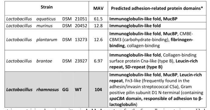
More predicted surface characteristics were analyzed for the four strains found to be adhesive to β-277
lactoglobulin. Predicted protein domains featuring LPxTG motif found for each strain are listed in 278
Table 1. Strains were analyzed for gene sequence resemblance with the spaCBA domain, known to be 279
responsible for adhesion to β-lactoglobulin for LGG WT (Guerin et al., 2016) but no homologue 280
sequence could be identified for any of the four adhesive strains. All strains are predicted to feature 281
immunoglobulin-like (Ig-like) fold domains, which are usually involved in binding or molecular 282
recognition processes (Bodelón et al., 2013). Other and more specific adhesion-related domains present 283
on the four adhesive strains studied as well as on LGG WT include MucBP (mucin-binding), 284
CBME/CBM3 (carbohydrate-binding), figbrinogen- and collagen-binding domains, cysteine- and 285
leucine-rich domains, and SD-repeat B-domain. Most of these domains are present once in the genome 286
of the adhesive strains (L. plantarum DSM 13273 is the only adhesive strain presenting 3 MucBP 287
domains) and are not repeated within a given protein. 288
The MucBP domain is the only domain with a known adhesive-related function (apart from the Ig-like 289
fold domain) which could be identified on L. aquaticus DSM 21051, the most adhesive strain to β-290
lactoglobulin. MucBP domains have been found predominantly in lactobacilli found naturally in 291
intestinal niches, which suggests that they play an important role in establishing host-microbial 292
interactions in the gut by binding mucus (Roos and Jonsson, 2002; Tassell and Miller, 2011). L. 293
plantarum DSM 13273 is the strain featuring the highest number of adhesion-related domains in its 294
genome (Table 1). This is also the only strain out of the four presenting fibrinogen- and collagen-295
binding domains. The fibrinogen-binding domain has been found to accommodate linear peptides with 296
a certain degree of ligand sequence variability (Ponnuraj et al., 2003) and therefore might be able to 297
interact with β-lactoglobulin. L. brantae DSM 23927 features leucine-rich repeats (LRRs) and SD-298
repeat (Sdr) domains (Table 1), both of them susceptible to play a role in adhesive interactions to β-299
lactoglobulin. LRRs have been found to provide a structural framework for the formation of protein-300
protein binding and interactions (Gay et al., 1991; Kobe and Kajava, 2001) and are likely to allow a 301
broad range of ligands (Kobe and Kajava, 2001). Sdr-repeat domains are surface proteins that play an 302
important role in Staphylococcus aureus adhesion and pathogenesis (McCrea et al., 2000; Wang et al., 303
2013). The protein containing Sdr-repeat domains may therefore be a good candidate for mediating 304
adhesion to β-lactoglobulin for the strain L. brantae DSM 23927. No other adhesion-related domain 305
than the Ig-like fold domain was identified on L. murinus DSM 20452 (Table 1), which would suggest 306
that the protein containing this domain would likely be the one involved in adhesive interactions with 307
β-lactoglobulin. 308
4 Discussion 309
The aim of this study was to evaluate and characterize adhesive interactions occurring between lactic 310
acid bacteria and β-lactoglobulin. A collection of 73 LAB strains was screened for their adhesive 311
behavior towards β-lactoglobulin and strains at the extreme of the adhesion spectrum i.e. a highly 312
adhesive and a poorly adhesive strains were studied in further depth. 313
Only four strains out of 73 were found to present adhesive affinities towards β-lactoglobulin. Therefore, 314
adhesion to β-lactoglobulin appears not to be a common characteristic of the LAB group. The 315
consequences of these adhesive interactions, when they occur, are not fully understood. However, it 316
could be hypothesized that strains featuring adhesive affinities towards whey proteins would be lost 317
during the drainage step of cheese manufacturing processes, alongside with whey expulsion from the 318
cheese network. It would be interesting to test the affinity of this same strain collection to other food 319
components in future work, in order to dispose of more comparison points to our study and to get a 320
better understanding of the importance of adhesion to β-lactoglobulin compared to adhesion to other 321
food components. Currently, the rare existing studies discussing bacterial adhesion to food components 322
other than β-lactoglobulin concern up to four strains at most at a time (Chumphon et al., 2016; De 323
Bellis et al., 2010; Tarazanova et al., 2017, 2018b, 2018a; Utratna et al., 2017), therefore failing to 324
provide an overview of adhesion to food components amongst wide bacterial groups such as the LAB 325
group. 326
The study performed by Tarazanova et al. (2017) is the only one to our knowledge that compares the 327
adhesion level of a wide number of strains (55) to food (casein-derived) components, however these 328
strains are all of the same species, Lactoccocus lactis. Out of 55, 30 to 40 strains presented adhesive 329
affinities towards casein-derived components, depending on their growth phase, and strains isolated 330
from a dairy environment presented much stronger binding of milk proteins versus strains isolated from 331
plants, suggesting a selective advantage (Tarazanova et al., 2017). However, this was not confirmed in 332
our case, as the four strains out of 73 that were originally isolated from dairy products, i.e. Lactobacillus 333
casei DSM 20011, Lactobacillus paracasei subsp. tolerans DSM 20258, Lactobacillus bifermentans 334
DSM 20003, and Lactobacillus kefiri DSM 20587, did not present more adhesive affinities towards β-335
lactoglobulin in average than the strains isolated from nondairy sources (data not shown). 336
The strain found to be the most adhesive to β-lactoglobulin, L. aquaticus DSM 21051, exhibited a 337
specific adhesive behavior when studied by AFM. The signature of the observed retraction curves was 338
identified as specific of biomolecules stretching, suggesting that the surface of L. aquaticus DSM 339
21051 features a strong affinity towards -lac. This has also been shown previously for the model strain 340
LGG WT by our team as well as for the mutant strain LGG welE, expolysaccharide-depleted and 341
known to adhere more to β-lactoglobulin than LGG WT due to its increased pili exposure (Guerin et 342
al., 2016, 2018a). A contrario, L. sharpeae DSM 20505 which screening results show not to adhere to 343
β-lactoglobulin presented retraction curves characteristic of a lack of adhesion to -lac when studied 344
by AFM (frequency of adhesive events was inferior to 5 %). Similarly, our team demonstrated 345
previously this same fact for the model strain non-adhesive to β-lactoglobulin, LGG spaCBA (Guerin 346
et al., 2016). Comparative results are presented in Table 2. 347
The adhesive behavior of L. aquaticus DSM 21051 towards -lactoglobulin appears relatively close to 348
the one of LGG welE in terms of frequency of adhesive events. The high specificity of the adhesion 349
phenomenon occurring between L. aquaticus DSM 21051 and β-lactoglobulin is highlighted by the 350
fact that the frequency of adhesion is almost twice as high as the one characterizing adhesive 351
interactions between LGG WT and β-lactoglobulin, whereas the frequency of adhesion of L. aquaticus 352
DSM 21051 on BSA is almost 4 times lower than the one occurring between LGG welE and BSA. The 353
mean adhesion force recorded on the last peak is also 3 times higher than the mean adhesion force 354
recorded for LGG WT and β-lactoglobulin, and higher than the highest adhesion force recorded on the 355
last peak for LGG welE and β-lactoglobulin, reaffirming the idea of a very strong specificity and 356
adhesion strength. When comparing the length of biomolecules stretched by adhesive interactions with 357
β-lactoglobulin, L. aquaticus DSM 21051 and LGG welE both exhibit molecules stretched up to 1µm 358
i.e. 3 times longer than the molecule stretched in the case of LGG WT (Table 2). The molecule 359
mediating adhesive interactions with β-lactoglobulin in the case of L. aquaticus DSM 21051 is 360
therefore comparable in length to LGG pili when stretched, which may explain the higher specificity 361
and adhesion strength found for L. aquaticus DSM 21051 compared to LGG WT, which pili are 362
partially hidden within the expolysaccharides layer (Guerin et al., 2016). 363
On the other hand, the frequency of adhesive events observed between L. sharpeae DSM 20505 and 364
β-lactoglobulin is inferior to 5 % and similar to the frequency of adhesive events observed on BSA for 365
both this strain and L. aquaticus DSM 21051. The frequency of adhesive events recorded when using 366
BSA-coated probes is also 4 times lower for L. sharpeae DSM 20505 than for LGG spaCBA (negative 367
control). Overall, L. sharpeae DSM 20505 has demonstrated very poor adhesive capacities towards β-368
lactoglobulin. However, when analyzed for predicted adhesion-related protein domains, this strain 369
revealed a total of 23 adhesion-related domains, 8 of which being different, including MucBP and 370
Gram positive pilin subunit D1 N-terminal, although no sequence homologue to the spaCBA domain 371
was found (data not shown). The spaCBA domain is known to mediate adhesion to β-lactoglobulin for 372
the piliated strain LGG WT (Guerin et al., 2016). This confirms that adhesive interactions with β-373
lactoglobulin are specific, and cannot be predicted accurately using only genomic predictions (the 374
functions of these domains may not be accurately predicted or they may not be expressed). 375
The gathering behavior observed by CLSM for the adhesive strains in the WPI solution also pledges 376
in favor of a specific bacterial adhesion to β-lactoglobulin for L. aquaticus DSM 21051. CLSM results 377
These observations are important as it was evidenced recently that physical properties of dairy 379
products, such as viscosity and gel hardness, are affected by bacterial surface properties in the case of 380
surface-engineered strains (Tarazanova et al., 2018a). In light of our results, it would be interesting to 381
see if that is also the case for wild strains presenting different surface properties inducing different 382
adhesive behaviors. Some peptides shown to be linked to bacterial aggregation were also recently 383
evidenced to be able to promote bacterial adhesion to functionalized surfaces and Caco-2-cells (Okochi 384
et al., 2017). This typical behavior was responsible for observed enhanced interactions between LAB 385
and the host intestinal mucosa (Okochi et al., 2017). Adhesive interactions with β-lactoglobulin leading 386
to the aggregation of L. aquaticus DSM 21051 and LGG WT cells might therefore be considered for 387
further study in order to determine whether they would promote such kind of behavior as well. 388
This work was performed in the continuity of previous studies, in which a method was developed 389
allowing screening a wide number of strains for their adhesive affinities towards biomolecules such as 390
dairy food components (Gomand et al., 2018), and which identified the bacterial surface molecules 391
(pili) involved in the adhesion of LGG to dairy components using AFM (Guerin et al., 2016). The 392
present study sought to go beyond bacterial species differences in revealing common adhesive 393
characteristics of Lactic Acid Bacteria in relation to dairy food components such as β-lactoglobulin. 394
We first looked for LAB species featuring adhesive affinities for β-lactoglobulin, then focused on the 395
molecular characteristics of this adhesion. We observed adhesion to β-lactoglobulin for few LAB (less 396
than 6% of our collection). However, for those which did feature adhesive affinities, some common 397
characteristics were pointed out that matched the characteristics previously identified on the model 398
strain LGG. These characteristics include the specificity of the affinity, as well as the impact on 399
bacterial spatial distribution in the matrix. The major findings of the present paper are that (i) Adhesion 400
to whey proteins is apparently not a common characteristic to the LAB group (few strains presented 401
adhesive affinities towards lactoglobulin), (ii) Strains featuring adhesive affinities towards β-402
lactoglobulin present common adhesive characteristics (specific β-lactoglobulin-adhesion domains 403
related to the specificity of the AFM signature), and (iii) Adhesion to β-lactoglobulin was shown to 404
strongly influence bacterial distribution in dairy matrices featuring this component (adhesive bacteria 405
gathered in flocs in whey matrices whereas non-adhesive bacteria distribute more homogeneously), 406
and could therefore modulate their accessibility and later delivery when designing functional foods 407
containing LAB with potential associated health effects. 408
According to these findings, food matrices could play a protective role on bacteria by influencing their 409
spatial distribution, which may prove especially useful for probiotic bacteria. Indeed, as bacteria 410
adhering to a component have been found to flocculate in the food matrix containing this component, 411
this could result in later heterogeneous delivery in the gastro-intestinal tract (GIT) which would impact 412
host colonization, but may also better protect bacterial survival until they reach the GIT. These findings 413
also pave the road to future experiments aiming generalizing bacterial adhesion characteristics to broad 414
bacterial groups, thus helping with practical food matrix design. It would therefore be interesting to 415
study the potential protective effect of components to which bacteria are adherent during critical steps 416
of the food manufacturing process, such as spray-drying during probiotic milk powder production. 417
Author contributions 418
FG, JG, JB, FB, CG conceived the research. FG, JG, JB, SEKC, DD, GF carried out the experiments. 419
FG, JB, JG, SEKC, DD, GF analyzed the data. FG, JG, JB wrote the manuscript. All authors 420
commented on the manuscript. 421
Funding 422
This article has been written as part of a LUE project (Lorraine Université d’Excellence). The authors 423
would like to thank the LUE initiative for having provided fundings and supported this work. 424
The authors declare no competing interest. 425
Acknowledgment 426
The wild type strain LGG ATCC53103 (WT) and the derivative mutant strains spaCBA CMPG 5357 427
(impaired in pili synthesis) were kindly provided by Dr Sarah Lebeer (Centre of Microbial and Plant 428
Genetics, K.U. Leuven, Leuven, Belgium, and Department of Bioscience Engineering, University of 429
Antwerp, Antwerp, Belgium). 430
Supplementary material 431
The Supplementary Material for this article can be found online at: … 432
Figure captions 433
Figure 1: Comparison of the adhesive properties of two strains (Lactobacillus aquaticus DSM 21051, 434
Lactobacillus sharpeae DSM 20505) for whey proteins isolates probed by Atomic Force Microscopy 435
(AFM): frequency of adhesive events occurring between whey proteins and L. aquaticus DSM 21051 436
(A1) and L. sharpeae DSM 20505 (B1) and representative examples of retraction curves obtained for
437
force measurements between L. aquaticus DSM 21051 and β-lactoglobulin (A2), L. aquaticus DSM
438
21051 and BSA (A3), L. sharpeae DSM 20505 and β-lactoglobulin (B2), and L. sharpeae DSM 20505
439
and BSA (B3).
440 441
Figure 2: Schematic description of Atomic Force Microscopy (AFM) with protein-coated tips and 442
bacteria-coated mica (A) 3D-AFM image of Lactobacillus aquaticus DSM 21051 recorded in liquid in 443
phosphate buffered saline (B) Interactions between β-lactoglobulin and L. aquaticus DSM 21051 444
explored by force measurement using atomic force microscopy: adhesions forces (C) and final rupture 445
length (D). Averages of adhesion forces and rupture lengths are precised in (C) and (D) with standard 446
errors. 447
448
Figure 3: Spatial distribution of L. aquaticus DSM 21051 and L. sharpeae DSM 20505 in MRS culture 449
medium [A1 and B1] and in whey protein isolate (WPI) solution [A2 and B2], imaged by confocal laser
450
scanning microscopy (CLSM). Bacterial concentration is 107 u.f.c./ mL. Bacteria cells are represented
451
in green on this figure whether they are viable or damaged (no difference is made here that would 452
depend on bacterial status). 453
Figure 4: Spatial distribution of LGG WT and LGG spaCBA in MRS culture medium [A1 and B1] and
454
in whey protein isolate (WPI) solution [A2 and B2], imaged by confocal laser scanning microscopy
455
(CLSM). Bacterial concentration is 107 u.f.c./ mL. Bacteria cells are represented in green on this figure
456
whether they are viable or damaged (no difference is made here that would depend on bacterial status). 457
459
Strain MAV Predicted adhesion-related protein domains* Lactobacillus aquaticus DSM 21051 61.5 Immunoglobulin-like fold, MucBP
Lactobacillus murinus DSM 20452 12.8 Immunoglobulin-like fold
Lactobacillus plantarum DSM 13273 12.6 Immunoglobulin-like fold, MucBP, CMBE-CBM3 (carbohydrate-binding), fibrinogen-binding, collagen-binding
Lactobacillus brantae DSM 23927 6.97 Immunoglobulin-like fold, Collagen-binding surface protein Cna-like (type B), Leucin-rich repeat, SD-repeat (type B)
Lactobacillus rhamnosus GG WT 104
Immunoglobulin-like fold, MucBP, Leucin-rich repeat, Fn3-like (frequently found in the adhesin/invasin streptococcal C5a), Gram positive pilin subunit D1 N-terminal (containing spaCBA domain, responsible of adhesion to β-lactoglobulin)
*proteins containing-domains written in bold characters for the four adhesive strains are likely to 460
mediate adhesive interactions with β-lactoglobulin 461
Table 1: Predicted proteins domains with LPxTG motif which may play a role in bacterial adhesion to 462
β-lactoglobulin. Domains present on L. rhamnosus GG (known to be adhesive to β-lactoglobulin) are 463
included as a reference. Proteins sequences used were those provided by Sun et al. (2015). 464
465
Adhesive events (%) Adhesion forces to β-lac (nN) Length of the stretched biomolecule (µm) Reference To β-lac To BSA Strains highly adhesive to β-lac L. aquaticus 82.6 ± 7.1 27.6 ± 10.4 1.43 ± 0.03 0.90 ± 0.01 / LGG WT 51.4 ± 9.9 13.1 ± 0.8 [0.13 ; 0.81] ± 0.01 0.39 ± 0.02 Guerin et al., 2016 LGG welE 84.1 ± 3.0 88.5 ± 2.5 [0.58 - 1.31] ± 0.01 0.93 ± 0.03 Guerin et al., 2016 Guerin et al., 2017 Strains
poorly adhesive to
β-lac
L. sharpeae 3.4 ± 1.5 2.5 ± 0.6 NS* / /
LGG spaCBA NS* / NS* / Guerin et al., 2016
*Frequency of adhesive events as found to be inferior to 5%. 466
Table 2: Comparison of the adhesive capabilities of five strains to β-lactoglobulin when studied by 467
Atomic Force Microscopy: L. aquaticus DSM 21051, L. sharpeae DSM 20505, and the model strains 468
LGG WT, LGG spaCBA (pili-depleted) and LGG welE (expolysaccharides-depleted). 469
References 471
Altschul, S. F., Gish, W., Miller, W., Myers, E. W., and Lipman, D. J. (1990). Basic local alignment 472
search tool. J. Mol. Biol. 215, 403–410. doi:10.1016/S0022-2836(05)80360-2. 473
Barnes, L., Adams, M. R., Watts, J. F., Zhdan, P. A., and Chamberlain, A. H. L. (2001). Correlated 474
XPS, AFM and bacterial adhesion studies on milk and milk proteins adherent to stainless steel. 475
Biofouling 17, 1–22. doi:10.1080/08927010109378460. 476
Bodelón, G., Palomino, C., and Fernández, L. Á. (2013). Immunoglobulin domains in Escherichia coli 477
and other enterobacteria: from pathogenesis to applications in antibody technologies. FEMS Microbiol. 478
Rev. 37, 204–250. doi:10.1111/j.1574-6976.2012.00347.x. 479
Burgain, J., Gaiani, C., Cailliez-Grimal, C., Jeandel, C., and Scher, J. (2013a). Encapsulation of 480
Lactobacillus rhamnosus GG in microparticles: Influence of casein to whey protein ratio on bacterial 481
survival during digestion. Innov. Food Sci. Emerg. Technol. 19, 233–242. 482
doi:10.1016/j.ifset.2013.04.012. 483
Burgain, J., Gaiani, C., Francius, G., Revol-Junelles, A.-M., Cailliez-Grimal, C., Lebeer, S., et al. 484
(2013b). In vitro interactions between probiotic bacteria and milk proteins probed by atomic force 485
microscopy. Colloids Surf. B Biointerfaces 104, 153–162. doi:10.1016/j.colsurfb.2012.11.032. 486
Burgain, J., Scher, J., Francius, G., Borges, F., Corgneau, M., Revol-Junelles, A. M., et al. (2014a). 487
Lactic acid bacteria in dairy food: Surface characterization and interactions with food matrix 488
components. Adv. Colloid Interface Sci. 213, 21–35. doi:10.1016/j.cis.2014.09.005. 489
Burgain, J., Scher, J., Lebeer, S., Vanderleyden, J., Cailliez-Grimal, C., Corgneau, M., et al. (2014b). 490
Significance of bacterial surface molecules interactions with milk proteins to enhance 491
microencapsulation of Lactobacillus rhamnosus GG. Food Hydrocoll. 41, 60–70. 492
doi:10.1016/j.foodhyd.2014.03.029. 493
Burgain, J., Scher, J., Lebeer, S., Vanderleyden, J., Corgneau, M., Guerin, J., et al. (2015). Impacts of 494
pH-mediated EPS structure on probiotic bacterial pili–whey proteins interactions. Colloids Surf. B 495
Biointerfaces 134, 332–338. doi:10.1016/j.colsurfb.2015.06.068. 496
Chumphon, T., Sriprasertsak, P., and Promsai, S. (2016). Development of rice as potential carriers for 497
probiotic Lactobacillus amylovorus. Int. J. Food Sci. Technol. 51, 1260–1267. doi:10.1111/ijfs.13079. 498
Comfort, D., and Clubb, R. T. (2004). A Comparative Genome Analysis Identifies Distinct Sorting 499
Pathways in Gram-Positive Bacteria. Infect. Immun. 72, 2710–2722. doi:10.1128/IAI.72.5.2710-500
2722.2004. 501
Conway, P. L., Gorbach, S. L., and Goldin, B. R. (1987). Survival of Lactic Acid Bacteria in the Human 502
Stomach and Adhesion to Intestinal Cells. J. Dairy Sci. 70, 1–12. doi:10.3168/jds.S0022-503
0302(87)79974-3. 504
De Bellis, P., Valerio, F., Sisto, A., Lonigro, S. L., and Lavermicocca, P. (2010). Probiotic table olives: 505
Microbial populations adhering on olive surface in fermentation sets inoculated with the probiotic 506
strain Lactobacillus paracasei IMPC2.1 in an industrial plant. Int. J. Food Microbiol. 140, 6–13. 507
doi:10.1016/j.ijfoodmicro.2010.02.024. 508
De Man, J. C., Rogosa, M., and Sharpe, M. E. (1960). A Medium for the Cultivation of Lactobacilli. 509
J. Appl. Bacteriol. 23, 130–135. doi:10.1111/j.1365-2672.1960.tb00188.x. 510
Douëllou, T., Montel, M. C., and Sergentet, D. T. (2017). Invited review: Anti-adhesive properties of 511
bovine oligosaccharides and bovine milk fat globule membrane-associated glycoconjugates against 512
bacterial food enteropathogens. J. Dairy Sci. 100, 3348–3359. doi:10.3168/jds.2016-11611. 513
Finn, R. D., Attwood, T. K., Babbitt, P. C., Bateman, A., Bork, P., Bridge, A. J., et al. (2017). InterPro 514
in 2017—beyond protein family and domain annotations. Nucleic Acids Res. 45, D190–D199. 515
doi:10.1093/nar/gkw1107. 516
Firstenberg-Eden, R. (1981). Attachment of Bacteria to Meat Surfaces: A Review. J. Food Prot. 44, 517
602–607. 518
Garrett, T. R., Bhakoo, M., and Zhang, Z. (2008). Bacterial adhesion and biofilms on surfaces. Prog. 519
Nat. Sci. 18, 1049–1056. doi:10.1016/j.pnsc.2008.04.001. 520
Gay, N. J., Packman, L. C., Weldon, M. A., and Barna, J. C. J. (1991). A leucine-rich repeat peptide 521
derived from the Drosophila Toll receptor forms extended filaments with a β-sheet structure. FEBS 522
Lett. 291, 87–91. doi:10.1016/0014-5793(91)81110-T. 523
Gomand, F., Borges, F., Burgain, J., Guerin, J., Revol-Junelles, A.-M., and Gaiani, C. (2019). Food 524
Matrix Design for Effective Lactic Acid Bacteria Delivery. Annu. Rev. Food Sci. Technol. 10, 285– 525
310. doi:10.1146/annurev-food-032818-121140. 526
Gomand, F., Borges, F., Salim, D., Burgain, J., Guerin, J., and Gaiani, C. (2018). High-throughput 527
screening approach to evaluate the adhesive properties of bacteria to milk biomolecules. Food 528
Hydrocoll. 84, 537–544. doi:10.1016/j.foodhyd.2018.06.038. 529
Guerin, J., Bacharouche, J., Burgain, J., Lebeer, S., Francius, G., Borges, F., et al. (2016). Pili of 530
Lactobacillus rhamnosus GG mediate interaction with beta-lactoglobulin. Food Hydrocoll. 58, 35–41. 531
doi:10.1016/j.foodhyd.2016.02.016. 532
Guerin, J., Burgain, J., Borges, F., Bhandari, B., Desobry, S., Scher, J., et al. (2017). Use of imaging 533
techniques to identify efficient controlled release systems of Lactobacillus rhamnosus GG during in 534
vitro digestion. Food Funct. doi:10.1039/C6FO01737A. 535
Guerin, J., Burgain, J., Francius, G., El-Kirat-Chatel, S., Beaussart, A., Scher, J., et al. (2018a). 536
Adhesion of Lactobacillus rhamnosus GG surface biomolecules to milk proteins. Food Hydrocoll. 82, 537
296–303. doi:10.1016/j.foodhyd.2018.04.016. 538
Guerin, J., Soligot, C., Burgain, J., Huguet, M., Francius, G., El-Kirat-Chatel, S., et al. (2018b). 539
Adhesive interactions between milk fat globule membrane and Lactobacillus rhamnosus GG inhibit 540
bacterial attachment to Caco-2 TC7 intestinal cell. Colloids Surf. B Biointerfaces 167, 44–53. 541
doi:10.1016/j.colsurfb.2018.03.044. 542
Halpin, R. M., O’Connor, M. M., McMahon, A., Boughton, C., O’Riordan, E. D., O’Sullivan, M., et 543
al. (2008). Inhibition of adhesion of Streptococcus mutans to hydroxylapatite by commercial dairy 544
powders and individual milk proteins. Eur. Food Res. Technol. 227, 1499. doi:10.1007/s00217-008-545
0872-4. 546
Hickey, C. D., Sheehan, J. J., Wilkinson, M. G., and Auty, M. A. E. (2015). Growth and location of 547
bacterial colonies within dairy foods using microscopy techniques: a review. Front. Microbiol. 6, 1–8. 548
doi:10.3389/fmicb.2015.00099. 549
Islam, M. T., Oishi, A., Machida, C., Ogura, A., Kin, S., Honjoh, K., et al. (2014). Combined effects 550
of selected food additives on adhesion of various foodborne pathogens onto microtiter plate and 551
cabbage leaves. Food Control 46, 233–241. doi:10.1016/j.foodcont.2014.05.034. 552
Jones, P., Binns, D., Chang, H.-Y., Fraser, M., Li, W., McAnulla, C., et al. (2014). InterProScan 5: 553
genome-scale protein function classification. Bioinformatics 30, 1236–1240. 554
doi:10.1093/bioinformatics/btu031. 555
Kobe, B., and Kajava, A. V. (2001). The leucine-rich repeat as a protein recognition motif. Curr. Opin. 556
Struct. Biol. 11, 725–732. doi:10.1016/S0959-440X(01)00266-4. 557
Laloy, E., Vuillemard, J.-C., El Soda, M., and Simard, R. E. (1996). Influence of the fat content of 558
Cheddar cheese on retention and localization of starters. Int. Dairy J. 6, 729–740. doi:10.1016/0958-559
6946(95)00068-2. 560
Lane, J. A., Mariño, K., Rudd, P. M., Carrington, S. D., Slattery, H., and Hickey, R. M. (2012). 561
Methodologies for screening of bacteria–carbohydrate interactions: Anti-adhesive milk 562
oligosaccharides as a case study. J. Microbiol. Methods 90, 53–59. doi:10.1016/j.mimet.2012.03.017. 563
Lebeer, S., Claes, I., Tytgat, H. L. P., Verhoeven, T. L. A., Marien, E., Ossowski, I. von, et al. (2012). 564
Functional Analysis of Lactobacillus rhamnosus GG Pili in Relation to Adhesion and 565
Immunomodulatory Interactions with Intestinal Epithelial Cells. Appl. Environ. Microbiol. 78, 185– 566
193. doi:10.1128/AEM.06192-11. 567
Lopez, C., Maillard, M.-B., Briard-Bion, V., Camier, B., and Hannon, J. A. (2006). Lipolysis during 568
Ripening of Emmental Cheese Considering Organization of Fat and Preferential Localization of 569
Bacteria. J. Agric. Food Chem. 54, 5855–5867. doi:10.1021/jf060214l. 570
Maresso, A. W., and Schneewind, O. (2008). Sortase as a Target of Anti-Infective Therapy. 571
Pharmacol. Rev. 60, 128–141. doi:10.1124/pr.107.07110. 572
McCrea, K. W., Hartford, O., Davis, S., Eidhin, D. N., Lina, G., Speziale, P., et al. (2000). The serine-573
aspartate repeat (Sdr) protein family in Staphylococcus epidermidis. Microbiology 146, 1535–1546. 574
doi:10.1099/00221287-146-7-1535. 575
Notermans, S., Dormans, J. A. M. A., and Mead, G. C. (1991). Contribution of surface attachment to 576
the establishment of micro‐organisms in food processing plants: A review. Biofouling 5, 21–36. 577
doi:10.1080/08927019109378226. 578
Okochi, M., Sugita, T., Asai, Y., Tanaka, M., and Honda, H. (2017). Screening of peptides associated 579
with adhesion and aggregation of Lactobacillus rhamnosus GG in vitro. Biochem. Eng. J. 128, 178– 580
185. doi:10.1016/j.bej.2017.10.004. 581
Ouwehand, A. C., Tuomola, E. M., Tölkkö, S., and Salminen, S. (2001). Assessment of adhesion 582
properties of novel probiotic strains to human intestinal mucus. Int. J. Food Microbiol. 64, 119–126. 583
doi:10.1016/S0168-1605(00)00440-2. 584
Piette, J. P., and Idziak, E. S. (1989). New method to study bacterial adhesion to meat. Appl. Environ. 585
Microbiol. 55, 1531–1536. 586
Pizarro-Cerdá, J., and Cossart, P. (2006). Bacterial Adhesion and Entry into Host Cells. Cell 124, 715– 587
727. doi:10.1016/j.cell.2006.02.012. 588
Ponnuraj, K., Bowden, M. G., Davis, S., Gurusiddappa, S., Moore, D., Choe, D., et al. (2003). A “dock, 589
lock, and latch” structural model for a staphylococcal adhesin binding to fibrinogen. Cell 115, 217– 590
228. doi:10.1016/S0092-8674(03)00809-2. 591
Pontefract, R. D. (1991). Bacterial Adherence: Its Consequences in Food Processing. Can. Inst. Food 592
Sci. Technol. J. 24, 113–117. doi:10.1016/S0315-5463(91)70033-3. 593
Proft, T., and Baker, E. N. (2009). Pili in Gram-negative and Gram-positive bacteria — structure, 594
assembly and their role in disease. Cell. Mol. Life Sci. 66, 613. doi:10.1007/s00018-008-8477-4. 595
Quinto, E. J., Jiménez, P., Caro, I., Tejero, J., Mateo, J., and Girbés, T. (2014). Probiotic Lactic Acid 596
Bacteria: A Review. Food Nutr. Sci. 05, 1765–1775. doi:10.4236/fns.2014.518190. 597
Roos, S., and Jonsson, H. (2002). A high-molecular-mass cell-surface protein from Lactobacillus 598
reuteri 1063 adheres to mucus components. Microbiol. Read. Engl. 148, 433–442. 599
doi:10.1099/00221287-148-2-433. 600
Schneewind, O., and Missiakas, D. (2014). Sec-secretion and sortase-mediated anchoring of proteins 601
in Gram-positive bacteria. Biochim. Biophys. Acta 1843, 1687–1697. 602
doi:10.1016/j.bbamcr.2013.11.009. 603
Servin, A. L., and Coconnier, M.-H. (2003). Adhesion of probiotic strains to the intestinal mucosa and 604
interaction with pathogens. Best Pract. Res. Clin. Gastroenterol. 17, 741–754. doi:10.1016/S1521-605
6918(03)00052-0. 606
Sun, X., and Wu, J. (2017). Food derived anti-adhesive components against bacterial adhesion: Current 607
progresses and future perspectives. Trends Food Sci. Technol. 69, 148–156. 608
doi:10.1016/j.tifs.2017.09.002. 609
Sun, Z., Harris, H. M. B., McCann, A., Guo, C., Argimón, S., Zhang, W., et al. (2015). Expanding the 610
biotechnology potential of lactobacilli through comparative genomics of 213 strains and associated 611
genera. Nat. Commun. 6, 8322. doi:10.1038/ncomms9322. 612
Tarazanova, M., Huppertz, T., Beerthuyzen, M., van Schalkwijk, S., Janssen, P., Wels, M., et al. 613
(2017). Cell Surface Properties of Lactococcus lactis Reveal Milk Protein Binding Specifically 614
Evolved in Dairy Isolates. Front. Microbiol. 8, 1691. doi:10.3389/fmicb.2017.01691. 615
Tarazanova, M., Huppertz, T., Kok, J., and Bachmann, H. (2018a). Altering textural properties of 616
fermented milk by using surface-engineered Lactococcus lactis. Microb. Biotechnol. 11, 770–780. 617
doi:10.1111/1751-7915.13278. 618
Tarazanova, M., Huppertz, T., Kok, J., and Bachmann, H. (2018b). Influence of lactococcal surface 619
properties on cell retention and distribution in cheese curd. Int. Dairy J. 85, 73–78. 620
doi:10.1016/j.idairyj.2018.05.003. 621
Tassell, M. L. V., and Miller, M. J. (2011). Lactobacillus Adhesion to Mucus. Nutrients 3, 613–636. 622
doi:10.3390/nu3050613. 623
Tripathi, P., Beaussart, A., Alsteens, D., Dupres, V., Claes, I., von Ossowski, I., et al. (2013). Adhesion 624
and Nanomechanics of Pili from the Probiotic Lactobacillus rhamnosus GG. ACS Nano 7, 3685–3697. 625
doi:10.1021/nn400705u. 626
Tripathi, P., Dupres, V., Beaussart, A., Lebeer, S., Claes, I. J. J., Vanderleyden, J., et al. (2012). 627
Deciphering the Nanometer-Scale Organization and Assembly of Lactobacillus rhamnosus GG Pili 628
Using Atomic Force Microscopy. Langmuir 28, 2211–2216. doi:10.1021/la203834d. 629
Utratna, M., Annuk, H., Gerlach, J. Q., Lee, Y. C., Kane, M., Kilcoyne, M., et al. (2017). Rapid 630
screening for specific glycosylation and pathogen interactions on a 78 species avian egg white 631
glycoprotein microarray. Sci. Rep. 7, 6477. doi:10.1038/s41598-017-06797-6. 632
Wang, X., Ge, J., Liu, B., Hu, Y., and Yang, M. (2013). Structures of SdrD from Staphylococcus aureus 633
reveal the molecular mechanism of how the cell surface receptors recognize their ligands. Protein Cell 634
4, 277–285. doi:10.1007/s13238-013-3009-x. 635
636 637
Figure 1 638
639 640
Figure 2 641
642 643 644
Figure 3 645
646 647
Figure 4 648