HAL Id: hal-02332224
https://hal.archives-ouvertes.fr/hal-02332224
Submitted on 24 Oct 2019
HAL is a multi-disciplinary open access
archive for the deposit and dissemination of
sci-entific research documents, whether they are
pub-lished or not. The documents may come from
teaching and research institutions in France or
abroad, or from public or private research centers.
L’archive ouverte pluridisciplinaire HAL, est
destinée au dépôt et à la diffusion de documents
scientifiques de niveau recherche, publiés ou non,
émanant des établissements d’enseignement et de
recherche français ou étrangers, des laboratoires
publics ou privés.
Distributed under a Creative Commons Attribution| 4.0 International License
Julie Noguerol, Pierre-Jean Roustan, Mikael N’taye, Léo Delcombel, Corinne
Rolland, Laura Guiraud, David Sagnat, Anissa Edir, Chrystelle Bonnart,
Alexandre Denadai-Souza, et al.
To cite this version:
Julie Noguerol, Pierre-Jean Roustan, Mikael N’taye, Léo Delcombel, Corinne Rolland, et al.. Sexual
dimorphism in PAR2-dependent regulation of primitive colonic cells. Biology of Sex Differences,
BioMed Central, 2019, 10 (1), pp.47. �10.1186/s13293-019-0262-6�. �hal-02332224�
R E S E A R C H
Open Access
Sexual dimorphism in PAR
2
-dependent
regulation of primitive colonic cells
Julie Noguerol
†, Pierre-Jean Roustan
†, Mikael N
’Taye, Léo Delcombel, Corinne Rolland, Laura Guiraud,
David Sagnat, Anissa Edir, Chrystelle Bonnart, Alexandre Denadai-Souza, Céline Deraison, Nathalie Vergnolle and
Claire Racaud-Sultan
*Abstract
Background: Sexual dimorphism in biological responses is a critical knowledge for therapeutic proposals. However, gender differences in intestinal stem cell physiology have been poorly studied. Given the important role of the protease-activated receptor PAR2in the control of colon epithelial primitive cells and cell cycle genes, we have
performed a sex-based comparison of its expression and of the effects of PAR2activation or knockout on cell
proliferation and survival functions.
Methods: Epithelial primitive cells isolated from colons from male and female mice were cultured as colonoids, and their number and size were measured. PAR2activation was triggered by the addition of SLIGRL agonist peptide in
the culture medium. PAR2-deficient mice were used to study the impact of PAR2expression on colon epithelial cell
culture and gene expression.
Results: Colonoids from female mice were more abundant and larger compared to males, and these differences were further increased after PAR2activation by specific PAR2agonist peptide. The proliferation of male epithelial
cells was lower compared to females but was specifically increased in PAR2knockout male cells. PAR2expression
was higher in male colon cells compared to females and controlled the gene expression and activation of key negative signals of the primitive cell proliferation. This PAR2-dependent brake on the proliferation of male colon
primitive cells was correlated with stress resistance.
Conclusions: Altogether, these data demonstrate that there is a sexual dimorphism in the PAR2-dependent
regulation of primitive cells of the colon crypt.
Keywords: Colon primitive cells, Sexual dimorphism, Protease-activated receptor Background
Cells in the different adult organs have a sexual identity influencing their behavior in physiology and pathophysi-ology [1]. For instance, neuronal survival is differently regulated between male and female developing brain in a hormone-independent way [2]. Furthermore, adult stem cells also display sexual differences in the responses to growth factors and cytokines [3]. Despite the implication of energetic and proliferative pathways, the mechanisms supporting this sexual dimorphism remain to be better understood.
We previously demonstrated that cell adhesion is implicated in the sexually dimorphic survival of leukemic stem cells [4]. Downstream of integrin engagement, Akt-dependent pathways were shown to control the survival
of male leukemic stem cells, whereas opposite GSK3
β-dependent pathways were required for females. This sexual dimorphism also influenced stem cell clonogenicity capacities and resistance to chemotherapy [4]. Import-antly, the dependency on either GSK3 or Akt pathways was switched in normal adherent male and female hematopoietic stem cells, respectively. This indicates the occurrence of plasticity in these sex-related signaling pathways [4].
Recently, Hudry and co-workers have shown that intestinal stem cells (ISC) in adult drosophila display a
© The Author(s). 2019 Open Access This article is distributed under the terms of the Creative Commons Attribution 4.0 International License (http://creativecommons.org/licenses/by/4.0/), which permits unrestricted use, distribution, and reproduction in any medium, provided you give appropriate credit to the original author(s) and the source, provide a link to the Creative Commons license, and indicate if changes were made. The Creative Commons Public Domain Dedication waiver (http://creativecommons.org/publicdomain/zero/1.0/) applies to the data made available in this article, unless otherwise stated.
* Correspondence:[email protected]
†Julie Noguerol and Pierre-Jean Roustan contributed equally to this work.
IRSD, Université de Toulouse, INSERM, INRA, ENVT, UPS, CHU Purpan, place du Dr Baylac, 31024 Toulouse Cedex 3, Toulouse, France
sexual dimorphism [5]. A master gene in sexual develop-ment and dosage compensation, Sxl, was found to control higher proliferative capacities of female ISC in gut homeo-stasis and regeneration. A large genetic investigation indi-cated that cell-intrinsic mechanisms such as carbohydrate metabolism and oxidation-reduction processes in males and cell cycle process in females were implicated in those sex differences. Interestingly, two genes were specifically found as positive regulators of the proliferation in female intestinal progenitors: a growth factor (imaginal disc growth factor 1) and an anti-protease (serpin 88Eb).
The dialogue between stem cells and their microenvir-onment is crucial for intestinal crypt homeostasis. Growth factors and proteases are key regulators of pro-liferation and differentiation of primitive cells, including stem cells and progenitors [6]. Indeed, we found
expres-sion of protease-activated receptors (PAR) PAR1 and
PAR2along the colon crypt and demonstrated that PAR2
plays a critical role in the survival of primitive cells cultured in 3D as colonoids [7]. Interestingly, the pro-survival role of PAR2was dependent on the activation of
GSK3β in a β-arrestin 2 complex and was associated with inhibition of cell proliferation. On the other hand, PAR1 activation triggered Akt activation and colonoid
growth [7]. Furthermore, we have shown that PAR1 is
implicated in the maturation and apoptotic behavior of primary colonoids treated by thrombin [8].
PAR expression is upregulated in digestive pathologies such as inflammation and cancer [9]. Moreover, a sexual dimorphism has been described in human digestive path-ologies, both in incidence and localization, indicating a poorer outcome for male patients developing inflammatory bowel diseases and colorectal cancer [10,11]. Interestingly, sexually dimorphic genes in prepubescent mouse intestine and colon are mostly linked to inflammation and cancer [12]. If this sexual dimorphism is related to PARs is unknown, and thus, it is crucial to get a better knowledge on the role of PARs in crypt homeostasis and their sex-dependent regulation.
Here, we investigated a potential sexual dimorphism in PAR2-dependent regulation of ISC. Indeed, the PAR2
-GSK3β pathway controlling ISC survival may pave the way to inflammation and cancer where GSK3β is overac-tivated [13]. Further, PAR2activity is known to control
the expression of cell cycle genes [14] and has been shown to display sexual dimorphism in vasodilatation and pruritus [15, 16]. The primary organoid model was chosen to investigate a potential sexual dimorphism in
PAR2-dependent regulation of ISC. Indeed, in that
culture conditions, isolated stem cells need to cope with stress and specific sex-related mechanisms can be highlighted [17]. Importantly, the stress-induced mecha-nisms control further lineage fidelity in tissue repair [18]. We first evaluated the survival and proliferative
capacities of stem cells and progenitors from murine male and female colons in the primary organoid model. Secondly, we measured the impact of PAR2activation or
knockout and related molecular pathways on those colonoids.
Methods
Antibodies and pharmacological inhibitors
Monoclonal antibodies: CD44 clone IM7 (Biolegend, Ozyme, Saint Quentin Yvelines, France; used at 1/200); Ki67 clone SP6 (Abcam, Paris, France; used at 1/500); GSK3β clone 7 (BD Transduction Laboratories; used at 1/2000). Polyclonal antibodies: PAR2antibody was from Santa Cruz
Biotechnology (Dallas, TX, USA; used at 1/100); P(Ser21/ 9)GSK3 (Cell Signaling Technology, Ozyme, Saint Quentin Yvelines, France; used at 1/50 for immunofluorescence and 1/1000 for Western blot); Alexa Fluor 488- and Alexa Fluor 555-conjugated secondary antibodies (Invitrogen Molecular Probes, Thermo Fisher Scientific, Illkirch, France; used at 1/ 1000). Pharmacological inhibitors: GSK3 inhibitor SB-216763 was from Tocris Bioscience (RD Systems, Lille, France); Rho kinase inhibitor Y-27632 was from Sigma (Saint-Quentin Fallavier, France).
Animals
C57BL/6 mice deficient for PAR2[19] and the WT
litter-mate C57BL/6 mice were maintained in the animal facil-ities (Anexplo platform, UMS US006/INSERM, Toulouse, France) under SPF conditions. Animals were maintained in ventilated cages (five mice per cage) in a specific patho-gen-free room at 20–24 °C and relative humidity (40– 70%) with a 12-h light/dark cycle and given free access to food and water. All animal experiments were conducted in accordance with the Guide for the Care and Use of La-boratory Animals of the European council and were re-ported in accordance with the ARRIVE guidelines.
PAR2+/− mice were inter-crossed to obtain littermates
of WT and KO genotypes. Six to ten weeks male and female mice were used in the experiments, and animals from both sexes with the same age were used simultan-eously. Animals were euthanized for abdominal laparotomy and colon sampling by a lethal overdose of pentobarbital i.p. followed by cervical dislocation.
Colonoids and PAR2stimulation
Colon crypts were isolated from the 2/3 ends of des-cendant colon of C57BL/6 male or female mice, WT, or PAR2KO (n = 13 experiments, each including the 4
ge-notypes, 2–3 mice pooled/phenotype). The colons were opened longitudinally, washed in phosphate-buffered saline (PBS), and incubated in PBS with EDTA (3 mM)
and Y-27632 (10μM) at 4 °C for 10 min, under orbital
shaking. Then, colons were gently shaken manually for 2 min at room temperature before incubation in 1 ml
DMEM F12 (Gibco, Thermo Fisher Scientific) with collagenase (C6885 Sigma, 5 mg/ml) for 5 min at 37 °C with periodic gentle shaking. Colons were then washed in cold PBS and transferred in PBS with EDTA 10 mM at 4 °C for 10 min, under orbital shaking. After transfer in cold PBS, colons were shaken vigorously for 2 min to isolate crypt fragments. Note that in some experiments (such as for cell sorting, see the paragraph below), male or female crypts have been also isolated by 75 min orbital shaking of colons at room temperature in PBS
with 9 mM EDTA plus 3 mM dithiothreitol and 10μM
Y-27632, followed by manual shaking for 2 min in PBS with 10μM Y-27632. Crypts were pelleted (43 g, 5 min), processed for transcriptome analysis, or resuspended in Matrigel for organoid culture.
One thousand bottom crypts were embedded in 25μl
Matrigel (EHS sarcoma tumor matrix, growth factor reduced, phenol red free, BD Biosciences) and seeded in 48-well plates or 8-well Lab-Tek (Thermo Fisher Scientific). Ten minutes after initiation of Matrigel polymerization at
37 °C, 250μl DMEM F12 supplemented with 100 U/ml
penicillin/streptomycin, 10 mM Hepes, 2 mM Glutamax, N2 (1/100), B27 (1/50) (all from Thermo Fisher Scientific), 100 ng/ml Wnt3a (RD Systems), 50 ng/ml EGF (Gibco, Thermo Fisher Scientific), 100 ng/ml noggin (Peprotech, Neuilly sur Seine, France), and 1μg/ml R-spondin 1 (RD Systems) was added. It should be noted that N2 and B27 additives contain progesterone and corticosterone and that DMEM F12 was used with phenol red since preliminary experiments have shown no difference in colonoid growth with or without this pH indicator (Additional file1).
Obtained colonoids in two wells by experimental condi-tion were observed daily using an Apotome microscope (Zeiss Axio-observer, HXP120) to follow their growth. Forty-eight hours after seeding, 3D cultures showed round shape structures whose size increased until the seventh day, when cultures were stopped. Medium was changed every 2 days. In some assays, colonoids were passaged at day 7 of culture through re-embedding in fresh Matrigel. For passage, colonoids were incubated for 30 min with Cell Recovery Solution (BD Transduction Laboratories, BD Biosciences, Le Pont de Claix, France) on ice. This step allows the dissociation of colonoids from Matrigel. Then, the entire colonoids from duplicate wells were pooled and gently re-suspended in ice-cold bovine serum albumin (BSA)-coated tubes containing DMEM F12 sup-plemented with Hepes, Glutamax, and penicillin/strepto-mycin as described above. After centrifugation (43 g, 10 min), colonoids were re-embedded in Matrigel and cul-tured in duplicate wells as described above.
PAR2 activation was triggered by a specific agonist
peptide SLIGRL from GenScript. One hundred micro-molar agonist peptide or its reversed sequence used as control (GenScript or Ezbiolab Inc., Carmel, IN, USA),
both dissolved in HBSS, were added to the colonoids every day from 48 h of seeding. At day 6, colonoids were counted at the microscope. Spheroid counting was con-ducted through bright field microscopy, and for each well of culture, four quadrants were analyzed along the entire depth of the Matrigel layer. The size of colonoids was evaluated after importation of apotome images into the Image J software.
Reverse transcriptase-polymerase chain reaction (RT-PCR)
Isolated crypts were conserved at − 80 °C in RP1 buffer (Macherey Nagel) until RNA extraction. Total RNAs from 1 × 10⁵ crypts were extracted using the NucleoS-pin® RNA/Protein kit (Macherey Nagel) according to the manufacturer’s instructions, including a DNAse (RNAse free) treatment 15 min at room temperature on column. Nucleic acid quantification and purity were assessed by the absorbance A260and the ratio A260/A280, respectively
(Nanodrop 2000, Thermo Fisher Scientific). One
micro-gram of RNA was reverse-transcribed in 20μl reaction
volume using the Maxima first strand kit and following the manufacturer’s instructions (Fermentas, Thermo Fisher Scientific). Quantitative PCR was prepared with LightCycler 480 DNA SYBR Green I Master reaction mix (Roche, Mannheim, Germany), and 15 ng cDNA was used as template for amplification (40 cycles, 60 °C)
using 0.6μM specific primers (Table 1). The run was
performed in two technical replicates on a LightCycler 480 Instrument (Roche). All primers used have PCR effi-ciency > 90%. Hprt and Gapdh were used as reference genes since these genes have already been used in
exper-iments where PAR2 or GSK3 expression/activity varied
[15, 20–22]. The delta Ct was calculated (Microsoft Excel software) from the means of reference gene and target gene duplicates. DdCt was used to perform
com-parisons between male and female or between PAR2WT
and PAR2 KO tissues. Comparative data shown were
calculated with Hprt as reference gene, and similar data were obtained with Gapdh as reference gene.
Immunostaining
Histological sections from frozen murine colons embed-ded in OCT were prepared. Tissues have been fixed with 4% formaldehyde. After three washes (3 × 10 min) in PBS plus 0.5% Triton X-100 and 1% BSA, slides were incubated overnight in a humid chamber with primary antibodies in PBS-Triton X-100-BSA. After three washes in PBS-Triton X-100-BSA, slides were then incubated with appropriated secondary fluorescent-coupled anti-bodies for 2 h at room temperature. After wash in PBS, actin staining was performed by adding Acti-stainTM 670 (Cytoskeleton, Inc.) for 30 min. Slides were finally washed three times in PBS, mounted in Prolong Gold-DAPI (Invitrogen Molecular Probes), and analyzed by confocal
laser scanning using Zeiss LSM710 (Leica Microsystems, Heerbrugg, Germany).
For immunocytostaining, colonoids were seeded in eight-well Lab-Tek and fixed in 2% paraformaldehyde (20 min), washed three times in PBS (15 min), and then permeabilized in PBS with 0.5% Triton X-100 (20 min). After two washes in PBS with 100 mM glycine (20 min),
blocking solution (7.7 mM NaN3, 1% BSA, 0.2% Triton
X-100, and 0.05% Tween-20, in PBS) was added for 90 min. Primary antibody was incubated overnight at 4 °C. After three washes in blocking solution (15 min), sec-ondary antibody was incubated for 45 min. Actin
stain-ing was performed by addstain-ing Acti-stainTM 670 for 30
min followed by three washes in PBS before mounting.
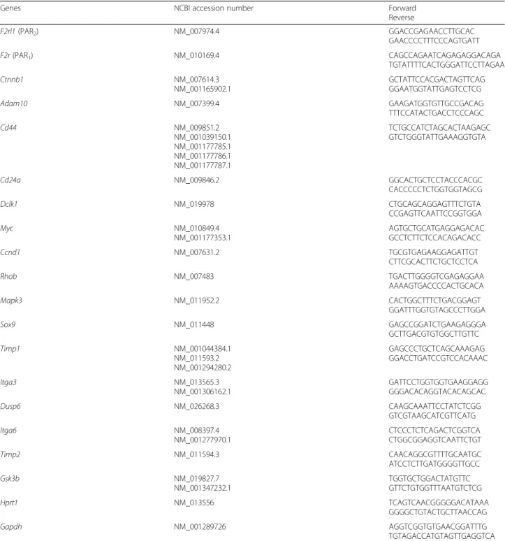
Table 1 Oligonucleotides used for quantitative RT-PCR. Official gene symbols, NCBI accession number of targeted transcripts, and forward and reverse oligonucleotide sequences are depicted
Genes NCBI accession number Forward Reverse F2rl1 (PAR2) NM_007974.4 GGACCGAGAACCTTGCAC GAACCCCTTTCCCAGTGATT F2r (PAR1) NM_010169.4 CAGCCAGAATCAGAGAGGACAGA TGTATTTTCACTGGGATTCCTTAGAA Ctnnb1 NM_007614.3 NM_001165902.1 GCTATTCCACGACTAGTTCAG GGAATGGTATTGAGTCCTCG Adam10 NM_007399.4 GAAGATGGTGTTGCCGACAG TTTCCATACTGACCTCCCAGC Cd44 NM_009851.2 NM_001039150.1 NM_001177785.1 NM_001177786.1 NM_001177787.1 TCTGCCATCTAGCACTAAGAGC GTCTGGGTATTGAAAGGTGTA Cd24a NM_009846.2 GGCACTGCTCCTACCCACGC CACCCCCTCTGGTGGTAGCG Dclk1 NM_019978 CTGCAGCAGGAGTTTCTGTA CCGAGTTCAATTCCGGTGGA Myc NM_010849.4 NM_001177353.1 AGTGCTGCATGAGGAGACAC GCCTCTTCTCCACAGACACC Ccnd1 NM_007631.2 TGCGTGAGAAGGAGATTGT CTTCGCACTTCTGCTCCTCA Rhob NM_007483 TGACTTGGGGTCGAGAGGAA AAAAGTGACCCCACTGCACA Mapk3 NM_011952.2 CACTGGCTTTCTGACGGAGT GGATTTGGTGTAGCCCTTGGA Sox9 NM_011448 GAGCCGGATCTGAAGAGGGA GCTTGACGTGTGGCTTGTTC Timp1 NM_001044384.1 NM_011593.2 NM_001294280.2 GAGCCCTGCTCAGCAAAGAG GGACCTGATCCGTCCACAAAC Itga3 NM_013565.3 NM_001306162.1 GATTCCTGGTGGTGAAGGAGG GGGACACAGGTACACAGCAC Dusp6 NM_026268.3 CAAGCAAATTCCTATCTCGG GTCGTAAGCATCGTTCATG Itga6 NM_008397.4 NM_001277970.1 CTCCCTCTCAGACTCGGTCA CTGGCGGAGGTCAATTCTGT Timp2 NM_011594.3 CAACAGGCGTTTTGCAATGC ATCCTCTTGATGGGGTTGCC Gsk3b NM_019827.7 NM_001347232.1 TGGTGCTGGACTATGTTC GTTCTGTGGTTTAATGTCTCG Hprt1 NM_013556 TCAGTCAACGGGGGACATAAA GGGGCTGTACTGCTTAACCAG Gapdh NM_001289726 AGGTCGGTGTGAACGGATTTG TGTAGACCATGTAGTTGAGGTCA
Then, after washes in PBS, slides were mounted in Pro-Gold DAPI and observed by confocal laser scanning (Zeiss LSM710).
For each staining, controls were made in the same conditions with no antibody, secondary antibody only, isotype control or pre-immune serum, and staining in PAR2KO tissue.
Western blotting
Male- or female-derived six wells of colonoid culture (48-well plates) were dissociated from Matrigel by incu-bation with Cell Recovery Solution as described above for passage. Then, colonoids were centrifuged (43g, 10 min) and lysed in Laemmli sample buffer 5x. After boiling for 10 min, proteins were resolved on polyacryl-amide SDS gels (SDS-PAGE) and transferred to nitrocel-lulose (membrane Hybond C-super, Merck Millipore). The membrane was blocked for 1 h at room temperature in Tris-buffer saline (TBS) containing 0.5% fat-free milk and 1% bovine serum albumin (BSA, Sigma). Then, mem-brane was probed overnight at 4 °C with the appropriate antibody in TBS-milk-BSA supplemented with 0.05% Tween. After incubation for 1 h at room temperature with secondary antibody coupled to horseradish peroxidase, detection was achieved using a chemiluminescent sub-strate (Amersham ECL Prime detection reagent) and visu-alized on ChemiDoc (Bio-Rad).
Cell sorting
Male or female crypts were isolated by 75 min orbital shaking at room temperature of 10 washed murine co-lons (2/3 ends of descendant colon) in PBS with 9 mM
EDTA plus 3 mM dithiothreitol and 10μM Y-27632,
followed by manual shaking for 2 min in PBS with
10μM Y-27632. Then, the crypt suspension (around 2 ×
105male crypts and 2.8 × 10⁵female crypts) was filtered through a 100-μm cell strainer and centrifuged (40g, 5 min, 4 °C). Individual epithelial cells were obtained after incubation of crypts at 37 °C with dispase (60,000 units/ml, BD Biosciences) and DNase I (20,000 units/ml, Sigma) for 4 min and shaking for 30 s. The suspension of individual cells (around 1.2 × 10⁶male cells and 0.6 × 10⁶female cells) was filtered through a 40-μm cell strainer in 1 ml of cold FCS. After centrifugation (1000g, 5 min, 4 °C), cells were suspended in DMEM F12 supplemented with 100 U/ml penicillin/streptomycin, 10 mM Hepes, 2 mM Glutamax, N2 (1/100), B27 (1/50), and N-acetylcysteine (NAC, 1 mM, Sigma).
For cell sorting, 9 × 10⁵ male and 3 × 10⁵ female cells were labeled for 45 min at 4 °C with antibodies from BD
Biosciences: CD31-FITC, CD45-FITC, CD326-APC,
CD44-BV421, and CD24-PE/CF594. Controls were incu-bated with above antibodies minus one or viability dye (eFluor 506, Thermo Fisher Scientific). CD326, CD44,
and CD24 antibodies were used to purify colonic cells into different subsets (CD326+ CD44+ CD24
high/medium/-low
) using a FACS ARIA-SORP (BD Biosciences). Sorted cells were collected in cold tubes containing 25μl of Matrigel. Around 1.2 × 10⁵ male and 2.5 × 10⁵ female CD44+CD24+ cells were collected and cultured as colo-noids (5000 cells per well, 48-well plate) as described above.
Statistical analysis and image/cell sorting processing
For each experiment (animals, crypts, colonoids, sorted cells), male and female were processed simultaneously. Student’s t (two-tailed) or ANOVA tests were used for experiment analysis. P values or adjusted P values (ANOVA) < 0.05 were considered to be significant, and the correction used for multiple comparisons is indi-cated on the figures. Number of colonoids and gene expression were calculated from the mean of duplicate assays in each experiment.
Apotome and confocal images were imported into the Image J software for analysis. Size of around 20 colo-noids was measured in each assay. Male and female colonoid size ranges were 25–80 μm and 30–120 μm,
respectively. A threshold ≥ 50 μm was taken for the
study of colonoid size since significant variations be-tween sexes and bebe-tween control/treatment assays were measured at this condition. Data of Ki-67 labeling in colonoids were calculated as ratio of positive Ki-67 nu-clei vs total nunu-clei counted in the larger diameter of colonoids whose size is representative of the male and female cultures.
Data of cell sorting were analyzed with the FlowJo software.
Results
Colonoid growth is sexually dimorphic and regulated by PAR2
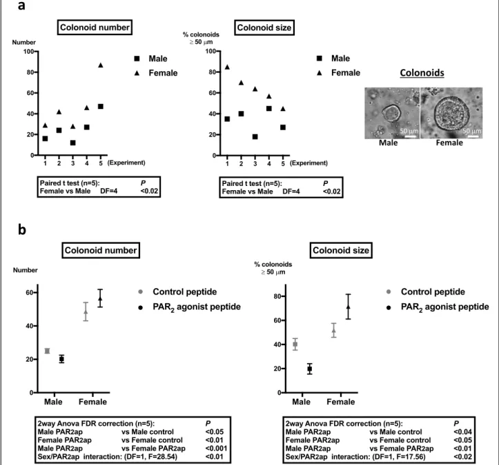
Colon crypts from male and female mice were embedded in Matrigel and grown as colonoids. At day 6 from initial seeding, despite identical numbers of crypts seeded, both the number and size of female mice-derived colonoids were significantly higher than those of male mice-derived colonoids (Fig. 1a). This higher size of female mice-de-rived colonoids was measured as soon as day 2 of culture and was maintained after re-embedding of colonoids in fresh Matrigel (Additional file2). These data suggest that female primitive epithelial cells have higher proliferation than male.
Since we have previously shown that PAR2 plays a
critical role in the control of ISC proliferation [7], we investigated the role of PAR2in the proliferation of colon
primitive cells from male and female mice. In agreement with our previous results, we measured a decrease in the number and size of colonoids from male mice treated by
the PAR2 agonist compared to control peptide (Fig.1b).
In contrast, treatment of colonoids from female mice by the PAR2agonist peptide increased their number and size
compared to the control (Fig. 1b). The PAR2 agonist
effects on colonoid growth were observed from 48 h post-treatment (Additional file3).
Altogether, these data show that the growth of colon
primitive cells is sexually dimorphic and that PAR2
activation further increases this difference.
PAR2controls the expression of key proliferative
regulators of colon primitive cells
In order to evaluate the impact of PAR2 on colonoid
growth, crypts from colons of PAR2 KO mice were
isolated. The absence of PAR2impaired colonoid culture
from both male and female mice (Additional file 4) as we have previously shown that PAR2is implicated in the
ISC survival [7]. However, labeling of cell proliferation marker Ki-67 was performed in surviving PAR2-deficient
Fig. 1 Growth characteristics of colonoids from male and female mice and impact of PAR2activation. a Colonoids were counted and measured
as described in the“Methods” section at day 6 after male and female colon crypts seeding in Matrigel. Representative colonoids are shown.
b Colonoids from male and female mice were stimulated daily with PAR2agonist peptide (SLIGRL-NH2, 100μM) or control peptide (LRGILS-NH2,
100μM) from day 2 to day 6 of culture. At day 6 of culture, colonoids were counted and their size measured. Results are mean ± SEM from n = 5 independent experiments
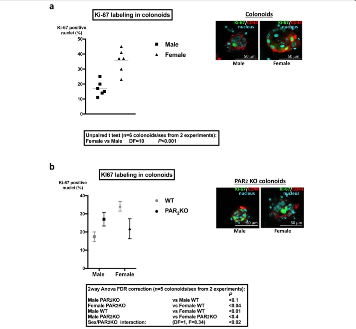
colonoids and WT colonoids. Whereas Ki-67 was expressed in a higher number of cells from female WT mice compared to males (Fig. 2a), PAR2 KO
female-de-rived colonoids showed decreased Ki-67 labeling
com-pared to WT (Fig. 2b). Conversely, PAR2 KO
male-derived colonoids showed a tendency to increase Ki-67
labeling compared to WT (Fig. 2b). As a consequence,
the percentages of Ki-67 positive nuclei were not
differ-ent between PAR2 KO colonoids from both sexes
(Fig. 2b). These results show that colonoids from female
WT mice contain a higher number of proliferative primitive cells (stem cells and progenitors) compared to WT males and suggest that PAR2may play a critical role
in that sexual dimorphism.
To investigate the role of PAR2 in the regulation of
male and female colon primitive cell proliferation, we analyzed the gene expression of key regulators of the Wnt, Notch, and EGF proliferative pathways, as well as
PAR2, in male and female colon crypts from WT or
PAR2KO mice. The gene expression of PAR1and some
Fig. 2 Cell proliferation in colonoids from male and female mice and impact of PAR2expression. a Ki-67 labeling in male and female colonoids at
day 6 of culture. Left panel: The percentage of Ki-67 positive nuclei was calculated as described in the“Methods” section by the ratio of positive
Ki-67 nuclei vs total nuclei in the larger diameter of colonoids. Right panel: Representative colonoid labeling of Ki-67 (green), CD44 (red, immaturity marker), and nuclei by DAPI (cyan) is shown. b Comparative Ki-67 labeling in PAR2WT and PAR2KO male and female colonoids at day
6 of culture. Right panel: Representative PAR2KO colonoid labeling of Ki-67 (green), CD44 (red, immaturity marker), and nuclei by DAPI (cyan) is
adhesion receptors was also studied given their implica-tion in the regulaimplica-tion of colon primitive cells [7].
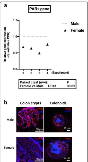
As shown in Fig. 3a, quantitative RT-PCR detected a lower level of PAR2(F2rl1) mRNA in colon crypts from
female WT mice compared to males, whereas PAR1
(F2r) was not differentially expressed (Additional file 5, n= 4, DF = 3, paired t test p < 0.2). In PAR2KO crypts,
PAR1 mRNA expression did not vary significantly
be-tween males and females (Additional file5, n = 4, DF = 3, paired t test p < 0.9). In the absence of suitable PAR2
antibody for quantification by Western blot, analysis of
PAR2 protein expression by immunostaining in colon
crypts and colonoids confirmed the lower expression in females compared to males (Fig.3b). These data demon-strate that PAR2 is differently expressed in male and
female colon epithelial cells.
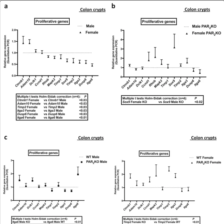
The expression of the proliferation enhancers, Ctnnb1 (β catenin, Wnt pathway) and Adam10 (Disintegrin and metalloprotease 10, Notch pathway), was higher in colon crypts from female mice compared to males (Fig.4a and Additional file5). In contrast, the expression of the prolifer-ation inhibitors, Timp2 (tissue inhibitor of metalloproteinases 2, EGF pathway) and Dusp6 (dual specificity phosphatase 6, Erk pathway), was lower in colon crypts from female mice compared to males (Fig. 4aand Additional file 5). Import-antly, other modulators of colon cell proliferation, the integ-rins alpha 6 (Itga6) and alpha 3 (Itga3), were expressed at a higher level in colon crypts from male mice compared to fe-males (Fig.4a and Additional file5). These data suggest that pathways important for ISC and progenitors proliferation (Wnt, Notch, EGF, integrins) are differently regulated in colon crypts from male and female mice.
In PAR2 KO crypts, the sexual dimorphism in the
expression of Ctnnb1 (β catenin), Adam10, Timp2,
Itga3, Dusp6, and Itga6 was abolished (Fig. 4b and
Additional file 5). Interestingly, the expression of
Sox9, a transcriptional factor playing a key role in male sex determination and stem cell proliferation, was reversed compared to WT since it was higher in
PAR2 KO female-derived crypts compared to PAR2
KO males (Fig. 4b and Additional file 5). Sox9 varied
in female PAR2 KO (6.60 ± 5.60 mean ± SD, fold
in-crease vs WT) but not in male PAR2 KO (0.85 ± 0.14
mean ± SD, fold increase vs WT). Analysis of signifi-cant variations of genes revealed that Itga6 in males and Timp2 in females were under the control of PAR2(Fig. 4c and Additional file5). Thus, in the
ab-sence of PAR2, the basal sexual dimorphism in Itga6
expression was reinforced, whereas Timp2 was spe-cifically upregulated in females. These data show
that PAR2 controls gene expression of important
regulators of the ISC and progenitor proliferation. Altogether, our data suggest that PAR2may play a
spe-cific and critical role in the control of proliferation in colon crypts from male and female mice.
The sexual dimorphism in colonoid growth is linked to metabolic and resistant phenotypes
We previously demonstrated the PAR2-dependent
regu-lation of the glycogen synthase kinase 3 (GSK3) in ISC [7]. Given the critical role of GSK3 to promote quies-cence and survival of primitive cells [4, 7], we investi-gated its expression and activation in colons from male and female mice.
Fig. 3 PAR expression in colon crypts and colonoids. a PAR2mRNA
expression in colon crypts from male or female mice was measured by qRT-PCR (n = 4 independent experiments). b Immunolabeling of PAR2
(red) in native colon crypts and cultured colonoids (day 6 of culture) from male and female mice. Actin (blue) was labeled by phalloidin. Results are representative of three independent experiments
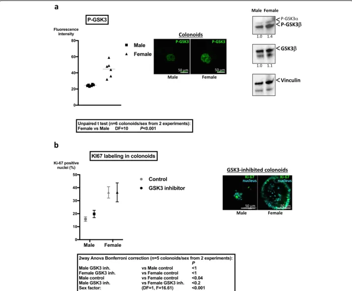
No significant difference in the mRNA expression of GSK3 (β isoform) between sexes was measured in colon crypts (relative expression female vs male 1.16 ± 0.19, mean ± SD, n = 3 independent experiments). However, the inhibited form of GSK3 (Pser21/9 GSK3) was higher expressed in colonoids from female mice showing that GSK3 is more active in colonoids from male mice
compared to females (Fig. 5a). Accordingly, Western
blot analysis showed increased serine 9 phosphorylated-GSK3β in female-derived colonoids compared to males (Fig.5a).
Incubation with the GSK3 inhibitor SB216763 induced a specific decrease of the number of colonoids from male mice compared to control (n = 4; male − 47% ± 11,
Fig. 4 Expression of proliferative signals for colon primitive cells in PAR2WT or KO male and female crypts. mRNA of male and female WT or
PAR2KO crypts were extracted, and the expression of key proliferative signals for colon primitive cells and their modulators was quantified by
RT-PCR. a Comparative data from male and female PAR2WT crypts. b Comparative data from male and female PAR2KO crypts. c Comparative data
from male PAR2KO crypts vs male PAR2WT crypts (left panel), and female PAR2KO crypts vs female PAR2WT crypts (right panel). Data are
mean ± SD, two-way ANOVA p < 0.001; female + 13% ± 26%, mean ± SD, two-way ANOVA ns). Moreover, this treatment abolished the difference in Ki-67 labeling
be-tween colonoids from both sexes (Fig. 5b). Thus, the
kinase GSK3 may be implicated in the sexual dimorph-ism of colonoid growth.
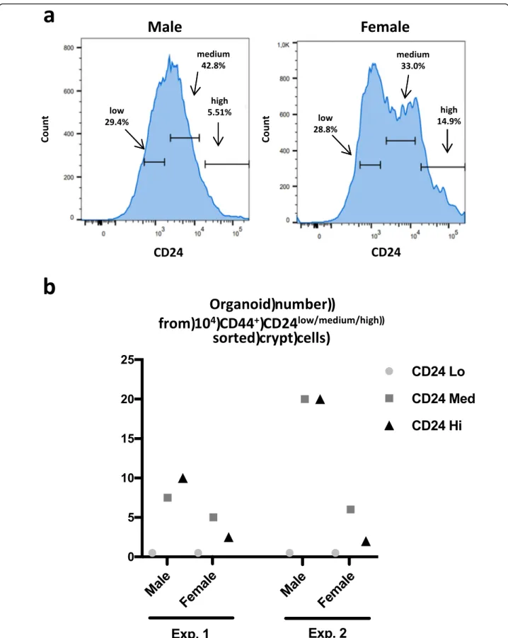
The above data suggest that proliferation and survival of colon primitive cells from male and female mice might be under the control of different metabolic path-ways. Thus, we evaluated their growth ability under high stress conditions such as cell sorting. The adhesion molecules CD44 and CD24 are markers of the colon
primitive cells and can be used for cell sorting protocols presenting the advantages to collect both slowly prolifer-ative/strongly resistant ISC and highly proliferative/ poorly resistant ISC [23,24]. Note that Cd44 and Cd24 gene expressions were not significantly different be-tween male and female colon crypts (male vs female n= 4, two-way ANOVA CD24 p < 0.3 and CD44 p < 0.9; Additional file 5). The cell sorting of colon primitive cells based on CD44 and CD24 allowed the collection of three cell populations CD44+
CD24low, CD44+CD24medium, and CD44+CD24high (Fig. 6a). Note that the fraction of CD44+CD24high cells was constantly higher in females
Fig. 5 Differential regulation of GSK3 in colon primitive cells from male and female mice. a Immunolabeling of Pser21/9 GSK3 in colonoids from male and female mice was performed at day 6 of culture. Fluorescence intensity of the brightest cross section of colonoids was quantified by Image J and is represented (n = 2 independent experiments). Representative images of immunolabeling of PserGSK3 are shown. Right panel: Western blot of Pser21/9 GSK3 and total GSK3β in colonoids is shown. Vinculin was used as loading control. Results are representative of two independent experiments. b Colonoids from male and female mice were incubated daily with GSK3 inhibitor (SB216763, 12.5μM) from day 2 to day 6 of culture. 67 labeling in male and female colonoids treated by GSK3 inhibitor was performed at day 6 of culture. The percentage of Ki-67 positive nuclei was calculated as described in the“Methods” section by the ratio of positive Ki-67 nuclei vs total nuclei in the larger diameter
Fig. 6 a A cell sorting of CD44+CD24+primitive cells was performed from male and female colon crypts. Three CD24 subpopulations were isolated as defined on the graphs (representative of three experiments). The percentage of each CD24 subpopulation is shown. b Sorted CD44+CD24high/medium/low
(percentage of CD44+CD24highcells vs entire CD44+CD24+ population: male 7.1 ± 2.3%, female 13 ± 3.2%, mean ± SD, n= 3, paired t test p = 0.05). After embedding in Matrigel, only the CD44+CD24medium and CD44+CD24high cells de-veloped as colonoids, cells from male mice showing greater efficacy (Fig.6b). This suggests male primitive cells may be more resistant to the cell sorting process.
Altogether, these data show that male and female colon primitive cells display a sexual dimorphism in their proliferation, metabolism, and adaptation to stress, which could be critically influenced by PAR2.
Discussion
This work shows that male and female colon primitive cells have different proliferation capacities and that this is controlled by the protease-activated receptor PAR2.
As colonoids, primitive cells from female mice displayed higher proliferation compared to males. Conversely, after complete epithelial cell dissociation, primitive cells from male mice produced more colonoids than females. Fur-thermore, PAR2 was shown to control key proliferative
pathways for colon primitive cells in both males and fe-males, although in different ways.
The strong proliferative capacities of ISC and progeni-tors from female mice allowed the development of a higher number and size of colonoids from bottom crypts, as compared to males. This observation is in accordance with the recent data of Zhou and collabora-tors [25]. Moreover, this sexual dimorphism in ISC and progenitor proliferation depends on intrinsic mecha-nisms since the culture of epithelial cells as colonoids
was exempt from stroma. Zhou and collaborators [25]
also found that proliferation of intestinal organoids was not influenced by estrogens. In drosophila, Hudry and collaborators [5] demonstrated that cell-intrinsic mecha-nisms linked to sex determination genes control cell cycle duration in female ISC. We found that important regulators of the ISC proliferation (β catenin/Wnt path-way, ADAM10/Notch pathway) displayed gene overex-pression in female-derived crypts compared to males. However, female- and male-derived crypts did not show significant differences in gene expression of immature markers CD44 and CD24 (our data) and in their size in vivo [25], suggesting sex-specific regulation of prolifera-tive and differentiation pathways at the progenitor level.
The crypt microenvironment is shaped by proteases through matrix proteolysis, release of growth factors, and receptor activation. The Wnt and Notch pathways are tightly controlled by this microenvironment. It is also true for the EGF pathway, a key regulator of the progenitor proliferation and differentiation [26], and our data show gene overexpression of inhibitors of this path-way (Dusp6, Timp2) in crypts from male mice compared to females. We have shown that the protease-activated
receptor PAR2was expressed in epithelial cells along the
male and female crypts, but at a higher level in males. PAR2was found required for survival of colonoids from
both male and female mice and supports sexual di-morphism in the expression of proliferative genes in the crypt. Thus, depending on its level of expression in the
crypt, PAR2 could promote the sexual dimorphism in
the proliferation of colon primitive cells.
However, PAR2activation had an opposite impact on
growth of colonoids from male and female mice. PAR2
slows down the growth of colonoids from male mice but increases it from females. The growth of male-derived colonoids is associated with an active status of the kinase GSK3, a key modulator of cell metabolism and prolifera-tion [27]. We have previously shown that the activation of
GSK3 was under the control of PAR2 in colonoids from
male mice [7]. It is therefore likely that the regulation of GSK3 represents a critical point in the sexual dimorphism of ISC function, as we previously demonstrated for leukemic stem cells [4]. Finally, a different subcellular localization of PAR2in intestinal epithelial cells between male and female
may also reflect differential presence of PAR2-activating
pro-teases in the epithelial microenvironment [28]. Further stud-ies would be necessary to investigate potential differential expression and activity of intestinal proteases in males and females.
The knockout of PAR2abolished the sexual dimorphism
in ISC proliferation and in gene expression of crypts described above. The protease-activated receptor PAR1is
co-expressed with PAR2in epithelial cells, and we
demon-strated that it oppositely regulates colonoid growth and GSK3 compared to PAR2 [7]. PAR1 was not differently
expressed at gene level in male and female crypts, WT or PAR2KO. This demonstrates that PAR2, but not PAR1, is
critical for the sexual dimorphism in ISC function. Com-pared to WT, the gene expression ofα6integrin increased
strongly in crypts from PAR2KO male mice. Since the
cel-lular balance betweenα6A andα6B variants influence cell
proliferation in the crypt [29], it is possible that the
prolif-erative isoform A of α6 integrin was differentially
expressed in PAR2KO crypts. Moreover, we measured an
increase of gene expression of TIMP2 in crypts from
PAR2 KO female mice compared to WT. Interestingly,
TIMP2 has been described to directly bind toα3integrin
(Itga3 also increased in PAR2KO female-derived crypts)
in a context of growth arrest [30]. Furthermore, Timp2, Itga3, and Sox9 which increased in PAR2KO females are
located on the same chromosome (17 human, 11 murine) where there is controlled sex reversal [31]. As demon-strated by Hudry and coll. in drosophila [5], crypt stem cells have to undergo sex reversal-related plasticity in physiological or pathological situations. A potential role of PAR2 in that process should be investigated. Altogether,
proliferation of both male and female colon primitive cells, in opposite ways, and possibly their plasticity.
An interesting observation was that both PAR2-deficient
male and female colon primitive cells become vulnerable in stressed conditions such as in vitro culture. We have shown previously that the PAR2/GSK3 pathway is critical in the
control of male colon primitive cell survival [7]. Moreover, integrins are key partners for PAR2and GSK3 to control
cell survival [4, 32], and we previously demonstrated that cell adhesion mechanisms related to the Rho kinase activa-tion influenced the PAR2/GSK3 pathway in colon primitive
cells [7]. Thus, it is possible that primitive cells in the male colon crypt have better adhesion capacities and resistance to stress leading to increased cell survival compared to females. In favor of this hypothesis are the lesser efficacy of the male crypt extraction and the better growth of sorted male CD24high/med epithelial cells, compared to females.
However, we measured great variations in the efficiency of male colonoid growth from sorted cells between the two experiments shown. Following a sorting protocol closed to ours, Yip and collaborators [24] have also obtained import-ant variations in organoid growth from male mice despite the presence of myofibroblastic growth factors in the medium culture that enhance survival and proliferation of primitive cells. This suggests that during the cell sorting process, important mechanisms aiming cells to cope with de-adhesion stress are set up. However, this setting-up,
in-volving probably PAR2 engagement which protects male
colon epithelial cells from anoïkis [7], might display variable efficacy depending on dispase incubation for the isolation of individual cells. Interestingly, dispase has been shown to induce a specific internalization of the alpha 6 integrin [33], which is overexpressed in male colon epithelial cells com-pared to females and whose expression is under the control
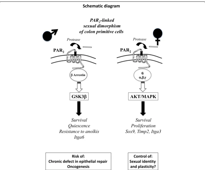
Fig. 7 Sexual dimorphism occurs in the PAR2-dependent regulation of colon primitive cells, which could have important implications in pathophysiology and therapy
of PAR2. Knowing that resistant primitive cells in the crypt
are in the vicinity of progenitors, specific sex-dependent mechanisms could be critical in that zone. The lower prolif-eration rate of male progenitors associated with their specific metabolism may provide an advantage for survival to stress compared to females. This observation may have important pathophysiological implications in in-testinal inflammation and cancer displaying a sexual dimorphism in incidence and location [10, 11]. Perspectives and significance
A sexual dimorphism has already been shown in PAR2
func-tions with increased PAR2-mediated vasodilatation and
pruritus associated with female sex. Our data show now that a sexual dimorphism occurs in the PAR2-dependent
regula-tion of colon primitive cells, which could have important implications in pathophysiology and therapy (Fig.7). Indeed, compared to females, the higher capacity of male-derived progenitor/stem cell coping with stress to develop PAR2
-dependent survival coupled with quiescence may be first ad-vantageous but can become deleterious during repeated ag-gressions such as chronic inflammation. As a consequence, there is a risk of chronic defect in epithelial repair and accu-mulation of mutations paving the way for oncogenesis. A crosstalk between integrins such as integrin α6 and PAR2
may play a critical role in that context. On the other hand, we show that PAR2controls the expression of genes
impli-cated in sexual dimorphism such as Sox9 and Timp2 sug-gesting that PAR2may be an important regulator of stem
cell sexual identity and plasticity. Additional files
Additional file 1:Colonoid culture with or without phenol red. (DOCX 52 kb)
Additional file 2:Time course of colonoid culture and passage. (DOCX 54 kb)
Additional file 3:Time course of PAR2-stimulated colonoid culture.
(PPTX 210 kb)
Additional file 4:Time course of PAR2KO colonoid culture. (DOCX 57 kb)
Additional file 5:DeltaCt of proliferative genes. (DOCX 72 kb)
Abbreviations
ADAM10:Disintegrin and metalloprotease 10; DCLK1: Doublecortin-like kinase 1; DUSP6: Dual specificity phosphatase 6; EGF: Epidermal growth factor; GSK3: Glycogen synthase kinase 3; ISC: Intestinal stem cell; NAC: N-Acetylcysteine; PAR: Protease-activated receptor; TIMP: Tissue inhibitor of metalloproteinases
Acknowledgements
We thank Dr. Paul Roustan and Dr. Michèle Allouche for helpful scientific discussion and critical reading of the manuscript. We thank Dr. Scott Magness for advices in the cell sorting of colon primitive cells and their culture in vitro. We thank Dr. Jean-Paul Motta for the statistical analysis of data. Acknowledgements are due to the members of the different core facilities: Marie Lulka, Tanguy Bernal, and Rachel Balouzat (animal housing, US006); Danièle Daviaud and Astrid Canivet (imaging, U1043); and Fatima L’Faqihi-Olive and Anne-laure Iscache (cell sorting, U1043).
Authors’ contributions
JN, PJR, MNT, LD, CR, LG, DS, AE, and CRS performed the experiments. JN, PJR, MNT, LD, CR, AE, and CRS analyzed the data. JN, PJR, MNT, LD, and CRS interpreted the results of the experiments. JN, PJR, MNT, LD, and CRS prepared the figures. JN, PJR, MNT, LD, CR, AE, CB, ADS, CD, NV, and CRS edited and revised the manuscript. All authors approved the final version of manuscript. CB, ADS, CD, NV, and CRS contributed to the conception and design of the research. CRS drafted the manuscript.
Funding
This work was supported by grants from the European Research Council (ERC- 310973 PIPE) to NV. This project was also supported through a“Fond Unique Interministeriel” (FUI) program by the Région Midi-Pyrénées (now Occitanie), Toulouse Métropole, the Banque Publique d’Investissement (BPI) de France. Equipment obtained from the use of Fonds Européens de Développement Régional (FEDER), and the region Occitanie were used in the present research program, as well as equipment funded by the Equipex “Investissements d’avenir” funds (ANR-11-EQPX-0003).
Availability of data and materials
The datasets generated and analyzed during the current study are available from the corresponding author on reasonable request.
Ethics approval and consent to participate
Animal samples: Protocol agreement PI-U1220-NV19. Protocol was approved by the local ethic committee from Toulouse, France (Animal care committee CEEA-122).
Consent for publication Not applicable
Competing interests
The authors declare that they have no competing interests.
Received: 30 April 2019 Accepted: 26 August 2019
References
1. Arnold AP, Chen X, Link JC, Itoh Y, Reue K. Cell-autonomous sex determination outside of the gonad. Dev Dyn. 2013;242:371–9. 2. Du L, Bayir H, Lai Y, Zhang X, Kochanek PM, Watkins SC, et al. Innate
gender-based proclivity in response to cytotoxicity and programmed cell death pathway. J Biol Chem. 2004;279:38563–70.
3. Markel TA, Crisostomo PR, Wang M, Wang Y, Lahm T, Novotny NM, et al. TNFR1 signaling resistance associated with female stem cell cytokine production is independent of TNFR2-mediated pathways. Am J Physiol Regul Integr Comp Physiol. 2008;295:R1124–30.
4. Bertrand J, Despeaux M, Joly S, Bourogaa E, Gallay N, Demur C, et al. Sex differences in the GSK3β-mediated survival of adherent leukemic progenitors. Oncogene. 2012;31:694–705.
5. Hudry B, Khadayate S, Miguel-Aliaga I. The sexual identity of adult intestinal stem cells controls organ size and plasticity. Nature. 2016;530:344–8. 6. Dempsey PJ. Role of ADAM10 in intestinal crypt homeostasis and
tumorigenesis. Biochim Biophys Acta. 2017;1864:2228–39.
7. Nasri I, Bonnet D, Zwarycz B, d'Aldebert E, Khou S, Mezghani-Jarraya R, et al. PAR2-dependent activation of GSK3β regulates the survival of colon stem/ progenitor cells. Am J Physiol Gastrointest Liver Physiol. 2016;311:G221–36. 8. Sébert M, Denadai-Souza A, Quaranta M, Racaud-Sultan C, Chabot S, Lluel P,
et al. Thrombin modifies growth, proliferation and apoptosis of human colon organoids: a protease-activated receptor 1- and protease-activated receptor 4-dependent mechanism. Br J Pharmacol. 2018;175:3656–68. 9. Vergnolle N. Clinical relevance of proteinase activated receptors (pars) in the
gut. Gut. 2005;54:867–74.
10. Coughlan A, Wylde R, Lafferty L, Quinn S, Broderick A, Bourke B, et al. DOCHAS Study. A rising incidence and poorer male outcomes characterise early onset paediatric inflammatory bowel disease. Aliment Pharmacol Ther. 2017;45:1534–41.
11. Koo JH, Leong RW. Sex differences in epidemiological, clinical and pathological characteristics of colorectal cancer. J Gastroenterol Hepatol. 2010;25:33–42.
12. Steegenga WT, Mischke M, Lute C, Boekschoten MV, Pruis MG, Lendvai A, et al. Sexually dimorphic characteristics of the small intestine and colon of prepubescent C57BL/6 mice. Biol Sex Differ. 2014;5:11.
13. Beurel E, Grieco SF, Jope RS. Glycogen synthase kinase-3 (GSK3): regulation, actions, and diseases. Pharmacol Ther. 2015;148:114–31.
14. Suen JY, Gardiner B, Grimmond S, Fairlie DP. Profiling gene expression induced by protease-activated receptor 2 (PAR2) activation in human kidney cells. PLoS One. 2010;5:e13809.
15. Hennessey JC, McGuire JJ. Attenuated vasodilator effectiveness of protease-activated receptor 2 agonist in heterozygous par2 knockout mice. PLoS One. 2013;8:e55965.
16. Yamaura K, Tomono A, Suwa E, Ueno K. Sex-related differences in SLIGRL-induced pruritus in mice. Life Sci. 2014;94:54–7.
17. Valentino RJ, Van Bockstaele E, Bangasser D. Sex-specific cell signaling: the corticotropin-releasing factor receptor model. Trends Pharmacol Sci. 2013;34:437–44.
18. Ge Y, Fuchs E. Stretching the limits: from homeostasis to stem cell plasticity in wound healing and cancer. Nat Rev Genet. 2018;19:311–25.
19. Lindner JR, Kahn ML, Coughlin SR, Sambrano GR, Schauble E, Bernstein D, et al. Delayed onset of inflammation in protease-activated receptor-2-deficient mice. J Immunol. 2000;165:6504–10.
20. Jones SM, Mann A, Conrad K, Saum K, Hall DE, McKinney LM, et al. PAR2 (Protease-Activated Receptor 2) Deficiency attenuates atherosclerosis in mice. Arterioscler Thromb Vasc Biol. 2018;38:1271–82.
21. Theeuwes WF, Gosker HR, Langen RCJ, Pansters NAM, Schols AMWJ, Remels AHV. Inactivation of glycogen synthase kinase 3β (GSK-3β) enhances mitochondrial biogenesis during myogenesis. Biochim Biophys Acta Mol Basis Dis. 2018;1864:2913–26.
22. Gobrecht P, Leibinger M, Andreadaki A, Fischer D. Sustained GSK3 activity markedly facilitates nerve regeneration. Nat Commun. 2014;31(5):4561. 23. King JB, von Furstenberg RJ, Smith BJ, McNaughton KK, Galanko JA, Henning
SJ. CD24 can be used to isolate Lgr5+ putative colonic epithelial stem cells in mice. Am J Physiol Gastrointest Liver Physiol. 2012;303:G443–52.
24. Yip HYK, Tan CW, Hirokawa Y, Burgess AW. Colon organoid formation and cryptogenesis are stimulated by growth factors secreted from
myofibroblasts. PLoS One. 2018;13:e0199412.
25. Zhou W, Davis EA, Li K, Nowak RA, Dailey MJ. Sex differences influence intestinal epithelial stem cell proliferation independent of obesity. Physiol Rep. 2018;6:e13746.
26. Wang Y, Kim R, Hinman SS, Zwarycz B, Magness ST, Allbritton NL. Bioengineered systems and designer matrices that recapitulate the intestinal stem cell niche. Cell Mol Gastroenterol Hepatol. 2018;5:440–53 e1. 27. Jellusova J, Cato MH, Apgar JR, Ramezani-Rad P, Leung CR, Chen C, et al. Gsk3 is a metabolic checkpoint regulator in B cells. Nat Immunol. 2017;18:303–12. 28. Rolland-Fourcade C, Denadai-Souza A, Cirillo C, Lopez C, Jaramillo JO,
Desormeaux C, et al. Epithelial expression and function of trypsin-3 in irritable bowel syndrome. Gut. 2017;66:1767–78.
29. Dydensborg AB, Teller IC, Basora N, Groulx JF, Auclair J, Francoeur C, et al. Differential expression of the integrinsα6Aβ4 and α6Bβ4 along the crypt-villus axis in the human small intestine. Histochem Cell Biol. 2009;131:531–6. 30. Jackson HW, Defamie V, Waterhouse P, Khokha R. TIMPs: versatile
extracellular regulators in cancer. Nat Rev Cancer. 2017;17:38–53. 31. Ogawa Y, Terao M, Hara S, Tamano M, Okayasu H, Kato T, et al. Mapping of
a responsible region for sex reversal upstream of Sox9 by production of mice with serial deletion in a genomic locus. Sci Rep. 2018;8:17514. 32. Chang LH, Pan SL, Lai CY, Tsai AC, Teng CM. Activated PAR-2 regulates
pancreatic cancer progression through ILK/HIF-α-induced TGF-α expression and MEK/VEGF-A-mediated angiogenesis. Am J Pathol. 2013;183:566–75. 33. Poumay Y, Leclercq-Smekens M, Grailly S, Degen A, Leloup R. Specific
internalization of basal membrane domains containing the integrin alpha 6 beta 4 in dispase-detached cultured human keratinocytes. Eur J Cell Biol. 1993 Feb;60(1):12–20.
Publisher’s Note
Springer Nature remains neutral with regard to jurisdictional claims in published maps and institutional affiliations.