HAL Id: hal-02170571
https://hal.archives-ouvertes.fr/hal-02170571
Submitted on 31 May 2021HAL is a multi-disciplinary open access
archive for the deposit and dissemination of sci-entific research documents, whether they are pub-lished or not. The documents may come from teaching and research institutions in France or abroad, or from public or private research centers.
L’archive ouverte pluridisciplinaire HAL, est destinée au dépôt et à la diffusion de documents scientifiques de niveau recherche, publiés ou non, émanant des établissements d’enseignement et de recherche français ou étrangers, des laboratoires publics ou privés.
Investigating the mode of action of the redox-active
antimalarial drug plasmodione using the yeast model
Pierre Mounkoro, Thomas Michel, Stéphanie Blandin, Marie-Pierre
Golinelli-Cohen, Elisabeth Davioud-Charvet, Brigitte Meunier
To cite this version:
Pierre Mounkoro, Thomas Michel, Stéphanie Blandin, Marie-Pierre Golinelli-Cohen, Elisabeth Davioud-Charvet, et al.. Investigating the mode of action of the redox-active antimalarial drug plas-modione using the yeast model. Free Radical Biology and Medicine, Elsevier, 2019, 141, pp.269-278. �10.1016/j.freeradbiomed.2019.06.026�. �hal-02170571�
Investigating the mode of action of the redox-active antimalarial drug plasmodione using the yeast model
Authors
Pierre Mounkoro1, Thomas Michel1, Stéphanie Blandin2, Marie-Pierre Golinelli-Cohen3, Elisabeth Davioud-Charvet4 & Brigitte Meunier1
Affiliation
1 Institute for Integrative Biology of the Cell (I2BC), CEA, CNRS, Univ. Paris Sud, Université Paris Saclay, 91198 Gif sur Yvette cedex, France
2 Université de Strasbourg, CNRS, Inserm, UPR9022/U1257, Mosquito Immune Responses (MIR), F-67000 Strasbourg, France
3 Institut de Chimie des Substances Naturelles (ICSN), CNRS, UPR 2301, Univ. Paris-Sud Université Paris Saclay, 91198 Gif-sur-Yvette cedex, France
4 Université de Strasbourg, Université de Haute-Alsace, Centre National de la Recherche Scientifique (CNRS), LIMA-UMR 7042, Team Bioorganic and Medicinal Chemistry, ECPM 25 Rue Becquerel, 67087 Strasbourg, France.
Corresponding author:
Brigitte Meunier, Institute for Integrative Biology of the Cell (I2BC), avenue de la Terrasse, 91198, Gif sur Yvette, France; brigitte.meunier@i2bc.paris-saclay.fr
Keywords: antimalarial drug, oxidative stress, mitochondrial respiratory chain, drug mode of action, yeast model
Summary
Malaria is caused by protozoan parasites and remains a major public health issue in subtropical areas.
Plasmodione is a novel early lead compound displaying
fast-acting antimalarial activity. Treatment with this redox active compound disrupts the redox balance of parasite-infected red blood cells. In vitro, the benzoyl analogue of plasmodione can act as a subversive substrate of the parasite flavoprotein NADPH-dependent glutathione reductase, initiating a redox cycling process producing ROS. Whether this is also true in vivo remains to be investigated. Here, we used the yeast model to investigate the mode of action of plasmodione and uncover enzymes and pathways involved in its activity. We showed that plasmodione is a potent inhibitor of yeast respiratory growth, that in drug-treated cells, the ROS-sensitive aconitase was impaired and that cells with a lower oxidative stress defence were highly sensitive to the drug, indicating that plasmodione may act via an oxidative stress.
We found that the mitochondrial respiratory chain flavoprotein NADH-dehydrogenases play a key role in plasmodione activity. Plasmodione and metabolites act as substrates of these enzymes, the reaction resulting in ROS production. This in turn would damage ROS-sensitive enzymes leading to growth arrest. Our data further suggest that plasmodione is a pro-drug whose activity is mainly mediated by its benzhydrol and benzoyl metabolites. Our results in yeast are coherent with existing data obtained in vitro and in Plasmodium falciparum, and provide additional hypotheses that should be investigated in parasites.
Introduction
Malaria is a mosquito-borne infectious disease caused by unicellular eukaryotic parasites of the genus Plasmodium. Among the five species known to infect humans, P. falciparum is responsible for the most severe forms of the disease, and P. falciparum and P. vivax are the most frequently encountered worldwide. Plasmodium parasites have a complex life cycle with different development stages that are adapted to their two hosts. In the human host, parasites mostly undergo asexual multiplication in red blood cells causing typical malaria symptoms, while a fraction of them differentiate in gametocytes that are the only forms able to infect female Anopheles mosquitoes upon a blood meal. Most drugs currently on the market target the symptomatic asexual forms, however an increasing effort is now focused on the transmissible forms aiming at malaria control and elimination. Of note, parasites have developed resistances to virtually all drugs used thus far to treat malaria, including to the most efficient drug artemisinin, underlying the need to find new drugs with different modes of action.
Plasmodione (PD) is a active against asexual blood stages
and early gametocytes [1-2]. PD kills parasites as fast as artemisinin, is effective on a large variety of drug-resistant parasites, and has demonstrated a low potential to generate genetic resistance [2]. Despite chemical similarity to atovaquone, PD does not target the same mitochondrial protein. Indeed transgenic parasites expressing the yeast dihydroorotate dehydrogenase, which uses fumarate instead of ubiquinone as electron acceptor and therefore renders the bc1 complex activity dispensable for parasite proliferation [3], presented unchanged sensitivity to plasmodione whereas they were highly resistant to the bc1 complex inhibitor atovaquone [4].
Plasmodione is a redox-active agent that disturbs the redox balance of the parasitized red blood cells [5]. Its mode of action (Fig.1A) was proposed to start with the benzylic oxidation of PD to form PDO (3-benzoylmenadione) by a mechanism yet to be elucidated. Benzylic oxidation can be catalysed by CYP450s and by free or protein-bound hemes via ferryl species (see for instance, [6-9]). Moreover numerous hemoproteins (including hemoglobin) can oxidize drugs [10]. While CYP450s are absent from Plasmodium and red blood cells, free heme and hemoproteins are abundant in infected erythrocytes and could mediate benzylic oxidation of PD to PDO. In addition, we previously proposed that in the presence of redox-cyclers, P. falciparum-infected erythrocytes could be electrochemically active, generating/transporting oxidized and reduced charged naphthoquinone species in different compartments, the anodic one for oxidation reactions (acidic vesicle/food vacuole network), and the cathodic one for reduction reactions (cytosol), possibly via iron complexation [1,11]. This particular situation is comparable to an electrochemical cell where the electrolytic milieu contains FeII and potentials values at the electrodes are at a scale that could be appropriate for plasmodione bioactivation, metabolite electrosynthesis and/or transport through membranes.
The PD metabolite PDO can act as a subversive substrate of flavoprotein(s), such as the parasite and/or human NADPH-dependent glutathione reductases [1]. In vitro, PDO is able to cycle with the parasite NADPH-dependent glutathione reductase and methemoglobin (metHb(FeIII)) [1,5]. In vivo, this continuous redox-cycle consuming NADPH and preventing the recycling of oxidised glutathione into reduced glutathione, would ultimately disturb the redox homeostasis of the parasitized red blood cells and lead to parasite death. Still, the nature of the in vivo target(s) of PD/PDO is not known and it cannot be excluded that other flavoenzymes are involved in this process, depending of the parasite life cycle stage.
The fact that PD has a strong and specific activity on infected erythrocytes, including hemichrome deposition on the surface of infected cells, suggests that its bioactivation and/or its redox cycling selectively occur in P. falciparum-parasitized red blood cells and not in normal cells. Several hypotheses could explain the specificity of PD towards infected erythrocytes, for instance: (1) a
higher rate of benzylic oxidation in the metHb(FeIII)-digesting parasitic stages, (2) large amounts of metHb(FeIII) accepting the electrons from PDO
red and contributing to much higher redox-cycling turnover (3) the presence of acidic vesicles and food vacuole in infected cells promoting ferrylhemoglobin formation upon superoxide dismutation.
Saccharomyces cerevisiae provides valuable genetic and biochemical tools and has proven to be instrumental for drug discovery, identification of drug targets or modeling mutations in human diseases (see for instance [12-14]). In this study, we used this yeast model to investigate the mechanism of action of PD and to identify enzymes and pathways that are involved in its redox-based activity.
Materials and Methods Growth media and yeast strains
The following growth media were used: YPD (1% yeast extract, 2% peptone, 3% glucose), YPEth (1% yeast extract, 2% peptone, 2% ethanol), YPGal (1% yeast extract, 2% peptone, 0.2% glucose, 2% galactose), YP10 (1% yeast extract, 2% peptone, 10% glucose), CSM medium (0.7% yeast nitrogen base, 2% glucose, 2% agar and 0.8 g/l of a complete supplement mixture minus uracil) supplied by Bio 101 (San Diego, CA, USA).
Two series of yeast strains were used for the growth assays: 1) BY4742 his3, leu2, lys2, ura3) and its derived isogenic deletion strains were from Euroscarf (Frankfurt, Germany); 2) AD1-9 that lacks several membrane transporters ( ura3, his1,
) was kindly provided by M. Ghislain, UCL, Belgium. From AD1-9, deletion and over-expressing mutants were constructed, respectively, by PCR-based deletion or by transformation with a multi-copy plasmid (yEP352) containing the genes of interest under the control of their own promoter.
Growth assays- drug sensibility test
Drug sensitivity was assessed by monitoring the inhibition of yeast cell proliferation. Yeast were grown in 5 mL culture medium YP10 or YPEth with increasing concentrations of drugs. Cultures were inoculated at an OD600nm of 0.2 and incubated at 28°C with (YPEth) or without (YP10) vigorous shaking for three days. OD600 nm were then measured. Data are presented for each strain and culture condition as the percentage of growth relative to control, i.e. untreated by drugs. The experiments were repeated at least twice and the data averaged. The final OD600nm of the untreated cultures of WT and mutant strains were similar.
Measurement of the components of the respiratory chain in yeast mitochondria.
Mitochondria were prepared as in [15]. Protein concentration was determined by Bradford method. bc1 complex concentration was determined from dithionite-reduced optical spectra, using mM -1.cm-1 at 562 nm minus 575 nm. Activity measurements were performed at room temperature. The measurements were repeated at least twice and averaged. IC50 values or mid-point inhibitor concentrations (inhibitor concentration required to obtain 50% inhibition of the activity) were determined by inhibitor titration.
O2 consumption activities (NADH-oxidase) were measured using a Clark-type oxygen electrode. Mitochondria were added at around 18 g of protein to 1 mL 0.7 M sorbitol, 50 mM potassium phosphate pH 7.5, 0.2 mM EDTA. Oxygen uptake activity was initiated by the addition of 0.8 mM NADH.
NADH dehydrogenase activities were measured by monitoring the rate of oxidation of NADH at 340 nm over a 1 to 5 min time-course. Measurements were performed in 1 mL of 10 mM potassium phosphate pH 7, 0.01% lauryl maltoside, 200 µM NADH and 2 mM KCN. Mitochondria were added at 60 to 240 g of protein per mL. The reaction was initiated by the addition 25-100 µM quinone or other electron acceptors.
bc1 complex activities were determined by measuring the reduction of cytochrome c (final concentration of 20 µM) at 550-540 nm over 1-min time-course in 1 mL of 10 mM potassium phosphate pH 7, 0.01% (w/v) lauryl-maltoside and 1 mM KCN. Mitochondria were added to obtain a final concentration of around 2 nM bc1 complex. The reaction was initiated by the addition of 20 M decylubiquinone and initial rates were measured. Activities are presented as the cytochrome c reduction rate per bc1 complex.
Aconitase and fumarase measurement using yeast cell extracts
The aconitase and fumarase activities were determined spectrophotometrically by monitoring the formation of cis-aconitate and fumarate at 240 nm and 25°C as described [16]. Briefly, cell extracts were prepared from 2.108 cells grown on YPGal, by glass bead breakage, aliquoted and frozen immediately in liquid nitrogen. The aconitase activities were measured in 50 mM potassium phosphate pH 7.4, 30 mM sodium isocitrate, 0.6 mM MnCl2. The fumarase activities were measured in 50 mM potassium phosphate, pH 7.4, 50 mM L-malic acid. Cell extracts were added at 150-250 µg of protein for a final volume of 1 mL. The absorbance changes were measured for 5-10 min, and the activity was calculated from the slope of the linear portion using = 3.6 mM-1.cm-1 for cis-aconitate and 2.44 mM-1.cm-1 for fumarate.
Aconitase measurement using porcine mitochondrial aconitase
210 mg of mitochondrial aconitase from porcine heart (Sigma) was dissolved in 20 mM Hepes pH 7.4 and loaded on a Hi Screen Capto Q ImpRes column (GE Healthcare). Proteins were eluted with a linear NaCl gradient (0–1 M). Fractions containing aconitase were pooled and concentrated on a Vivaspin 30K concentrator (Sartorius). Aconitase was reactivated by incubation for 1h at room temperature under anaerobic conditions in a glove box (Jacomex, O2<9 ppm) with 5X-Mohr's salt and Na2S and loaded on a NAP-5 gel filtration column (GE Healthcare) equilibrated with 10 mM Hepes pH 7.4, 40 mM KCl and 3 mM MgCl2. Aconitase activity was measured in degazed 100 mM Tris–HCl pH 7.4, 20 mM cis-aconitate at 37°C. The absorbance changes were measured for 15 min, and the activity was calculated from the slope of the linear portion using 240 = 3.6 mM-1.cm-1. The specific activity of purified mitochondrial aconitase was of 2334 -1.
Results
Effect of PD on yeast growth and respiratory function.
We first tested whether PD, a potent inhibitor of Plasmodium proliferation, could inhibit yeast growth in respiratory and fermentative conditions. For this, we used strain AD1-9 that lacks several membrane transporters, which renders the cells more sensitive to compounds as previously shown for atovaquone and other anti-malaria drugs [17,18]. We also compared the inhibitory activity of PD to that of artesunate (ART), currently the most active antimalarial drug. Yeast respiratory growth was inhibited by both PD and ART at low M concentrations (Fig.2A). However, in contrast to ART, PD had no effect on the fermentative growth up to 80 M.
We then checked whether the respiratory growth inhibition could be caused by the disruption of the respiratory activity. To that end, we monitored the effect of PD treatment on cell O2 consumption in the presence or absence of the uncoupling agent carbonyl cyanide m-chlorophenyl hydrazone (CCCP) at 20 µM. CCCP releases the respiratory chain activity from the control of the mitochondrial membrane potential. Cells, pre-grown in respiratory medium, were incubated for 24h in respiratory medium with a dose of PD that fully abolished growth. The O2 consumption rate of the PD-treated and untreated cells was then monitored and compared in the presence or absence of CCCP (Fig.2B). As expected, in untreated cells, the addition of CCCP resulted a 2-fold increase of the O2 consumption rate. PD treatment mildly decreased O2 consumption rate and did not abolish the 2-fold increase in rate upon CCCP addition. These results indicated that PD treatment did not severely impair the respiratory function, even at a concentration where the respiratory growth was completely blocked. By comparison, treatment with an inhibitory dose of ART resulted in a significant decrease in O2 consumption and a complete loss of the response to CCCP, suggesting that the mitochondrial membrane was impaired. This is in agreement with previous reports [19,20] and with the severe loss of viability we observed in cell cultures treated with ART (less than 15% viable cells). By contrast, PD-treatment did not impair yeast survival.
PD action via oxidative stress
As the inhibition of respiratory growth by PD could not be explained by an impaired respiratory function, we then checked whether PD inhibitory action could be caused, at least in part, by the generation of an intracellular oxidative stress.
First, we monitored the effect of the absence of key enzymes of the oxidative stress defence, namely the superoxide dismutases Sod1 and Sod2 and the catalase Ctt1, on the sensitivity of the respiratory growth to PD (Fig.3A). We compared four strains from the BY4742 series: control, sod1, sod2 and ctt1. The BY4742 control strain is resistant to PD because, contrarily to AD1-9, the cells contain a full set of membrane transporters that are very active in pumping out exogenous compounds. Still, the deletion of SOD2, and, to a lesser extend of SOD1 and CTT1, rendered the cells sensitive to PD. This clearly indicates that PD growth inhibitory activity is highly exacerbated in cells with lower oxidative stress defence, and thus that PD could act, at least in part, via the generation of oxidative stress. This is coherent with previous data showing a more oxidizing glutathione-dependent redox potential in intact living parasites treated with PD [5]. As a comparison, we observed that deletions of SOD1, SOD2 and CTT1 had no or minor effect on the sensitivity to ART in the same conditions (not shown).
Second, we tested the effect of PD treatment on aconitase activity. This enzyme of the tricarboxylic acid (TCA) cycle is located in the mitochondrial matrix and contains a [4Fe-4S] (Fe-S) cluster that is exposed to solvent and easily damaged by oxidative stress. As a control, we monitored the activity of fumarase, a TCA enzyme that is not sensitive to oxidative damage. AD1-9 cells were cultured for 24h in YPGal in the absence or in presence of 12 M PD. In YPGal with a good aeration, yeast cells can use both respiration and fermentation, and thus display a lower sensitivity to PD. At 12 M PD in YPGal, growth was decreased to 35% of the control while, at the same concentration, it was fully inhibited in YPEth (Fig.2A). As shown in Fig.3B, PD treatment resulted in a 50% decrease in aconitase activity whereas fumarase activity was not significantly affected, further indicating that PD treatment triggers an oxidative stress.
Effect of deletion and overexpression of NADH-dehydrogenase encoding genes on PD inhibitory activity
The current model suggests that PD, or its metabolites, could disturb the redox balance of the parasitized red blood cells by acting as a subversive substrate of the parasite glutathione reductase, a NADPH-dependent flavoenzyme [1]. In yeast, we observed that PD inhibitory activity required an active respiratory function. Thus we hypothesized that flavoenzymes directly associated with the respiratory function might be involved in PD activity. The respiratory chain NADH-dehydrogenases (NDHs) appeared as best candidates. Yeast, as Plasmodium, lacks a mitochondrial complex I, which is replaced by NDHs. Yeast has three NADH-dehydrogenases or type II NADH:quinone oxidoreductases (NDHs): Nde1 and Nde2 (external NDHs) that are located at the inter-membrane side of the inner mitochondrial membrane, and Ndi1 (internal NDH), located at the matrix side. To assess the role of NDHs in PD activity, we tested the effect of the deletion and over-expression of NDH-encoding genes on the sensitivity of respiratory growth to PD.
We reasoned that if NDH activity is required for the inhibitory activity of PD, cells with higher NDH content would be more sensitive to PD whereas cells with lower NDH content would be less sensitive. We evaluated the effect of the deletion and over-expression of NDE1 and NDE2. We did not test NDI1 as its deletion results in a severe respiratory growth defect.
AD1-9 cells transformed with a multi-copy NDE1 or NDE2-bearing plasmid and nde1 and nde2 mutants issued from the same strain were tested for their sensitivity to PD.
The deletion of NDE1 resulted in a major loss of sensitivity to PD while that of NDE2 had no effect (Fig.4A). In parallel, we observed a three-fold decrease in NDH activity in mutant nde1, and only a slight decrease in mutant nde2 (Fig.4B). This was in agreement with a previous publication reporting that the deletion of NDE2 had no effect on the rate of oxidation of cytosolic NADH whereas the deletion of NDE1 had a severe effect [21], an observation that can be explained by the much higher content in Nde1 as compared to Nde2 (and Ndi1) in cells as revealed by the analysis of the protein steady-state levels [22].
Increased NDE2 gene copy number resulted in a clear increase in PD sensitivity in control cells (Fig.4C), which correlates with a significant increased NDH activity (Fig.4B). Similarly, when cells lacking NDE1 (thus with low NDH activity) were transformed with multi-copy plasmids bearing NDE1 or NDE2 (Fig.4C), a marked increase in PD sensitivity was observed.
Intriguingly, transformation of the control cells with the multicopy plasmid bearing NDE1 only led to a small increase in NDH activity (Fig.4B) and, by consequent, had no or little effect on PD sensitivity (Fig.4C). We hypothesize that in the control cells containing a large amount of Nde1 (compared to Nde2), the level of Nde1 is tightly regulated and could not be further increased using a plasmid bearing NDE1 gene under the control of its own promoter.
Taken together, our data show a strong correlation between NDH activity and PD sensitivity of yeast growth.
We obtained similar results with ART: deletion of NDE1 resulted in a decreased ART susceptibility and over-expression of NDE2 led to increased sensitivity while deletion of NDE2 and over-expression of NDE1 had no or minor effects (data not shown). NDH-encoding gene dosage effect on ART sensitivity has been previously reported [23].
To test whether other yeast flavoenzymes could be involved in PD activity, we transformed the nde1 mutant with multi-copy plasmids in order to overexpress a set of potential candidate genes, namely LPD1, MCR1 and GLR1. LPD1 encodes the mitochondrial matrix lipoamide dehydrogenase. P.
falciparum possesses two distinct dihydrolipoamide dehydrogenases (LipDH), one in the mitochondria and the other in the apicoplast [24]. Recombinant P. falciparum mitochondrial LipDH displays kinetics similar to other members of the disulphide oxidoreductase family including the Trypanosoma cruzi enzyme, which is known to be able to use menadione as substrate [25]. MCR1 encodes the mitochondrial NADH-cytochrome b5 reductase. As in in vitro assays, P. falciparum glutathione reductase was shown to reduce slowly PD and very efficiently its metabolite PDO, which initiated redox-cycling [1], we also included GLR1 encoding the yeast glutathione reductase that is located both in the cytoplasm and mitochondria.
Over-expression of these three genes resulted in a slight increase in PD sensitivity, suggesting that these enzymes could also contribute to PD inhibitory activity (Fig.4D).
PD and its putative metabolites as substrates of NADH-dehydrogenases
We found that yeast growth sensitivity to PD was modulated by the dosage of the NDH encoding genes. We then tested whether PD and derived compounds could act as substrates of NDH. The rate of NADH-oxidation was monitored using mitochondrial samples, NADH as electron donor and increasing concentrations of PD and six derived compounds, PDO, PD-bzol, 6-OH-PD, 7-OH-PD, PD-epoxide and benzoxanthone [26], as possible electron acceptors. The chemical structures are presented in Fig.1. Decylubiquinone (DQ), a synthetic electron acceptor, was used as a positive control. As shown in Fig.5, PD, PDO and PD-bzol can act as substrates for NDH. The rates of NADH-oxidation using PDO and PD-bzol as electron acceptors were higher than with DQ. PD was the least efficient electron acceptor and the reaction seemed inhibited at high PD concentration (100 M). These substrate activities were in agreement with previous observations made with the P. falciparum NADPH-dependent glutathione reductase [1]. Moreover bioactivation through benzylic oxidation of PD, leading to a benzhydrol (PD-bzol) or a benzoyl (PDO) analogue was shown to have a significant impact on redox properties and to markedly favor methemoglobin (FeIII) reduction in the coupled assay with the human NADPH-dependent glutathione reductase [27,28].
By contrast, 6-OH-PD, 7-OH-PD, PD-epoxide and benzoxanthone did not act as NDH substrates. The same assay was performed with ART but no activity of the drug was detected on NADH-oxidation, suggesting that, while yeast sensitivity to PD and ART correlated with NDH activity, the modes of action of the two drugs are different.
ROS production initiated by PD and its metabolites
To evaluate whether the NDH activity initiated by PD, PD-bzol and PDO could be associated with ROS production, we monitored the O2 consumption by mitochondria using NADH as an electron donor in the presence of KCN. In this condition, the respiratory chain was fully blocked by KCN that inhibits cytochrome oxidase, and no O2 consumption was observed (Fig.6A). Upon addition of PD-bzol, O2 was consumed, indicating that PD-bzol reacting with NDHs resulted directly or indirectly in O2 consumption and most likely in ROS production. Addition of SOD and catalase stopped the observed O2 consumption. The sole scavenging of superoxideand hydrogen peroxide by SOD and catalase, respectively, could not explain this complete inhibition. The NADH-oxidation initiated by PDO and monitored as in Fig.5 was not inhibited by SOD and catalase (data not shown). Thus that cannot account for the stop in O2 consumption. As crude mitochondria samples were used in the assays, a more complex mechanism might take place, resulting in the observed inhibition upon addition of SOD and catalase.
The rates of SOD and catalase-sensitive O2 consumption were also estimated with PD and PDO (Fig.6B). The NADH - PD-bzol and NADH - PDO oxido-reduction reactions resulted in a ROS production 6-7 fold higher than the NADH - PD reaction. Of note, the O2 consumption rate initiated
by PD-bzol and PDO was higher than the rate of O2 consumption by the respiratory chain (i.e. in the absence of KCN and of these compounds). By contrast, no SOD and catalase-sensitive O2 consumption was observed after addition of 6-OH-PD, 7-OH-PD, PD-epoxide, benzoxanthone or ART to cyanide-inhibited mitochondria in the presence of NADH. Taken together, these results suggest that the mechanism of action of plasmodione is dependent on the production of PD-bzol and/or PDO that could redox-cycle with NDHs, potentially leading to a large production of ROS. We then monitored the effect of the PD metabolites on the growth of control, sod1, sod2 and ctt1 cells (Fig.6C). As for PD, the absence of Sod1 and Sod2 rendered the cells highly sensitive to PD-bzol and PDO, which seems coherent with ROS production initiated by these compounds, although we cannot exclude that sod1 and sod2 mutants might be more sensitive to other, possibly non-oxidative, insults. sod2 cells, but not sod1, were to a lesser extend sensitive to PD-epoxide and benzoxanthone. These two compounds are thus likely to induce also a stress, possibly but not necessarily oxidative, localised in the mitochondria and via a mechanism different from that of PDO and PD-bzol.
None of the mutants, sod1, sod2 or ctt1, were sensitive to 6-OH-PD and 7-OH-PD (data not shown).
Direct effect of plasmodione putative metabolites PDO and PD-bzol on aconitase
We then checked whether PD, PDO, PD-bzol and 7-OH-PD could directly impair the activity of purified aconitase (Fig.7). Exposure to PDO and PD-bzol resulted in a decrease of aconitase activity in a concentration dependent manner, with a mid-point inhibitory concentration of 5 M while PD and 7-OH-PD had a much milder effect. The effect of PDO on aconitase activity was also tested in anaerobic conditions in a glove box (Jacomex, O2<9 ppm, data not shown). The same decrease in activity was observed indicating that the aconitase impairment was not caused by an indirect effect via ROS production but by a direct effect. While the mechanism of direct inhibition needs to be elucidated, we hypothesize that it may pertain to iron chelation of aconitase [4Fe-4S] prosthetic group. One iron of the [4Fe-4S] group is not ligated to a protein residue, and thus can bind to hydroxyl groups of substrates or water. This iron is particularly labile and can be attacked by anionic oxidants leading to cluster degradation and enzyme inactivation [29]. Moreover, similar cluster degradation and protein inactivation was observed when purified aconitase was treated by iron chelator such as EDTA and bathophenanthroline disulfonate (BPS) [30] and ferrozine [31]. As it was observed that PDO, under its reduced form has an iron-binding site and an iron-chelating capacity (PDOred, Fig.1) [5], we propose that PDO, and presumably PD-bzol, could directly affect aconitase activity in vitro by either chelating the exposed iron of the enzyme Fe-S cluster (converting the active [4Fe-4S] cluster into the inactive [3Fe-4S] cluster) or by binding this iron and preventing the binding of the substrate to the aconitase active site.
Therefore, in cells cultured in the presence of PD, the observed impairment of aconitase activity could result from both a direct reaction of the PD metabolites with the Fe-S cluster, and an indirect effect via PD-derived ROS.
Inhibition of the bc1 complex by PD metabolites
Finally, we tested whether PD metabolites could inhibit the bc1 complex. Several quinone antagonist inhibitors of the complex are known, for instance the antimalarial atovaquone that tightly binds into the Qo-site of the bc1 complex [32], or ELQ-271 that is active against Toxoplasma gondi, binding into the bc1 complex Qi-site [33,34].
We monitored the bc1 complex activity (measured as cytochrome c reduction rate at 550-540 nm) using decylubiquinol as substrate, with increasing concentrations of compounds (Fig.8). PDO, PD-bzol and benzoxanthone had no effect up to 100 M. PD had a very mild inhibitory effect (with mid-point inhibition at around 120 M). 6-OH-PD, 7-OH-PD and PD-epoxide inhibited the reaction with mid-point inhibitory concentrations of around 5 M. As a comparison, we previously observed that the mid-point inhibitory concentration for atovaquone was in the nanomolar range [35]. Thus PD and its metabolites are not or weak inhibitors of the bc1 complex. This result is in good agreement with previous report evidencing that PD itself would not act as atovaquone by inhibiting P. falciparum bc1 complex in parasitized red blood cells [4].
We then checked whether mutations in the Qo- and Qi-sites known to cause resistance to atovaquone and ELQ-271, respectively, would affect the inhibitory efficiency of 6-OH-PD and 7-OH-PD. The inhibitor titration was repeated using mitochondrial samples prepared from cells carrying the Qo-site mutation Y279S or the Qi-site mutation M221Q. No resistance was observed. This suggests that 6-OH-PD and 7-6-OH-PD do not share the binding site of atovaquone or ELQ-271, or bind at both sites as does ELQ-400 [18].
Discussion
PD mode of action in yeast
Here we used the yeast model to investigate the mode of action of plasmodione, a novel antimalarial lead-compound. We found that the respiratory function was required for the growth inhibition by PD. But, in contrast to the antimalarial drug atovaquone, PD did not block the respiratory chain and had no or only a very weak inhibitory effect on the respiratory chain bc1 complex. These results are in agreement with a previous report showing that, in P. falciparum, PD sensitivity, contrarily to atovaquone sensitivity, was not affected by the expression of yeast dihydroorotate dehydrogenase that renders the bc1 complex activity dispensable for the parasite proliferation [2]. By contrast, we demonstrated that PD inhibitory activity is mainly mediated by the mitochondrial respiratory chain NADH-dehydrogenases (NDHs). Deletion of NDE1 significantly lowered the NDH activity in cells and resulted in a major decrease in PD sensitivity, whereas overexpression of NDE2 led to an increased NDH activity and increased PD sensitivity, indicating that NDHs are key for the growth inhibition caused by PD. Overexpression of other mitochondrial flavoenzymes, Mcr1, Lpd1 and Glr1, slightly increased yeast sensitivity to PD suggesting that they might play a minor role in PD activity and that PD can interact with multiple flavoenzymes.
We also provide a series of evidences indicating that PD inhibitory activity is mediated, in a large part, through the production of reactive oxygen species, in agreement with observations made in P. falciparum [5]. First, growth sensitivity to PD was markedly increased in strains that lack key components of the antioxidant defence, Sod1 and Sod2, the cytoplasmic and mitochondrial superoxide-dismutases, respectively, and, to a lower extent, catalase. Second, the reaction of PD and its metabolites with mitochondrial NDHs in vitro, led to ROS production. Third, PD treatment of cell culture resulted in an impaired activity of aconitase, a mitochondrial enzyme whose Fe–S cluster is notoriously sensitive to ROS. Taken together, these observations suggest that PD treatment generates an oxidative stress that results in growth arrest.
Seven putative PD metabolites were previously synthesized and their antimalarial activities were monitored [26]. All compounds, except PDO, were efficient inhibitors of P. falciparum proliferation, with IC50s below 1 M suggesting that these metabolites could contribute to the antimalarial
efficiency of the parent drug PD. Of note, the lower efficiency of PDO in parasite culture assays might be due to a lower ability to penetrate into the cells or to the observed high adsorption on serum proteins (E. D-C, unpublished data).
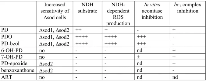
Of these putative PD metabolites, six were tested using the yeast model. Our results are summarised in Table 1. Increased sensitivity of sod cells NDH substrate NDH-dependent ROS production In vitro aconitase inhibition bc1 complex inhibition PD sod1, sod2 ++ + - ±
PDO sod1, sod2 ++++ ++++ +++
-PD-bzol sod1, sod2 ++++ ++++ +++
-6-OH-PD no - - nd +
7-OH-PD no - - ± +
PD-epoxide sod2 - - nd +
benzoxanthone sod2 - - nd
-ART no - - nd nd
Table 1: Effect of PD, PD-metabolites and ART on sod cells growth, on enzyme activities and ROS production. nd: not determined. +, activity; -, no activity
Taken together, our data suggest that most of PD activity on yeast would be mediated through two of its metabolites: PDO and PD-bzol. These two metabolites present a similar profile to PD, but with higher efficiency than PD, in agreement with our current model where PD is a prodrug (Fig.1). (1) As PD, they are alternative substrates of NDH, and are even better substrates than the electron acceptor decylubiquinone, and (2) through this activity, they generate ROS, but in both cases, they are by far more efficient than PD. (3) Cells depleted for Sod1 and Sod2 are highly sensitive to PDO and PD-bzol; sod1 growth was fully inhibited at 6 M PDO and PD-bzol but not abolished at 30
M PD. (4) In contrast to PD, PDO and PD-bzol can directly inhibit aconitase in vitro.
Other metabolites showed milder activities. Cells lacking the mitochondrial Sod2 were also sensitive to PD-epoxide and benzoxanthone, but these metabolites did not act as NDH substrates, suggesting that they may contribute to the generation of oxidative stress in the mitochondria through a different mechanism. Finally, three additional metabolites can inhibit the bc1 complex activity, namely 6-OH-PD, 7-OH-PD and PD-epoxide. Interestingly, the main PD metabolite present in the urine of mice treated with PD was 6-OH-PD and its glucuronic ester derivative [26]. Thus they might also contribute to the antimalarial activity of the parent drug although the three compounds are much weaker inhibitors of the bc1 complex than atovaquone.
The data we have obtained with the yeast model are coherent with the redox cycling model initially proposed (Fig.1) but using NDH instead of glutathione reductase as the main redox-cycling enzyme. The inhibition of yeast growth caused by PD would be explained as follows:
(1) PD, once uptaken by the yeast cells, is oxidised generating PD-bzol and/or PDO. In yeast, as in P. falciparum, there is no CYP450. The reaction could be catalysed by free or protein-bound hemes via ferryl species.
(2) PD-bzol and PDO bind to NDH and accept electrons from NADH via the co-factor FAD. The binding of a menadione into NDH has been modelled in silico [36]. PD-bzol and PDO may bind in a similar manner. (3) Once reduced, PD-bzol and PDO react with O2 to produce superoxide. (4)
Re-oxidized compounds bind again to and are reduced by NDH in a redox-cycling mode. PDO and PD-bzol binding to NDH might also result in the accumulation of flavosemiquinone intermediates, which could react with O2, producing superoxide. The superoxide generated by PDO (and PD-bzol) radicals and NDH flavosemiquinone damage ROS-sensitive enzymes, in particular enzymes containing Fe-S clusters that are likely the first targets of oxidative stress. In addition, the continuous oxidation of NADH results in NADH/NAD imbalance in the cells, possibly affecting the NADPH/NADP balance, increasing the oxidative stress and leading to growth arrest.
Our different assays using the yeast model also indicated that PD and ART do not share the same mode of action. Both compounds block the respiratory growth and their activity is exacerbated by the overexpression of NDH enzymes. However, in contrast to PD and PD metabolites, ART is not a substrate of NDHs in yeast; deletions of Sod1, Sod2 and catalase do not increase yeast sensitivity to ART; ART affects mitochondrial integrity, and ART blocks the fermentative growth.
From yeast to P. falciparum
Like yeast, Plasmodium does not possess a mitochondrial (protonmotive) complex I but a single type-II NDH called NDH2. Menadione can be a substrate of the parasite NDH2 [37]. Thus it seems possible that PD, PD-bzol and/or PDO could also enter into a redox cycle with P. falciparum NDH2. However there is a discrepancy between the expression profile of NDH2 and the parasite stages on which PD is most active. Transcriptomics and proteomics analyses [38-41] suggest that NDH2 is present at relatively low levels over the parasite asexual cycle, with very low expression at the ring stage where PD is most effective. NDH2 reaches its highest levels of expression during gametocytogenesis [42] at a stage where energy metabolism relies on a functional TCA cycle [43-45], while we showed that PD activity is limited to young gametocytes [2].
Still the decreased efficiency of PD at the mid to late gametocyte stages could be due to a lower ability to uptake or bioactivate PD, and the high efficiency of PD at ring stage might not be in contradiction with a possible redox-cycling role of NDH2 as PD might require only a low amount of the enzyme to be effective at killing the parasite, especially if in parallel, antioxidant enzymes are at low levels and thus cannot shield the ROS-sensitive cellular components. Moreover if NDH2 has a high catalytic activity and its diaphorase activity is increased by PD and/or its metabolites, the depletion of cellular NADH, that might result in a decreased NADPH/NADP+ ratio that could further exacerbate the oxidative stress.
In addition, other flavoproteins of both parasite and/or erythrocyte origin may play a role in mediating PD antimalarial activity, especially when they are present at high levels. For instance, the human NADPH-dependent glutathione reductase and methemoglobin are major proteins of the parasitized red blood cells during hemoglobin digestion at the trophozoite stage where they could sustain most of PD redox cycling. Of note, these proteins may be localised in different compartments, within the parasite or in the host erythrocyte. Indeed, moving of drug metabolites between different compartments of the erythrocyte and parasite has already been reported in the case of other redox cyclers with antimalarial properties, such as indolone-N-oxides [46]. In order to explore the role of these enzymes in the antimalarial activity of PD, gene-dosage experiments will be carried out with parasites expressing NDH2 and other flavoenzymes at different levels.
Importantly, ROS-sensitive Fe-S cluster containing enzymes in the parasite, as in the yeast model, could be damaged by PD-produced ROS and by iron chelation by PD metabolites PDO and PD-bzol. It was reported that the TCA cycle, while not needed for parasite survival in asexual blood stages, is
essential for gametocyte and mosquito stages. Aconitase knock-out parasites arrest as gametocytes and fail to transmit to mosquitoes [44], suggesting that ROS damage to aconitase could block or decrease parasite transmission. Thus the effect of PD and other benzylmenadiones should be tested on parasite transmission to mosquitoes.
In conclusion, our study of the mode of action of PD in the yeast model has generated data that are coherent with the model for PD bioactivation and mode of action and with previous observations obtained in vitro and in parasites. It provided additional experimental support to the redox cycling and ROS-producing properties of PD and its metabolites, and identified two metabolites that are more active than PD and could mediate most of PD activity in Plasmodium parasites. In addition, we uncovered the key role of the respiratory chain NADH-dehydrogenases in PD activity in yeast, which provides novel hypotheses to be tested in parasites and during parasite transmission to mosquitoes.
Acknowledgements
The work was supported by the French National Research Agency (ANR) (PlasmoPrim project to B.M, E.D.-C., M-P.G-C. and S.A.B.), by the Laboratoire d’Excellence (LabEx) ParaFrap (grant LabEx ParaFrap ANR-11-LABX-0024 to E.D.-C. and S.A.B.), by the ERC Starting Grant Nº260918 to S.A.B., and by funding from CNRS (E.D.-C. and S.A.B), Inserm (S.A.B), and the University of Strasbourg (E.D.-C. and S.A.B). We are grateful to N. Fisher for his comments on the manuscript.
Legends to figures
Figure 1:(A) Model for plasmodione mode of action in parasitized red blood cells (adapted from [5]). (Step 1) The benzylic oxidation of plasmodione (PD) generates the 3-benzoylmenadione metabolite (PDOox),
a reaction likely catalysed by free or protein-bound hemes in the parasite. (Step 2) PDOox acts as a subversive
substrate of a flavoenzyme and is reduced through the consumption of NAD(P)H, generating PDOred. (Step 3)
Then, PDOred transfers an electron to methemoglobin (metHb(FeIII)), the major form of hemoglobin in the
parasite acidic vacuole, or to O2 leading to high rate of production of superoxide radical anions. (Step 4) At
acidic pH typically found in the parasite food vacuole (H3O+), these radicals dismutate spontaneously to
hydrogen peroxide (H2O2) [47]. (Step 5) The continuous NAD(P)H consumption in the presence of oxygen in
the course of the redox-cycling produces a steady flux of hydrogen peroxide, which reacts with oxyHb(FeII)
and metHb(FeIII) to generate ferrylHb(FeIV=O) leading to (Step 6) hemichrome deposits specifically in
membranes of ring-infected red blood cells (iRBCs). Elevation of hemichrome, a known marker for RBC aging, increases their removal by macrophage phagocytosis [48]. (Step 7) In the parasite, an oxidative phenolic coupling reaction from PDOred might generate a benzoxanthone derivative that was shown to inhibit hemozoin
formation both in a spectrophotometric assay coupled to mass spectrometry and in parasitized RBC [5]. Note that in normal RBCs, where oxyhemoglobin (oxyHb(FeII)) is the major protein, autooxidation and superoxide
production occur slowly in the 3 months-life time of RBCs. In Plasmodium-parasitized RBC, oxyHb(FeII)from
the host is uptaken by the parasite and quickly oxidized into metHb(FeIII) in the acidic food vacuole.
MetHb(FeIII) is digested faster than oxyHb(FeII) by the parasite proteases [49] and is thus its main nutrient
source. Consequently, the reduction of metHb(FeIII) to oxyHb(FeII) by PDO
red (Step 3) is expected to slow
down the parasite growth rate by food deprivation [1,5], and also to improve O2 transport by iRBCs. (B)
Chemical structures of four others putative plasmodione metabolites. (C) Reduced PDO presenting an iron (III) chelation site.
Figure 2: Effect of plasmodione (PD) and artesunate (ART) on yeast growth and respiration. (A) Effect on respiration- and fermentation-supported growth. Yeast AD1-9 cells freshly grown on YPD plates were
inoculated in respiratory medium YPEth or in fermentative medium YP10 with increasing concentrations of PD or ART at an initial OD600nm of 0.2. The cultures were incubated three days at 28°C with vigorous agitation
for YPEth and without agitation for YP10 cultures. The OD600nm were then recorded. For each growth
condition, YP10 or YPEth, the data are presented as percentage of the OD600nm of the untreated cultures. The
growth experiments were repeated at least twice and the data averaged. Error bars represent standard deviation. (B) Effect of PD and ART treatment on cell O2 consumption. Cells were grown for 24h in respiratory medium
YPEth to an OD600nm of around 5, and then incubated for another 24h in YPEth without inhibitor (control) or
with 30 M of PD or 6 M of ART. The cells were then harvested. O2 consumption rates with 20 M CCCP
(with CCCP) and without (w/o CCCP) were recorded. The data, normalised per OD600nm, are presented as %
of control rates (rates without CCCP of untreated cells). Each measurement was repeated at least three times and averaged. Error bars represent standard deviation.
Figure 3: Effect of plasmodione (PD) on the respiratory growth of yeast mutants with lower oxidative stress defence and on the activity of the oxidative stress marker aconitase.
(A) Effect of deletion of oxidative stress defence genes on the sensitivity of respiratory growth to PD. Control and mutant sod1, sod2 and ctt1 strains were grown in YPEth medium with various concentrations of PD. OD600nm were measured after three days. For each strain, the data are presented as percentage of the OD600nm
of the untreated culture. The experiments were repeated at least twice and the data averaged. Error bars represent standard deviation. (B) Effect of PD treatment on aconitase and fumarase activities. The activities were assessed spectrophotometrically at 240 nm. Total cell extracts were obtained from a one day culture in YPGal with or without 12 M PD. The reaction was started by addition of the cell extracts to potassium phosphate buffer containing isocitrate as substrate for the aconitase or malate as substrate for the fumarase. Values represent the averages of five experiments, and error bars, standard deviation. ***, p < 0.001 according to two-tailed Student’s t test.
Figure 4: Effect of flavoenzyme-encoding gene dosage on plasmodione sensitivity. (A and C) Effect of deletion (A) and over-expression (C) of the NADH-dehydrogenase encoding genes NDE1 and NDE2 on PD sensitivity. Control and mutants were grown in YPEth medium with various concentrations of PD. OD600nm
were measured after three days. For each strain, the data are presented as percentage of the OD600nm of the
untreated culture. The experiments were repeated at least twice and the data averaged. (A) AD1-9 control and isogenic deletion mutants nde1 and nde2. ; (C) AD1-9 control and AD1-9 transformed with a multicopy plasmid bearing NDE1 (pNDE1) or NDE2 (pNDE2); (B) NDH activity in the deletion mutants nde1 and nde2, in the over-expressing cells pNDE1 and in pNDE2, and in their respective controls. The rate of NADH oxidation was measured at 340 nm. Mitochondria were added at 60 to 240 g protein per mL. The reaction was initiated by the addition of 100 M decylubiquinone. Experiments were repeated at least twice and data averaged. The error bars represent standard deviation. **, p < 0.005; ***, p < 0.001 according to two-tailed Student’s t test. (D) Effect of the over-expression of flavoprotein encoding genes on PD sensitivity in a nde1 background. Deletion mutant nde1 and derived strains over-expressing NDE1 (pNDE1), NDE2 (pNDE2), GLR1 (pGLR1), LPD1 (pLPD1) or MCR1 (pMCR1) were grown in YPEth medium with increasing concentration of PD. OD600nm were measured after three days. For each strain, the data are presented as
percentage of the OD600nm of the untreated culture. The experiments were repeated at least twice and the data
averaged.
Figure 5: NADH-dehydrogenase activity initiated by plasmodione and its metabolites. NADH oxidation was measured at 340 nm. Mitochondria were added at around 60 g protein per mL. The reaction was initiated by the addition decylubiquinone (DQ), PD, PD-bzol or PDO as substrate at 25, 50 and 100 M. The experiments were repeated at least twice and the data averaged. The error bars represent standard deviation.
Figure 6: SOD and catalase-sensitive O2 consumption activities initiated by plasmodione and its putative
(A) O2 consumption of cyanide-inhibited mitochondria initiated by PD, PD-bzol and PDO. The trace shows
the O2 consumption by mitochondria (added at 18 g.mL) in the presence of 2 mM KCN and 0.8 mM NADH.
PD-bzol was added to a final concentration of 50 M. SOD and catalase were added each at a final concentration of 300 U.mL. (B) Rates of SOD and catalase-sensitive O2 consumption initiated by PD, PD-bzol
and PDO. O2 consumption was monitored as in (A). In white, rate of O2 consumption of cytochrome oxidase
active mitochondria (monitored in the same conditions but in absence of KCN and PD, PD-bzol or PDO). The averages of at least two experiments are shown, with error bars for standard deviation. *, p < 0.05 according to two-tailed Student’s t test. (C) Effect of deletion of oxidative stress defence genes on the sensitivity to putative PD metabolites. Strains were grown in YPEth medium with various concentrations of PD derivatives. OD600nm were measured after three day culture. For each strain, the data are presented as percentage of the
OD600nm of the untreated culture. The experiments were repeated at least twice and the data averaged. The error
bars represent standard deviation.
Figure 7: Effect of plasmodione and its putative metabolites PDO, PD-bzol and 7-OH-PD on purified aconitase activity. mitochondrial aconitase (33 ng.mL-1) was incubated for 20 min at room temperature
with drugs at different concentrations. The reaction was started by addition of cis-aconitate and activity was monitored spectrophotometrically at 240 nm at 37°C. The activity is expressed as percentage of the activity obtained after incubation of the enzyme in the absence of drugs. The average of four measurements is shown with error bars for standard deviation.
Figure 8: Inhibition of the bc1 complex activity by putative plasmodione metabolites. Activities were
determined by measuring the reduction of cytochrome c at 550-540 nm over 1-min time-course Mitochondria were added to obtain a final concentration of around 2 nM bc1 complex. Activity was initiated by the addition
of decylubiquinol and initial rates were measured. The measurements were repeated with various concentrations of drugs. Each point corresponds to the average of at least two measurements, with the error bars representing standard deviation.
References
[1] T. Müller, L. Johann, B. Jannack, M. Brückner, D.A. Lanfranchi, H. Bauer, C. Sanchez, V. Yardley, C. Deregnaucourt, J. Schrével, M. Lanzer, R.H. Schirmer, E. Davioud-Charvet, Glutathione reductase-catalyzed cascade of redox reactions to bioactivate potent antimalarial 1,4-naphthoquinones - A new strategy to combat malarial parasites, J. Am. Chem. Soc. 133 (2011) 11557–11571. doi:10.1021/ja201729z.
[2] K. Ehrhardt, C. Deregnaucourt, A.A. Goetz, T. Tzanova, V. Gallo, P. Arese, B. Pradines, S.H. Adjalley, D. Bagrel, S. Blandin, M. Lanzer, E. Davioud-Charvet, The redox cycler plasmodione is a fast-acting antimalarial lead compound with pronounced activity against sexual and early asexual blood-stage parasites, Antimicrob. Agents Chemother. 60 (2016) 5146–5158. doi:10.1128/AAC.02975-15.
[3] H.J. Painter, J.M. Morrisey, M.W. Mather, A.B. Vaidya, Specific role of mitochondrial electron transport in blood-stage Plasmodium falciparum, Nature. 446 (2007) 88–91. doi:10.1038/nature05572.
[4] K. Ehrhardt, E. Davioud-Charvet, H. Ke, A.B. Vaidya, M. Lanzer, M. Deponte, The antimalarial activities of methylene blue and the 1,4-naphthoquinone
3-[4-(Trifluoromethyl)benzyl]-menadione are not due to inhibition of the mitochondrial electron transport chain, Antimicrob. Agents Chemother. 57 (2013) 2114–2120.
doi:10.1128/AAC.02248-12.
[5] M. Bielitza, D. Belorgey, K. Ehrhardt, L. Johann, D.A. Lanfranchi, V. Gallo, E. Schwarzer, F. Mohring, E. Jortzik, D.L. Williams, K. Becker, P. Arese, M. Elhabiri, E. Davioud-Charvet, Antimalarial NADPH-consuming redox-cyclers as superior glucose-6-phosphate
dehydrogenase deficiency copycats, Antioxid. Redox Signal. 22 (2015) 1337–1351. doi:10.1089/ars.2014.6047.
[6] R. V. Subba-Rao, M. Alexander, Bacterial and fungal cometabolism of 1,1,1-trichloro-2,2-bis(4-chlorophenyl)ethane (DDT) and its breakdown products, Appl. Environ. Microbiol. 49 (1985) 509–516.
[7] J. Segrestaa, P. Vérité, F. Estour, S. Ménager, O. Lafont, Improvement of a biomimetic porphyrin catalytic system by addition of acids, Chem. Pharm. Bull. (Tokyo). 50 (2002) 744– 748. doi:10.1248/cpb.50.744.
[8] T. Nakano, N. Agatsuma, S. Kodama, H. Kakuda, D. Dolphin, A biomimetic study of cytochrome P450 related oxidations of toluenes using synthetic hemin, Bull.Chem.Soc.Jnp. 69 (1996) 3513–3521.
[9] P.P. Kelly, A. Eichler, S. Herter, D.C. Kranz, N.J. Turner, S.L. Flitsch, Active site
diversification of P450cam with indole generates catalysts for benzylic oxidation reactions, Beilstein J. Org. Chem. 11 (2015) 1713–1720. doi:10.3762/bjoc.11.186.
[10] T. Spolitak, P.F. Hollenberg, D.P. Ballou, Oxidative hemoglobin reactions: Applications to drug metabolism, Arch. Biochem. Biophys. 600 (2016) 33–46.
doi:10.1016/j.abb.2016.04.007.
[11] D. Belorgey, D.A. Lanfranchi, E. Davioud-Charvet, dependent glutathione reductase-catalyzed redox cyclers as antimalarial agents, Curr Pharm Des. 19 (2013) 2512–2528. [12] S. Ottilie, G.M. Goldgof, C.M. Calvet, G.K. Jennings, G. LaMonte, J. Schenken, E. Vigil, P.
Kumar, L.-I. McCall, E.S.C. Lopes, F. Gunawan, J. Yang, Y. Suzuki, J.L. Siqueira-Neto, J.H. McKerrow, R.E. Amaro, L.M. Podust, J.D. Durrant, E.A. Winzeler, Rapid chagas disease drug target discovery using directed evolution in drug-sensitive yeast, ACS Chem. Biol. (2016) acschembio.6b01037. doi:10.1021/acschembio.6b01037.
[13] J.-P. Lasserre, A. Dautant, R.S. Aiyar, R. Kucharczyk, A. Glatigny, D. Tribouillard-Tanvier, J. Rytka, M. Blondel, N. Skoczen, P. Reynier, L. Pitayu, A. Rotig, A. Delahodde, L.M. Steinmetz, G. Dujardin, V. Procaccio, J.-P. di Rago, Yeast as a system for modeling mitochondrial disease mechanisms and discovering therapies, Dis. Model. Mech. 8 (2015) 509–526. doi:10.1242/dmm.020438.
[14] N. Fisher, C.K. Castleden, I. Bourges, G. Brasseur, G. Dujardin, B. Meunier, Human disease-related mutations in cytochrome b studied in yeast., J. Biol. Chem. 279 (2004) 12951–8. doi:10.1074/jbc.M313866200.
[15] C. Lemaire, G. Dujardin, Preparation of respiratory chain complexes from Saccharomyces cerevisiae wild-type and mutant mitochondria: activity measurement and subunit
composition analysis, Methods Mol Biol 432: 65-81.
[16] A. Lalève, C. Vallières, M.-P. Golinelli-Cohen, C. Bouton, Z. Song, G. Pawlik, S.M. Tindall, S. V. Avery, J. Clain, B. Meunier, The antimalarial drug primaquine targets Fe–S cluster proteins and yeast respiratory growth, Redox Biol. 7 (2016) 21–29.
doi:10.1016/j.redox.2015.10.008.
Saccharomyces cerevisiae of cytochrome b mutations conferring resistance to atovaquone in Pneumocystis jiroveci, Antimicrob. Agents Chemother. 47 (2003) 2725–2731.
doi:10.1128/AAC.47.9.2725.
[18] Z. Song, B.I. Iorga, P. Mounkoro, N. Fisher, B. Meunier, The antimalarial compound ELQ-400 is an unusual inhibitor of the bc1 complex, targeting both Qo and Qi sites, FEBS Lett. (2018). doi:10.1002/1873-3468.13035.
[19] J. Wang, L. Huang, J. Li, Q. Fan, Y. Long, Y. Li, B. Zhou, Artemisinin directly targets malarial mitochondria through its specific mitochondrial activation., PLoS One. 5 (2010) e9582. doi:10.1371/journal.pone.0009582.
[20] C. Sun, B. Zhou, The antimalarial drug artemisinin induces an additional, Sod1-supressible anti-mitochondrial action in yeast, Biochim. Biophys. Acta - Mol. Cell Res. 1864 (2017) 1285–1294. doi:10.1016/j.bbamcr.2017.04.014.
[21] M.A.H. Luttik, K.M. Overkamp, P. Kötter, S. De Vries, J.P. Van Dijken, J.T. Pronk, The Saccharomyces cerevisiae NDE1 and NDE2 genes encode separate mitochondrial NADH dehydrogenases catalyzing the oxidation of cytosolic NADH, J. Biol. Chem. 273 (1998) 24529–24534. doi:10.1074/jbc.273.38.24529.
[22] F. Gomes, E.B. Tahara, C. Busso, A.J. Kowaltowski, M.H. Barros, nde1 deletion improves mitochondrial DNA maintenance in Saccharomyces cerevisiae coenzyme Q mutants, Biochem. J. 449 (2013) 595–603. doi:10.1042/BJ20121432.
[23] W. Li, W. Mo, D. Shen, L. Sun, J. Wang, S. Lu, J.M. Gitschier, B. Zhou, Yeast model uncovers dual roles of mitochondria in action of artemisinin, PLoS Genet. 1 (2005) e36. doi:10.1371/journal.pgen.0010036.
[24] P.J. McMillan, L.M. Stimmler, B.J. Foth, G.I. McFadden, S. Müller, The human malaria parasite Plasmodium falciparum possesses two distinct dihydrolipoamide dehydrogenases, Mol. Microbiol. 55 (2005) 27–38. doi:10.1111/j.1365-2958.2004.04398.x.
[25] K. Blumenstiel, R. Schöneck, V. Yardley, S.L. Croft, R.L. Krauth-Siegel, Nitrofuran drugs as common subversive substrates of Trypanosoma cruzi lipoamide dehydrogenase and
trypanothione reductase, Biochem. Pharmacol. 58 (1999) 1791–1799. doi:10.1016/S0006-2952(99)00264-6.
[26] L. Feng, D.A. Lanfranchi, L. Cotos, E. Cesar-Rodo, K. Ehrhardt, A.-A. Goetz, H. Zimmermann, F. Fenaille, S.A. Blandin, E. Davioud-Charvet, Synthesis of plasmodione metabolites and 13 C-enriched plasmodione as chemical tools for drug metabolism investigation, Org. Biomol. Chem. 16 (2018) 2647–2665. doi:10.1039/C8OB00227D.
[27] L. Johann, D.A. Lanfranchi, E. Davioud-Charvet, M. Elhabiri, A Physico-biochemical study on potential redox-cyclers as antimalarial and antischistosomal drugs, Curr. Pharm. Des. 18 (2012) 3539–3566. doi:10.2174/138161212801327284.
[28] P. Sidorov, I. Desta, M. Chessé, D. Horvath, G. Marcou, A. Varnek, E. Davioud-Charvet, M. Elhabiri, Redox polypharmacology as an emerging strategy to combat malarial parasites, ChemMedChem. 11 (2016) 1339–1351. doi:10.1002/cmdc.201600009.
[29] H. Beinert, M.C. Kenneday, C.D. Stout, Aconitase as sulfur protein, enzyme, and iron-regulatory protein, Chem. Rev. 96 (1996) 2335–2373.
[30] M.C. Kennedy, M.H. Emptage, J.L. Dreyer, H. Beinert, The role of iron in the activation-inactivation of aconitase, J. Biol. Chem. 258 (1983) 11098–11105.
[31] O. Gawron, A. Waheed, A.J. Glaid, A. Jaklitsch, Iron and aconitase activity, Biochem. J. 139 (2015) 709–714. doi:10.1042/bj1390709.
[32] D. Birth, W.-C. Kao, C. Hunte, Structural analysis of atovaquone-inhibited cytochrome bc1 complex reveals the molecular basis of antimalarial drug action, Nat. Commun. 5 (2014) 4029. doi:10.1038/ncomms5029.
[33] J.S. Doggett, A. Nilsen, I. Forquer, K.W. Wegmann, L. Jones-Brando, R.H. Yolken, C. Bordón, S. a Charman, K. Katneni, T. Schultz, J.N. Burrows, D.J. Hinrichs, B. Meunier, V.B. Carruthers, M.K. Riscoe, Endochin-like quinolones are highly efficacious against acute and latent experimental toxoplasmosis, Proc. Natl. Acad. Sci. U. S. A. 109 (2012) 15936–41. doi:10.1073/pnas.1208069109.
[34] P.H. Alday, I. Bruzual, A. Nilsen, S. Pou, R. Winter, C. Ben Mamoun, M.K. Riscoe, J.S. Doggett, Genetic evidence for cytochrome b Qi site inhibition by
4(1H)-quinolone-3-diarylethers and antimycin in Toxoplasma gondii, Antimicrob. Agents Chemother. 61 (2017) e01866-16. doi:10.1128/AAC.01866-16.
[35] C. Vallières, N. Fisher, B. Meunier, Reconstructing the Qo site of Plasmodium falciparum bc1 complex in the yeast enzyme, PLoS One. 8 (2013) e71726.
doi:10.1371/journal.pone.0071726.
[36] J.N. Blaza, H.R. Bridges, D. Aragão, E.A. Dunn, A. Heikal, G.M. Cook, Y. Nakatani, J. Hirst, The mechanism of catalysis by type-II NADH:quinone oxidoreductases, Sci. Rep. 7 (2017) 40165. doi:10.1038/srep40165.
[37] C.K. Dong, V. Patel, J.C. Yang, J.D. Dvorin, M.T. Duraisingh, J. Clardy, D.F. Wirth, Type II NADH dehydrogenase of the respiratory chain of Plasmodium falciparum and its inhibitors, Bioorganic Med. Chem. Lett. 19 (2009) 972–975. doi:10.1016/j.bmcl.2008.11.071.
[38] L. Florens, M.P. Washburn, J.D. Raine, R.M. Anthony, M. Grainger, J.D. Haynes, J.K. Moch, N. Muster, J.B. Sacci, D.L. Tabb, A.A. Witney, D. Wolters, Y. Wu, M.J. Gardner, A.A. Holder, R.E. Sinden, J.R. Yates, D.J. Carucci, A proteomic view of the Plasmodium falciparum life cycle, Nature. 419 (2002) 520–526. doi:10.1038/nature01107.
[39] E. Lasonder, Y. Ishihama, J.S. Andersen, A.M.W. Vermunt, A. Pain, R.W. Sauerwein, W.M.C. Eling, N. Hall, A.P. Waters, H.G. Stunnenbergt, M. Mann, Analysis of the
Plasmodium falciparum proteome by high-accuracy mass spedrometry, Nature. 419 (2002) 537–542. doi:10.1038/nature01111.
[40] F. Silvestrini, Z. Bozdech, A. Lanfrancotti, E. Di Giulio, E. Bultrini, L. Picci, J.L. DeRisi, E. Pizzi, P. Alano, Genome-wide identification of genes upregulated at the onset of
gametocytogenesis in Plasmodium falciparum, Mol. Biochem. Parasitol. 143 (2005) 100– 110. doi:10.1016/j.molbiopara.2005.04.015.
[41] T.D. Otto, D. Wilinski, S. Assefa, T.M. Keane, L.R. Sarry, U. Böhme, J. Lemieux, B. Barrell, A. Pain, M. Berriman, C. Newbold, M. Llinás, New insights into the blood-stage transcriptome of Plasmodium falciparum using RNA-Seq, Mol. Microbiol. 76 (2010) 12–24. doi:10.1111/j.1365-2958.2009.07026.x.
[42] N. Lang-Unnasch, A.D. Murphy, Metabolic changes of the malaria parasite during the transition from the human to the mosquito host, Annu. Rev. Microbiol. 52 (2002) 561–590. doi:10.1146/annurev.micro.52.1.561.
[43] A. Srivastava, N. Philip, K.R. Hughes, K. Georgiou, J.I. MacRae, M.P. Barrett, D.J. Creek, M.J. McConville, A.P. Waters, E. Ashley, M. Dhorda, R. Fairhurst, C. Amaratunga, P. Lim,
C. Homewood, Z. Bozdech, M. Llinas, B. Pulliam, E. Wong, J. Zhu et al. Stage-specific changes in Plasmodium metabolism required for differentiation and adaptation to different host and vector environments, PLOS Pathog. 12 (2016) e1006094.
doi:10.1371/journal.ppat.1006094.
[44] H. Ke, I.A. Lewis, M. Llina, H. Ke, I.A. Lewis, J.M. Morrisey, K.J. Mclean, S.M. Ganesan, H.J. Painter, M.W. Mather, M. Jacobs-Lorena, M. Llinás, A.B. Vaidya, Genetic investigation of tricarboxylic acid metabolism during the Plasmodium falciparum life cycle, Cell Rep. 11 (2015) 164–174. doi:10.1016/j.celrep.2015.03.011.
[45] J.I. MacRae, M.W. Dixon, M.K. Dearnley, H.H. Chua, J.M. Chambers, S. Kenny, I. Bottova, L. Tilley, M.J. McConville, Mitochondrial metabolism of sexual and asexual blood stages of the malaria parasite Plasmodium falciparum, BMC Biol. 11 (2013) 67. doi:10.1186/1741-7007-11-67.
[46] H. Ibrahim, A. Pantaleo, F. Turrini, P. Arese, J.-P. Nallet, F. Nepveu, Pharmacological properties of indolone-N-oxides controlled by a bioreductive transformation in red blood cells, Med. Chem. Commun. 2 (2011) 860–869.
[47] B.J. Reeder, The Redox Activity of Hemoglobins: From physiologic functions to pathologic mechanisms, Antioxid. Redox Signal. 13 (2010) 1087–1123. doi:10.1089/ars.2009.2974. [48] P. Arese, F. Turrini, E. Schwarzer, Band 3 /complement-mediated recognition and removal
of normally senescent and pathological Human Erythrocytes, Cell. Physiol. Biochem. (2005) 133–146.
[49] H. Jiang, L. Chen, S. Herzberg, X. Shen, R. Hilgenfeld, K. Nagarajan, T. Hogg, C.L.
Schmidt, M. Wecke, C. Blohmke, structural and functional characterization of falcipain-2, a hemoglobinase from the malarial parasite Plasmodium falciparum, J. Biol. Chem. 281 (2006) 25425–25437. doi:10.1074/jbc.m603776200.
CF3 O O CF3 O O O O O HO CF3 Flavoenzyme
oxyHb(FeII) metHb(FeIII)
H2O2
oxyHb(FeII) / metHb(FeIII)
ferrylHb(FeIV=O)
Inhibition of hemozoin formation hemichrome deposits in parasitized RBC membranes Active removal of parasitized RBC by phagocytosis H3O+
metHb(FeIII) depletion
and parasite developement arrest CF3 O O O CF3 O O HO CF3 O O HO CF3 O O OH NADPH NADP+ 1 2 3 7 4 Plasmodione (PD) PD-epoxide
PDOred Benzoxanthone
PD-bzol 6-OH-PD 7-OH-PD A B PDOox Fe CF3 O OH O Fe3+ - PDO complex C CF3 O OH O . O2.- O2 5 6 not digestible digestible, promotes
0 20 40 60 80 100 0 6 12 18 40 80 Drug (µM) PD—YP10 PD—YPEth ART—YPEth ART—YP10 0 50 100 150 200 Control PD ART Sans cccp Avec cccp W/O CCCP with CCCP A B
B A 0.00 0.04 0.08 0.12 fumarase aconitase untreated treatedPD-treated untreated *** 0 20 40 60 80 100 0 10 20 30 Plasmodione (µM) ctt1 sod1 sod2 control
D B 0.0 0.2 0.4 0.6 0.8 1.0 ** *** *** 0 20 40 60 80 100 0 3 6 9 12 Plasmodione (µM) A control nde1 nde2 0 20 40 60 80 100 0 2 4 6 Plasmodione (µM) C control pNDE1 pNDE2 0 20 40 60 80 100 0 3 6 9 12 15 Plasmodione (µM) nde1 pNDE1 pNDE2 pGLR1 pMCR1 pLPD1
Substrate M) 0 0.2 0.4 0.6 0.8 0 50 100 PDO PD-bzol DQ PD
0 50 100 150 200 0 300 600 900 1200 PD-bzol SOD + catalase Time (s) NADH A C 0 50 100 0 6 12 18 Benzoxanthone (µM) 0 50 100 0 6 12 18 PD-epoxide (µM) 0 50 100 0 6 12 18 PD-bzol (µM) 0 50 100 0 6 12 18 PDO (µM) control ctt1 sod1sod2 0.0 0.4 0.8 1.2 1.6 B * *
PDO 7-OH-PD PD PD-bzol 0 20 40 60 80 100 120 0 25 50 75 100 500 1000 Drug (µM)
0 25 50 75 100 0 5 10 15 20 Drug (µM) PD-epoxide 6-OH-PD 7-OH-PD