HAL Id: hal-00994974
https://hal.inria.fr/hal-00994974
Submitted on 8 Dec 2014
HAL is a multi-disciplinary open access
archive for the deposit and dissemination of
sci-entific research documents, whether they are
pub-lished or not. The documents may come from
teaching and research institutions in France or
abroad, or from public or private research centers.
L’archive ouverte pluridisciplinaire HAL, est
destinée au dépôt et à la diffusion de documents
scientifiques de niveau recherche, publiés ou non,
émanant des établissements d’enseignement et de
recherche français ou étrangers, des laboratoires
publics ou privés.
Julien Pansiot, Lionel Reveret, Edmond Boyer
To cite this version:
Julien Pansiot, Lionel Reveret, Edmond Boyer. Combined Visible and X-Ray 3D Imaging. Medical
Image Understanding and Analysis, Jul 2014, London, United Kingdom. pp.13-18. �hal-00994974�
Combined Visible and X-Ray 3D Imaging
Julien Pansiot [email protected] Lionel Reveret [email protected] Edmond Boyer [email protected]INRIA Grenoble Rhône-Alpes 655 Avenue de l’Europe, 38334 Saint Ismier cedex France
Abstract
This paper considers 3D imaging of moving objects and introduces a technique that exploits visible and x-ray images to recover dense 3D models. While recent methods such as tomography from cone-beam x-ray can advantageously replace more expensive and higher-dose CT scanners, they still require specific equipment and immobilised pa-tients. We investigate an alternative strategy that combines a single x-ray source and a set of colour cameras to capture rigidly moving samples. The colour cameras allow for coarse marklerless motion tracking, which is further refined with the x-ray information. Once the sample poses are correctly estimated, a dense 3D attenuation model is recon-structed from the set of x-ray frames. Preliminary results on simulated data compared to ground-truth as well as actual in-vivo experiments are presented.
Keywords: motion capture; tomography; x-ray; colour video.
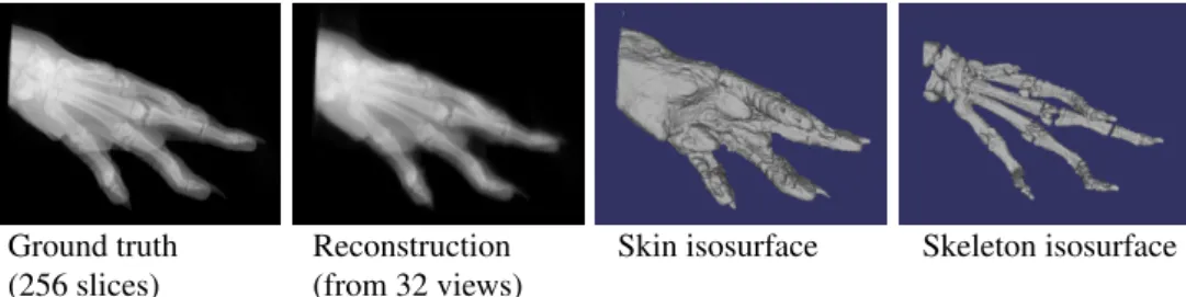
Ground truth Reconstruction Skin isosurface Skeleton isosurface (256 slices) (from 32 views)
Figure 1: Rat paw volumetric reconstruction from a sparse set of simulated visible and x-ray images (32 frames from a rotational motion). Left-to-right: ground truth CT volume, volumetric reconstruction from 32 views rendered as planar x-ray, isosurface for soft tissues interface, and skeleton interface.
1
Introduction and related work
The capture of movements to study functional analysis has gained great attention in the re-cent years with the improvement of acquisition systems. In that respect, the ability to capture simultaneously the motion of both internal structures such as the skeleton and external sur-faces is a challenge with promising applications. Whilst a range of solutions are available
c
2014. The copyright of this document resides with its authors. It may be distributed unchanged freely in print or electronic forms.
to the recovery of surface information. Conversely, radiography allows for the capture of inner structures in motion, but is mostly restricted to 2 dimensions. In this paper we proceed one step towards full volumetric 3D motion capture by investigating the case of rigidly mov-ing samples, relymov-ing on a combination of visible and x-ray cameras, and assummov-ing no prior knowledge on the captured sample.
Our aim is to recover dense 3D models of moving objects. The existing two main strate-gies for radiography are the regular planar x-ray and the Computed Tomography (CT) scan-ner. While the latter produces high-resolution dense 3D models, it suffers three main draw-backs: higher radiation dose for the patient [10], higher cost and, more importantly here, immobility of the patient during the procedure. For these reasons, specific procedures have been developed to generate volumetric attenuation models from a limited number of regular cone-beam x-ray sources, such as full 3D models from isocentric/orbital C-arm [10], and increasingly well resolved models for breast tomosynthesis [11]. Model-based methods us-ing bi-planar x-ray beams [1] even allow motion capture [2], however such methods require strong prior models and usually manual intervention.
In order to compute a dense 3D attenuation model while limiting the patient dose, al-lowing for motion capture and incidentally reducing cost, we propose a novel approach that combines regular colour cameras with standard x-ray imagery to acquire a rigidly but freely moving markerless sample. Our framework keeps all equipment static, eliminating the need for specific isocentric C-arms or other tightly controlled systems [10]. The approach does not use prior anatomic models either, hence this is neither a registration problem [11], nor a model fitting issue [1] and it allows for unknown shapes. The paradigm we follow here does not consider motion as noise but, on the contrary, as one key to 3D reconstruction.
The closest approach to ours has been proposed by Sidky et al. [9] where a reconstruction method for any type of calibrated x-ray imaging device under the assumption of limited angles and/or number of views is presented. Schumacher et al. [8] also proposed a method that combines reconstruction and motion correction in SPECT imaging, without requiring colour cameras. This method is however tailored for motion correction and hence appears to cater only for a limited range of motion. The use of colour video for motion detection during e.g. SPECT has been covered for example by [6], but requires markers. A markerless method has been proposed by [4], but is again targeted to small motion correction.
To the best of our knowledge, our approach is the first to use motion, through the com-bination of visible and x-ray images, to perform 3D radiography. Potential medical appli-cations include affordable dental 3D imaging, 3D imaging in confined environments, and, when non-rigidities will be handled, joints inner dynamics.
The remainder of this paper is composed of the following: in Section2we describe our reconstruction method, in Section3we present our results from both simulated and in-vivo data, before concluding in Section4.
2
Proposed method
The proposed method relies on a single x-ray source combined with a set of regular colour cameras. The colour cameras are used to build a 3D surface model that is tracked over time. The motion estimation of this model is refined using the x-ray images. Once the motion is known, a dense 3D attenuation model can be reconstructed from the x-ray data. The overall pipeline is illustrated in Figure2.
pose estimation pose refinement segmentation reconstruction segmentation
segmentation visual hulls
3D mesh visible img.
x-ray img. silhouettes
initial pose
calibration silhouettes
3D model refined pose
Figure 2: Overview of the proposed pipeline for combined visible and x-ray 3D imaging.
2.1
Calibration
Both visible and x-ray cameras are fully calibrated in world coordinate system prior to ac-quisition. This operation is performed in two stages to minimise the x-ray usage. Firstly, the eight video cameras are fully calibrated (intrinsic and pose) on their own, using a moving LED wand within a single bundle adjustment framework. Secondly, a lightly coloured lead ball is displaced within the entire capture volume, triangulated by the visible cameras, before the x-ray camera is finally fully calibrated knowing the 3D from calibrated video cameras.
2.2
Initial pose estimation
Since the sample can move freely within the capture volume, it is necessary to estimate its pose prior to reconstruction. Using the colour cameras only, the silhouettes of the sample segmented from the background are processed with a polyhedral visual hull algorithm [3] to generate a 3D mesh of the sample per frame (see fig. 2). The remaining meshes of the sequence are then registered to a reference pose model, e.g. the first frame, with a robust Iterative Closest Point (ICP) implementation with outlier detection. This provides an initial pose, i.e. rotation and translation, estimation for each frame and with respect to the reference frame.
2.3
Pose refinement
Since the visual hull meshes exhibit artefacts due to the limited number of colour cameras, the initial pose estimation is corrupted. To improve the pose, we exploit directly the x-ray silhouettes, segmented from the x-ray background light.
To this aim, we rely on the assumption that if the poses were perfectly registered, the volume reprojection on the x-ray plane should equate perfectly to the x-ray silhouettes. For simplicity, we consider the model to be static and captured by moving cameras, with intrinsic parameters from the initial calibration, and extrinsic from the initial pose estimation. A cost function penalising differences between the original x-ray silhouettes and the reprojected model is used within a gradient descent framework to refine the poses iteratively. If C is the set of N camera parameters, I the set of N segmented images, M a mesh, ‘proj’ the camera projection operator, and ‘vh’ the visual hull operator, we try to estimate:
arg min C N
∑
i=1 |projCi(M) − Ii| ! st. M= vhC(I). (1)ble and x-ray systems, providing more robust results.
2.4
Reconstruction
Once the pose is known, the Kaczmarz method [5], also referred to as Algebraic Recon-struction Technique (ART) is used to reconstruct iteratively the volumetric attenuation. For enhanced performance, the views are ordered such that two successive points of view are most orthogonal, and the reconstruction takes places only within the x-ray visual hull.
Given the volume X being reconstructed, the observed images A with a saturated attenu-ation Asat, and a relaxation parameter λi, we exploit the projection matrix sparseness by pre-computing each camera ray defined as the list of the N intersected voxels in the reconstructed volume p and their associated weights w. For each ray Ri= {(pi,1,wi,1), ..., (pi,N,wi,N)} associated to the image pixel value Ai, we then compute the norm, the current integrated attenuation on the ray xi, and the weighted difference with the observed pixel value, di:
kRik2= N
∑
j=1 w2i, j, xi= N∑
j=1 wi, jX[pi, j] , di= λi Ai− xi kRik2 . (2)The voxel attenuations are then updated as follows:
Xn+1[pi, j] = 0 if Xn[pi, j] + diwi, j<0 (positivity constraint), Xn[pi, j] if di<0 and Ai= Asat(prevent saturation artefacts), Xn[pi, j] + diwi, j in other cases.
(3)
The relaxation parameter λiis weighted for each pose based on the leave-one-out score calculated as in eq. (1) in order to favour poses which appear more reliably estimated.
Since the amount and nature of the observed data may render the problem locally ill-posed, high-frequency noise is observed in the results. Using the assumption that living organisms can be modeled by a set of relatively homogeneous tissues, a 3D adaptation of Rudin et al. seminal non-linear Total Variation (TV) [7] is enforced on the model in between iterations for regularisation, as proposed by [9]. This improves dramatically the results, as illustrated in Figure3.
3
Experiments and results
3.1
Simulated data
The combined x-ray and visible images can be simulated by rendering real volumetric CT data using raycasting. In this experiment, we rendered the CT model of a rat paw (2563 voxels), simulating a system of eight colour cameras and one x-ray device. The combined set of 2D images was then processed through the whole pipeline. The effective projection resolution for both systems was 600×600 pixels. The model was artificially moved continu-ously for 32 frames, following roughly a 180-degree rotation. This allowed for comparison with ground-truth data. Figures1and3show the original model as well as the reconstructed volume.
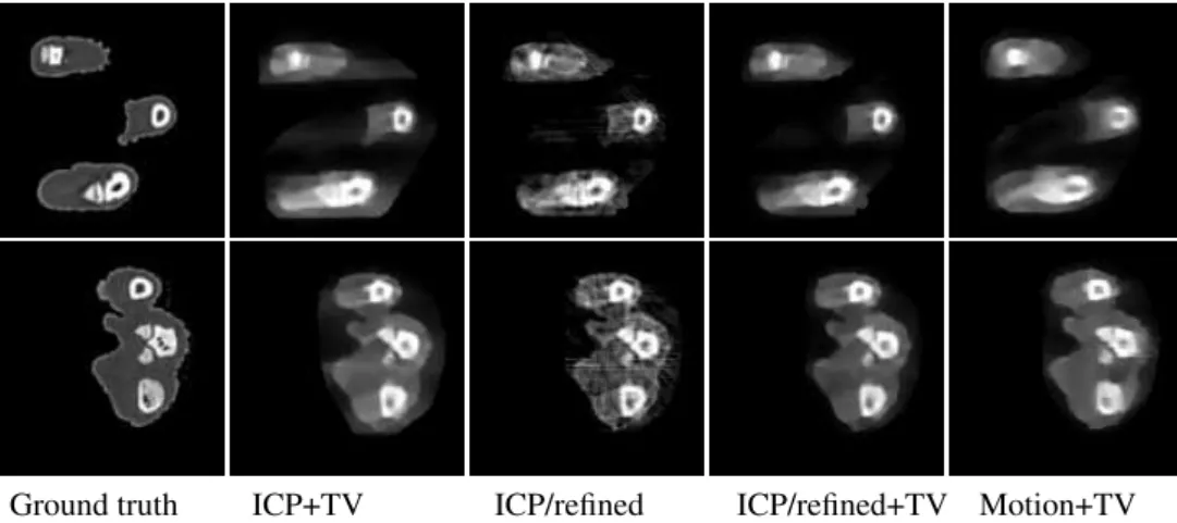
Ground truth ICP+TV ICP/refined ICP/refined+TV Motion+TV
Figure 3: Two rat paw reconstructed slices from simulated visible and x-ray images (32 frames). Left-to-right: ground-truth CT data (256 slices), motion estimated with ICP and TV regularisation, motion refined but without TV, motion refined and TV, true motion and TV. Slight motion estimation errors are clearly corrected by the refinement stage, but not as well as with true motion. TV regularisation reduces significantly the high-frequency noise.
3.2
In-vivo experiment
A system composed of eight colour cameras (2048×2048 pixels) and one Siemens AR-CADIS Avantic x-ray C-arm (1024×1024 pixels) was deployed for this preliminary exper-iment. The reconstruction volume (2563 voxels) covered a cube of 20×20×20 cm, ie. a resolution of 0.78mm/voxel.
Three volunteers (one of them shown here) were asked to perform roughly a 180-degree wrist circumduction with fingers kept as rigid as possible within the working volume, during which 32 frames were captured. The colour cameras recorded at 30 fps, whilst the x-ray acquired at 10 fps to minimise the dose, both being only coarsely synchronised. Figure4
illustrates the preliminary results of the proposed method.
Figure 4: Human hand reconstruction with a combined visible & x-ray system (32 frames). Left-to-right: volume rendered as planar x-ray, soft tissues thresholding, skeleton threshold-ing, skeleton thresholding without pose refinement: some phalanges are thicker, the distal noisier. The soft tissues exhibit a concavity that cannot be modelled solely by visual hulls.
tribute this difference to the fast shutter noise in x-ray images (in particular in the metacarpal area) as well as non strictly rigid motion (on the finger tips and the thumb in particular). The former should be improved by carefully planned x-ray acquisition parameters.
4
Conclusion and future work
In this paper we have shown that a multi-camera system can advantageously complement a single static x-ray imaging device and enable 3D motion imaging of rigidly moving samples. Future work includes the analysis of the achievable quality based on the recorded motion and non-rigid motion handling using rigid-by-part modelling.
Acknowledgments
This project was funded by KINOVIS (ANR-11-EQPX-0024) and MORPHO (ANR-10-BLAN-0206), with help from Amiqual4Home (ANR-11-EQPX-0002).
References
[1] S. Benameur, M. Mignotte, H. Labelle, and J. A. De Guise. A hierarchical statistical modeling approach for the unsupervised 3-D biplanar reconstruction of the scoliotic spine. Biomedical Engineering, IEEE Transactions on, 52(12):2041–2057, 2005.
[2] E. L. Brainerd, D. B. Baier, S. M. Gatesy, T. L. Hedrick, K. A. Metzger, S. L. Gilbert, and J. J. Crisco. X-ray reconstruction of moving morphology (XROMM): precision, accuracy and applications in comparative biomechanics research. Journal of Experimental Zoology Part A: Ecological Genetics and Physiology, 313 (5):262–279, 2010.
[3] J.-S. Franco and E. Boyer. Exact polyhedral visual hulls. In BMVC, pages 329–338, 2003.
[4] B. F. Hutton, A. Z. Kyme, Y. H. Lau, D. W. Skerrett, and R. R. Fulton. A hybrid 3-D reconstruction/registration algorithm for correction of head motion in emission tomography. Nuclear Science, IEEE Transactions on, 49 (1):188–194, 2002.
[5] S. Kaczmarz. Angenäherte auflösung von systemen linearer gleichungen. Bulletin International de l’Académie Polonaise des Sciences et des Lettres. Classe des Sciences Mathématiques et Naturelles. Série A, Sciences Mathématiques, pages 355–357, 1937.
[6] J. E. McNamara, P. H. Pretorius, K. Johnson, J. M. Mukherjee, J. Dey, M. A. Gennert, and M. A. King. A flexible multicamera visual-tracking system for detecting and correcting motion-induced artifacts in cardiac SPECT slices. Medical physics, 36(5):1913–1923, 2009.
[7] L. I. Rudin, S. Osher, and E. Fatemi. Nonlinear total variation based noise removal algorithms. Physica D: Nonlinear Phenomena, 60(1):259–268, 1992.
[8] H. Schumacher, J. Modersitzki, and B. Fischer. Combined reconstruction and motion correction in SPECT imaging. Nuclear Science, IEEE Transactions on, 56(1):73–80, 2009.
[9] E. Y. Sidky, C.-M. Kao, and X. Pan. Accurate image reconstruction from few-views and limited-angle data in divergent-beam ct. Journal of X-ray Science and Technology, 14(2):119–139, 2006.
[10] J. H. Siewerdsen, D. J. Moseley, S. Burch, S. K. Bisland, A. Bogaards, B. C. Wilson, and D. A. Jaffray. Volume CT with a flat-panel detector on a mobile, isocentric C-arm: pre-clinical investigation in guidance of minimally invasive surgery. Medical physics, 32(1):241–254, 2005.
[11] G. Yang, J. H. Hipwell, D. J. Hawkes, and S. R. Arridge. A nonlinear least squares method for solving the joint reconstruction and registration problem in digital breast tomosynthesis. In MIUA, pages 87–92, 2012.