HAL Id: hal-01727385
https://hal.archives-ouvertes.fr/hal-01727385
Submitted on 9 Mar 2018
HAL is a multi-disciplinary open access
archive for the deposit and dissemination of
sci-entific research documents, whether they are
pub-lished or not. The documents may come from
teaching and research institutions in France or
abroad, or from public or private research centers.
L’archive ouverte pluridisciplinaire HAL, est
destinée au dépôt et à la diffusion de documents
scientifiques de niveau recherche, publiés ou non,
émanant des établissements d’enseignement et de
recherche français ou étrangers, des laboratoires
publics ou privés.
Venous Nicking Without Arteriovenous Contact: The
role of the arteriolar microenvironment in arteriovenous
nickings
Michel Paques, Aurélie Brolly, Jonathan Benesty, Nicolas Lermé, Edouard
Koch, Florence Rossant, Isabelle Bloch, Jean-François Girmens
To cite this version:
Michel Paques, Aurélie Brolly, Jonathan Benesty, Nicolas Lermé, Edouard Koch, et al.. Venous
Nick-ing Without Arteriovenous Contact: The role of the arteriolar microenvironment in arteriovenous
nickings. JAMA Ophthalmology, American Medical Association 2015, 133 (8),
�10.1001/jamaoph-thalmol.2015.1132�. �hal-01727385�
Venous Nicking Without Arteriovenous Contact
The Role of the Arteriolar Microenvironment in Arteriovenous
Nickings
Michel Paques, MD, PhD; Aurélie Brolly, MD; Jonathan Benesty, MD, PhD; Nicolas Lermé, PhD; Edouard Koch, MD, PhD; Florence Rossant, PhD; Isabelle Bloch, PhD; Jean-François Girmens, MD
A
rteriovenous nickings (AVNs) may cause branch reti-nal vein occlusion,1and their prevalence correlates witharterial pressure2
and with lacunar stroke.3
Thus, AVNs are surrogates of the retinal and cerebral microcirculations. However, despite its clinical importance, the pathophysiol-ogy of AVNs has received limited attention. This may be be-cause there is a general acceptance of the concept postulat-ing that the vein is crushed by the bypasspostulat-ing artery. This belief persists despite the fact that histologies have consistently re-ported that venous changes were related to perivascular changes described either as glial proliferation,4glial edema,5
or extracellular deposits.6Furthermore, clinical studies of AVNs
using high-resolution imaging have failed to evidence exter-nal compression of veins7
or parietal thickening of arterioles.8
Clinical studies of AVNs are impaired by the extreme vari-ability of their presentation and the difficult access of the ar-teriovenous interface. In this regard, of interest are cases of ve-nous remodeling occurring where an artery and a vein are close yet not overlapping8,9
because the arteriovenous interface is directly observable. Here, we analyzed such cases using
high-resolution imaging to better understand the relationship be-tween the arteriovenous interface and venous changes.
Methods
This institutional clinical study, conducted from January 2013 to September 2014, was carried out according to the prin-ciples outlined in the Declaration of Helsinki. Approval of the Ethics Committee of the Saint-Antoine Hospital (Paris, France) was obtained. Seven adult patients showing venous remod-eling proximal to arterioles were recruited. Each patient re-ceived full oral and written information and gave written con-sent prior to inclusion.
Infrared and 488-nm reflectance imaging and optical co-herence tomography were obtained using combined scan-ning laser ophthalmoscope–optical coherence tomography (Spectralis; Heidelberg Engineering). Adaptive optics (AOs) in-frared fundus images were obtained with a commercially avail-able flood imaging AO camera (rtx1 camera; Imagine Eyes) using a previously described procedure.8The AO camera
illumi-IMPORTANCEArteriovenous nickings (AVNs) in the retina are the cause of retinal vein occlusions and are also surrogates of cerebrovascular aging. The prevalent mechanistic model of AVNs stating that arteries crush veins remains somewhat unchallenged despite the lack of evidence other than fundus photographs. Here, we observed that venous nicking may be observed in the absence of physical contact with an arteriole.
OBSERVATIONS This observational study, conducted from January 2013 to September 2014, included 7 patients showing remodeling of a venous segment close to a retinal arteriole without arteriovenous overlap were imaged by adaptive optics imaging. Affected venous segments showed a variable association of nicking, narrowing, deviation, and opacification. Venous segments were deviated toward the arterioles in 6 of the 7 cases. The degree of venous narrowing ranged from 40% to 77%, while at these sites, the width of the intervascular space ranged from 16 μm to 42 μm. Similar features were identified in typical AVNs.
CONCLUSIONS AND RELEVANCEArteriovenous nickings do not necessarily involve an arteriovenous compression. Instead, the topology of venous changes suggests a retractile process originating in the intervascular space. These findings have important implications for the understanding of retinal vein occlusions and of cerebrovascular aging.
JAMA Ophthalmol. doi:10.1001/jamaophthalmol.2015.1132
Published online May 21, 2015.
Supplemental content at jamaophthalmology.com
Author Affiliations: Quinze-Vingts Hospital, DHU SightMaintain, INSERM-DHOS CIC 1423, Paris, F-75012 and Université Pierre et Marie Curie-Paris 6, France (Paques, Brolly, Benesty, Koch, Girmens); Institut Supérieur d’Electronique de Paris, Paris, France (Lermé, Rossant); Institut Mines Telecom, Telecom ParisTech, CNRS LTCI, Paris, France (Bloch).
Corresponding Author: Michel Paques, MD, PhD, Clinical Investigation Center 1423, Quinze-Vingts Hospital, 28 rue de Charenton, 75012 Paris, France (mp@cicoph.org) .
nates the fundus with a noncoherent light delivered by a su-perluminescent diode at 840 nm, covering a 4° × 4° area (ie, approximately 1.2 mm × 1.2 mm in emmetropic eyes). For im-age acquisition, the gaze of the left eye was oriented with an external target, which allowed the exploration of the fundus of the right eye up to approximately 30° from the disc.
Arteries and veins were semiautomatically segmented from AO images using a custom software running under Matlab (Mathworks).10The segmentation algorithm is based on the
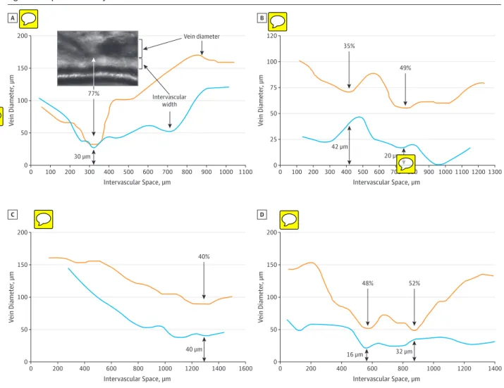
de-tection of 4 curves parallel to the central reflection, which are refined using a deformable model incorporating a parallelism constraint. Graphic representations of the venous diameter and of the width of the intervascular space (ie, the distance be-tween the venous lumen and the outer limit of the arteriolar wall) along a given venous segment were generated.
Results
Seven cases of focal venous remodeling without arteriove-nous crossing from 7 patients were documented. Their mean age was 65.9 years (range, 54-80); 3 were women; and 4 had arterial hypertension. Three were fellow eyes of branch reti-nal vein occlusion. Part of the data from case 4 was previ-ously reported.8
The regions of interest were located along tem-poral arcades, up to 3 mm from the disc. The affected vessels had a diameter of 100 μm to 150 μm. There was unequivocal deviation of the vein toward the arteriole in 6 of the 7 cases. Adaptive optics imaging allowed a high-power view of the ar-teriolar wall, the arteriovenous interface, and the venous mor-phology (Figure 1 and eFigure 1 in the Supplement). Nicking of the vein at the side opposite to the arteriole was noted in 3 cases (1, 4, and 5). Adaptive optics imaging could delineate the area between the outer boundary of the arteriole and the ve-nous lumen. At sites of maximal narrowings, the reduction of
venous diameter ranged from 40% to 77%, while the width of the intervascular space ranged from 16 μm to 42 μm (Figure 2; eFigure 1 in the Supplement).
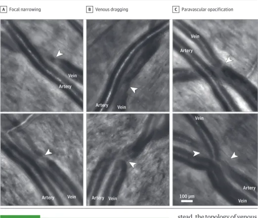
We then examined AO images of common presentations of AVNs (that is, within arteriovenous crossing) to search for similar venous changes. For this, we retrospectively exam-ined AO images of AVNs occurring at arteriovenous crossings from other eyes to determine whether similar changes may be present (Figure 3). Focal narrowing, venous dragging toward the arteriole, and paravascular opacification in AVNs were pre-sent; some combined several of these features.
Discussion
There has been a longstanding yet unsubstantiated consen-sus among clinicians about the compressive nature of the ar-teriovenous conflict underlying AVNs. Our findings support the conclusions of histology and in vivo studies suggesting that the paradigm of arterial crushing as the cause of venous nick-ing stems from a misinterpretation of fundus photographs.4
We indeed found here that veins close to arterioles, but with-out detectable arteriovenous contact, may undergo marked phenotypical changes comprising nicking, narrowing, opaci-fication, and/or dragging. Nicking of the venous wall on the side opposite to the arteriole, venous dragging toward the ar-teriole, and the presence of an arteriovenous gap up to 40 μm do not fit easily with the hypothesis of an arterial compres-sion. Similar findings were retrospectively evidenced in AVNs occurring with arteriovenous overlap from other patients, which suggest that classic AVNs may also imply indirect arte-riovenous interaction.
Our findings suggest that the interaction between arter-ies and veins leading to AVNs is mediated by the paravascular milieu. Such hypothesis of an indirect arteriovenous interac-Figure 1. Adaptive Optics Images of Arteriovenous Interfaces of Venous Nickings
A B C D Artery Artery Artery Artery Vein Vein Vein Vein
In each panel, the magnification is shown to the right of the original adaptive optics image. The arteriolar wall (between white arrowheads) does not enter in contact with the venous lumen. The black arrowheads indicate peak venous narrowing (bars = 125 μm; see also eFigure 1 in the Supplement).
tion has been previously proposed.11Our results suggest that
retraction of the intervascular space plays a key role in ve-nous changes; however, this somewhat conflicts with histol-ogy findings, which would rather imply its expansion. There-fore, the various ophthalmoscopic presentations of AVNs may result from the combined effects of hypertrophy and retrac-tion of the intervascular space. The common adventicia and/or the inner limiting membrane may provide a scaffold transmit-ting retractile forces. The lower mechanical resistance of veins would explain their predominant deviation.
The process that initiates such arteriovenous interaction remains to be characterized. Molecular compounds in the cir-culation and/or derived from the metabolism of arteries may leak into the arteriolar microenvironment and modify glial, mi-croglial, and/or venous metabolism. The factors mediating such arteriovenous interaction may either be immunomodulators or vasomodulators. It is known that AVNs are related to bio-logical markers of inflammation12
and that veins are respon-sive to vasoactive compounds.13Chronically increased
ve-nous tone could induce rearrangement of the scaffold of smooth muscle cells of veins, similarly to what is observed in
arterioles during arterial hypertension.14The role of venous
flow turbulence in this process is unclear because laminar flow may be present even in severe venous narrowings (eFigure 2 in the Supplement).
There is indirect evidence that AVNs may regress,15
sug-gesting that AVNs have a dynamic component. Therefore, fur-ther investigations may address the natural history of AVNs. Histology of arteriovenous crossings may address glial cells, the arrangement of intracellular actin networks, and/or the ex-tracellular matrix. The presence of vasoactive or inflamma-tory metabolites, in particular small and/or lipophilic molecules,11which may cross the blood-retinal barrier, may also
be explored. It is unclear whether experimental studies will be contributive because we have as of yet failed to identify AVNs in rodent and nonhuman primate eyes (M.P.; unpub-lished data; 2013). Given the proximity of the central artery and vein within the optic nerve, it is plausible that central retinal vein occlusion may be related to a similar interaction. It may also be of interest to document whether similar changes oc-cur in the brain.
Figure 2. Morphometric Analysis of Arteriovenous Interfaces
200 150 100 50 0 0 100 200 300 800 900 1000 1100 V ein Diameter , µm Intervascular Space, µm 600 700 400 500 100 300 500 700 900 1100 A Vein diameter Intervascular width 77% 100 120 50 75 25 0 0 200 800 1000 1200 1300 V ein Diameter , µm Intervascular Space, µm 600 400 B 30 µm 35% 49% 42 µm 20 µm 200 150 100 50 0 0 200 800 1000 1200 1400 1600 V ein Diameter , µm Intervascular Space, µm 600 400 C 200 100 150 50 0 0 200 800 1000 1200 1400 V ein Diameter , µm Intervascular Space, µm 600 400 D 40 µm 48% 52% 40% 16 µm 32 µm
Variations of lumen diameter (orange lines) and of intervascular space (blue lines) were measured along venous segments. The black arrows point to venous narrowings.
Conclusions
In summary, this study found that arteriovenous nickings do not necessarily involve an arteriovenous compression.
In-stead, the topology of venous changes suggests a retractile pro-cess originating in the intervascular space. These findings have important implications for the understanding of retinal vein occlusions and cerebrovascular aging.
ARTICLE INFORMATION
Submitted for Publication: December 17, 2014; final revision received March 12, 2015; accepted March 16, 2015.
Published Online: May 21, 2015. doi:10.1001/jamaophthalmol.2015.1132. Author Contributions: Drs Paques and Brolly had full access to all of the data in the study and take responsibility for the integrity of the data and the accuracy of the data analysis. All authors had access to data and analysis and participated in the redaction of the manuscript.
Study concept and design: Paques, Benesty, Lermé,
Rossant.
Acquisition, analysis, or interpretation of data:
Paques, Brolly, Koch, Bloch, Girmens.
Drafting of the manuscript: Paques, Brolly, Benesty,
Rossant.
Critical revision of the manuscript for important intellectual content: Paques, Lermé, Koch, Bloch,
Girmens.
Obtained funding: Paques.
Administrative, technical, or material support:
Benesty, Lermé, Koch, Rossant.
Study supervision: Paques, Bloch.
Conflict of Interest Disclosures: All authors have completed and submitted the ICMJE Form for Disclosure of Potential Conflicts of Interest. Dr Paques is a consultant for Imagine Eyes, the manufacturer of the adaptive optics camera used in
this study, and has received personal fees from Merck Serono. Dr Girmens has received grants and/or personal fees from Allergan, Bayer, Novartis, and Sanofi–Fovea Pharmaceuticals. No other disclosures were reported.
Funding/Support: This study was supported by the Institut National de la Santé et de la Recherche Médicale (Contrat d’Interface 2011), the Agence Nationale de la Recherche (ANR-09-TECS-009 and ANR-12-TECS-0015-03), and the Association Contre l’OVR.
Role of the Funder/Sponsor: The funders had no role in the design and conduct of the study; collection, management, analysis, and interpretation of the data; preparation, review, or approval of the manuscript; and decision to submit the manuscript for publication.
Previous Presentation: This study was presented in part at the 29th Annual Jules Gonin Meeting; September 6, 2014; Zurich, Switzerland.
REFERENCES
1. Klein R, Klein BE, Moss SE, Meuer SM. The epidemiology of retinal vein occlusion: the Beaver Dam Eye Study. Trans Am Ophthalmol Soc. 2000; 98:133-141.
2. Wong TY, Klein R, Nieto FJ, et al. Retinal microvascular abnormalities and 10-year
cardiovascular mortality: a population-based case-control study. Ophthalmology. 2003;110(5): 933-940.
3. Hanff TC, Sharrett AR, Mosley TH, et al. Retinal microvascular abnormalities predict progression of brain microvascular disease: an atherosclerosis risk in communities magnetic resonance imaging study.
Stroke. 2014;45(4):1012-1017.
4. Seitz R. The Retinal Vessels: Comparative
Ophthalmoscopic and Histologic Studies on Healthy and Diseased Eyes. Saint Louis, MO: CV Mosby Co;
1964.
5. Kimura T, Mizota A, Fujimoto N, Tsuyama Y. Light and electron microscopic studies on human retinal blood vessels of patients with sclerosis and hypertension. Int Ophthalmol. 2005;26(4-5):151-158. 6. Jefferies P, Clemett R, Day T. An anatomical study of retinal arteriovenous crossings and their role in the pathogenesis of retinal branch vein occlusions. Aust N Z J Ophthalmol. 1993;21(4):213-217.
7. Kumagai K, Tsujikawa A, Muraoka Y, et al. Three-dimensional optical coherence tomography evaluation of vascular changes at arteriovenous crossings. Invest Ophthalmol Vis Sci. 2014;55(3): 1867-1875.
8. Koch E, Rosenbaum D, Brolly A, et al. Morphometric analysis of small arteries in the human retina using adaptive optics imaging:
Figure 3. Adaptive Optics Images of Classic Arteriovenous Nickings (With Arteriovenous Crossings)
Focal narrowing
A B Venous dragging C Paravascular opacification
Artery Artery Artery Artery Artery Artery Vein Vein Vein Vein Vein Vein Vein 100 µm
The arrowheads show the site of focal narrowing (A), dragging of the vein toward the artery (B), and paravascular opacification (C).
relationship with blood pressure and focal vascular changes. J Hypertens. 2014;32(4):890-898. 9. Wise GN, Dollery CT, Henkind P. The Retinal
Circulation. New York, NY: Harper & Row Publishers
Inc; 1971: 231.
10. Lermé N, Rossant F, Bloch I, et al. Segmentation of retinal arteries in adaptive optics images. International Conference on Pattern Recognition. 2014; 574-578.
11. Fraenkl SA, Mozaffarieh M, Flammer J. Retinal vein occlusions: the potential impact of a dysregulation of the retinal veins. EPMA J. 2010;1 (2):253-261.
12. Klein R, Sharrett AR, Klein BE, et al. Are retinal arteriolar abnormalities related to atherosclerosis? the Atherosclerosis Risk in Communities Study.
Arterioscler Thromb Vasc Biol.
2000;20(6):1644-1650.
13. Raffetto JD, Barros YV, Wells AK, Khalil RA. MMP-2 induced vein relaxation via inhibition of
[Ca2+]e-dependent mechanisms of venous smooth muscle contraction: role of RGD peptides. J Surg Res. 2010;159(2):755-764.
14. Mulvany MJ. Small artery remodeling in hypertension. Curr Hypertens Rep. 2002;4(1):49-55. 15. Liew G, Campbell S, Klein R, et al. Ten-year longitudinal changes in retinal microvascular lesions: the atherosclerosis risk in communities study. Ophthalmology. 2011;118(8):1612-1618.