HAL Id: tel-01529536
https://tel.archives-ouvertes.fr/tel-01529536
Submitted on 31 May 2017HAL is a multi-disciplinary open access
archive for the deposit and dissemination of sci-entific research documents, whether they are pub-lished or not. The documents may come from teaching and research institutions in France or abroad, or from public or private research centers.
L’archive ouverte pluridisciplinaire HAL, est destinée au dépôt et à la diffusion de documents scientifiques de niveau recherche, publiés ou non, émanant des établissements d’enseignement et de recherche français ou étrangers, des laboratoires publics ou privés.
G4-Hunter : a new algorithm for G-quadruplexes
prediction’s
Amina Bedrat
To cite this version:
Amina Bedrat. G4-Hunter : a new algorithm for G-quadruplexes prediction’s. Human genetics. Université de Bordeaux, 2015. English. �NNT : 2015BORD0197�. �tel-01529536�
THÈSE PRÉSENTÉE
POUR OBTENIR LE GRADE DE
DOCTEUR DE
L’UNIVERSITÉ DE BORDEAUX
ÉCOLE DOCTORALE
: Sciences de la Vie et de la Santé
SPÉCIALITÉ :
Bioinformatique
Par :
Amina Bedrat
G4-Hunter : un nouvel algorithme
pour la prédiction des
G-quadruplexes
Sous la direction de :
Dr. Jean-Louis Mergny
Soutenue le : 06/11/2015
Mention : Très honorable
Membres du jury :
Pr. Roger Marthan
- Université de Bordeaux (France)
Président
Dr. Geneviève Pratviel
- Université de Toulouse (France)
Rapporteur
Pr. Jean-Pierre Perreault
- Université de Sherbrooke (Canada) Rapporteur
Dr. Patrizia Alberti
- CNRS/ INSERM/ MNHN (France) Examinateur
Dr. Marie Beurton-Aimar
- Université de Bordeaux (France) Examinateur
G4-Hunter : un nouvel algorithme pour la prédiction des G-quadruplexes.
Des séquences compatibles avec la formation de G4 sont présentes au niveau de certaines régions clé du génome telles que les extrémités des chromosomes, mais également les régions de commutation de classe des immunoglobulines, les promoteurs de certains gènes dont des oncogènes et des séquences transcrites. Plus de 370 000 cibles potentielles ont été prédites lors des analyses bioinformatique du génome humain. Cependant, ces prédictions ne sont pas exhaustives étant limitées par la formulation des algorithmes de prédictions utilisés. En effet, les séquences recherchées suivent la formule consensus suivante G3+N(1−7)G3+N(1−7)G3+N(1−7)G3+. Ainsi, en apportant plus de souplesse dans la description du quadruplex nous pourrons identifier et localiser plus de cibles potentielles. C’est pourquoi, nous proposons un nouvel algorithme G4-Hunter qui permettra l’identification la plus exhaustive possible de séquences cibles en prenant en compte la totalité de la région et non plus uniquement la cible potentielle. Par ailleurs, une étude expérimentale à grande échelle (sur une centaine de séquences cibles) a été menée afin de valider et tester la robustesse de G4-Hunter. A l’aide de ce nouvel outil, nous avons pu identifier de nouvelles séquences cibles non identifiées par les approches déjà existantes au sein des génomes humain, HIV et Dictyostelium discoideum.
Mots clés : [Algorithme, Bioinformatique, G-quadruplex, ADN, Mitochondrie.]
G4-Hunter: a new algorithm for G-quadruplexes prediction’s.
Biologically relevant G4 DNA structures are formed throughout the genome including immunoglobulin switch regions, promoter sequences and telomeric repeats. They can arise when single-stranded G-rich DNA or RNA sequences are exposed during replication, transcription or recombination. Computational analysis using predictive algorithms suggests that the human genome contains approximately 370 000 potential G4-forming sequences. These predictions are generally limited to the standard G3+N(1−7)G3+N(1−7)G3+N(1−7)G3+ description. However, many stable G4s defy this description and escape this consensus; this is the reason why broadening this description should allow the prediction of more G4 loci. We propose an objective score function, G4-hunter, which predicts G4 folding propensity from a linear nucleic acid sequence. The new method focus on guanines clusters and GC asymmetry, taking into account the whole genomic region rather than individual quadruplexes sequences. In parallel with this computational technique, a large scale in vitro experimental work has also been developed to validate the performance of our algorithm in silico on hundred of different sequences. G4-hunter exhibits unprecedented accuracy and sensitivity and leads us to reevaluate significantly the number of G4-prone sequences in the human genome. G4-hunter also allowed us to predict potential G4 sequences in HIV and Dictyostelium discoideum, which could not be identified by previous computational methods.
Keywords : [Algorithm, Bioinformatic, G-quadruplex, DNA, Mitochondria.]
Inserm U1212 (U869)
Resum´e
Les acides nucl´eiques (ADN et ARN) sont des composants cellulaires essentiels au d´eveloppement et au fonctionnement des organismes dont toutes atteintes (mutation, d´el´etion, insertion ...) sont impliqu´ees dans de nombreuses pathologies.
Depuis 1879, la composition des acides nucl´eiques est connue : sucre (d´esoxyribose et ribose respectivement pour ADN et ARN), phosphate et bases azot´ees (Ad´enosine A, Thymine T, Cytosine C, Guanine G), ce n’est qu’en 1953 que James Watson et Francis Crick d´eterminent la structure de l’ADN. Ils proposent le mod`ele de la double-h´elice, qui est l’appariement de deux brins anti parall`ele par des liaisons hydrog`ene entre les bases A-T et G-C.
Cependant, les acides nucl´eiques peuvent adopter des structures diff´erentes. Elles sont dues aux diff´erents arrangements possibles entre les bases azot´ees tels que l’appariement Watson-Crick invers´e, l’appariement bancal et les liaisons Hoogsteen ou Hoogsteen invers´e.
Parmi ces structures nous retrouvons:
• Simple-brin; structure tige-boucle ou ´epingle `a cheveux, • Double-brins; conformation A, B et Z de l’ADN,
• Triplexes; form´es par l’insertion d’un simple-brin dans le sillon d’une double h´elice et constitu´es d’appariement de type Watson-Crick et Hoogsteen,
• Quadruplexes; repliement d’un, deux ou quatre brins d’ADN ou ARN.
Ainsi, les s´equences ADN ou ARN riches en guanines peuvent adopter une structure diff´erente de la double-h´elice classique, et se replier en structures appel´ees quadruplexes (”G4”). Cette conformation repose sur la formation de quartets de guanines, o`u quatre guanines coplanaires ´etablissent un r´eseau cyclique de liaisons hydrog`ene. Il a ´et´e d´emontr´e que la stabilit´e de ces structures est assur´ee par la nature et la concentration du cation (K+, Na+)
pr´esent entre les quartets cons´ecutifs.
Par ailleurs, ces structures pr´esentent un polymorphisme structurel important dˆu entre autre `a la mol´ecularit´e et l’orientation des brins.
Les G-quadruplexes peuvent ˆetre constitu´es de quatre (intermol´eculaires), de deux (dimer) ou d’un seul brin (mono- ou intramol´eculaire) d’ADN.
L’orientation des brins est ´egalement responsable de la diversit´e structurale des G4. Quatre possibilit´es sont d´ecrites; (i) les quatre brins sont orient´es dans la mˆeme direction (G4 parall`ele), (ii) un des brins est orient´e dans la direction oppos´ee des autres (G4 “hybride” ou (3+1)),
iv
(iii) les deux brins voisins ou diam´etralement oppos´es sont orient´es dans une direction et les 2 autres dans la direction oppos´ee (G4 anti parall`ele).
Des s´equences compatibles avec la formation de G4 sont pr´esentes au niveau des r´egions r´ep´et´ees des t´elom`eres, trouv´ees aux extr´emit´es des chromosomes, et le promoteur de nombreux oncog`enes tel que : c-myc, c-kit [51][50]. Il est suppos´e qu’elles jouent un rˆole important dans la r´egulation de la transcription, la translation[222] et la r´egulation de l’expression des g`enes. Les G4 ne sont pas uniquement sp´ecifiques aux eucaryotes. Ainsi, des s´equences avec le potentiel de former des G4 ont ´et´e identifi´ees au sein des virus. En effet, le g´enome du papillomavirus (HPVs) pr´esente sept s´equences avec un potentiel de former un G4 stable.
Les G-quadruplexes semblent jouer ainsi un rˆole dans de nombreux processus cellulaires tels que le contrˆole de l’expression g´enique, la r´eplication ou l’´epissage et repr´esenteraient ainsi des cibles th´erapeutiques d’int´erˆet. D’o`u la n´ecessit´e d’avoir `a disposition des outils fiables permettant d’identifier les s´equences `a fort potentiel in silico.
Historiquement, le premier algorithme de pr´ediction d´evelopp´e est Quadparser [19].
Quadparser1 repose sur l’identification du motif pr´ed´efini,
d(G3+N(1−7)G3+N(1−7)G3+N(1−7)G3+) (N= A, T, C ou G). Ainsi les s´equences
iden-tifi´ees sont des G4 intramol´eculaires, avec trois quartets et plus, sans discontinuit´e dans les blocs de guanine et des boucles de 7 bases. Les s´equences pr´edites peuvent potentiellement former un G4 en conditions physiologique (100mM KCl, 10mM Tris-HCl (pH 7.4))
Ecrit en language C, Quadparser identifie de nouvelles s´equences `a partir de donn´ees g´enomiques au format FASTA. Le format de sortie et les motifs recherch´es peuvent ˆetre personnalis´es. Ainsi, plus de 370000 s´equences ont ´et´e identifi´ees lors de l’analyse du g´enome humain.
D’autres algorithmes accessible en ligne peuvent ´egalement ˆetre utilis´es. QGRS Mapper (Quadruplex forming G-Rich Sequences) est l’un de ces algorithmes [92]. L’interface d’analyse permet `a l’utilisateur de moduler le motif recherch´e en intervenant sur le nombre de guanines constituant les quartets (au minimum 2 selon les r`egles de l’algorithme), la taille de la s´equence recherch´ee et non seulement la taille des boucles (0 `a 36 par d´efaut ) mais aussi la composition de celle-ci et cela par de simple expression r´eguli`ere. L’algorithme attribue `a chacune des s´equences identifi´ees un score. Le calcule du score est d´ependant de trois principes :
• Les petites boucles sont plus communes que les longues, • Les G4 ont tendance `a avoir des boucles de mˆeme tailles,
• Plus le nombre de bloc de guanine est ´elev´e plus le G4 est stable.
Ainsi, plus le score est ´elev´e plus la s´equence pr´edite a un potentiel G4 ´elev´e.
QGRS Mapper, ´ecrit en PHP et JAVA (pour les graphiques), analyse des sequences d’ADN et d’ARN en utilisant les donn´ees de NCBI ou des s´equences fournis par l’utilisateur au format brute ou FASTA. Les pages de sortie regroupent un ensemble d’information sous forme de tableaux et de graphiques en pr´ecisant la localisation des exons ainsi que la localisations des sequences identifi´ees.
v
L’interface web Quadfinder2, permet non seulement de pr´edire de nouveaux G4 `a l’aide de
Quadparser mais ´egalement de d´efinir la stabilit´e thermodynamique grˆace au Bayesian learning algorithme. Contrairement aux algorithmes d´evelopp´es jusqu’ici, ddiQFP (duplex-derived interstrand Quadruplex Forming Potentiel) impl´ement´e en Perl, permet d’identifier des G4 intermol´eculaires pr´esents au sein de sites cibl´es par l’h´elicase pif1 [100].
Beaudoin et al proposent un algorithme d´edi´e `a la recherche de G4 au sein des ARN [97]. Le programme calcule le score d’une s´equence ARN en fonction du score des blocs de guanine/-cytosine. Cet algorithme est caract´eris´e par une grande sensibilit´e et sp´ecificit´e et comme un outil compl´ementaire au outils d´ej`a existants.
Ces ´etudes bioinformatiques ont identifi´e plusieurs s´equences et d´emontr´e comment les quadruplexes sont distribu´es dans le g´enome. La diversit´e structurelle des quadruplexes observ´es, propose un motif diff´erent de celui recherch´e par ces algorithmes ainsi le nombre de quadruplexes pr´edit peut ˆetre plus ´elev´e.
En effet, ces ´etudes se basent sur des informations structurelles (recherche de motif) et des analyses comparatives de la localisation des ces s´equences [84]. L’analyse est r´ealis´ee uniquement sur de petites s´equences ce qui limite la d´etection de s´equences avec de longues boucles qui peuvent former des G4 [16]. La s´equence c-myc retrouv´ee au sein du promoteur de ce mˆeme g`ene pr´esente un polymorphisme. En effet, dans certaines condition salines c-myc ne suit pas le motif rechercher [20]. De mˆeme des s´equences avec plus de trois quartets peuvent ´egalement exister [101].
Les m´ethodes de calcule de score quant `a elles se basent sur des caract´eristiques de la s´equence tels que la longueur de la boucle le nombre de groupe de guanines ou les s´equences avoisinantes. Clairement ces outils pr´edisent de nouvelles s´equences en fonction de caract´eristiques de s´equences d´ej`a existantes mais leurs pr´edictions sont limit´ees et un grand nombre de faux n´egatifs sont g´en´er´es. La recherche d’un nouvel outils de pr´ediction et donc une n´ec´essaire.
Nous proposons un nouvel outil qui, • Favorise les blocs de guanines,
• Favorise l’asym´etrie G/C (recherche de G4 dans les deux brins),
• Repousse les limites des outils existent (longueur de la boucle, nombre de quartets), • Recherche de motifs insolites.
A partir d’une id´ee simple, l’algorithme recherche des s´equences formant des G4 par calcule de score, ce score d´epende uniquement de la composition en bases de la s´equence. En effet les bases de la s´equence sont converties en chiffres (de -4 `a +4 selon le nombre de guanines cons´ecutives) et le score repr´esente la moyenne d’une fenˆetre de 25 `a 100 nucl´eotides. Plus la valeur absolue du score est ´elev´e plus la s´equence est susceptible de former un G4.
vi
J’ai tout d’abord impl´ement´e l’algorithme en Python prend en entr´ee un fichier au format FASTA (qui peut comprendre une ou plusieurs s´equence FASTA) et produit deux fichiers texte (un fichier qui repr´esente les scores de chaque fenˆetres et un fichier qui regroupe les s´equences chevauchantes avec leurs nouveau score ) et une repr´esentation graphique des scores obtenus.
j’ai ensuite analys´e un ensemble de s´equences formant ou non un G4, issue de la litt´erature et valid´es exp´erimentalement, afin de valider notre algorithme. Le score, pour une fenˆetre de 25 nucl´eotides, est calcul´e pour ces deux groupes de s´equences (G4, non G4) et pour une (P value
<0.001) nous retrouvons une diff´erence significative entre les deux distributions du score.
J’ai ´egalement analys´e l’ADN mitochondrial avec l’algorithme que nous avons d´evelopp´e et compar´e les s´equences ainsi obtenus avec celles identifi´ees par Quadparser. Cent soixante sept candidats ont ´et´e identifi´e (avec un score > 1 et une fenˆetre d’analyse de 25 nucl´eotides). En revanche, lors de l’analyse avec Quadparser (G= 2 `a 5 et N = 1 `a 7) 81 candidats ont ´et´e identifi´es dont 23 uniquement identifi´e par cet algorithme. Ces s´equences de plus petite taille que notre fenˆetre d’analyse (< 25 nucl´eotides), ont un score inf´erieur `a 1.
Une fois pr´edite il est n´ecessaire de valider exp´erimentalement la capacit´e des s´equences `a former un G4. De nombreuses techniques sont d´ecrites. Au sein de notre ´equipe, nous utilisons principalement (i) les techniques de spectroscopie UV: d´enaturation thermique (Thermal merlting-Tm) [103], spectres de diff´erence thermiques (Thermal Difference Spectra TDS) [102], spectre de diff´erences isotherme (Isothermal Difference Sperctra IDS) [109] et Dichro¨ısme Circulaire (CD) [107], (ii) R´esonance Magn´etique Nucl´eaire (RMN) [22] (iii) et un test de fluorescence d´evelopp´e au sein de l’´equipe, le test de la thioflavine T [104].
J’ai r´ealis´e une ´etude exp´erimentale de grande ampleur qui consiste `a tester exp´erimentalement tous les nouveaux candidats. Ainsi 75% des s´equences pr´edites for-ment un G4 pour score >1.
24% des candidats dont le score est entre 1 et 1.25 ne forme pas de G4 ou de conformation inconnue (nos tests biophysique n’ont pas ´et´e concluants) . Au del`a d’un score de 1.5 les s´equences ont tendance `a former un G4 stable et au del`a d’un score de 2 toutes les s´equences forment des G4 stables.
Le nombre de G4 form´es au sein de la mitochondrie est plus grand que le nombre de G4 pr´edit par Quadparser. Ainsi, le nouvel algorithme r´epond aux conditions d´ej`a pos´ees et propose une nouvelle liste de G4 test´ee et valid´ee exp´erimentalement. A partir de cette ´etude, nous avons choisi la valeur 1 comme valeur seuil du score pour les nouvelles recherches.
En collaboration avec Amrane S., nous avons identifi´e 10 candidats suite `a l’analyse d’un alignement de 1684 g´enomes d’HIV-1. La r´egion conserv´ee riche en guanine du promoteur du HIV-1, connue pour r´eguler sa transcription, forme un G4 antiparall`ele stable [189]. Nous avons aussi brevet´e cette s´equences pour son potentiel d’inhiber le virus d’HIV.
vii
Enfin nous avons d´evelopp´e un nouvel outil bioinformatique d´edi´e `a la recherche de nouveaux G4, sa fiabilit´e est d´emontr´e par une analyse des deux g´enomes : la mitochondrie et HIV-1. Ainsi, Nous souhaitons pour la suite de cette th`ese proposer une interface graphique qui rend l’utilisation de notre algorithme facile, am´eliorer les param`etres de recherche ( fenˆetre et seuil du score ), rechercher de nouveaux G4 dans d’autre g´enomes tels que Dictyostelium sp.,
Acknowledgements
I would like to thank the entirely and especially Merciful. I thank my parents and my brother, the true love.
I thank my PhD supervisor, the big boss ever.
I thank Marie-No¨el B.A. & Patricia T., the best souls that a foreigner can meet. I thank my traveling friend, my lab brother and ma cherie.
I thank every single person I meet in my life. I thank all the members of my thesis committee.
Contents
State of the art 1
1 Guanine Quadruplexes 3
1.1 Folding and Topology of G-quadruplexes . . . 5
1.2 G-quadruplex structural polymorphism . . . 8
1.2.1 Strands number & orientations . . . 8
1.2.2 Loop conformations . . . 8
1.2.3 G-quadruplex and bulges . . . 10
1.2.4 RNA quadruplexes . . . 10
1.3 Biological functions of G-quadruplexes . . . 11
1.3.1 Localization of G-quadruplexes . . . 11
1.3.2 G-quadruplexes and ligands . . . 14
1.3.3 G-Quadruplexes in vivo . . . 15
1.4 Conclusion . . . 16
2 Computational detection 17 2.1 Bioinformatics . . . 18
2.1.1 Algorithm . . . 18
2.1.2 Data identification and structuration . . . 19
2.2 Pattern-matching algorithms . . . 21
2.2.1 Quadparser . . . 21
2.2.2 QGRS Mapper & QGRS-H Predictor & QGRS-Conserve . . . 22
2.2.3 Quadfinder . . . 25
2.3 Sliding window approaches . . . 25
2.3.1 G4P calculator & QFP algorithm . . . 25
2.3.2 ddiQFP . . . 26
2.4 Score calculation . . . 27
2.4.1 cG/cC score calculator . . . 27
2.5 Conclusion . . . 28
Material and methods 29 3 Experimental detection 31 3.1 Products . . . 32
3.1.1 Buffers . . . 32
xii Contents
3.2 Absorbance . . . 32
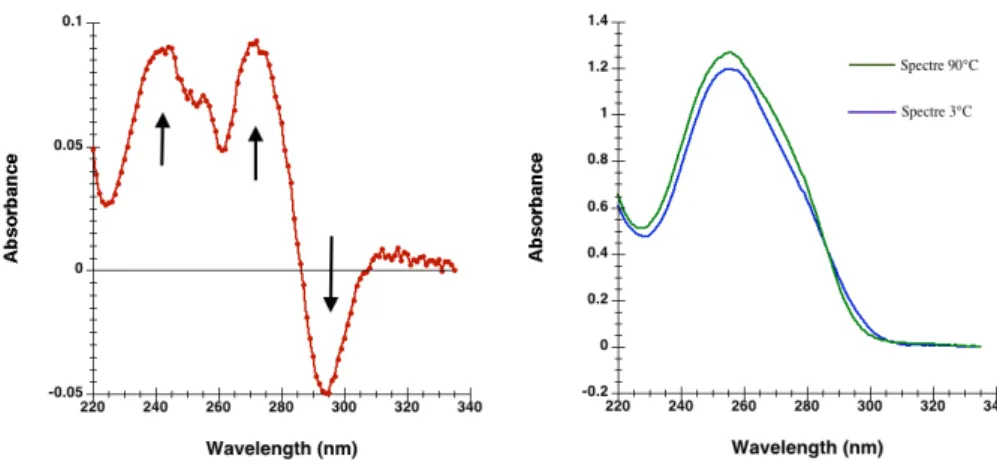
3.2.1 Thermal Difference Spectrum (TDS) . . . 33
3.2.2 Isothermal Difference Spectrum (IDS) . . . 33
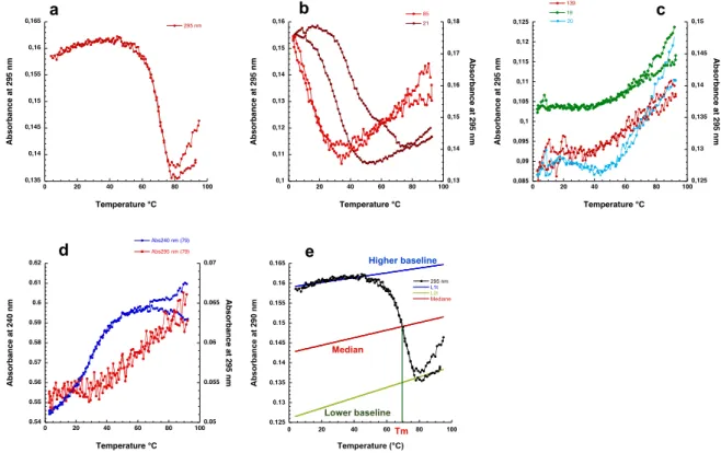
3.2.3 Thermal melting . . . 35
3.3 Circular Dichroism (CD) . . . 36
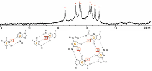
3.4 Nuclear Magnetic Resonance (1D NMR) . . . 37
3.5 Thioflavin T test . . . 39
3.6 Biophysical evaluation . . . 41
3.6.1 Interpretation of the Thermal difference spectra . . . 41
3.6.2 Interpretation of the Thermal melting transition . . . 41
3.6.3 Interpretation of the Circular Dichroism spectra . . . 41
3.6.4 Interpretation of the Isothermal Difference Spectra . . . 44
3.6.5 Interpretation of the Thioflavin T test . . . 44
3.6.6 Interpretation of the NMR Spectra . . . 44
3.7 Conclusion . . . 45
Development and validation of a new algorithm: G4-Hunter 47 4 Reevaluation of quadruplex propensity with G4-Hunter 49 4.1 Mitochondrial genome . . . 51
4.1.1 Structure . . . 51
4.1.2 Replication and transcription . . . 55
4.1.3 Function . . . 55
4.2 G-quadruplexes & Human mitochondrial DNA . . . 57
4.2.1 Human mitochondrial genome . . . 57
4.2.2 G-quadruplexes and mitochondria . . . 57
4.3 Article: Reevaluation of quadruplex propensity with G4-Hunter . . . 60
4.4 Algorithm performance analysis (Receivers Operating Characteristic) . . . 103
4.5 Conclusion . . . 106
Application to pathogens 107 5 G4s in Viruses, is there a hidden link? 109 5.1 Introduction . . . 109
5.1.1 Function of G-quadruplexes in different pathogens . . . 110
5.1.2 G-quadruplexes as therapeutic targets . . . 112
5.1.3 G-quadruplex-forming oligonucleotides with antiviral activity . . . 112
5.1.4 Objectives . . . 113
5.2 Hunting new G-quadruplexes in HIV . . . 115
5.2.1 Human Immunodeficiency Virus . . . 115
5.2.2 G-quadruplexes and HIV: the hidden link . . . 119
5.2.3 Potential functions of these G4s . . . 126
5.2.4 New conserved G4 sequences in vpr and env regions . . . 131
5.3 Hunting new G-quadruplexes in Ebola and Marburg viruses . . . 133
5.4 Patent 1: Nucleic acids acting as decoys for the treatment of lentivirus infection. 137 5.5 Patent 2: Methods and pharmaceutical compositions for the treatment of filovirus infections. . . 165
Contents xiii Conclusion 179 Conclusion 181 Annexes 185 Bibliography 235 Bibliography . . . 236
List of Figures
1.1 Nucleic acids components & different duplex DNA structures. . . 4
1.2 Triplex DNA structure. . . 6
1.3 I-motif DNA structure . . . 6
1.4 G-quadruplex DNA structure . . . 7
1.5 G-quadruplex polymorphism. . . 7
1.6 Bulges in G-Quadruplexes: broadening the Definition of G-Quadruplex-forming sequences . . . 9
1.7 Human telomeric G4 polymorphism. . . 9
1.8 G-quadruplexes found in the promoter regions . . . 12
1.9 The G-quadruplex structure formed within the 5’UTR of the NRAS mRNA. . . 12
2.1 Computational workflow of QGRS-H Predictor. . . 24
2.2 QGRS-Conserve algorithm stages. . . 24
3.1 Thermal Difference Spectrum (TDS) . . . 34
3.2 Exemple of Temperature of Melting determination. . . 34
3.3 Circular Dichroism wavelength spectra of G-quadruplexes. . . 34
3.4 Principle of 1D NMR spectra interpretation . . . 38
3.5 Principle of the ThT assay . . . 38
3.6 Interpretation of the Thermal Difference Spectra profiles. . . 40
3.7 Interpretation of the Thermal melting profiles . . . 40
3.8 Interpretation of the Circular Dichroism spectra. . . 42
3.9 Interpretation of the Isothermal Difference Spectra. . . 42
3.10 Interpretation of the Thioflavin T test. . . 43
3.11 Interpretation of the NMR graphs. . . 43
4.1 Mitochondrial genome architectures. . . 50
4.2 A structural model of the mitochondrial nucleoid. . . 50
4.3 The asymmetric and strand-coupled models of mtDNA replication. . . 53
4.4 Human mtDNA genome . . . 54
4.5 Human mitochondrial deletion spectra. . . 56
4.6 Example of ROC curve . . . 104
4.7 ROC curves with different area under curve’s (AUC) values . . . 104
5.1 Schematic model of the role of the pilE G-quadruplex (G4) in N. gonorrhoeae pilin antigenic variation. . . 110
xvi List of Figures
5.3 HIV-1 replication cycle. . . 114
5.4 Score calculation of the NC 001802 HIV-1 sequence. . . 118
5.5 Score calculation for 2177 aligned HIV-1 sequences. . . 118
5.6 LOGO representation of the different HIV-1 PQS. . . 121
5.7 Matrix organization of aligned sequences and principal of score calculation for aligned sequences. . . 123
5.8 In vivo validation of G-quadruplex formation . . . 124
5.9 Genomic structure of HIV-1 provirus and the conserved G-rich region of HIV-1 promoter. . . 128
5.10 Putative G-forming regions in the HIV-1 nef coding region. . . 129
5.11 New Potential quadruplex sequences in the HIV-1 genome. . . 130
5.12 Presentation of filamentous 970 nm-long Ebolavirus . . . 133
List of Tables
2.1 Different databases, webservers and tools for predicting G-quadruplex motifs. Methods are sorted according to the type of search . . . 20 5.1 HIV-1 genome organization and products . . . 116 5.2 PQS obtained by G4-Hunter score calculation of the NC 001802 HIV-1 sequence. 117 5.3 Conserved HIV-1 prone motifs and the in vitro conclusion. . . 120 5.4 Filoviridae family, all the species from Ebolavirus and Marburgvirus genus. . . . 134 5.5 G-rich sequences extracted from the literature and their G4-Hunter score . . . . 190 5.6 Selected oligonucleotides that do not form G-quadruplexes in vitro and their score
with G4-Hunter. . . 197 5.7 G-rich sequences in the human mitochondrial genome predicted by G4-Hunter for
a window of 25 nucleotides and score higher than 1 . . . 199 5.8 Selected oligonucleotides with a potential quadruplex-forming. All the sequences
are experimentally validated and their score with G4-Hunter is shown. . . 203 5.9 Number of hits by kbp of the sequenced genome obtained with the G4-Hunter . . 204
Chapter 1
Guanine Quadruplexes
Contents1.1 Folding and Topology of G-quadruplexes . . . . 5
1.2 G-quadruplex structural polymorphism . . . . 8
1.2.1 Strands number & orientations . . . 8 1.2.2 Loop conformations . . . 8 1.2.3 G-quadruplex and bulges . . . 10 1.2.4 RNA quadruplexes . . . 10
1.3 Biological functions of G-quadruplexes . . . . 11
1.3.1 Localization of G-quadruplexes . . . 11 1.3.2 G-quadruplexes and ligands . . . 14 1.3.3 G-Quadruplexes in vivo . . . 15
1.4 Conclusion . . . . 16
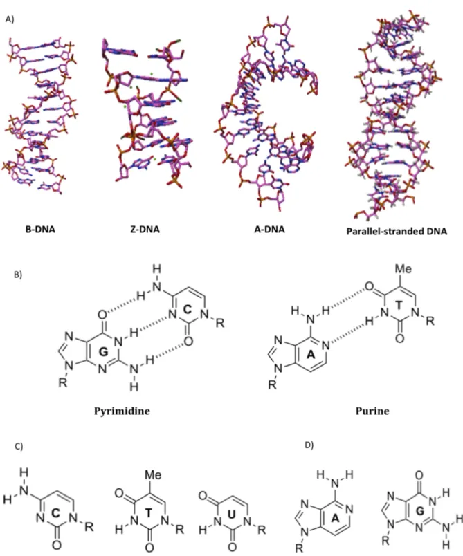
The story of DNA structure is as varied as it is interesting. The beauty and the simplicity of the DNA double-helix structure are all that is needed for a basic comprehension of cellular genetics. This double-helix was proposed by J.D. Watson & F.H.C. Crick in 1953 [1], based on the rules of Chargaff and the diffraction images obtained from R.E. Franklin [2]. DNA is usually represented by a right-handed double-helix formed with two anti-parallel strands held together by complementary base pairing (Fig.1.1-A). This pairing is made through Hydrogen bonds between the donor and acceptor ”Watson-Crick” nucleotides sites of the bases A-T and C-G (Fig.1.1-B). The succession of base pairs (Fig.1.1-C & -D) defines the genetic information needed by the cells to accomplish their vital functions. The helix is stabilized by important staking interactions between consecutive bases. B-DNA has always been regarded as the biologically relevant structure; however DNA can adopt a wide variety of conformations including highly distorted A-form (observed under conditions of low hydration), Z-form (favoured at high salts concentration) and parallel-stranded DNA (Fig.1.1-A).
4 Chapter 1. Guanine Quadruplexes
Figure 1.1: Nucleic acids components & different duplex DNA structures. (A) Models of
the A, B and Z DNA. B and A forms are right-handed helices, B form (PDB : 1BNA) is characterized by a helical turn every 10 base pairs (3.4 nm) with 0.34 nm by base pair. A form (PDB : 440D) is more compact with 11 base pairs per turn. Z form (PDB : 4FS5) is left-handed with a zig-zag like back-bone. Parallel-stranded duplex DNA (PDB : 1JUU) are maintained by reverse Watson-Crick base pairing between A-T or alternating, symmetrical self-pairs of Gsyn-Gsyn (B) Canonical base pairing
(Watson-Crick) of nucleotides within the double-helix of DNA (A-T & C-G). (C & D) Schematic structures of the nucleobases constituting DNA and RNA.
1.1. Folding and Topology of G-quadruplexes 5
Since Watson and Crick study, it is now well established that DNA can adopt different secondary structures. DNA can folds into a vast variety of hairpin, triplex, i-motif and G-Quadruplex structures containing non-canonical base pairs, triads or quartets.
Some of these non-canonical base pairing schemes are called ”Hoogsteen” as they were highlighted by Karst Hoogsteen using X-ray diffraction [3]. They can be established within guanine-rich DNA sequences between guanines via four Hydrogen bonds involving the ”Watson Crick” and ”Hoogsteen” edges (Fig.1.2-A & -B).
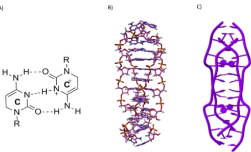
Triplex DNA consists of a double-stranded DNA (dsDNA) (with one purine-rich and one pyrimidine-rich strand) and a single-stranded triplex-forming oligonucleotide (TFO) that binds to the major groove of the duplex through Hoogsteen or reverse Hoogsteen bonding with the purine-rich strand [4, 5] (Fig.1.2). Cytosine-rich sequences may adopt an i-motif structure (Fig.1.3) at acidic pH, which consists of two parallel-stranded DNA duplexes held together in an antiparallel orientation by intercalated, hemiprotonated cytosine–cytosine+ base pairs [6].
The G-quadruplexes are formed by guanine-rich sequences. This structure is based on the stacking of several tetrads, which were first identified in 1962 as the basis for the aggregation of 5’-guanosines monophosphate (GMP) [7]. Today, the interest for G4 is growing with thousands of reports on structure, detection within cells and function [8]. For many years G-quadruplexes were considered as a structural curiosity, but now come up in various areas, ranging from biology and medical biology to supramolecular chemistry and nanotechnology. In this chapter, I will describe the main characteristics of the non-canonical DNA ”G-quadruplex” structures.
1.1 Folding and Topology of G-quadruplexes
G-quadruplexes are four stranded structures formed by guanine-rich DNA (or RNA) sequences. The association of four guanines by a network of eight Hydrogen bonds generates a planar structure called a G-quartet or G-tetrad (Fig.1.4-A) and the stacking of several G-quartets forms a G-quadruplex (Fig.1.4-B & -C) [7, 9]. The primary building block of this structure, the G-quartet, is composed of four coplanar guanines that interact with each other via Hoogsteen base pairs. The G-quartets are maintained by the presence of monovalent cations, mainly the metallic cations such as K+, and to a lesser degree Na+ [10], and others
such as: Rb+, Sr2+, Ca2+ and Pb2+[11]. The most common mode of cation binding is the
”sandwich” mode where a cation is positionned between two G-quartets and coordinated to eight carbonyl O6 atoms, thereby minimizing their repulsion and providing stability to the quartet [12].
G-quadruplexes are highly polymorphic depending on the nucleic acid sequence and experi-mental conditions. The four strands serving as columns supporting the G-tetrad core can be in different orientation, G4 can be formed in an intermolecular or intramolecular fashion (Fig.1.5-Top). The loops connecting these strands (Fig.1.5-Down) can also adopt different conformations [13].
6 Chapter 1. Guanine Quadruplexes
Figure 1.2: Triplex DNA structure. Schematic representation of (A & B) G-GC and A-AT triplets
(PDB : 1BWG), involved in purine and pyrimidine triplexes, respectively. (B) Triplex DNA adapted from the structure (PDB : 1BWG). The triplex above is called a purine triplex as the TFO is purine rich. Other triplex may be formed with pyrimidine oligonucleotides, based on the formation of C+-GC
and T-AT triplets .
Figure 1.3: I-motif DNA structure. (A) A hemiprotonated cytosine–cytosine+ base pair. (B & C) Structure of the d(A2C4) intermolecular i-motif (PDB : 1YBL).
1.1. Folding and Topology of G-quadruplexes 7
Figure 1.4: G-quadruplex DNA structure.(A) Presentation of a G-quartet formed by the association
of four guanines. (B) G-quadruplex nucleic acids structure. (C) The intramolecular model of G4 formed by the stacking of three G-quartets adapted from the human telomeric DNA sequence (PDB : 1KF1).
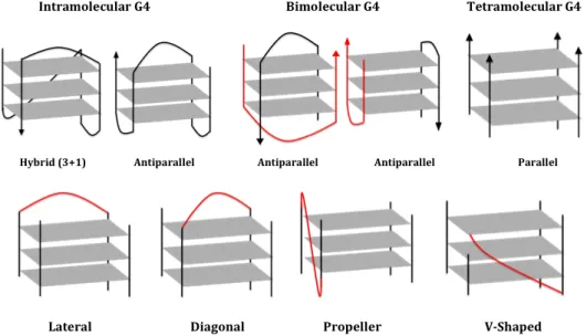
Figure 1.5: G-quadruplex polymorphism. Schematic representation of different G4 conformations
according to the molecularity (intra-, bi-and tetra-molecular), strand orientations (parallel, anti-parallel and hybrid (3+1)) (top). Schematic representation of different loop conformations within G4 (lateral, diagonal, propeller and V shaped) (down).
8 Chapter 1. Guanine Quadruplexes
1.2 G-quadruplex structural polymorphism
The stacked G-quartets linked by the phosphate backbone constitute the relatively invariant core of all G4 structures. Different parameters have been described to explain the structural diversity of G4 such as the number of strands, their orientations, the conformation of loops and the nature of cations.
1.2.1 Strands number & orientations
G-quartets can be assembled in an intramolecular (monomolecular) fashion, where one strand containing several blocks of guanines is able to fold back and form a G4 structure. G4 can also be formed from two-, three- or four-strands, resulting in intermolecular structures that can adopt a wide variety of conformations (Fig.1.5) [8].
Within each strand, the glycosidic conformations of guanines can be either syn or anti. The relative orientations of strands are geometrically related with the glycosidic conformation of guanines [8]. The possibilities are: (i) Parallel G4 (four strands identically oriented) (ii) ”3+1” Hybrid G4: three strands oriented at the same direction and in the opposite direction, (iii) ”2+2” Anti-parallel G4: two strands in one direction, the others in the reverse orientation. These antiparallel G4 can be subdivided into two distinct classes: two parallel strands can be adjacent or diagonal, and this will result in very different structures and groove sizes [14]. These backbone strands are connected by linkers commonly called loops.
1.2.2 Loop conformations
Monomolecular and bimolecular structures generally present 2 or 3 loops. The loops are linkers connecting G-stretches that support the G-tetrad core. Four conformations are distinguishable: (i) lateral or edgewise loops connecting two anti-parallel adjacent strands, (ii) diagonal loops connecting two opposing antiparallel strands, (iii) ”Propeller” or Double-chain reversal loops (N-shape) connecting two adjacent parallel strands, and (iv) V-shaped or snap-back loop connecting two corners of G-tetrad core in which a support column is missing (the wedges of G-quartet).
Furthermore, loop residues can form base-pairing alignments, which in turn stack with the terminal G-tetrads, further stabilizing G-quadruplex structures [14]. The loop conformations is closely linked to strand orientations of a G4 and depends on the size and the sequence of the linkers. Different studies pointed the effect of loops length and composition on the G4 formation and stability. They indicate that: (i) loops consisting of a single nucleotide are generally N-shaped and promotes parallel stable G4 formation; (ii) loops of ≥ 3 nucleotides (nt) promotes an antiparallel conformation with a decrease in stability [15] and (iii) a sequences with a loop of more than 9 nt may still form a G4 [16, 17].
1.2. G-quadruplex structural polymorphism 9
Figure 1.6: Bulges in G-Quadruplexes: broadening the definition of G-Quadruplex-forming Se-quences. The bulges can be formed in many different situations within G-quadruplexes, thus making some G4
sequences defying the standard description. Adapted from [13].
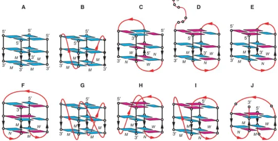
Figure 1.7: Schematic structure of human telomeric G-quadruplexes. (A) Tetrameric parallel-stranded
G4 observed for the single-repeat human telomeric sequences d(TTAGGG) and d(TTAGGGT) in K+solution. (B)
Dimeric parallel-stranded G4 observed for the two-repeat human telomeric sequence d(TAGGGTTAGGGT) in a K+ containing crystal and in K+ solution. (C) Dimeric antiparallel-stranded G4 observed for two-repeat human
telomeric sequence d(TAGGGTTAGGGT) in K+ solution. (D) Asymmetric dimeric (3 + 1) G4 observed for the
three-repeat human telomeric sequence d(GGGTTAGGGTTAGGGT) in Na+ solution. (E) Asymmetric dimeric
(3 + 1) G4 association observed for the three-repeat human telomeric sequence d(GGGTTAGGGTTAGGGT) and the single-repeat human telomeric sequence d(TAGGGT) in Na+ solution and in K+ solution
(unpub-lished results). (F) Basket-type form observed for d[A(GGGTTA)3GGG] in Na+ solution. (G) Propeller-type form observed for d[A(GGGTTA)3GGG] in a K+ containing crystal. (H) (3 + 1) Form 1 observed for
d[TA(GGGTTA)3GGG] in K+ solution. (I) (3 + 1) Form 2 observed for d[TA(GGGTTA)3GGGTT] in K+
10 Chapter 1. Guanine Quadruplexes
1.2.3 G-quadruplex and bulges
The knowledge accumulated on G4 structures led to the formulation of a consensus motif, which is capable of forming an intramolecular G4 used in the search for Putative Quadruplex Sequences (PQS).
To date, a G-quadruplex is often considered to be formed by a sequence containing four tracts of two, three or more continuous guanines connected by linkers, in which the G-tracts would form continuous columns supporting the G-tetrad core, while the linkers would form loops connecting the corners of the G-tetrad core [19]. This motif is widely used and most of G4 structures resolved are conform to this consensus. Nevertheless, it has been shown that several examples defy the description of this consensus, such as the sequence Pu24 from the human c-myc promoter [20] and c-kit87up from the human c-kit promoter [21]. These sequences exhibit discontinuous arrangement of guanines in one column of the G-tetrad core, despite the presence of four G-tracts each having at least two or three continuous guanines [20, 21]. While a loop connects two corners of the tetrad core, bulges are projections of bases from the G-tetrad core: they connect two non-adjacent guanines of the same strand within the G-G-tetrad core. Recently, Phan et al [13] have shown that many different bulges can exist in G4 structures. They vary in their sequence, size, position and numbers within the G4. This result alters the common view on the ability of many sequences to form G-quadruplexes. Expanding this description should help to identify more potential G4 forming sequences. It could also open the possibilities of exploiting bulges as recognitions elements for interaction between G-quadruplexes and other molecules. In the same view NMR-based solution structures have been reported for several sequences in which G-quartets are connected by four loops. The guanines involved in the G-tetrads include some isolated guanines but can exclude guanines from the G-tract (e.g. c-myc23456 and c-kit1) [22, 20].
1.2.4 RNA quadruplexes
Coding and non-coding RNA containing several blocks of guanines are able to fold into G-quadruplexes. It has also been shown that many G4 RNA possess remarkable stability [23, 24]. In addition, the formation of G4 RNA seems all more relevant since RNA is generally single-stranded. Such structures might play key roles in the gene regulation, translation, genomic stability and disease. The human fragile X syndrome is caused by: (i) the loss of FMRP protein the ability to bind RNA, or (ii) the repression of the gene FMR1 coding this protein. The in vitro selection showed that the FMRP protein bind to G4-prone sequences [25]. Bioinformatics studies demonstrated that the 5’ and 3’ untranslated regions (UTRs) of mRNA are G-rich. Guanine-rich UTRs can fold into G4, which may play a role in the regulation of translation, mutation and degradation of RNA [26, 19]. Recent studies reported that telomeric repeat-containing RNA (TERRA) folds into a parallel G4 conformation; additionally, the parallel RNA G4 tends to associate and form higher-order structures [27]. TERRA could play important roles in regulating telomerase and in chromatin remodeling during cell development [28].
1.3. Biological functions of G-quadruplexes 11
1.3 Biological functions of G-quadruplexes
1.3.1 Localization of G-quadruplexes
Both prokaryotic [29] and eukaryotic genomes from yeast to human are rich in potential G4-forming motifs [30]. Among these are repetitive and functionally essential chromosomal do-mains, including telomeres, rDNA and the immunoglobulin heavy chain switch regions of higher vertebrates. Many minisatellite and microsatellite repeats are G-rich and have the potential to form G4 DNA. G-rich regions are also found within specific single-copy genes.
Telomeres
Telomeres are specialized nucleoprotein complexes that cap and protect the extremities of linear eukaryotic chromosomes from degradation [31]. During each cell division, telomeric DNA shortens by 50-200 base pairs, until a critical size is reached, causing cell division arrest. In most eukaryotes, telomeric DNA consists of a tandem array of a short motif of 5–8 nts, that includes two, three or four consecutive and highly conserved guanines (Fig 1.7)[32]. The GGGTTA motif is found in many phylogenetically distant organisms, including vertebrates [33], several fungi [34] and slime molds [35]. Variants of this motif are found in many other organisms: parasites [36], plants [37], algae [38], nematodes [39] and yeasts [40]. Such a structure might be important for telomere biology and a good target for drug design [14]. Since then, numerous structural studies were able to demonstrate the complexity of human telomeric sequence folding and conclude that this motif can be folded into parallel, hybrid or antiparallel G-quadruplexes depending on experimental conditions and exact sequence [18].
Telomeric repeats are normally capped by a protein complex that identifies them as telomeres rather than damaged DNA and protect them from misguided cellular efforts at repair that are potentially destabilizing [41]. The removal of this telomere-capping complex results in telomere instability that can be countered by drugs that stabilize G4 structures [42]. The shortening process is countered by telomerase [43], an enzyme required for the proliferation of stem cells and germline cells, as well as most cancer cells [44]. Telomerase is highly over-expressed in many cancer cell types whereas it is only expressed at low levels in normal somatic cells, allowing cancerous cells to be replicated indefinitely. Folding of telomeric DNA into G4 seems to influence the extent of telomere elongation in vitro and might therefore act as a negative regulator of elongation in vivo [45]. Therefore, G-quadruplexes formed by human telomeric DNA have been considered as promising anticancer targets [46].
Promoters & UTRs
Guanine-rich tracts are observed in critical segments of eukaryotic and prokaryotic genomes. Genome-wide computational analysis has revealed that there is significant enrichment in G-quadruplex motifs in gene promoter regions extending up to 1kb upstream of the transcription start sites. These putative promoter G-quadruplex-forming regions are strongly associated with nuclease hypersensitivity sites, and could be involved in gene regulation at the transcriptional level [49].
12 Chapter 1. Guanine Quadruplexes
Figure 1.8: The six hallmarks of cancer shown with the associated G-quadruplexes found in the promoter regions of these genes. The various G-quadruplexes differ by folding pattern, number
of tetrads, loop size and constituent bases. Adapted from [47].
Figure 1.9: The G-quadruplex structure formed within the 5’UTR of the NRAS mRNA
is proposed to act as a translational repressor element. The G-quadruplex segment is shown in red, and the 5’ cap and 3’ poly(A) tail are omitted for simplicity. Adapted from [48].
1.3. Biological functions of G-quadruplexes 13
Following transient opening of the duplex (a process necessary for transcription, replication and recombination), these guanine-rich tracts have the potential to form G-quadruplexes. This stimulated investigations on the role of G-quadruplex formation in transcriptional regulation of the promoters (Fig 1.8) of c-myc [50] c-kit [51], bcl-2 [52], VEGF [53] and HIF-1 [54] oncogenes. Another well-known G-rich region is the insulin gene-linked polymorphic region (ILPR), located 363 basepair (pb) upstream of the human insulin gene. Both inter- and intramolecular G-quadruplex formation in the ILPR can influence transcriptional activity of the human insulin gene [55].
The cell-surface-receptor tyrosine kinase. Platelet-derived growth factor receptor β
(PDGFR-β), plays an essential role in cellular growth, proliferation, differentiation, and development
[56]. The promoter of the human PDGFR-β gene has previously been characterized: the G-quadruplex formed [57] there could be a potential target for drug development in the treatment of cancer, fibrotic disorders [58] and other diseases.
Several studies have demonstrated that the information required for post-transcriptional con-trol is located mainly in the 5’ and 3’ UTRs of mRNA [59] (Fig 1.9). The secondary structures formed in 5’ and 3’ UTRs can also serve as regulatory elements by acting as target sites for RNA-binding factors such as proteins or by interacting directly with the translation machinery. Bioinformatic searches have recently identified up to 3000 PQS in the 5’-UTRs of the human gene. A conserved intramolecular G-quadruplex motif within the human NRAS proto-oncogene 5’-UTR, can modulate gene expression at the translational level [60]. These results open op-portunities for small molecule identification in order to stabilize 5’-UTR RNA G-quadruplex formation and therefore inhibit translation of oncogenes. We find in the literature other onco-genic 5’-UTR RNAs such as TRF2, fi1, bcl-2 and jun capable of forming G-quadruplexes[14].
Minisatellites & Aptamers
Minisatellites consist of head-to-tail arrays of identical or slightly polymorphic 10-100-bp
long motifs. They are present in prokaryote and eukaryote genomes and might constitute as much as 10% of the human genome. It has been shown that they may cause many diseases by influencing gene expression, modifying coding sequences within genes or generating fragile sites [61]. The large number of minisatellite loci, the variety of their nucleotide sequences, and their persistence in the genome suggest that they likely possess biological roles. Some sequences, such as the human CEB25 minisatellite loci are susceptible to form G-quadruplex structures. This 3-quartets parallel G-quadruplex can be formed in long sequence contexts, providing evi-dence for a pearl-necklace of G-quadruplexes formed by single-stranded CEB25 minisatellite [62].
Aptamers are artificial oligonucleotides (DNA or RNA) selected in vitro; they bind a broad
range of relevant targets with high affinity and specificity [63]. They consist of relatively small nucleic acid sequence ( i.e. usually composed by 15 to 45 bases), that bind to their target with high specificity and with a dissociation constant in the nanomolar to micromolar range. They can be selected against any target such as: small metabolites, peptides, large proteins, nucleic acids and even whole cells.
14 Chapter 1. Guanine Quadruplexes
Moreover, aptamers are easily synthesized and alterable. They are highly used in biotech-nology and biological imaging. Some aptamers are characterized for their utility in cancer or viral infections. Indeed aptamers act as inhibitors with great promise, in particular as targeting ligands for the treatment or diagnosis of cancer, HIV and other diseases [64]. They are also characterized by their structure; the folding of these aptamers play an important role in the carried function and their interaction with the targets. Many aptamers fold into G-quadruplex structures. The best known example is the Thrombin Binding Aptamer (TBA) [65, 66]. It forms a monomeric G-quadruplex with two quartets connected by two ”TT” loops and a central ”TGT” loop. Since then, a lot of G-rich aptamers have been isolated.
Renaud de la Faverie et al. analyzed 33 aptamer sequences, extracted from the literature, to determine their topologies. They concluded using different tests, that these aptamers fold into G-quadruplexes [67]. Romanucci V. et al designed a bimolecular G-quadruplex aptamers based on Hotoda’s sequences d(TG3AG) [68]. These molecules showed significant binding to HIV envelope glycoproteins gp120 and gp41 and represent the first attractive anti-HIV bimolecular G-quadruplexes.
1.3.2 G-quadruplexes and ligands
The examples presented above suggest that the G4s play roles in cells, either at the level of regulation of gene expression, replication, recombination, or division. It is important to develop means to characterize, visualize, induce, stabilize or localize this G4s in cellulo or in vivo. There are a number of ligands, proteins and antibodies, characterized by different chem-ical structures, already reported in the literature for their ability to bind and stabilize the G-quadruplex structures. In the past two decades, hundreds of small molecules with diverse chemical structures and physicochemical properties have been prepared and examined for their abilities to interact with G-quadruplexes [69]. Many of them show in vitro and in vivo activities, and at least one (Quarfloxin) has entered clinical trials.
Proteins Numerous proteins are able to interact with G4 by: (i) binding to G4 that stabilize
the structure, (ii) by unfolding, destabilizing and cleaving the G4, or (iii) inducing G4 formation [70]. The Bloom’s and Werner’s syndrome proteins are DNA structure-specific helicases. They unwind a variety of very stable G-quadruplexes DNA [71]. The most studied S.cerevisiae Pif1 protein can affect the genomic stability of the G-rich minisatellite CEB1, thus providing evidence of their formation in vivo [72].
Small molecules Studies of G-quadruplex ligands are now among those at the head of drug
discovery, and many databases that include comprehensive information of G-quadruplex ligands are available [73]. The concept that quadruplex-binding small molecules can stabilize telomeric quadruplexes, has been widely used to discover small molecules with anti-cancer activity via indirect inhibition of the telomerase enzyme complex [74, 75].
1.3. Biological functions of G-quadruplexes 15
Most G4 ligands target the invariant part of the structure, the G-quartet, and also possess a large aromatic surface favourable to the interaction with the large hydrophobic surface consti-tuted by the G-quartet. G-quadruplex ligands may be considered as potential anticancer agents because of their telomerase and/or oncogene transcription inhibition potential.
Antibodies Two antibodies (Sty3, Sty49) are able to recognize G4s formed by the telomeric
repeats of Stylonychia lemnae, with Kd of 125 pM and 3–5 nM respectively [76]. A single-chain variable fragment (scFv1) antibody selected by phage display exhibits a competitive selection to a human parallel intramolecular DNA G-quadruplex with high affinity [77]. The antibody strongly discriminates between G-quadruplex and duplex DNA. More recently, the same team used an antibody to demonstrate the existence of G-quadruplexes in human cells. This will be detailed in the next paragraph.
1.3.3 G-Quadruplexes in vivo
Since the first description of G-quadruplexes in vitro, key questions have been whether such structures occur in vivo, and what their function might be. The first direct evidence for the presence of G4 DNA structures in vivo came from studies using G4 DNA-specific antibodies to detect intermolecular structures at ciliate telomeres where their formation and dissolution are cell cycle regulated [76]. Many researcher attempt to localize G-quadruplexes in vivo. In 2009 [78], the use of the porphyrine derivative NMM in live Neisseria gonorrhoeae showed significantly decreased Antigenic Variation (AV).
Another indirect proof of G-quadruplexes in vivo role was performed in the yeast S.
cerevisiae [42]. This study demonstrated that G4 DNA can contribute to telomere capping
which supports the idea that telomere G4 DNA can play a positive role in telomere regulation
in vivo. The evolutionary conservation of the G4 DNA motif and its association with specific
genomic features supports the hypothesis that G4 DNA has in vivo functions that are under evolutionary constraint [79]. In addition [80], genome-wide chromatin immunoprecipitation was used to determine the in vivo binding sites of the Saccharomyces cerevisiae Pif1 DNA helicase, a potent unwinder of G4 structures in vitro. The slowing of replication near the Pif1-binding site and the stimulation of DNA breakage suggest that G4 structures form
in vivo and that they are resolved by Pif1 to prevent replication fork stalling and DNA breakage.
Genetic studies performed on human cells, strengthened the evidence for the existence of G4s in vivo. Pyridostatin is a small G4 ligand, which generates DNA damage at specific genomic loci, leading to cell cycle arrest and transcription down-regulation of several genes that contain PQS [81]. Recent studies used antibodies to detect G-quadruplex DNA in mammalian cells. Biffi et al [82] have employed a specific antibody to quantitatively visualize DNA G-quadruplex structures in human cells. The BG4 antibody binds with high selectivity and low nanomolar affinity to DNA G-quadruplex structures and does not have any preference to any particular G-quadruplex conformation. The BG4 localization at the chromosomal ends confirms the presence of G4 structures at human telomeres.
16 Chapter 1. Guanine Quadruplexes
Furthermore, the observation of the BG4 dispersed across the chromosomes demonstrates that G4 also forms outside telomeric regions. This study came out with the information that probably most G4 formation occurs during DNA replication and that G4 are modulated dynamically within the cell cycle.
In parallel, another team developed a murine monoclonal antibody called 1H6 [83]. This has also been used to visualize G4 in human and murine cells, providing additional support for the existence of G4 DNA in vivo. 1H6 exhibits strong staining in most human and murine cells. This staining indicates the abundance of G4s structure and that the 1H6 antibody is a valuable tool for further studies on the role of G4 DNA.
1.4 Conclusion
G-quadruplexes are prevalent tetra-stranded structures that are formed in both DNA and RNA. They are involved in fundamental biological processes such as transcription and transla-tion. Their structural polymorphism, their presence at different locations within the genome, their association with a number of cancer-related genes and their utilization as potential thera-peutic targets, attracted researchers from various areas. Different computational approaches have been developed for specifying the potential of a nucleotide sequences to fold into G-quadruplexes structure. In the next chapter, I will describe the major bioinformatic tools and algorithms developed to predict and analyze sequences with G-quadruplex forming potential.
Chapter 2
Computational detection
Contents2.1 Bioinformatics . . . . 18
2.1.1 Algorithm . . . 18 2.1.2 Data identification and structuration . . . 19
2.2 Pattern-matching algorithms . . . . 21
2.2.1 Quadparser . . . 21 2.2.2 QGRS Mapper & QGRS-H Predictor & QGRS-Conserve . . . 22 2.2.3 Quadfinder . . . 25
2.3 Sliding window approaches . . . . 25
2.3.1 G4P calculator & QFP algorithm . . . 25 2.3.2 ddiQFP . . . 26
2.4 Score calculation . . . . 27
2.4.1 cG/cC score calculator . . . 27
2.5 Conclusion . . . . 28
Most biological G4 structures studies have combined in silico predictions with biophysical evidence of G4 folding in vitro. Computational approaches can nonetheless be a good indicator of the potential of a sequence to form G4 structure, and then, can be used to distinguish sequences that have no potential to form G4-structures from other G4-forming sequences. Different search algorithms have been developed on criteria based on a variety of biophysical experiments. Se-quences information alone is not enough to make sure a motif may or may not form a quadruplex, but it is the starting point for the discovery of new G-quadruplexes that should be confirmed using biophysical and biochemical techniques. [84]. The algorithm development started with pattern matching techniques, followed by a sliding window approaches and score calculation. In this chapter, I will bring an overview of the current algorithms used to predict G-quadruplex formation but before this a bioinformatics section should be of interest.
18 Chapter 2. Computational detection
2.1 Bioinformatics
Nowadays, computer science is omnipresent. In biology, it is used in a wide variety of purposes and experimental devices: data archiving, data processing, sequences analysis and predictions etc. Bioinformatics emerged in the 80’s as a new discipline where biology, informatics and data processing were fused. Thereby bioinformatics is not only the application of computer science to biology, it is a wholly owned branch of biology.
Current bioinformatics focuses on the study of DNA/RNA sequences and protein folding. It is used for storage or data management and also in the interpretation of such data, whence the data analysis of a sequence might determine the biological function of the gene.
When used to tackle new problems, bioinformatics rely on multiple steps: • determination of the logical steps required to solve the problem (algorithm) • identification and structuration of the data (i.e. ”objects” used by a program); • implementation of the algorithm using the appropriate programming languages.
2.1.1 Algorithm
Once the problem is defined and the data involved are identified, one needs to find the calculation mechanism (algorithm) that allows to solve the problem (formula for calculating solutions of an equation, exploring a search tree, etc. ).
Algorithm: It consists in a set of logic procedures or formula for solving a problem. The
word derives from the name of the mathematician, Mohammed ibn-Musa al-Khwarizmi, who was part of the royal court in Baghdad and who lived from about 780 to 850. His work is the likely source for the word algebra as well1.
Before validation an algorithm, one should wonder if it is: • Easy to implement (to write using a programing language)? • Correct: does it answer the question in all the cases? • Efficient enough?
Implementation of the algorithm needs an appropriate programming language. Since computers can handle only binary (machine language : succession of only 0 and 1), it is therefore necessary to use a programming language to write legibly the instructions to be executed by the computer; The programming language is the intermediary between the human and the computer. It allows to write in a language close to the machine, but intelligible to the human, whereas, the computer language is a set of consecutive actions that a computer must run.
2.1. Bioinformatics 19 Programming language: Generally, a program is a simple text file, written in any
program-ming language by a specific text editor and called a source file containing lines called source code. This source file is then compiled into machine language to be readable by the processor and executed to solve the problem. The compilation is a phase carried out by the computer itself through another program called compiler.
Different processes and tasks are involved in bioinformatics analysis. Several program have been written for various applications using every available language. There are different levels of programming languages: (i) high-level programing language are easily understood by the user, such as C# , C++, Pascal, Java, Python and Perl. (ii) and the low-level programming language are the one close to the machine language such as assembly.
One can also distinguish Compiled and Interpreted languages. In the compiled languages, the source code is reduced to a machine-specific instructions before being saved as an executable file. Interpreted languages are saved and executed in the same initial format. The accuracy of each categories and language have been analyzed [85]. The compiled programs generally run faster than the interpreter ones but it is usually easier to develop application in an interpreted environment.
Perl and Python are often called Script languages and form a group of intermediate languages. Indeed, when executed, they are compiled in an intermediate representation (without creating an intermediate file) and then interpreted. Both languages have large free libraries, suitable for web scripting and pipeline implementation [86, 87].
2.1.2 Data identification and structuration
Data is a set of qualitative or quantitative variables values, information and knowledge, which are closely related concepts. Data is collected and analyzed to create information suitable for making decisions. Considering a program or an algorithm data should be formatted in a special way. A software is divided into two categories: programs (algorithms) and data can exist in a variety of forms: numbers or text on paper, bytes stored in electronic memory or statement stored in human mind. Data files are the files that store the database information. In bioinformatics, the most known and used data files are FASTA files.
What is a FASTA file? The FASTA format is a text-based format that represents either
nucleotide sequences or peptide sequences. The simplicity of FASTA format makes it easy to manipulate using any scripting languages. A sequence record in a FASTA format consists of a single-line description (sequence name) preceded by a greater-than (”>”) symbol, followed by line(s) of sequence data (bases). The number of sequences in the input data is determined by the number of lines beginning with a ”>”.
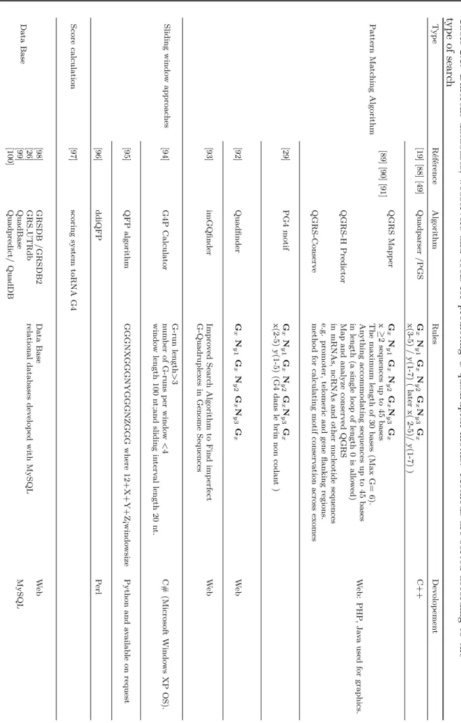
20 Chapter 2. Computational detection Table 2.1: Differen t databases, w ebserv ers and to ols for predicting G-qu adruplex motifs. Metho ds are sorted according to th e typ e of searc h T yp e R ´ef ´erence Algorithm R ules Dev olop emen t P atte rn Matc hing Algorithm [19 ][88 ][49 ] Quadparser /PGS G x N y1 G x N y2 G xN y3 G x C++ x(3-5) / y(1-7) ( la te r x( 2-5)/ y(1-7) ) [89 ][90 ][91 ] QGRS Mapp er G x N y1 G x N y2 G xN y3 G x x ≥2 sequences up to 45 bases W eb: PHP ,Ja va used for graphics. The maxim um length of 30 bases (Max G= 6). An ything accommo dating sequence s up to 45 bases in length (a single lo op of length 0 is allo w ed) QGRS-H Predictor Map and analyze conserv ed QGRS in mRNAs, ncRNAs and other nucleotide sequences e.g. promoter, telome ric and gene flanking regions. QGRS-Conserv e metho d for calculating motif conserv ation across exomes [29 ] PG4 motif G x N y1 G x N y2 G xN y3 G x x(2-5) y(1-5) (G4 dans le brin non co dan t ) [92 ] Quadfinder G x N y1 G x N y2 G xN y3 G x W eb [93 ] imGQfinder Impro ved Searc h Algorithm to Find imp erfect W eb G-Quadruplexes in Genome Sequences Sliding windo w approac hes G-run length > 3 [94 ] G4P Calculator num ber of G-runs per windo w < 4 C# (Mi cro soft Windo ws XP OS). windo w length 100 nt;and sli ding in terv al length 20 nt. [95 ] QFP algorithm GGGNX GGGNYGGGNZGGG where 12+X+Y+Z¡windo wsize Python and av ailable on req uest [96 ] ddiQFP P erl Score calculation [97 ] scoring system toRNA G4 Data Base [98 ] GRSDB /GRSDB2 Data Base W eb [26 ] GRS UTRdb relational databases dev elop ed with MySQL [99 ] QuadBase MySQL [100 ] Quadpredict/ QuadDB
2.2. Pattern-matching algorithms 21
2.2 Pattern-matching algorithms
Mainly three types of computational approaches based on string pattern matching have been used in the literature for analyzing the genomic distribution of PQS. The aim of those algorithms is to search for putative-quadruplexes sequences that follow the motif of the form equation 2.1
Gx Ny1 Gx Ny2 GxNy3 Gx (2.1)
In other words, these algorithms are looking for four runs of x guanines separated by three loops of y length. From 2004, a dozen of algorithms appeared [19, 89, 88, 29, 92, 94, 95, 96, 97] (Table 2.1). They all play with the parameters (x, y) of equation (2.1) and the sequences tested. In this section, the major and most cited algorithms for G4 prediction are described.
2.2.1 Quadparser
Huppert et al. [19] developed a simple rule describing sequences that may form G-quadruplexes based on the primary DNA sequence. They later investigated the frequency of these sequences and the distribution of the length of the loops joining the tetrads [88].
These methods specifies a regular expression that the PQS should take and consider: • The strand stoichiometry by limiting the analysis to intramolecular PQS;
• The tetrad number and focusing only on sequences capable of forming three (and later two) or more G-tetrad stacks;
• The presence of mutations or deletions. In this version the discontinuity in the blocks of guanines (presence of bulges) is not tolerated (mutations and deletions in the human telomeric sequence d(GGTTAG)n results in the decrease in the stability of this sequence);
• The length of the loops is considered between 1 to 7 nucleotides (later up to 12).
The potential quadruplex sequence is then a sequence with four runs of guanines of more than three bases long, separated by loops of 1 to 7 of any nucleotides (equation 2.2). The authors proposed an algorithm named Quadparser written in C. Quadparser can rapidly analyze large amounts of genomic data, in FASTA file format, by searching the pattern and report on the number, position of the identified sequences in the output file.
G3+ N1,7 G3+ N1,7 G3+N1,7 G3+
N means any nucleotides (ACGT) (2.2)
The number of PQS using the folding rule is then calculated considering that:
• when there is more than one possible quadruplex from one sequence it is counted as one (e.g. c-myc d(AGGGTGGGGAGGGTGGGG) could form G4 using either the first or the forth G of the two bolded blocks)
22 Chapter 2. Computational detection
• when there are more than four runs of d(GGG), it count the maximum number of G4 that could be formed at any given time using consecutive G-runs (i.e. d(GGGTTA)8 would
yield a count of 2)
• both G and C-rich patterns are taken into account, meaning that G-quadruplex could be formed in the complementary strand to that for which the sequence was obtained.
The search for prevalent intramolecular G4 in the human genome by Quadparser [88] demon-strates that it contains a large number (376000) of potential sequences. These sequences may be used for a variety of purposes to examine particular genes or other regions of interest.
2.2.2 QGRS Mapper & QGRS-H Predictor & QGRS-Conserve
QGRS Mapper (Quadruplex forming G-Rich Sequences) is another pattern-matching
al-gorithm [92] that aims to predict the presence of G4 in nucleotides entries according to the formula (equation 2.1). QGRS-mapper is a web-based server that generates detailed informa-tion on composiinforma-tion and distribuinforma-tion of PQS in any nucleotides sequence in FASTA format. The program provides options to search the entire NCBI, Gene Entrez, RefSeq GeneBank database in order to retrieve the desired gene/nucleotide sequence for analysis, and to provide a user-friendly interface to define the minimum number of tetrads and a maximum length of the loops. The motif again involves four groups of guanines separated by three loops following the restric-tions below:
• at least two tetrad are required;
• the default G4 size is 30 nucleotides considering G-groups of a maximum size of 6 while sequences up to 45 nucleotides can be found;
• arbitrary or specified composition of the loops: the user can specify the composition of the loops by a regular expression (e.g.: T{3,5} ⇒ loops of three to five consecutive T), • at most one of loops is allowed to have a length of zero.
In addition to searching for G4 sequences, the tool provides a score that evaluates PQS for its likelihood to form a stable G-quadruplex. The scoring method is dependent on the users parameters (loops length and composition, sequence length...), based on previous studies and uses the following principles for the score calculation: (i) shorter loops are more common than longer ones, (ii) G-quadruplexes are generally of equal loops size and (iii) the greater the number of guanines, the more stable is the sequence.
The highest possible score is 105 corresponding to (GGGGGGT)4. This score allows also
the elimination of all possible overlapping sequences by selecting the motif with the highest score. QGRS-Mapper is a flexible and comprehensive tool. The web-based program, written in PHP and java, takes a nucleotides sequence and analyses it for the presence of putative G4. The sequences are mapped to locations such as promoter and poly(A)assuming the original sequences information are provided by the user otherwise this step is omitted. The score is then assigned to PQS for their ability to form G4 and overlapping sequences are eliminated. Finally the results can be displayed in three forms: GeneView, DataView and GraphicsView.