HAL Id: hal-01655963
https://hal.sorbonne-universite.fr/hal-01655963
Submitted on 5 Dec 2017HAL is a multi-disciplinary open access archive for the deposit and dissemination of sci-entific research documents, whether they are pub-lished or not. The documents may come from teaching and research institutions in France or abroad, or from public or private research centers.
L’archive ouverte pluridisciplinaire HAL, est destinée au dépôt et à la diffusion de documents scientifiques de niveau recherche, publiés ou non, émanant des établissements d’enseignement et de recherche français ou étrangers, des laboratoires publics ou privés.
A safer, urea-based in situ hybridization method
improves detection of gene expression in diverse animal
species
Chiara Sinigaglia, Daniel Thiel, Andreas Hejnol, Evelyn Houliston, Lucas
Leclère
To cite this version:
Chiara Sinigaglia, Daniel Thiel, Andreas Hejnol, Evelyn Houliston, Lucas Leclère. A safer, urea-based in situ hybridization method improves detection of gene expression in diverse animal species. Developmental Biology, Elsevier, In press, 434 (1), pp.15-23. �10.1016/j.ydbio.2017.11.015�. �hal-01655963�
A safer, urea-based in situ hybridization method improves detection of gene
expression in diverse animal species
Chiara Sinigaglia1,3,*, Daniel Thiel2, Andreas Hejnol2, Evelyn Houliston1, Lucas Leclère1
1 Sorbonne Universités, UPMC Univ. Paris 06, CNRS, Laboratoire de Biologie du
Développement de Villefranche-sur-mer (LBDV), 06230 Villefranche-sur-mer, France.
2 Sars International Centre for Marine Molecular Biology, University of Bergen,
Thormøhlensgate 55, 5006 Bergen, Norway.
3 Present address: Institut de Génomique Fonctionnelle de Lyon (IGFL), École Normale
Supérieure de Lyon, CNRS UMR 5242 - INRA USC 1370, 69364 Lyon cedex 07, France.
Abstract
In situ hybridization is a widely employed technique allowing spatial visualization of gene expression in fixed specimens. It has greatly advanced our understanding of biological processes, including developmental regulation. In situ protocols are today routinely followed in numerous laboratories, and although details might change, they all include a hybridization step, where specific antisense RNA or DNA probes anneal to the target nucleic acid sequence. This step is generally carried out at high temperatures and in a denaturing solution, called hybridization buffer, commonly containing 50% (v/v) formamide – a hazardous chemical. When applied to the soft-bodied hydrozoan medusa Clytia hemisphaerica, we found that this traditional hybridization approach was not fully satisfactory, causing extensive deterioration of morphology and tissue texture which compromised our observation and interpretation of results. We thus tested alternative solutions for in situ detection of gene expression and, inspired by optimized protocols for Northern and Southern blot analysis, we substituted the 50% formamide with an equal volume of 8 M urea solution in the hybridization buffer. Our new protocol not only yielded better morphologies and tissue consistency, but also notably improved the resolution of the signal, allowing more precise localization of gene expression and reducing aspecific staining associated with problematic areas. Given the improved results and reduced manipulation risks, we tested the urea protocol on other metazoans, two brachiopod species (Novocrania anomala and Terebratalia transversa) and the priapulid worm Priapulus caudatus, obtaining a similar reduction of aspecific probe binding. Overall, substitution of formamide by urea during in situ hybridization offers a safer alternative, potentially of widespread use in research, medical and teaching contexts. We encourage other workers to test this approach on their study organisms, and hope that they will also obtain better sample preservation, more precise expression patterns and fewer problems due to aspecific staining, as we report here for Clytia medusae and Novocrania and Terebratalia developing larvae.
Keywords
Introduction
In Situ Hybridization (ISH) is a widely employed and powerful technique, allowing localization of specific nucleotide sequences in DNA or RNA strands within cells or tissues. The coupling of genetic and histological information provides a highly informative view of spatial gene expression. Nucleic acids have the fundamental property of pairing to complementary sequences, which in the case of ISH probes are exogenously synthesized and labeled through the incorporation of chemically modified nucleotides, allowing for the detection of known target sequences in the endogenous gene or mRNA. The technique was developed in the 60s (Pardue and Gall, 1969), and has since proven invaluable in cell and developmental biology research, as well as in medical diagnostics.
ISH has been successfully applied to animals, plants and bacteria, and over the years numerous protocols have been developed, tailored to specific needs, such as the detection of non-coding RNA, or for different sample types including whole embryos, tissue sections, or cell preparations. A number of alternative approaches can be used for labeling and subsequent detection of probes. Historical use of probes incorporating radioactive nucleotides has today been largely superseded by safer alternatives, involving nucleotides linked to biotin or Digoxigenin (DIG) (Tautz and Pfeifle, 1989), or to fluorescent tags.
The hybridization step, central to the process, is carried out at high temperatures, promoting the breaking of hydrogen bonds and destabilizing the nucleic acid strands. Usually a temperature in the 55° to 65°C range is chosen, aiming at a compromise between sensitivity and specificity of the probe-target annealing reaction. Temperatures as high as 72°C can be used (Blackshaw, 2013). The ideal temperatures for denaturation and annealing depend on the nature of the target nucleic acid strands: denaturation or melting temperatures (Tm) are determined by the base composition of the target sequence (C-G Watson-Crick bonds are more stable than A-T), while ideal hybridization temperatures fall about 25°C below Tm (Marmur and Doty, 1961). Unfortunately, the long incubations usually performed to allow probe penetration into the specimen increase the risk of nucleic acid degradation and tissue damage in the samples, and much effort has been therefore devoted to finding methods to lower hybridization temperatures. Early reports modified parameters such as salt concentration, pH, and solvents to modulate the efficiency and stringency of the hybridization process, and to lower the reaction temperature by destabilizing the structured organization of DNA or RNA molecules. Various organic solvents found to effectively destabilize nucleic acid structure included guanidinium chloride, salicylate, formamide, dimethyl sulfoxide (DMSO), N,N‟-dimethylformamide (DMF), a variety of alcohols (for
example see Rice and Doty, 1957; Marmur and Ts‟o, 1961; Hamaguchi and Geiduschek, 1962; Herskovits, 1962; Levine et al., 1963), urea and several of its derivatives, or sodium hydroxide, used in the first hybridization in situ experiments on Xenopus oocytes (Pardue and Gall, 1969). The first protocols for ISH indeed achieved nucleic acid denaturation either with high temperatures or with NaOH or salts (John et al., 1969; Buongiorno-Nardelli and Amaldi, 1970; Barsacchi and Gall, 1972; Gall, 2016). Only few years later, researchers favored instead the addition of 50-70% formamide in the hybridization buffer, recently found to lower hybridization temperature and efficiently denature DNA/RNA (see for example (Barbera et al., 1979; Bauman et al., 1980; Gerhard et al., 1981; Hafen et al., 1983; Levine et al., 1983; Braissant and Wahli, 1998; Brown, 1998). This organic solvent was considered particularly useful, for its ability of denaturing and renaturing DNA at room temperature (Hutton, 1977; Marmur and Ts‟o, 1961; McConaughy et al., 1969), a property that allowed the generation of the first DNA-RNA hybrids (Bonner et al., 1967).
Formamide is today standardly employed in different hybridization methods, either in situ hybridization or Northern and Southern blotting, and generally provides reliable results. However, it is also a very hazardous chemical, causing both short term effects such as respiratory tract irritation, headache and nausea, and long term damage to internal organs and to reproduction ((Fail et al., 1998; George et al., 2002, 2000; Gleich, 1974; Stula and Krauss, 1977; Merkle and Zeller, 1980; Kennedy and Short, 1986), see also Table 1 for further details). It is rapidly absorbed orally, via inhalation or skin contact, and since in experimental animals formamide has been shown to have embryotoxic and teratogenic effects (Merkle and Zeller, 1980; George et al., 2000, 2002), pregnant women are considered to be particularly at risk (European Chemical Agency. Proposal for identification of a substance as a CMR CAT 1A or 1B, PBT, vPvB or a substance of an equivalent level of concern. Formamide). Moreover, the generally high reaction temperatures used for ISH pose an additional threat, since augmented evaporation increases the risk of inhalation. These hazards mean that handling of samples and of waste has to be carefully controlled and managed (CICAD 31: N,N-Dimethylformamide, 2001).
The ISH technique is a well-established method for detecting gene expression in the hydrozoan Clytia hemisphaerica, and has proven extremely useful for understanding embryonic development, oogenesis, and the biology of adult structures such as the tentacle bulb (Chevalier et al., 2006; Denker et al., 2008; Leclère et al., 2012; Lapébie et al., 2014; Kraus et al., 2015). Unfortunately, the prolonged, high-temperature, hybridization step is rather aggressive for the medusa form, particularly for the fragile umbrella, rich in extracellular matrix. While other
morphological features of the medusa, such as the feeding manubrium, the gonads and the tentacle bulbs (see Fig. 1A and 1B), retain their overall structural integrity, the umbrella becomes deformed and shrunken (Fig. 1C), thus impairing the study of finer elements, such as the nervous system network underlying the umbrellar epithelia. This limitation, which different fixation methods could not overcome, prompted us to question the standard hybridization step. We searched for alternative hybridization buffer composition that could improve sample preservation and, we set to target formamide as the most abundant and aggressive reagent. During the early years of research on nucleid acids, urea was identified as an efficient organic solvent (Herskovits, 1963), still mainly employed as a denaturing agent in PAGE (Polyacrylamide Gel Electrophoresis) methods (Summer et al., 2009). Urea and formamide share similar properties, and have been long successfully employed as equivalents in a number of techniques (Kourilsky et al., 1971), including Fluorescent In Situ Hybridization (FISH) on bacteria (for a recent report see (Fontenete et al., 2016)), protein denaturation (e.g. (Lim et al., 2009)), or as clearing agents for tissue imaging (e.g. ScaleS and ClearT methods respectively, reviewed in (Azaripour et al., 2016)).
Here, we present a formamide-free in situ hybridization protocol for the hydrozoan Clytia hemisphaerica, in which the use of urea as a denaturing agent not only improves the overall morphology of specimens, but can also improve the sensitivity of the detection. This substitution allows for a safer and easier procedure too, with reduced risks both for the operator and the environment. In addition, by successfully assessing gene expression in two developing brachiopods, Novocrania anomala and Terebratalia transversa, and in embryos of the priapulid worm Priapulus caudatus, we show that this alternative urea-containing hybridization buffer can represent a general useful option for in situ hybridizations in other metazoan species.
Methods
Animal culture/collection
The Clytia hemisphaerica (Linnaeus, 1767) Z4B strain used in this study is cultured in artificial sea water, under controlled conditions of temperature (20°C), pH and water flow, in our in-house aquarium system (Houliston et al., 2010). Medusae were fed with newly hatched Artemia and grown until fully mature (2-3 weeks from release from gonozooid polyp) before fixation.
Priapulus caudatus (Lamarck, 1816) collection was performed as described in (Martín-Durán and Hejnol, 2015), while collection of Terebratalia transversa (Sowerby, 1846) and Novocrania
anomala (O. F. Müller, 1776) was done as in (Santagata et al., 2012) and (Martín-Durán et al., 2016), respectively.
Clytia hemisphaerica in situ hybridization protocol
Protocols were adapted from (Lapébie et al., 2014) and from Takeda et al. (https://doi.org/10.1101/140160) for chromogenic (CISH) and fluorescent in situ hybridization (FISH), respectively.
Medusae were relaxed and fixed on ice with a pre-chilled solution of 3.7% formaldehyde plus 0.4% glutaraldehyde in 1X PBS (Phosphate-Buffered Saline), for two hours (CISH fixation) or fixed for 36 hours at 18°C with 3.7% formaldehyde in HEM buffer (0.1M HEPES pH 6.9, 50mM EGTA pH 7.20, 10mM MgSO4). Specimens were washed thoroughly with 1X PBST (1X PBS
plus 0.1% Tween-20), and stepwise dehydrated to 100% methanol, and finally stored at -20°C. Samples were re-hydrated for 15 minutes with 50% methanol/ PBST, followed by three PBST washes.
The traditional hybridization solution that we used for comparison contained a 50% volume of formamide, plus other reagents commonly employed in hybridization mixes (final proportions are provided; further details about reagents are included in the Supplementary Material #2): 5X SSC (Saline Sodium Citrate, a buffer solution at pH 7.00); 1% dextran powder (which acts as a volume-excluding polymer to concentrate the probe, thus promoting hybridization rate); 50 µg/ml of tRNA (a blocking agent, reducing non-specific binding); 50 µg/ml of heparin (which reduces background staining; Singh and Jones, 1984); 1% SDS (Sodium Dodecyl Sulfate, a detergent permeabilizing membranes; see Shain and Zuber, 1996), and milliQ purified H2O to volume (Fig.
1C, 2A, 2C, 2E, 2G, 2I, 2K, 2M, 2O, 2P, 2R, 2T). In our new protocol, the formamide was substituted with an equivalent volume of freshly prepared urea solution (8M urea, dissolved in milliQ H2O), so that the final hybridization mix contained 4M urea, plus the same reagents listed
above (5X SSC; 1% dextran; 50 µg/ml tRNA; 50 µg/ml heparin; 1% SDS), see Figures 1D, 2B, 2D, 2F, 2H, 2J, 2L, 2N, 2Q, 2S and 2U. The concentration of urea was determined on the basis of available literature. In particular, Simard et al. (2001) demonstrated that for Northern blot a 2-4M urea-containing solution provided the best signal, while higher concentrations significantly decreased the sensitivity of the hybridization. Similar values were found by Søe and colleagues (2011), who showed that a 4M urea-containing hybridization buffer provided the best detection of low-copy miRNAs in mouse brains. Samples were gradually transferred to the hybridization solution at room temperature. First they were incubated for 10 minutes in a 50% hybridization buffer/ PBST solution, then for 20 minutes with hybridization buffer alone, and then
pre-hybridized at 58°C for two hours. Probes (details about synthesis are provided in Supplementary Materials #1) were added to a final concentration of 0.1-1 ng/µl, and hybridized at 58°C for 72 hours (previous experiments demonstrated that 48 hour incubations can produce satisfactory results, in particular for strongly expressed genes). Samples were then transferred to progressively stringent washes, still at 58°C, as follows: three washes of 30 minutes with (4M urea, 0.1% Tween-20, 5X SSC, milliQ H2O), likewise with (2M urea, 0.1% Tween-20, 2X SSC,
milliQ H2O), and finally twice for 30 minutes with (0.1% Tween-20, 2X SSC, milliQ H2O).
For CISH detection, samples were transferred to MABT (Maleic Acid Buffer, containing Tween-20), and washed twice with MABT (20 minutes), pre-treated with blocking solution (MABT/ 1% Blocking Reagent (Roche)) for 1 hour, and finally incubated with the appropriate antibody (anti-DIG-AP, 1:2000 in blocking solution) overnight at 4°C. Samples were then thoroughly washed with MABT (six washes of 15 minutes) and transferred to NTMT buffer (NaCl, Tris-HCl at pH 9.5, MgCl2, Tween-20). The signal was detected with a colorimetric NBT/BCIP reaction (0.08 mg/ml
of Nitro Blue Tetrazolium and 0.1 mg/ml of 5-Bromo-4-Chloro-3-Indolyl-Phosphate), which was then stopped with a rapid milliQ H20 rinse, followed by a 1X PBS wash. Samples were post-fixed
with 3.7% formaldehyde in 1X PBS for 30 minutes, rinsed with 1X PBS, and finally transferred to glycerol for imaging and long-term storage.
For FISH detection, samples were transferred to MABT, and incubated overnight with the appropriate antibody, peroxidase conjugated (anti-DIG-POD, 1:2000 in blocking solution). Samples were then washed twice in fresh color reaction buffer (0.0015% H202 in PBS) for 30
minutes. Signal was developed with fluorophore-conjugated tyramide kit (1:400 in color reaction buffer) for one hour. Samples were washed with 1X PBS, stained with Hoechst 33528 (from Sigma, used at 0.3 μg/ml in 1X PBS) for 30 minutes, rinsed with 1X PBS, and finally transferred to Citifluor AF1 (Electron Microscopy Sciences) for storage and imaging. The complete protocol is provided in the Supplementary Material #2.
Novocrania, Terebratalia and Priapulus in situ hybridization protocols
The protocol was adapted from (Hejnol, 2008), with the following modifications: proteinase K digestion (before hybridization) was followed by a post-fixation with 3.7% formaldehyde and 0.2% glutaraldehyde in PBST, the hybridization solution contained 1% dextran, and, finally, the formamide in the hybridization buffer and stringent-wash buffer, was replaced by 8M urea (with a final concentration of 4M urea). The complete protocol is provided in the Supplementary Material #3.
Image acquisition and processing
Full-sized medusae images were taken on Leica M205 FA and M165 FC stereomicroscopes. Colorimetric images were taken on a Zeiss Axio Imager A2. All fluorescent images were taken on a SP8 Leica confocal microscope, using the same image acquisition and laser parameters. The composite images of medusae (Fig. 1C and 1D) were assembled with Photoshop CS5, as follows: images were converted to black-and-white, transformed into outlines with the Stylize filter, opacity was reduced to 20%, and the resulting images were superimposed, generating global shapes. Colorimetric ISH images were adjusted with Photoshop CS5, while fluorescent images were processed with the same noise reduction parameters using the default settings of the proprietary Leica software (LasX). Novocrania, Terebratalia and Priapulus embryos were imaged with Axiocam, on an Axioscope 2, and processed with the Axiovision software.
Results
Improved medusa morphology following in situ hybridization
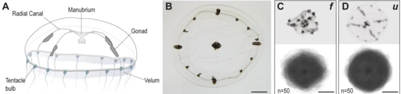
The medusa stage of the hydrozoan Clytia hemisphaerica (Fig. 1A) displays tetraradial symmetry, with four radial canals running from the oral manubrium to the ring of tentacle bulbs at the bell margin, and four gonads positioned on the radial canals. The transparent umbrella is composed of an exumbrellar layer and a subumbrella, separated by a thick layer of acellular mesoglea. The central manubrium, leading to the digestive pouch, is rather short and is organized according to the general tetraradial symmetry. A circular canal follows the periphery of the bell, connecting the four radial canals and the tentacle bulbs, numbering 16 in the adult animal. A thin velum, a bi-layered epithelium typical of hydrozoan medusae, extends from the bell margin and contributes to swimming (Leclère et al., 2016). The aldehyde-based fixation process used routinely for ISH reliably preserved the shape and the size of the live animal, which at the adult stage measures about 1 cm in diameter (Fig. 1B). However, the standard, formamide-based, in situ hybridization treatment invariably caused an extensive shrinking of the umbrella, with a marked alteration of body proportions. The umbrella appeared folded and heavily shrunken, while the more conspicuous elements, such as the manubrium, the gonads and the tentacle bulbs did not appear to be significantly affected, allowing the overall organization of the medusa to be maintained (Fig. 1C). The composite image in Fig. 1C, generated by the superposition of 50 animals, highlights their irregular morphology, and the
damage to the umbrella. This deformation, along with a general stiffening of tissues occurring during the processing, impaired the analysis of gene expression and the recognition of fine features. In contrast, the urea-based treatment preserved a better medusa morphology, as shown by the superposition of 50 different jellyfish (Fig. 1D), although the shrinking could not be completely avoided. The improved preservation of tissues, coupled with a more flexible consistency of medusae following the urea-based protocol, greatly facilitated the observation of our specimens.
A more sensitive technique for gene expression analyses
Nervous system-related genes provide a reliable way to assess the precision of mRNA detection, given their cell type-specific expression. We thus chose to compare the expression patterns for mcol3/4a (minicollagen 3/4a in Denker et al., 2008), RFamide (also called pp5), and drgx and six3/6 (Kraus et al., 2015). In all cases, formamide and urea variants gave comparable expression patterns, demonstrating that 4M urea could efficiently substitute for the 50% formamide during hybridization steps (results are shown in Figure 2). The urea-based protocol produced sharper staining patterns compared to the formamide one, particularly in the case of isolated cells, as for example showed by mcol3/4a expression in nematoblasts (the cells that will produce the nematocysts) at the base of the manubrium, and in the tentacle bulbs (compare Fig. 2A and 2B, 2E and 2F), where single positive cells could be more easily distinguished within the tissues.
For the genes tested, the urea method proved to be overall more sensitive than the traditional protocol. Testing a 10-fold reduced probe concentration (0.05 ng/µl), the mcol3/4a signal could no longer be detected in the manubrium using the formamide method (Fig. 2C), while clear staining could still be obtained using urea (Fig. 2D). We cannot currently assess whether the poorer staining was due to probe degradation, which could for example occur if the deionized formamide had lost purity (in our experiments, recently opened bottles of deionized formamide were used, kept at 4°C) but not in the urea- based hybridization solution.
The greater sensitivity of the urea method was particularly relevant for RFamide expression, where, using the same probe and color development conditions, we could reveal an unexpected complexity of the neural network in the manubrium, in the circular canal, and in the subumbrella (manubrium shown in Fig. 2I and 2J, circular canal in Fig. 2K and 2L).
An increased signal-to-noise ratio
Side-by-side comparison of staining patterns produced by formamide or urea protocols highlighted the tendency of the formamide-based hybridization method to generate aspecific staining of some medusa structures. In the case of fluorescent in situ hybridization, which is a highly sensitive method, the aspecific staining generated by the formamide protocol was in some cases so strong as to mask the true signal (for instance compare staining of RFamide neurons in the subumbrellar and radial canal regions in Fig. 2P and 2Q). Extensive previous experience with the colorimetric method had revealed a range of reproducible non-specific signals, including diffuse staining of the tentacle bulbs (exemplified by mcol3/4a detection in Fig. 2E, 2G), and superficial, punctate staining of the margin of the velum (see for example the drgx detection in Fig. 2M and six3/6 in Fig. 2O), and on the surface of gonads (as illustrated by drgx detection in Fig. 2T). The non-specific nature of the signal was easily recognizable for its rapid appearance and its superficial localization over epithelia, and further confirmed using control sense-strand probes (see the control for mcol3/4a in Fig. 2G, which shows a strong signal in the tentacle bulb and in the endoderm of tentacle). These strong signals forced premature arrest of the detection reaction, with the risk of under-developing the „true‟ expression patterns. These issues were resolved using the urea method, as demonstrated by the sharp signal obtained at the level of the tentacle bulb with the mcol3/4a probe, where the endoderm of the tentacle appears clear of background (Fig. 2F), and the absence of spots on the surface of velum and gonad (Fig. 2N and 2U, respectively). Moreover, control ISH performed in parallel with a sense probe (for mcol3/4a, Fig. 2H) produced a clean result, devoid of any non-specific staining.
To summarize, the urea ISH protocol for Clytia medusae not only allowed us to identify more confidently sites of gene expression, but also to continue the color development reaction until details of the expression patterns had been completely revealed.
The urea method is a reliable alternative for multiple species
The modified, urea-based, ISH protocol detailed in this study is safe and simple to implement, and we wondered whether it could be generally applicable across metazoan species.
We thus tested it on different animals and on different developmental stages, posing various experimental challenges. We included two Brachiopoda species (Novocrania anomala and Terebratalia transversa) and a priapulid worm (Priapulus caudatus), for which in situ hybridization protocols had been successfully established, but where gene expression analysis were at times hindered by aspecific staining. Similarly to Clytia, we used the already published
nervous system-related genes, nk2.1 and otx (orthodenticle), as a reference (Martín-Durán et al., 2016, 2012; Martín-Durán and Hejnol, 2015).
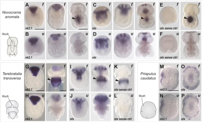
In all three species, the urea-based hybridization buffer produced a specific signal, compatible with the results already described using the formamide-based hybridization buffer particularly in P. caudatus, where the resolution appeared substantially equivalent. Examples included expression of nk2.1 in the oral ectoderm territory (formamide and urea-protocol shown in Fig. 3M and 3N, respectively), and of otx in the oral and ventral side (Fig. 3O and 3P, formamide and urea respectively). A decisive difference was observed for older stages of the two brachiopod larvae, where the central part of the body includes a typical site of aspecific staining, associated with the shell-secreting gland - shown by the formamide in situ hybridization images for nk2.1 (Fig. 3A and 3G) and otx (Fig. 3C, 3E, 3I, 3K). This strong staining was never observed for specimen processed using the urea protocol (compare Fig. 3B, 3D, 3H, 3J). The aspecific nature of the glandular staining was confirmed by the signal seen in the corresponding sense-probe formamide control (see Fig. 3E and 3K), which was markedly absent in the urea-control (Fig. 3F and 3L). A possible explanation for this difference is that the shell-gland staining results from probe trapping, a common artifact, and that urea buffers could improve the washing and reduce the retention of reagents due to greater tissue permeabilization.
These results demonstrate not only that our urea protocol for in situ hybridization can be successfully applied to other species, but importantly that it can prove very useful in reducing different types of non-specific signal associated with particular structures, as exemplified by the adult medusa and the late brachiopod larvae.
Discussion
The urea-based in situ hybridization protocol described in this study represents a powerful alternative to the standard formamide-based in situ hybridization techniques. In both the hydrozoan medusa Clytia hemisphaerica and two brachiopod species, Novocrania anomala and Terebratalia transversa, the urea-based hybridization buffer effectively boosted signal detection, reduced aspecific staining and improved specimen morphology, allowing for a more confident interpretation of results.
The common in situ hybridization techniques routinely employed to assess gene expression in numerous metazoan models, such as Xenopus, Danio, Nematostella or Mus, typically employ a
hybridization buffer composed of a 50% volume of formamide. Formamide is a dangerous chemical, especially at high temperatures, which increase evaporation and the risk of exposure to hazardous vapors. Extreme care is therefore needed during manipulation and for waste disposal, which can be particularly problematic in some field work or teaching contexts. For these safety and environmental reasons several reports have recently questioned the extensive use of formamide, asking if a less toxic option could be found (see Table 1 for a safety comparison of formamide and urea, and Table 2 for a survey of available literature). These studies were mostly aimed at medically-oriented research, for example for developing in vivo pathogen diagnostic techniques.
For ISH, formamide is widely used as a denaturing agent, but in other types of gene expression analysis, such as Northern, Southern or Western blots, urea is often employed for denaturing both proteins and nucleic acids. Indeed, the denaturing properties of urea have been known and investigated since the 1960s, and the efficiency of urea and formamide in hybridizations to RNA probes has already thoroughly been tested for blotting applications (Simard et al., 2001). It was demonstrated not only that urea could effectively replace formamide in detecting RNA, but also that the best results were obtained when urea concentrations were comprised between 2M and 4M. Above this value the sensitivity of detection was markedly reduced, probably due to the high viscosity of the solution (Hutton, 1977). Our observations in the laboratory paralleled this finding, with the urea-based hybridization solution being more viscous than the formamide one. This difference might be relevant while adapting the protocol to new organisms.
Formamide and urea share similar denaturing properties and are broadly employed for numerous applications. Nevertheless, their mechanisms of action are still incompletely understood, probably due to the multiple factors affecting the efficiency of the reaction. Formamide lowers the melting temperature of DNAs by 2.4- 2.9°C (per mole of formamide) - with an efficiency depending on the properties of the target nucleic acid strands, such as their G+C content, the helix topology and the state of hydration (Blake and Delcourt, 1996). This solvent weakens hydrogen bonds, ultimately allowing for lower hybridization temperatures, maintaining similar high stringencies (Casey and Davidson, 1977; Sadhu et al., 1984; Robertson and Vora, 2012). Generally, the more concentrated the formamide, the higher is the stringency of reaction, but it was shown that, similarly to urea, an excess of solvent causes a dramatic drop in probe binding and signal detection (Manz et al., 1992; Bond and Banfield, 2001). Non-specific signal is a common artifact, and stringency can be further improved through several post-hybridization washes, which remove the excess probe and disrupt any incorrectly paired
duplexes, for example weak base pairs between Guanine and Uracil in RNA (Uhlenbeck et al., 1971; Lomant and Fresco, 1975). The stringency of the washing buffer can be further controlled by lowering the concentration of salt, instead of using formamide, thus reducing the volume of toxic waste (Lathe, 1985).
Another factor affecting the signal-to-noise ratio in ISH is the purity of the formamide in the hybridization buffer, and numerous protocols recommend using deionized formamide. The reason for choosing a deionized solvent is that formamide solutions become acidic with time, due to the hydrolytic breakdown of formamide to formic acid and ammonium formate, responsible for attacking the phosphodiester bonds of RNA strands and degrading the larger RNA molecules in particular (Chow and Broker, 1989). The purification step removes those breakdown products, which will then take time to reform (this is why, for sensitive applications, it is generally recommended to use freshly opened bottles or to deionize the formamide from older ones). Our, previous experience did not show any difference between deionized or non-deionized formamide, and in the Clytia laboratory we routinely use recently opened bottles of deionized formamide, kept at 4°C.
Due to the widespread use of formamide, fewer studies have addressed the mechanism of action of urea. Urea can substantially lower the melting temperature of DNA, with values approaching 2°C reduction per mole of urea (Hutton, 1977), and thus slightly lower than the decrease that can be obtained with formamide. Recently it was shown that, as hypothesized previously, urea can interact with water and with both the polar and nonpolar components of nucleotides. It forms multiple hydrogen bonds with the RNA bases, and generates stacking interactions with them, ultimately disrupting the base-pair interactions and causing a destabilization of the structure of the RNA molecules (Herskovits and Bowen, 1974; Priyakumar et al., 2009; Lambert and Draper, 2012).
In our test species, the sensitivity of detection increased when formamide was replaced by urea, in line with a similar result previously observed for a FISH protocol developed for detecting Helicobacter pylori in gastric biopsies (Fontenete et al., 2013). This might be due to an additional permeabilization action of urea (Lim et al., 2009; Huang et al., 2011), which could enhance probe penetration in the tissues. An enhanced permeabilizing activity for urea could explain the improved detection we obtained in urea-treated medusae, for example demonstrated using low mcol3/4a probe concentrations (see Fig. 2C and 2D).
The urea alternative appears to be a reliable option for routine in situ hybridization, and has already been successfully tested in multiple species, including C. hemisphaerica, N. anomala, T.
transversa, P. caudatus (this study), and also the scyphozoan jellyfish Aurelia aurita (M. Manuel and T. Condamine, personal communication), the acoel Hofstenia miamia (L. Ricci, personal communication), and mouse oocytes (M.H. Verlhac and M. Manil-Segalen, personal communication). Despite broad reproducibility, we would recommend performing an initial comparison between the two protocols, in order to verify the reproducibility of the gene expression patterns detected.
Overall, substitution of formamide by urea in situ hybridization offers a safer alternative protocol, potentially useful in a broad spectrum of research, medical and teaching contexts. We encourage other workers to test this approach on their study organisms, and hope that they will also obtain more informative and sharp expression patterns, as we saw with Clytia hemisphaerica, Novocrania anomala and Terebratalia transversa.
Acknowledgements
We are grateful to Sandra Chevalier for suggesting focusing on the hybridization buffer, Gonzalo Quiroga-Artigas for thorough testing of the urea-protocol, Antonella Ruggiero for troubleshooting of the fluorescent in situ protocol, Muriel Jaeger and A. Ruggiero for initial identification and ISH with Clytia pp5, Stefania Castagnetti and Michael Schubert for kindly sharing their stereoscopes. Research at the Observatoire Océanologique de Villefranche-sur-mer was supported by the ANR grant ANR-13-PDOC-0016 “MEDUSEVO”. Research at the Sars International Centre was supported by the FP7-PEOPLE-2012-ITN grant no. 317172 “NEPTUNE” and the European Research Council Community’s Framework Program Horizon 2020 (2014–2020) ERC grant agreement 648861.
References
Azaripour, A., Lagerweij, T., Scharfbillig, C., Jadczak, A.E., Willershausen, B., Van Noorden, C.J.F., 2016. A survey of clearing techniques for 3D imaging of tissues with special reference to connective tissue. Prog. Histochem. Cytochem. 51, 9–23. doi:10.1016/j.proghi.2016.04.001.
Barbera, E., Caliani, M.J., Pagés, M., Alonso, C., 1979. Chromosomal DNA denaturation and reassociation in situ. Exp. Cell Res. 119, 151–162. doi:10.1016/0014-4827(79)90344-6 Barsacchi, G., 1972. Chromosomal localization of repetitive DNA in the newt, Triturus. J. Cell
Biol. 54, 580–591. doi:10.1083/jcb.54.3.580.
Bauman, J.G.J., Wiegant, J., Borst, P., van Duijn, P., 1980. A new method for fluorescence microscopical localization of specific DNA sequences by in situ hybridization of
fluorochrome-labelled RNA. Exp. Cell Res. 128, 485–490. doi:10.1016/0014-4827(80)90087-7.
Berndt, A., Kosmehl, H., Celeda, D., Katenkamp, D., 1996. Reduced formamide content and hybridization temperature results in increased non-radioactive mRNA in situ hybridization signals. Acta Histochem. 98, 79–87. doi:10.1016/S0065-1281(96)80053-5.
Blake, R.D., Delcourt, S.G., 1996. Thermodynamic effects of formamide on DNA stability. Nucleic Acids Res. 24, 2095–2103. doi:10.1093/nar/24.11.2095.
Blackshaw, S., 2013. High-throughput RNA in situ hybridization in mouse retina. Methods in Molecular Biology 935, 215-226. doi: 10.1007/978-1-62703-080-9-15.
Bond, P.L., Banfield, J.F., 2001. Design and performance of rRNA targeted oligonucleotide probes for in situ detection and phylogenetic identification of microorganisms inhabiting acid mine drainage environments. Microb. Ecol. 41, 149–161. doi:10.1007/s002480000063. Bonner, J., Kung, G., Bekhor, I., 1967. A method for the hybridization of nucleic acid molecules
at low temperature. Biochemistry 6, 3650–3653. doi:10.1021/bi00864a005.
Braissant, O., Wahli, W., 1998. A simplified in situ hybridization protocol using non-radioactively labeled probes to detect abundant and rare mRNAs on tissue sections. Biochemica 1, 10– 16.
Brown, C., 1998. In situ hybridization with riboprobes: an overview for veterinary pathologists. Vet. Pathol. 35, 159–167. doi:10.1177/030098589803500301.
Buongiorno-Nardelli, M., Amaldi, F., 1970. Autoradiographic detection of molecular hybrids between rRNA and DNA in tissue sections. Nature 225, 946–948. doi:10.1038/225946a0.
Casey, J., Davidson, N., 1977. Rates of formation and thermal stabilities of RNA:DNA and DNA:DNA duplexes at high concentrations of formamide. Nucleic Acids Res. 4, 1539–1552. doi:10.1093/nar/4.5.1539.
Chevalier, S., Martin, A., Leclère, L., Amiel, A., Houliston, E., 2006. Polarised expression of FoxB and FoxQ2 genes during development of the hydrozoan Clytia hemisphaerica. Dev. Genes Evol. 216, 709–720. doi:10.1007/s00427-006-0103-6.
Chow, L.T., Broker, T.R., 1989. Mapping the genetic organization of RNA by electron
microscopy, in: Methods in Enzymology. pp. 239–261. doi:10.1016/0076-6879(89)80105-3. CICAD 31: N,N-Dimethylformamide, 2001. , World Health Organization Geneva.
Denker, E., Manuel, M., Leclère, L., Le Guyader, H., Rabet, N., 2008. Ordered progression of nematogenesis from stem cells through differentiation stages in the tentacle bulb of Clytia hemisphaerica (Hydrozoa, Cnidaria). Dev. Biol. 315, 99–113.
doi:10.1016/j.ydbio.2007.12.023.
European Chemical Agency. Proposal for identification of a substance as a CMR CAT 1A or 1B, PBT, vPvB or a substance of an equivalent level of concern. Formamide.
Fail, P.A., George, J.D., Grizzle, T.B., Heindel, J.J., 1998. Formamide and Dimethylformamide: Reproductive Assessment by Continuous Breeding in Mice. Reprod. Toxicol. 12, 317–332. doi:10.1016/S0890-6238(98)00011-2.
Fontenete, S., Carvalho, D., Guimarães, N., Madureira, P., Figueiredo, C., Wengel, J., Azevedo, N.F., 2016. Application of locked nucleic acid-based probes in fluorescence in situ
hybridization. Appl. Microbiol. Biotechnol. 100, 5897–5906. doi:10.1007/s00253-016-7429-4.
Fontenete, S., Guimarães, N., Leite, M., Figueiredo, C., Wengel, J., Azevedo, N.F., 2013. Hybridization-based detection of Helicobacter pylori at human body temperature using advanced Locked Nucleic Acid (LNA) probes. PLoS One 8, 1–11.
doi:10.1371/journal.pone.0081230.
Fontenete, S., Leite, M., Guimarães, N., Madureira, P., Ferreira, R.M., Figueiredo, C., Wengel, J., Azevedo, N.F., 2015. Towards fluorescence in vivo hybridization (FIVH) detection of H. pylori in gastric mucosa using advanced LNA probes. PLoS One 10, 1–22.
doi:10.1371/journal.pone.0125494.
Gall, J.G., 2016. The origin of in situ hybridization - A personal history. Methods 98, 4–9. doi:10.1016/j.ymeth.2015.11.026.
George, J.D., Price, C.J., Marr, M.C., Myers, C.B., Jahnke, G.D., 2002. Evaluation of the developmental toxicity of formamide in New Zealand white rabbits. Toxicol. Sci. 69, 165– 74.
George, J.D., Price, C.J., Marr, M.C., Myers, C.B., Jahnke, G.D., 2000. Evaluation of the
developmental toxicity of formamide in Sprague-Dawley (CD) rats. Toxicol. Sci. 57, 284–91. Gerhard, D.S., Kawasaki, E.S., Bancroft, F.C., Szabo, P., 1981. Localization of a unique gene by
direct hybridization in situ. Proc. Natl. Acad. Sci. U. S. A. 78, 3755–3759.
Gleich, J., 1974. Proceedings: The influence of simple acid amides on fetal development of mice. Naunyn. Schmiedebergs. Arch. Pharmacol. 282, suppl 282:R25.
Hafen, E., Levine, M., Garber, R.L., Gehring, W.J., 1983. An improved in situ hybridization method for the detection of cellular RNAs in Drosophila tissue sections and its application for localizing transcripts of the homeotic Antennapedia gene complex. EMBO J. 2, 617–23. Hamaguchi, K., Geiduschek, E.P., 1962. The effect of electrolytes on the stability of the
deoxyribonucleate helix. J. Am. Chem. Soc. 84, 1329–1338. doi:10.1021/ja00867a001. Hejnol, A., 2008. In situ protocol for embryos and juveniles of Convolutriloba longifissura. Protoc.
Exch. doi:10.1038/nprot.2008.201.
Herskovits, T.T., 1963. Nonaqueous solutions of DNA; denaturation by urea and its methyl derivatives. Biochemistry 2, 335–340. doi:10.1021/bi00902a027.
Herskovits, T.T., 1962. Nonaqueous solutions of DNA: factors determining the stability of the helical configuration in solution. Arch. Biochem. Biophys. 97, 474–484. doi:10.1016/0003-9861(62)90110-8.
Herskovits, T.T., Bowen, J.J., 1974. Solution studies of the nucleic acid bases and related model compounds. Solubility in aqueous urea and amide solutions. Biochemistry 13, 5474–83. Houliston, E., Momose, T., Manuel, M., 2010. Clytia hemisphaerica: a jellyfish cousin joins the
laboratory. Trends Genet. 26, 159–167. doi:10.1016/j.tig.2010.01.008.
Huang, E., Talukder, S., Hughes, T.R., Curk, T., Zupan, B., Shaulsky, G., Katoh-Kurasawa, M., 2011. BzpF is a CREB-like transcription factor that regulates spore maturation and stability in Dictyostelium. Dev. Biol. 358, 137–146. doi:10.1016/j.ydbio.2011.07.017.
Hutton, J.R., 1977. Renaturation kinetics and thermal stability of DNA in aqueous solutions of formamide and urea. Nucleic Acids Res. 4, 3537–3555. doi:10.1093/nar/4.10.3537.
Jang, T.-S., Weiss-Schneeweiss, H., 2015. Formamide-free genomic in situ hybridization allows unambiguous discrimination of highly similar parental genomes in diploid hybrids and allopolyploids. Cytogenet. Genome Res. 146, 325–331. doi:10.1159/000441210.
John, H.A., Birnstiel, M.L., Jones, K.W., 1969. RNA-DNA hybrids at the cytological level. Nature 223, 582–587. doi:10.1038/223582a0.
Kennedy, G.L., Short, R.D., 1986. Biological effects of acetamide, formamide, and their monomethyl and dimethyl derivatives. CRC Crit. Rev. Toxicol. 17, 129–182. doi:10.3109/10408448609023768.
Kourilsky, P., Leidner, J., Tremblay, G.Y., 1971. DNA-DNA hybridization on filters at low temperature in the presence of formamide or urea. Biochimie 53, 1111–1114. doi:10.1016/S0300-9084(71)80201-8.
Kraus, J.E.M., Fredman, D., Wang, W., Khalturin, K., Technau, U., 2015. Adoption of conserved developmental genes in development and origin of the medusa body plan. Evodevo 6, 23. doi:10.1186/s13227-015-0017-3.
Lambert, D., Draper, D.E., 2012. Denaturation of RNA secondary and tertiary structure by urea: simple unfolded state models and free energy parameters account for measured m-values. Biochemistry 51, 9014–26. doi:10.1021/bi301103j.
Lapébie, P., Ruggiero, A., Barreau, C., Chevalier, S., Chang, P., Dru, P., Houliston, E., Momose, T., 2014. Differential responses to Wnt and PCP disruption predict expression and
developmental function of conserved and novel genes in a cnidarian. PLoS Genet. 10. doi:10.1371/journal.pgen.1004590.
Lathe, R., 1985. Synthetic oligonucleotide probes deduced from amino acid sequence data. J. Mol. Biol. 183, 1–12. doi:10.1016/0022-2836(85)90276-1.
Lawson, T.S., Connally, R.E., Vemulpad, S., Piper, J.A., 2012. Dimethyl formamide-free, urea-NaCl fluorescence in situ hybridization assay for Staphylococcus aureus. Lett. Appl. Microbiol. 54, 263–266. doi:10.1111/j.1472-765X.2011.03197.x.
Leclère, L., Copley, R.R., Momose, T., Houliston, E., 2016. Hydrozoan insights in animal development and evolution. Curr. Opin. Genet. Dev. 39, 157–167.
doi:10.1016/j.gde.2016.07.006.
Leclère, L., Jager, M., Barreau, C., Chang, P., Le Guyader, H., Manuel, M., Houliston, E., 2012. Maternally localized germ plasm mRNAs and germ cell/stem cell formation in the cnidarian Clytia. Dev. Biol. 364, 236–248. doi:10.1016/j.ydbio.2012.01.018.
Levine, L., Gordon, J. A., Jencks, W.P., 1963. The relationship of structure to the effectiveness of denaturing agents for deoxyribonucleic acid. Biochemistry 2, 168–175.
doi:10.1021/bi00901a030.
Levine, M., Hafen, E., Garber, R.L., Gehring, W.J., 1983. Spatial distribution of Antennapedia transcripts during Drosophila development. EMBO J. 2, 2037–46.
Lim, W.K., Rosgen, J., Englander, S.W., 2009. Urea, but not guanidinium, destabilizes proteins by forming hydrogen bonds to the peptide group. Proc Natl Acad Sci U S A 106, 2595– 2600. doi:10.1073/pnas.0812588106.
Lomant, A.J., Fresco, J.R., 1975. Structural and energetic consequences of noncomplementary base oppositions in nucleic acid helices. Prog Nucleic Acid Res Mol Biol 15, 185–218. doi:10.1016/S0079-6603(08)60120-8.
Manz, W., Amann, R., Ludwig, W., Wagner, M., Schleifer, K.-H., 1992. Phylogenetic
oligodeoxynucleotide probes for the major subclasses of proteobacteria: problems and solutions. Syst. Appl. Microbiol. 15, 593–600. doi:10.1016/S0723-2020(11)80121-9. Marmur, J., Doty, P., 1961. Thermal renaturation of deoxyribonucleic acids. J. Mol. Biol. 3, 585–
594. doi:10.1016/S0022-2836(61)80023-5.
Marmur, J., Ts‟o, P.O.P., 1961. Denaturation of deoxyribonucleic acid by formamide. Biochim. Biophys. Acta 51, 32–36. doi:10.1016/0006-3002(61)91013-7.
Martín-Durán, J.M., Hejnol, A., 2015. The study of Priapulus caudatus reveals conserved molecular patterning underlying different gut morphogenesis in the Ecdysozoa. BMC Biol. 13, 1–20. doi:10.1186/s12915-015-0139-z.
Martín-Durán, J.M., Janssen, R., Wennberg, S., Budd, G.E., Hejnol, A., 2012. Deuterostomic development in the protostome Priapulus caudatus. Curr. Biol. 22, 2161–2166.
doi:10.1016/j.cub.2012.09.037.
Martín-Durán, J.M., Passamaneck, Y.J., Martindale, M.Q., Hejnol, A., 2016. The developmental basis for the recurrent evolution of deuterostomy and protostomy. Nat. Ecol. Evol. 1, 5. doi:10.1038/s41559-016-0005.
Matthiesen, S.H., Hansen, C.M., 2012. Fast and non-toxic in situ hybridization without blocking of repetitive sequences. PLoS One 7, 1–8. doi:10.1371/journal.pone.0040675.
McConaughy, B.L., Laird, C.D., McCarthy, B.J., 1969. Nucleic acid reassociation in formamide. Biochemistry 8, 3289–3295. doi:10.1021/bi00836a024.
Merkle, J., Zeller, H., 1980. Studies on acetamides and formamides for embryotoxic and teratogenic activities in the rabbit. Arzneimittelforschung. 30, 1557–62.
Pardue, M.L., Gall, J.G., 1969. Molecular hybridization of radioactive DNA to the DNA of cytological preparations. Proc. Natl. Acad. Sci. U. S. A. 64, 600–4.
doi:10.1073/pnas.64.2.600.
Priyakumar, U.D., Hyeon, C., Thirumalai, D., Mackerell, A.D.J., 2009. Urea destabilizes RNA by forming stacking interactions and multiple hydrogen bonds with nucleic acid bases. J. Am. Chem. Soc. 131, 17759–61. doi:10.1021/ja905795v.
Rice, S.A., Doty, P., 1957. The thermal denaturation of desoxyribose nucleic acid. J. Am. Chem. Soc. 79, 3937–3947. doi:10.1021/ja01572a001.
Robertson, K.L., Vora, G.J., 2012. Locked nucleic acid flow cytometry-fluorescence in situ hybridization (LNA flow-FISH): a method for bacterial small RNA detection. J. Vis. Exp. e3655. doi:10.3791/3655.
Sadhu, C., Dutta, S., Gopinathan, K.P., 1984. Influence of formamide on the thermal stability of DNA. J. Biosci. 6, 817–821. doi:10.1007/BF02716841.
Santagata, S., Resh, C., Hejnol, A., Martindale, M.Q., Passamaneck, Y.J., 2012. Development of the larval anterior neurogenic domains of Terebratalia transversa (Brachiopoda) provides insights into the diversification of larval apical organs and the spiralian nervous system. Evodevo 3, 3. doi:10.1186/2041-9139-3-3.
Shain, D.H., Zuber, M., 1996. Sodium dodecyl sulfate (SDS)-based whole-mount in situ hybridization of Xenopus laevis embryos. J. Biochem. Biophys. Methods 31, 185–188. doi:10.1016/0165-022X(95)00030-U.
Simard, C., Lemieux, R., Côté, S., 2001. Urea substitutes toxic formamide as destabilizing agent in nucleic acid hybridizations with RNA probes. Electrophoresis 22, 2679–2683.
doi:10.1002/1522-2683(200108)22:13<2679::AID-ELPS2679>3.0.CO;2-L.
Singh, L., Jones, K.W., 1984. The use of heparin as a simple cost-effective means of controlling background in nucleic acid hybridization procedures. Nucleic Acids Res. 12, 5627–38. Søe, M.J., Møller, T., Dufva, M., Holmstrøm, K., 2011. A sensitive alternative for microRNA in
situ hybridizations using probes of 2‟-O-methyl RNA + LNA. J. Histochem. Cytochem. 59, 661–672. doi:10.1369/0022155411409411.
Stula, E.F., Krauss, W.C., 1977. Embryotoxicity in rats and rabbits from cutaneous application of amide-type solvents and substituted ureas. Toxicol. Appl. Pharmacol. 41, 35–55.
doi:10.1016/0041-008X(77)90052-7.
Summer, H., Grämer, R., Dröge, P., 2009. Denaturing urea polyacrylamide gel electrophoresis (Urea PAGE). J. Vis. Exp. doi:10.3791/1485.
Takeda, N., Kon, Y., Quiroga Artigas, G., Lapébie, P., Barreau, C., Koizumi, O., Kishimoto, T., Tachibana, K., Houliston, E., Deguchi, R., 2017. Jellyfish oocyte maturation inducing hormone and neuroendocrine regulation of reproduction. bioRxiv. 140160; doi: https://doi.org/10.1101/140160.
Tautz, D., Pfeifle, C., 1989. A non-radioactive in situ hybridization method for the localization of specific RNAs in Drosophila embryos reveals translational control of the segmentation gene hunchback. Chromosoma 98, 81–85. doi:10.1007/BF00291041.
Uhlenbeck, O.C., Martin, F.H., Doty, P., 1971. Self-complementary oligoribonucleotides: Effects of helix defects and guanylic acid-cytidylic acid base pairs. J. Mol. Biol. 57, 217–229. doi:10.1016/0022-2836(71)90342-1.
Figures
Figure 1. The morphology of Clytia medusae is better preserved after urea-based in situ hybridization (u), with respect to the classical, formamide-based, protocol (f).
(A) Anatomy of Clytia medusa: the tetraradial symmetry is evident in the four radial canals emerging from the manubrium (stomach and mouth part), crossing the umbrella, from which the four gonads develop. An acellular, thick, mesoglea separates exumbrella and subumbrella layers. (B) Oral view of a medusa, post-fixation. The fixation process preserves morphology and size of the living animal. (C, D) Sample individual pictures (top) and composite images generated by the superposition of 50 images (bottom) of medusae following formamide-based (C) or urea-based (D) hybridization steps. Scale bars, 1 mm.
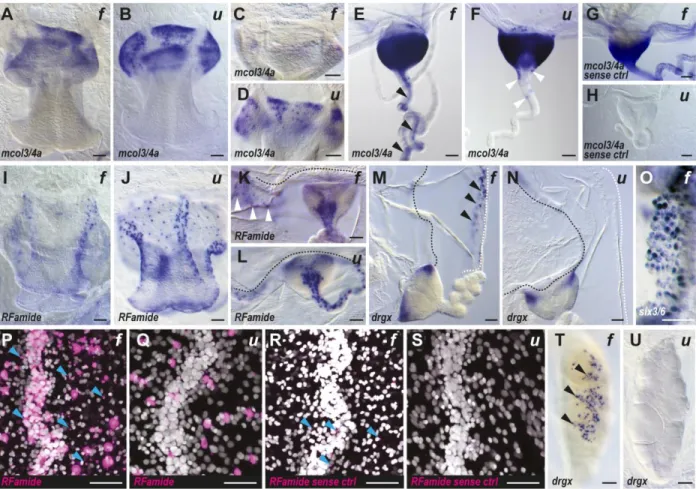
Figure 2. Improved sensitivity and signal-to-noise ratio with the urea-based in situ hybridization method on Clytia medusa.
(A-O,T,U) Colorimetric and (P-S) fluorescent (signal in magenta, nuclear Hoechst staining in white) in situ hybridization results, comparing formamide-based (f) and urea-based (u) methods. Antisense (A-F, I-Q, T-U) and sense (G, H, R, S) probe gene names are indicated at the bottom left of each picture. The pictures show: (A, B, I, J) whole manubrium with oral opening at the bottom; (C, D) aboral, basal, side of the manubrium; (E-H, K, N) tentacle, tentacle bulbs and portions of the bell margin (black dotted line in L-N) and velum (white dotted line in M and N); (P-S) portions of radial canals running from top to bottom and nearby subumbrella; (O) high magnification of velum margin; (T, U) female gonad with oocytes. Black arrows point to aspecific staining seen after formamide-based ISH in: the tentacle endoderm (in E), velum epithelium (in M) and gonad ectoderm (T). White arrows highlight specific single cell staining produced with the urea-based method in the tentacle ectoderm (in F) and radial canal (in K). Blue arrows indicate few examples of aspecific fluorescent staining (in P and R). In (C and D) the concentration was decreased by 10 fold compared to (A and B), notice how the specific mcol3/4a signal at the base of the manubrium is lost with the formamide method (C), while it is still detectable with the urea
method (D). The aspecific staining on tentacle using mcol3/4a sense control probe, as shown in (G), was obtained in many but not all analyzed medusae. (P-R) the four fluorescent images were taken with the same settings. Scale bars, 50 µm
Figure 3. In situ hybridization on developmental stages of two brachiopods and a priapulid worm using either formamide (f) or urea-based (u) methods. A-F Novocrania anomala, G-L Terebretalia transversa, M-P Priapulus caudatus. Antisense (A-D, G-J, M-P) and sense control (E, F, K, L) probe gene names indicated on the bottom left of each panel. Developmental stages from left to right: (A-D) early gastrula, late gastrula and late larva; (E-J): early gastrula and late larva; (K-L): late larva; M-P: late gastrula. In all drawings and images, embryos and larvae are oriented with the anterior pole on top. Black arrows indicate aspecific signal, frequently seen with the traditional formamide method in the shell gland of Novocrania anomala (right images in A, C, E) and Terebretalia transversa (right images in G, I, K) late larvae. Scale bars, 50 µm.
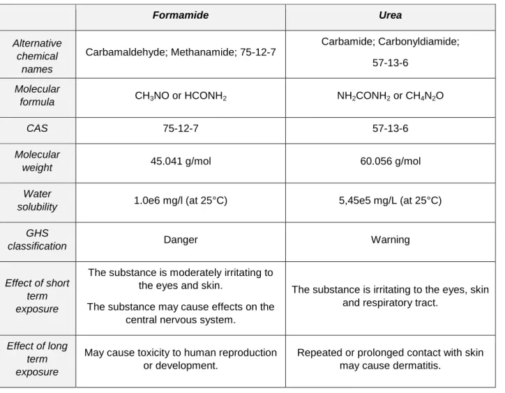
Table 1. Overview of properties and health risks of formamide, the most diffuse denaturing agent, and of urea, a safer alternative (source www.ncbi.nlm.nih.gov/pccompound).
Formamide Urea Alternative chemical names Carbamaldehyde; Methanamide; 75-12-7 Carbamide; Carbonyldiamide; 57-13-6 Molecular
formula CH3NO or HCONH2 NH2CONH2 or CH4N2O
CAS 75-12-7 57-13-6
Molecular
weight 45.041 g/mol 60.056 g/mol
Water
solubility 1.0e6 mg/l (at 25°C) 5,45e5 mg/L (at 25°C)
GHS
classification Danger Warning
Effect of short term exposure
The substance is moderately irritating to the eyes and skin.
The substance may cause effects on the central nervous system.
The substance is irritating to the eyes, skin and respiratory tract.
Effect of long term exposure
May cause toxicity to human reproduction or development.
Repeated or prolonged contact with skin may cause dermatitis.
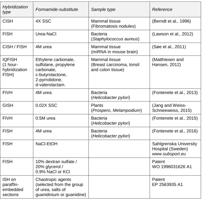
Table 2. A survey of the recent reports focused on formamide-alternatives for in situ hybridization. Hybridization types: CISH- Chromogenic In Situ Hybridization, FISH- Fluorescent In Situ Hybridization, FIVH- Fluorescent In Vivo Hybridization, GISH - Genomic In Situ Hybridization.
Hybridization
type Formamide-substitute Sample type Reference
CISH 4X SSC Mammal tissue
(Fibromatosis nodules)
(Berndt et al., 1996)
FISH Urea-NaCl Bacteria
(Staphylococcus aureus)
(Lawson et al., 2012)
CISH / FISH 4M urea Mammal tissue
(miRNA in mouse brain)
(Søe et al., 2011) IQFISH (1 hour-hybridization FISH) Ethylene carbonate, sulfolane, propylene carbonate, c-butyrolactone, 2-pyrrolidone, d-valerolactam. Mammal tissue
(Breast carcinoma, tonsil and colon tissue)
(Matthiesen and Hansen, 2012)
FIVH 4M urea Bacteria
(Helicobacter pylori)
(Fontenete et al., 2013)
GISH 0.02X SSC Plants
(Prospero, Melampodium)
(Jang and Weiss-Schneeweiss, 2015)
FIVH 0.5M urea Bacteria
(Helicobacter pylori)
(Fontenete et al., 2015)
FISH 4M urea Bacteria
(Helicobacter pylori)
(Fontenete et al., 2016)
FISH NaCl-EtOH Sahlgrenska University
Hospital (Sweden) www.subsport.eu FISH 10% dextran sulfate /
20% glycerol / 0.9% NaCl or KCl Patent WO 1996031626 A1 ISH on paraffin-embedded sections Chaotropic agents (selected from the group of urea, salts of
guanidinium or guanidine)
Patent
Supplementary Material_1
Additional information regarding probe synthesis.
• Genes
Gene name Probe name CDS Lenght Accession
number Minicollagen 3/4a
(C. hemisphaerica)
mcol3-4a 821 bp EU024529
DRGX (Dorsal Root Ganglia Homeobox) (C. hemisphaerica) drgx 1021 bp LN611639 pp5 (C. hemisphaerica) RFamide 1566 bp KX496951 Six3/6 (C. hemisphaerica) six3/6 1189 bp LN611635 Nk2.1 (N. anomala) nk2.1 978 bp KF946068 Otx (N. anomala) otx 807 bp KF946066 Nk2.1 (T. transversa) nk2.1 972 bp KF946076 Otx (T. transversa) otx 822 bp HQ679622 Nk2.1 (P. caudatus) nk2.1 715 bp KP013757 Otx (P. caudatus) otx 822 bp JX430801
• Probe synthesis (Clytia hemisphaerica)
Probes used in this study were synthesized from cDNA clones retrieved from our EST collection, and the DIG-labeled RNA probes were synthesized using the T7 RNA polymerase kit from Promega (according to vector and orientation of insert, SP6 or T3 polymerases could be necessary), purified using the ProbeQuant G-50 MicroColumns (GE Healthcare) and stored at -20°C.
For probe synthesis, add in order:
Notes DIG labelling mix RNA
(Roche, #11277073910)
2 µl 10X solution with: 10 mM
ATP, CTP, GTP (each), 6.5 mM UTP, 3.5 mM DIG-11-UTP.
RNasin Ribonuclease Inhibitor (Promega, #N2111)
0.5 µl Protects RNA from
degradation by RNases A, B and C. DTT (Promega, #P1171) 2 µl (stock 100mM)
Dithiothreitol, necessary for the correct functioning of the ribonuclease inhibitor. Transcription Optimized 5X Buffer
(Promega, #P1181)
4 µl
DNA template (1-2 µg) To calculate
RNase-free H2O To volume T7 RNA polymerase (Promega, #9PIP207) 0.5 µl DNA-dependent RNA polymerase. Total volume 20 µl
- Let the synthesis run for 2-5 hours at 37°C, then stop the reaction by digesting the DNA template: add 1.5 µl of RQ1 RNase-free DNase (Promega, #M6101) and incubate at 37°C for 30 minutes.
- Add 30 µl of RNase free H2O, and purify using the illustra ProbeQuant G-50 Micro Columns
(GE Healthcare, #28-9034-08).
- Check 1 µl on 1% agarose gel, and quantify (with a nanodrop). Add 50 µl of deionized formamide (Molecular Biology, Biosolve BV, # 06812335) for storage at -20°C.
• Probe synthesis (brachiopods and priapulid)
Genes were cloned into the pGEM-T Easy vector (Promega), and probes were synthesized using the MEGAscript SP6 kit or the MEGAscript T7 kit (Invitrogen, Thermo Fisher Scientific), using the DIG-labeled nucleotides Digoxigenin-11-UTPs (Roche Diagnostics).
Supplementary Material_2
Detailed protocol for Clytia hemisphaerica.
Step Solution Duration Temperature Comment
Relaxation 10 min On ice
Fixation (CISH) 3.7% formaldehyde plus 0.4% glutaraldehyde in 1X PBS (Phosphate-Buffered Saline, pH 7.4)2 hours On ice Pre-chilled solution.
Fixation (FISH) 3.7% formaldehyde in HEM buffer (0.1M HEPES pH 6.9, 50mM EGTA pH 7.20, 10mM MgSO4)36 hours 18°C Renew solution
once.
Wash 1X PBST (1X PBS plus 0.1% Tween-20) 5 x 10 min On ice
Wash 50% 1X PBST / 50% Methanol 1 x 10 min On ice
Wash 100% Methanol 1 x 10 min On ice
Storage 100% Methanol -20°C Can be directlyre-hydrated.
Wash 50% Methanol / 50% 1X PBST 1 x 5 min RT
Wash 1X PBST 3 x 5 min RT
Acetylation step 0.1M Triethanolamine (TEA) in 1X PBST5 min RT Optional step.
Acetylation step0.25% Acetic anhydride in (0.1M TEA in 1X PBST)
5 min RT Optional step.
Wash 1X PBST 10 min RT Optional step.
Transfer 50% Hybridization Buffer (4M urea, 5X SSC, 1% dextran, 50 µg/ml of tRNA, 50 µg/ml of heparin, 1% SDS, and milliQ H2O) /50% 1X PBST
1 X 10 min RT
Transfer 100% Hybridization buffer 20 min RT
Pre-hybridization
100% Hybridization buffer 2 hours minimum
58°C Pre-hybridizing
overnight can improve the results.
Hybridization Probe in hybridization buffer 48-72 hours 58°C Heat probe mix at 95°C for 5 min. Spin down and quickly apply probe, without letting it cool down.
Stringent wash 4M urea, 0.1% Tween-20, 5X SSC, milliQ H2O 3 x 30 min 58°C
Stringent wash 2M urea, 0.1% Tween-20, 2X SSC, milliQ H2O 3 x 30 min 58°C
Stringent wash 0.1% Tween-20, 2X SSC, milliQ H2O 2 x 30 min 58°C
Wash MABT buffer (Maleic Acid Buffer, containing 0.1% Tween-20)2 X 20 min RT
Block Blocking buffer (1% blocking solution, in MABT buffer)1 hour RT CISH detection
Antibody incubation Anti-DIG-AP (1:2000, in blocking buffer) Overnight 4°C
Wash MABT buffer 6 x 15 min RT
Wash NTMT minus buffer (NaCl, Tris-HCl at pH 9.5, Tween-20) 3 x 10 min RT Optional.
Wash NTMT buffer (NaCl, Tris-HCl at pH 9.5, MgCl2, Tween-20) 2 x 10 min RT
Colour development NBT/BCIP in NTMT buffer (0.08 mg/ml of Nitro Blue Tetrazolium and 0.1 mg/ml of 5- Bromo-4-Chloro-3-Indolyl-Phosphate) Until appropriate RT/4°C
Rinse milliQ H2O RT
Post-fixation 3.7% formaldehyde in 1X PBS 1 x 30 min RT
Wash 1X PBS
Clarify & Mount 50% 1X PBS/ 50% glycerol
Clarify & Mount 70% glycerol FISH detection
Antibody incubation Anti-DIG-POD (1:2000, in blocking buffer) Overnight 4°C
Wash Reaction buffer(0.0015% H202 in 1x PBS)
2 x 30 min RT Freshly prepared.
Signal developmentFuorophore-conjugated tyramide kit
(1:400, in reaction buffer)
1 hour RT Keep in dark.
Rinse 1X PBS RT Keep in dark.
Nuclear stainingHoechst 33528 (in 1X PBS) 30 minutes RT Keep in dark.
Rinse 1X PBS RT Keep in dark.
Storage and mountNote: the urea buffer tends to be more viscous, and more prone to evaporation. We recommend sealing the samples carefully.
• Main reagents
Company #
Hybridization buffer
Formamide – deionized. Molecular Biology Biosolve BV; The Netherlands 06812335
Urea Sigma U5378
Dextran sulfate sodium salt, Mr ~200''00 Sigma 67578-25G
tRNA, from baker’s yeast Sigma 10109509001
Heparin sodium Thermofisher 10239840
Antibody
Anti-Digoxigenin-AP, Fab fragments AP = Alkalyne Phosphatase
Roche Applied Sciences (Sigma- Aldrich)
11093274910 Anti-Digoxigenin-POD, Fab fragments
POD = Horseradish Peroxidase
Roche Applied Sciences (Sigma- Aldrich)
11207733910
Colour reaction
BCIP/NBT Color Development Substrate Promega S3771
TSA Plus Fluorescence Amplification kit Cyanine 3/5
Supplementary Material_3
Detailed protocol for brachiopod and priapulid species.
In situ hybridization protocol for: T. transversa, N. anomala and P. caudatus.
Step Solution Duration Temperature Comment
Relaxation 7.4% MgCl2x6H2Oin sea water
10- 15 min Room temperature (RT)
Fixation 4% paraformaldehyde (PFA) in 1X PBST(1X PBS plus 0.1% Tween-20)
1 hour RT
Wash 1X PBST 7 x 5 min RT
Storage Methanol Minimum24-48 hours -20°C Can be stored up to several months.
Wash 1X PBST 5 x 5 min RT
Permeabilize 10 µg/ml proteinase K in 1X PBST Species/ stage specific*1 RT No shaking.
Arrest of digestion 2 mg/ml glycine in 1X PBST 2 x 5 min RT
Wash 1% Triethanolamine (TEA) in 1X PBST5 min RT
Wash Acetic anhydride *2 in 1% TEA in 1X PBST5 min RT
Wash Acetic anhydride *3 in 1% TEA in 1X PBST 5 min RT
Wash 1X PBST 2 x 5 min RT
Fixation 3.7% formaldehyde in 1X PBST (+ 0.2% glutaraldehyde*4) 1 hour RT Depends on hybridization buffer*4.
Wash 1X PBST 3 x 5 min RT
Heating 1X PBST 10 min 80°C
Wash 1X PBST 5 min RT
Transfer Hybridization buffer (see legend for recipe)5 min RT
Transfer Hybridization buffer 10 min RT
Pre-hybridization
Hybridization buffer Overnight 67°C Hybridization
temperature, and probe concentration need to be adjusted according to species and probe.*5
Hybridization Probe in hybridization buffer 24- 72 hours67°C
Wash Hyb-wash buffer(see legend for recipe)
15 min 67°C
Wash Hyb-wash buffer 45 min 67°C
Stringent wash 75% Hyb-wash buffer / 25% 2X SSC 30 min 67°C
Stringent wash 50% Hyb-wash buffer / 50% 2X SSC 30 min 67°C
Stringent wash 25% Hyb-wash buffer / 75% 2X SSC 30 min 67°C*1 10 min P. caudatus, 9 min T. transversa
*2 1.5 µl acetic anhydride per 500 µl 1X PBST/TEA *3 4.5 µl acetic anhydride per 500 µl 1X PBST/TEA
*4 Glutaraldehyde is only necessary for urea-containing buffer, not for the formamide-based buffer.
*5 In the case of T. transversa and P. caudatus, for nk2.1 and otx, we incubated at 66°C and used probes at a concentration of 1 ng/µl.
Note: the urea buffer tends to be more viscous, and more prone to evaporation. We recommend sealing the samples carefully.
Stringent wash 2X SSC 30 min 67°C
Stringent Wash 0.2X SSC 3 x 20 min 67°C
Stringent Wash 75% 0.2X SSC / 25% 1X PBST 10- 15 min RT
Wash 50% 0.2X SSC / 50% 1X PBST 10- 15 min RT
Wash 25% 0.2X SSC / 75% 1X PBST 10- 15 min RT
Wash 1X PBST 10- 15 min RT
Wash/Block 1x PBSTx (1X PBS + 0.2% Triton-X100 + 0.1% BSA (Bovine Serum Albumine))2 x 15 min RT
Rinse Maleic acid buffer 5 min RT
Block Blocking buffer (Boehringer-Mannheim blocking buffer, in maleic acid buffer)1 hour RT
Antibody incubation Anti-DIG-AP (1:5000) in blocking buffer Overnight 4°C
Wash 1X PBSTx 5 x 15 min RT
Wash 1X PBST 5 x 30 min RT
Wash NTMT minus buffer(NaCl, Tris-HCl at pH 9.5, Tween-20) 3 x 10 min RT