HAL Id: hal-01002243
https://hal.archives-ouvertes.fr/hal-01002243
Submitted on 29 May 2020
HAL is a multi-disciplinary open access
archive for the deposit and dissemination of
sci-entific research documents, whether they are
pub-lished or not. The documents may come from
teaching and research institutions in France or
abroad, or from public or private research centers.
L’archive ouverte pluridisciplinaire HAL, est
destinée au dépôt et à la diffusion de documents
scientifiques de niveau recherche, publiés ou non,
émanant des établissements d’enseignement et de
recherche français ou étrangers, des laboratoires
publics ou privés.
Copyright
Nutritional strategies to counteract muscle atrophy
caused by disuse and to improve recovery
Hugues Magne, Isabelle Savary-Auzeloux, Didier Remond, Dominique
Dardevet
To cite this version:
Hugues Magne, Isabelle Savary-Auzeloux, Didier Remond, Dominique Dardevet. Nutritional strategies
to counteract muscle atrophy caused by disuse and to improve recovery. Nutrition Research Reviews,
Cambridge University Press (CUP), 2013, 26 (2), pp.149-165. �10.1017/S0954422413000115�.
�hal-01002243�
Nutritional strategies to counteract muscle atrophy caused by disuse
and to improve recovery
Hugues Magne
1,2*, Isabelle Savary-Auzeloux
1,2, Didier Re´mond
1,2and Dominique Dardevet
1,21Clermont Universite´, Universite´ d’Auvergne, Unite´ de Nutrition Humaine, BP 10448, F-63000 Clermont-Ferrand, France
2INRA, UMR 1019, UNH, CRNH Auvergne, F-63000 Clermont-Ferrand, France
Abstract
Periods of immobilisation are often associated with pathologies and/or ageing. These periods of muscle disuse induce muscle atrophy which could worsen the pathology or elderly frailty. If muscle mass loss has positive effects in the short term, a sustained/uncontrolled muscle mass loss is deleterious for health. Muscle mass recovery following immobilisation-induced atrophy could be critical, particularly when it is uncompleted as observed during ageing. Exercise, the best way to recover muscle mass, is not always applicable. So, other approaches such as nutritional strategies are needed to limit muscle wasting and to improve muscle mass recovery in such situations. The present review discusses mechanisms involved in muscle atrophy following disuse and during recovery and emphasises the effect of age in these mechanisms. In addition, the efficiency of nutritional strategies proposed to limit muscle mass loss during disuse and to improve protein gain during recovery (leucine supplementation, whey proteins, antioxidants and anti-inflammatory compounds, energy intake) is also discussed.
Key words:Muscular atrophy: Disuse: Muscle recovery: Ageing: Nutrition: Protein: Leucine
Introduction
Chronic periods of reduced physical activity occur throughout the life, following injuries or trauma, after pro-longed immobilisation (casting or extended bed rest), or as a natural result of ageing. The primary effect of muscle disuse in such situations is the progressive loss of skeletal muscle, even in the absence of a diagnosed disease(1 – 3). For example, ageing itself accelerates inactivity-induced atrophy(4), hence contributing to the decline of muscle function and physical activity in older adults(5). Since skel-etal muscle is the major reservoir of body proteins and amino acids that the organism could mobilise to cope with traumatic, infectious or nutritional stresses, sustained and uncontrolled muscle mass loss is then deleterious for maintaining optimal health status. After a period of immobilisation, the rehabilitation phase usually leads to a complete muscle mass recovery. Even though mechanisms involved during disuse-induced atrophy have been exten-sively studied, mechanisms involved in the recovery phase have not been thoroughly investigated. Exercise is known to be the best way to maintain and/or gain muscle mass even in elderly individuals(6 – 8), but it is not
always applicable in all situations. Thus, other approaches such as nutritional strategies are needed to limit muscle wasting and to improve muscle mass recovery in such situ-ations. The purpose of the present review is to concentrate on mechanisms involved in muscle atrophy following disuse and during recovery and to discuss the efficiency of potential nutritional strategies to limit muscle mass loss during disuse and to improve muscle mass gain during recovery in such situations of immobilisation.
Protein turnover during muscle disuse
Due to a decrease in mechanical stimuli, muscle disuse induces an alteration in skeletal muscle protein turnover associated with a negative N balance(9 – 12). A decreased synthesis and/or an increased breakdown of myofibrillar proteins (which represent about 85 % of the muscle fibre volume) explain muscle atrophy and the subsequent muscle dysfunction(13,14). Although the direct measurement of muscle protein breakdown is not possible in vivo, surro-gate markers such as proteolytic enzyme activities or mRNA have been used to describe myofibrillar proteolysis during immobilisation. Experiments with immobilisation
* Corresponding author: Dr Hugues Magne, email hugues.magne@clermont.inra.fr
Abbreviations: Akt, protein kinase B; BCAA, branched-chain amino acid; EAA, essential amino acid; eIF, eukaryotic initiation factor; FoxO, forkhead O transcription factor; MAFbx, muscle ubiquitin ligase muscle atrophy F-box; mTOR, mammalian target of rapamycin; MuRF1, muscle RING-finger protein-1; PI3K, phosphoinositide 3-kinase; S6K1, S6 kinase 1; UPS, ubiquitin – proteasome system.
qThe Authors 2013
conducted in human subjects and animals indicated that muscle disuse induces an early increase in protein breakdown, within the first 24 h(15,16). The extent of this increase differs considerably according to the model of immobilisation used, varying from 50 to 200 %(15,17 – 21). All the proteolytic systems, which target different com-ponents of muscle architecture, seem to be involved. The main proteolytic system activated during immobilisation is the ubiquitin – proteasome system (UPS). This is, some-how, not very surprising as the UPS is responsible for the breakdown of the major contractile proteins, i.e. actin and myosin(22 – 24). In this pathway, substrates are tagged by covalent attachment of multiple ubiquitins and then recognised and degraded by the 26S proteasome. Both
steps are activated during bed rest in humans(15,17) or
immobilisation-induced atrophy in animals(25 – 29). Animal models of immobilisation also clearly demonstrated that the UPS is then rapidly normalised during the recovery period in adults(27,29). The same kinetics has been observed in aged animals immobilised by casting(30,31); however, in these experiments the UPS was activated to a lesser extent: the proteasome chymotrypsin-like activity increased only by 53 % in old rats, while a 138 % increase was reported in adult rats immobilised using the same
model(28). As immobilisation-induced muscle atrophy
remained similar at both ages, with 21 and 23 % in old and adult rats, respectively, this indicates that additional proteolytic mechanisms leading to muscle loss during disuse may take place during ageing(32,33). Calpains are proteases involved in the early steps of disassembly of sarcomeric proteins(22,34,35). The role of the calpain path-way in response to immobilisation was demonstrated
in vivo and in vitro(12,26,36) and confirmed in mice by
using inhibitors of calpain activity which prevented sarcomere structure disruption(37). In healthy volunteers submitted to limb immobilisation, an early (24 h) increase of mRNA coding for calpains-1 and -2 has been
observed(15). Growing evidence also suggests a central
role of the lysosomal pathway (i.e. autophagy processes) in immobilisation-induced atrophy, despite its low
contri-bution to overall protein breakdown and weak
participation in the degradation of myofibrillar proteins(22). Key molecules of this pathway (i.e. Beclin 1 or LC3) are nevertheless over-expressed in response to muscle disuse within the first 14 d of immobilisation(38) and cathepsin mRNA, and activities which are protease-specific to this pathway are also increased(26,39 – 41). Despite autophagy-lysosome substrates in skeletal muscle being still poorly identified, this pathway could nevertheless play a crucial role during disuse as its activation is associated with the activation of the forkhead O transcription factors (FoxO) which are known to activate the muscle RING-finger pro-tein-1 (MuRF1) and the specific muscle ubiquitin ligase muscle atrophy F-box (MAFbx) atrogenins (atrogens) involved in the activation of UPS proteolysis(41).
Protein synthesis is the most dynamic variable in the pro-tein balance and it changes several-fold throughout the
day, as demonstrated with direct measurement(10).
How-ever, when studied in the fasted state, contradictory results have been reported with either a reduced basal-fasted rate of muscle protein synthesis(42,43), or an unchanged rate of protein synthesis(44 – 47). When muscle protein synthesis was decreased, this change occurred early during the immobilisation period (, 10 d) and then protein synthesis stabilised, i.e. remained decreased at the same level for
the next 21 d of immobilisation(48). Conversely, data
regarding the postprandial state agreed that immobilised skeletal muscle exhibits a decrease in responsiveness of muscle protein synthesis to amino acids across a wide range of amino acid concentrations. Indeed, provision of low and very high concentrations of amino acids (43 and 261 mg/kg per h, respectively) failed to restore muscle pro-tein synthesis to that seen in the non-immobilised muscle(49). Immobilisation may thus induce a real resist-ance of skeletal muscle protein synthesis to the stimulatory effect of food intake(10,49), a condition defined as ‘anabolic resistance’(49). Under conditions of anabolic resistance, the normal anabolic response to amino acids is blunted and the subsequent postprandial gain in muscle proteins may be less important, resulting in muscle atrophy(50). This explains why a normal diet (i.e. which in theory covers the recommended daily amino acid requirements) is a diet not adapted any more to overcome anabolic
resist-ance(51) and why animals or individuals fed such a diet
present this decrease in postprandial protein synthesis when experiencing prolonged immobilisation. Recently, this anabolic resistance has also been demonstrated in
aged animals immobilised by casting(31), which could
exacerbate the age-related ‘normal’ blunted response of muscle anabolism to food intake (see the section on Ageing in the present review). The rate of protein synthesis is dependent on the stimulation and the efficiency of the anabolic phosphoinositide 3-kinase (PI3K)/protein kinase B (Akt)/mammalian target of rapamycin (mTOR) pathway. Immobilisation induces a decrease in Akt protein con-tent(52), but its activity is either decreased(53,54) or unchanged(55,56), which, in both cases, leads to an imbal-ance of protein synthesis as protein breakdown is increased. Muscles from animals with immobilised hind-limbs exhibit a decrease in S6 kinase 1 (S6K1) protein content(57), and a decrease in mRNA translation, a critical step of protein synthesis(58) dependent on the activation of mTOR. However, data from recent disuse investigations in human subjects have failed to demonstrate a decrease in activation of key proteins in the PI3K/Akt/mTOR pathway during both the postabsorptive and the postprandial states, suggesting that the decrease in protein synthesis may be
regulated by downstream translation, initiation and
elongation factors independent of PI3K/Akt/mTOR
signal-ling(48,49). Confirming this hypothesis, data have shown
that 10 or 21 d bed rest in humans did not affect the
phosphorylation state of S6K1 and 4E-BP1 proteins in the postabsorptive state(48). This demonstrates the complexity of the regulation involved, which may include: (1) species differences in protein metabolism; (2) altered kinetics of protein synthesis during immobilisation; and (3) the nutri-tional state (postabsorptive or postprandial) during which the measure is performed.
Two possible links could exist between skeletal muscle protein breakdown and protein synthesis (Fig. 1). The first one is the specific MAFbx (a main component of the UPS) which is known to orientate eukaryotic initiation factor (eIF)-3 subunit f (eIF3-f) to degradation by the pro-teasome(59). Thus, part of the decreased protein synthesis observed during immobilisation could indirectly result from the activation of the protein breakdown pathway(s). The second ones are the FoxO transcription factors that regulate the specific muscle ubiquitin ligases MAFbx and MuRF1 directly or indirectly(60,61). Over-expression of FoxO1 and FoxO3 increases the expression of MuRF1 and MAFbx and then reduces muscle mass during immobil-isation(62,63). Similarly, immobilisation for 1 week increased
FoxO1 activity and MuRF1 and MAFbx levels(64). Several
experiments have demonstrated a connection of the reduced activation of the PI3K/Akt/mTOR pathway and the decrease in skeletal muscle protein synthesis with the
dephosphorylation of FoxO factors and the increased expression of proteolytic genes(52,62,65 – 67). The results from immobilisation experiments are consistent with these data, as Akt kinase is known to phosphorylate the FoxO factors, which are then unable to translocate into the nucleus and to induce the transcription of proteolytic genes. Interestingly, while activities of Akt and mTOR were both reduced after 6 h or 1 week of immobilisation, the expression level of both MuRF1 and MAFbx ligases was reduced only after 1 week of immobil-isation, suggesting that protein synthesis is affected earlier than protein degradation(64) and is the primary driver of immobilisation-induced atrophy.
Cellular turnover during muscle disuse
Muscle atrophy that occurs in immobilised muscles is also associated with a loss of functional muscle cells(68 – 70). This observation has been linked to an increase in apop-totic processes in various animal models of immobili-sation(28,69 – 72)(Fig. 2). This increased apoptosis seems to be more detectable in slow-type fibres(68,69,73), which could partly explain why this fibre type is more likely to be altered by immobilisation. Among the multiple path-ways leading to cellular apoptosis, mitochondria-mediated
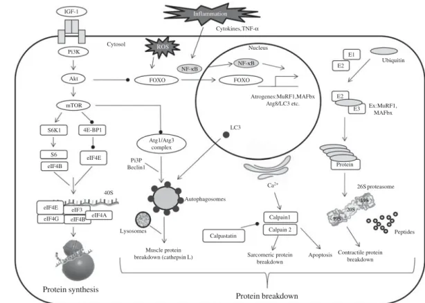
IGF-1 Pi3K Akt mTOR S6K1 4E-BP1 FOXO Atrogenes:MuRF1,MAFbx Atg8/LC3 etc. Peptides Protein E1 E2 Ubiquitin Ex:MuRF1, MAFbx FOXO E2 E3 Cytosol Nucleus Atg1/Atg3 complex Autophagosomes LC3 26S proteasome Ca2+ Calpain1 Calpain 2 Protein breakdown Protein synthesis S6 eIF4E eIF4B 40S eIF4B eIF4E eIF4G eIF3 eIF4A Calpastatin Apoptosis Sarcomeric protein breakdown Pi3P Beclin1 Lysosomes Muscle protein
breakdown (cathepsin L) Contractile proteinbreakdown
ROS
NF-κB NF-κB Cytokines,TNF-α
Inflammation
Fig. 1. Simplified pathways regulating muscle protein metabolism. Maintenance of muscle mass depends on muscle protein synthesis which is normally balanced with muscle protein breakdown. Muscle proteolysis involves three main pathways (ubiquitin-dependent system, lysosomal and calpain-dependent pathways). Regulation involves complex processes between systems. PI3K, phosphoinositide 3-kinase; ROS, reactive oxygen species; Akt, protein kinase B; FOXO, forkhead O transcription factor; MuRF1, muscle RING-finger protein-1; MAFbx, muscle ubiquitin ligase muscle atrophy F-box; Atg, autophagy-related; LC3, light chain 3; E1, ubiquitin-activating enzyme; E2, ubiquitin-conjugating enzyme; E3, ubiquitin ligase; mTOR, mammalian target of rapamycin; S6K1, S6 kinase 1; 4E-BP1, factor 4E binding protein 1; eIF, eukaryotic initiation factor; PI3P, phosphatidylinositol 3-phosphate.
apoptosis is deeply involved in the atrophy of disused
muscle(28,29,74,75) and to the same extent whatever the
age(28 – 30). To our knowledge no data on apoptosis
in response to muscle disuse by itself and without any pathology are available in humans.
Muscle size is dependent on the number and volume of muscle fibres, which is directly related to satellite cells. These quiescent and undifferentiated cells are activated from the quiescent state upon appropriate stimulatory signals and undergo active proliferation and myogenic differentiation. Mechanical unloading, as observed during immobilisation, has been shown to reduce the number of satellite cells, probably due to impaired function and/or activity of these satellite cells(76 – 78). This could be explained by a reduction in the amount and/or activity of the myogenic regulatory factors responsible for the activation of satellite cells and this has been shown in casted animals with myf5(28,30)and in suspended animals with myoD and myogenin(79,80) (Fig. 2). As a relationship between muscle fibre size and myonuclear number exists, the myonuclear domain has been defined as ‘cyto-plasmic volume to nucleus ratio’ and refers to a capacity
of the cell to supply transcriptional demands(81). This myonuclear domain is normally constant, but decreased during immobilisation particularly in slow-type fibres, hence probably contributing to the muscle atrophy(82 – 84).
Metabolic abnormalities associated with disuse
Physical inactivity is associated with a disruption of glucose homeostasis. Thus, insulin resistance appears during
pro-longed periods of immobilisation(85,86). However, the
specific molecular mechanisms underlying this association remain unclear(87). This insulin resistance has been found even in young healthy men with and without a known predisposition to diabetes(88 – 90), confirming the role of immobilisation by itself. This decreased insulin sensitivity appears early during the immobilisation period (after only 3 – 5 d)(91 – 96). Since insulin is known to stimulate pro-tein synthesis in gastrocnemius muscles(97), this decreased
insulin sensitivity could contribute to the anabolic
resistance observed. Moreover, muscle disuse induces a metabolic shift towards utilisation of glucose away from fat as evidenced by increased gene expression of proteins
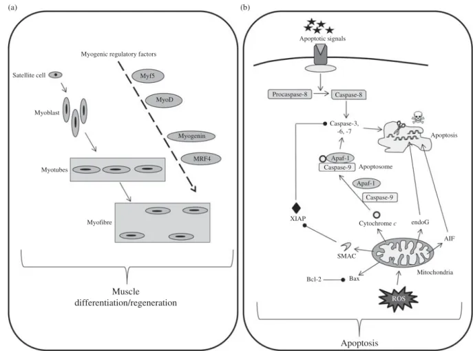
Mitochondria Satellite cell Myofibre MyoD Myf5 MRF4 Myogenin Myoblast Myotubes Muscle differentiation/regeneration Myogenic regulatory factors
Apoptotic signals Procaspase-8 Caspase-8 Caspase-3, -6, -7 Apoptosome Apoptosis Bax Bcl-2 Cytochrome c Apaf-1 Caspase-9 Caspase-9 Apaf-1 SMAC XIAP Apoptosis endoG AIF ROS (a) (b)
Fig. 2. Simplified pathways regulating muscle cellular balance. Cell number in the skeletal muscle is regulated by cell differentiation/regeneration (involving myogenic regulatory factors acting in cascades) (a) and cell apoptosis (b). Cell apoptosis has been simplified and normally involves three different pathways, i.e. external, internal caspase-dependent and internal caspase-independent. Myf5, myogenic factor 5; MyoD, myogenic differentiation antigen; MRF4, myogenic regulatory factor 4; Apaf-1, apoptotic protease activating factor 1; endoG, endonuclease G; XIAP, X-linked inhibitor of apoptosis protein; AIF, apoptosis inducing factor; Bcl-2, B-cell lymphoma 2; Bax, Bcl-2-associated X protein; ROS, reactive oxygen species.
involved in glycolysis and decreased expression of proteins involved in b-oxidation in hindlimb atrophied soleus muscle(98). These metabolic changes are important because they affect performance and may affect recovery processes.
Furthermore, growing evidence suggests that skeletal muscle atrophy is positively correlated with oxidative stress in various situations of muscle disuse(99 – 106)despite there being no clear evidence to determine if it is a primary cause or a latter consequence of disuse. This oxidative stress is characterised by an increase in reactive oxygen
species in skeletal muscle of both animals(107,108) and
humans(106,109). It has been postulated that this oxidative stress may play an important role in the process of muscle atrophy during disuse(75). Even if its origin remains to be clearly determined(110), one of the possible mechan-isms could be a decreased production of endogenous
antioxidant defences in response to
immobilis-ation(12,111 – 113), due to the decrease in protein synthesis associated with muscle atrophy(12,114). In rodents,
immobi-lised muscles exhibit either a decrease(101,115) or an
increase(30) in muscle glutathione concentration. The
activity of key enzymes in glutathione metabolism such as glutathione reductase and glutathione peroxidase are increased(116), unaffected or down-regulated(117). When reactive oxygen species increase in skeletal muscle, DNA fragmentation, lipid peroxidation and protein oxidation can result, which can lead to apoptosis, depending on the severity of the oxidative stress(69,118,119). As an example, a large increase in carbonylated protein content was demonstrated in human subjects following 35 d of bed
rest(109). When carbonylated, proteins are preferentially
degraded by the 20S proteasome without
ubiquitina-tion(120)and there is evidence that these damaged proteins
are more efficiently and rapidly scavenged by proteolytic degradation than their non-oxidised forms to avoid accumulation of damaged functional proteins(120 – 122). Thus, an increase in protein carbonylation could lead to an increase in proteolysis, hence contributing to muscle atrophy. Moreover, oxidative stress is an important activa-tor of key proteases (for example, calpain and caspase-3)
in skeletal muscle(123,124) whose activities have been
demonstrated to be increased in immobilised muscle(125). Finally, excess production of reactive oxygen species can also up-regulate NF-kB activity, which in turn may also enhance protein degradation by the UPS(126 – 128).
Finally, immobilisation-induced atrophy is associated with local inflammation, as demonstrated in human subjects and animals(30,39). This inflammatory response parallels tissue infiltration by macrophages(129 – 131). These inflammatory cells are able to produce the cytokine TNF-a which could later activate the inflammatory NF-kB pathway(75,110). Up-regulation of other pro-inflammatory cytokines (for example, IL-1b and IL-6) has also been
observed in response to muscle immobilisation(39). As
these cytokines are able to activate apoptosis and
proteolysis in muscle(132,133), this immobilisation-induced inflammation may participate directly to the generation of muscle atrophy. Indeed, it has been well demonstrated under other atrophying conditions that inflammation is the main negative regulator of skeletal muscle protein synthesis(134 – 136).
Protein and cellular turnover during the muscle recovery phase
After a catabolic state, muscle mass recovery is a key factor in maintenance of the health and autonomy of individuals. Two main processes are necessary to ensure an efficient recovery, namely a gain in muscle fibre number and muscle protein accretion. The appearance of new muscle fibres has been observed in human subjects as soon as 4 d after recovery and muscle mass loss has been demon-strated to be fully recovered after 14 – 40 d in animals depending on the model studied(28,54,137,138). As mentioned previously, muscle regeneration and subsequent muscle fibre gain depend on the number and activity of satellite cells, and on their upstream regulators, i.e. the myogenic regulatory factors. Subsequent to 2 weeks of hindlimb
unloading, a study(39) explored inflammatory cytokines,
apoptotic, or proteolytic pathways during the early (1 and 5 d) and later (14 d) stages of the regrowth process. It demonstrated that at early stages, muscle repair is
mediated via the decrease in mitochondrial-driven
apoptosis and proteolysis. Despite full muscle mass recov-ery, inflammation pathways were still activated until later stages of muscle remodelling(39). These results and others(106,139,140)demonstrated that inflammatory processes could be indispensable for an efficient remodelling and recovery of skeletal muscle, as inflammatory cytokines are needed to activate proliferation of muscle cells. The rapid recovery of muscle proteolysis and apoptosis has also been observed in several other studies(28,29,141,142).
To generate the positive N balance required for protein accretion during recovery, a further increased protein syn-thesis, decreased proteolysis, or simultaneous changes in both processes is required. Only a few hours are needed for normalisation of the immobilisation-induced
deregu-lated genes involved in protein turnover(143), hence
improving whole-body protein synthesis and N
reten-tion(98). Muscle protein breakdown is normalised early
during the recovery period in adults(27,28). Similarly, in adults, muscle protein synthesis is increased during the
recovery period following immobilisation(144) via an
increase of the Akt/mTOR pathway and downstream sig-nalling(52,54,145,146) whereas it is only normalised in aged animals(31). These results were confirmed in a study con-ducted in mice showing that mice heterozygous for mTOR with about 50 % reduction in total mTOR protein in skeletal muscle and other tissues failed to fully replete muscle mass during recovery following hindlimb immo-bilisation, whereas wild-type mice fully recovered(147).
Nutrition: a key factor for preserving muscle from atrophy and improving recovery?
The maintenance of muscle mass during prolonged periods of immobilisation and the improvement of muscle mass gain during recovery are partly dependent on muscle protein turnover. Indeed, the anabolic resistance induced by immobilisation could represent the primary driver of muscle protein loss and subsequent atrophy. This phenomenon corresponds to an increased threshold of anabolism, i.e. a most important and more sustained amount of anabolic stimulators are required to obtain a stimulation of protein synthesis similar to the one before immobilisation (Fig. 3, adapted from Dardevet et al.(51)). Nutritional strategies could be used during immobilisation and recovery to overcome this anabolic resistance. Two approaches are possible. The first one consists of exceed-ing this increased threshold (i.e. providexceed-ing more anabolic factors, i.e. nutrients to achieve stimulation of protein syn-thesis), and the second one consists of decreasing the threshold (i.e. to restore muscle sensitivity to the stimu-latory effect of normal food intake) (Fig. 4, adapted from Dardevet et al.(51)).
Nutritional intervention to exceed the increased anabolic threshold during immobilisation
Amino acid/protein supplementation. Protein and
consti-tutive amino acids are known to be robust regulators
of protein metabolism(148 – 150). Therefore, the use of diet-ary protein and/or amino acid supplementation could be an efficient countermeasure to increase muscle anabolism
during prolonged immobilisation and so to limit
muscle atrophy.
As a normal diet is unable to maintain normal and sustained protein synthesis because of the increased threshold of anabolism, it is reasonable to think that increasing normal protein intake in bedridden patients
might limit immobilisation-induced atrophy. Stuart
et al.(47) investigated the effects of increasing dietary pro-tein intake isoenergetically from minimum requirements of 0·6 – 1·0 g/kg per d on whole-body protein synthesis during a 7 d bed rest study. They demonstrated that the decrease in protein synthesis induced by bed rest was pre-vented by the higher dietary protein intake and that N bal-ance was improved. However, and unfortunately, muscle
mass was not evaluated in this trial(47). The protein
source appeared to be a main determinant of the efficiency of nutritional interventions. Thus, casein has been referred to as a ‘slow’ protein because of the modest but prolonged increase in plasma amino acid concentrations following its ingestion. By contrast, whey proteins are defined as ‘fast’ proteins because they are rapidly digested and lead to a large, albeit temporary, rise in plasma amino acid levels(151). Data indicate that the peak in plasma amino acids appeared later (150 min v. 75 min) and remained elevated longer after soya protein ingestion than whey
Physiological situation Immobilisation
Anabolic stimulators Anabolic stimulators Meal Meal (a) (b)
Fig. 3. Anabolism threshold under normal (a) and bed rest (b) conditions. Muscle mass is regulated by the anabolism threshold which determines the efficiency of meal intake on muscle anabolism. Under bed rest conditions, anabolic resistance appears, i.e. the muscle anabolism threshold is increased, thus leading to poor efficiency of meal intake on muscle protein accretion.
Anabolic stimulators Anabolic stimulators Meal Meal (a) (b)
Fig. 4. Nutritional strategies to overcome immobilisation-induced anabolic resistance. Two nutritional strategies could be proposed to overcome anabolic resist-ance during immobilisation: to increase the effect of anabolic factors (a) and/or to decrease/restore the anabolism threshold (b).
protein ingestion(152,153); this indicates that soya protein is more of an ‘intermediate’ protein in terms of digestion rate, based on plasma peak amino acid concentrations. In animals, dietary supplementation with 20 % soya protein hydrolysates reduced proteolytic pathways and was further able to prevent immobilisation-induced atrophy when compared with a 20 % casein control diet(154). As casein is the standard protein source used in animal diets, these results confirm that a normal diet is not suitable to limit muscle mass loss under conditions of disuse.
Essential amino acid (EAA) supplementations have also been proposed as a possible countermeasure in humans. Two studies of a 28 d bed rest trial did not show any ben-efit with such supplementations on either muscle mass or
strength(155,156). Another study tested EAA and
carbo-hydrate supplementation during a 28 d bed rest trial in healthy subjects. Volunteers received a mix of 15 g EAA and 30 g sucrose (added for palatability) three times per d during the whole bed rest period. EAA supplementation increased protein synthesis throughout the study and main-tenance of lean body mass was observed(157). However, it is noticeable in this study that protein supplementation was associated with an increased energy intake and thus a synergic effect cannot be excluded(157). Similar results with the same limitation (i.e. an increased energy intake) have been obtained by others in healthy males(158). In this study, subjects consumed 16·5 g EAA and 30 g sucrose three times per d(158). Altogether, these results suggest that EAA supplementation alone is unable to sufficiently stimulate muscle protein synthesis to counteract the dele-terious effect of immobilisation.
Branched-chain amino acids (BCAA) and particularly leucine play an important role in the stimulation of post-prandial muscle protein synthesis(148 – 150). Leucine is known as a ‘nutrient signal’ as it reduces muscle protein breakdown and further stimulates muscle protein syn-thesis(159 – 161). The ‘leucine signal’ stimulates the mTOR pathway, activity and enhances eIF4E-binding protein 1 phosphorylation and the association of eIF4E with eIF4G both in vitro and in vivo(162 – 164). These signalling path-ways are known to become resistant to amino acid (i.e. leucine) stimulation in the case of chronic inflammation or oxidative stress(134,135,165), phenomena that also occur during immobilisation. Consequently, BCAA, and more particularly leucine, supplementation has been tested to limit muscle atrophy during immobilisation based on the hypothesis that increasing BCAA/leucine intake could overcome anabolic resistance. BCAA supplementation was compared with an equimolar mixture of non-EAA during a 14 d bed rest study in men; BCAA supplemen-tation resulted in a 1·2 g/kg per d protein intake during the bed rest period but the protein turnover still decreased with both types of supplementation and muscle mass was not preserved by the nutritional intervention(166). Similarly, sixteen women were subjected to bed rest for 60 d; they were divided into a control group, which maintained
ambulatory protein intake, and into a nutrition intervention group in which protein intake was increased from 1·2 to 1·6 g/kg per d. It is noticeable that this supplement included 0·18 g leucine/kg per d whereas recommended leucine consumption could be estimated at 0·05 g/kg per d based on the RDA. No effect of this treatment was observed(167,168). In women participating in a 60 d bed rest study, protein intake was increased during bed rest from 1 to 1·45 g/kg per d, with BCAA supplementation (3·6 g free leucine/d, 1·8 g free isoleucine/d, and 1·8 g
free valine/d)(169). This study used a transcriptomic
approach and muscle mass was not studied but, interest-ingly, the counteracting effect of BCAA supplementation with leucine enrichment appeared limited since most of the genes up-regulated were moderately down-regulated after the nutrition intervention(169). A leucine supplemen-tation was also tested in immobilised animals. Young rats received 2·7 g leucine/kg per d for 3 d before immobilis-ation and during 7 d of immobilisimmobilis-ation. Neither protein synthesis nor insulin sensitivity was modified with the supplemented diet, but skeletal muscle wasting was atte-nuated via inhibition of ubiquitin ligases(170), suggesting that leucine supplementation alone may be a more effec-tive intervention than BCAA supplementation.
Supplementation with other amino acids has been tested in the prevention of immobilisation-induced atrophy. Oral supplementation with taurine was tested in immobilised rats as a countermeasure to muscle atrophy. The rationale of this study was that taurine has been proposed to protect muscle function in a variety of diseases through diverse
mechanisms(171). This study demonstrated that taurine
content was markedly decreased in immobilised muscles and that a high daily dose of taurine supplementation (5 g/kg per d) fully prevented this decrease but without preventing muscle atrophy(172). Because of assumed ergo-genic effects, short-term creatine supplementation has also been proposed for limiting muscle atrophy during immo-bilisation. Young adults received 20 g creatine/d during muscle immobilisation in a cross-over trial; this resulted in a maintained lean tissue mass when compared with the placebo group(173). Finally, cysteine supplementation showed a positive effect in limitation of immobilisation-induced atrophy, an effect attributed to the suppression of muscle protein ubiquitination and the normalisation of the ratio of reduced glutathione:oxidized glutathione (GSH:GSSG) in muscle(174).
Altogether, these results suggest that increasing protein intake could be an efficient measure to counteract immo-bilisation-induced atrophy, most probably by overcoming the anabolism threshold. Free amino acid supplemen-tation does not exhibit a clear effect in the maintenance of muscle mass since trials conducted with EAA and BCAA supplementation failed to preserve muscle mass during immobilisation. Additional data on taurine, creatine and cysteine supplementation are required to conclude definitively on their beneficial effects.
Nutritional interventions to reduce the anabolism threshold during immobilisation
Antioxidant and anti-inflammatory compounds.
Oxida-tive stress and inflammation situations have been
demonstrated to have a negative impact on muscle protein turnover, particularly by decreasing protein synthesis. As immobilisation is associated with local inflammation and oxidative stress, it may be postulated that these phenom-ena lead to an increased anabolism threshold, translating into the anabolic resistance of skeletal muscle to food intake. Antioxidant supplementation has been shown to be an effective countermeasure to fight oxidative stress in a wide variety of tissue types and conditions(110,175), and it is speculated that this might be an efficient approach
to reduce muscle wasting associated with muscle
disuse(176). Similar results have been obtained with
anti-inflammatory compounds(136) that improve muscle mass
maintenance under atrophying conditions.
Particular attention has been given to polyphenols and their antioxidant properties. Among polyphenols, resveratrol, which is naturally found in grapes, berries, groundnuts and red wine, has been extensively studied for its health benefits. Resveratrol has been shown to mediate cardiomyocyte survival following simulated hypoxia – reperfusion by up-regulating both the antioxidant thioredoxin and the anti-apoptotic protein Bcl-2(177,178), thus underscoring its potential to act as both an antioxidant and anti-apoptotic compound. Recent data suggest that resveratrol induces the transcription of two key antioxidant enzymes, catalase(179,180), and Mn superoxide dismutase (MnSOD)(181 – 183). Adult rats (aged 6 months) were then supplemented with this polyphenol (12·5 mg/kg per d) for 3 weeks, including 2 weeks of muscle immobilisation. Despite a limitation in the functional decrements and the
oxidative stress levels associated with muscle
immobilisation, resveratrol was unable to suppress body weight loss and muscle mass loss(184). In another study, rats were fed a resveratrol-supplemented diet in a dose equivalent to 400 mg/kg per d before unloading and 2 weeks of muscle immobilisation. Resveratrol treatment significantly reduced disuse atrophy by 26 and 10 % without and with normalisation on body weight, respectively. This effect was attributed to the prevention of the decrease of the glutathione:glutathione disulfide ratio, a biomarker of oxidative stress(185). Translated into human doses, this represents a daily intake of 4·5 g/d, a dose for which potential side effects have to be studied before the relevance of its clinical use. These two studies tend to demonstrate that limitation of oxidative stress is able to prevent the metabolic,
and muscle deconditioning caused by mechanical
unloading only when a high dose is used. In another study, immobilised mice were fed a diet supplemented with 0·05 % tea catechins containing 81 % polyphenols (i.e. reported as animal food intake of 1·2 – 1·3 mg polyphenols, i.e. 46 – 50 mg/kg). The supplemented diet was consumed
14 d before immobilisation and during the 10 d of
immobilisation. The supplemented diet significantly
inhibited the decrease in tetanic force observed in the soleus muscle in response to immobilisation, but did not suppress muscle atrophy, therefore suggesting that tea catechins affect skeletal muscle function rather than skeletal mass, and prevent the decrease in muscle strength due to disuse(186).
Amelioration of disuse muscle atrophy following admin-istration of various other antioxidants (i.e. vitamin E or soya protein extract) has been published(14,101,187,188). However, contradictory results have been obtained regard-ing the impact of vitamin E on muscle atrophy. Some authors highlighted no effect of vitamin E
supplemen-tation(99), whereas others have shown that vitamin E
reduced muscle atrophy(176). Surprisingly in this study, its protective effect did not depend on its antioxidant func-tion, but probably on a direct modulation of muscle
pro-teolysis(176). Dietary curcumin was used in rodents to
prevent immobilisation-induced atrophy but without any effect on muscle mass atrophy(29,189). Farid et al.(189) reported that N-acetylcysteine did not suppress muscle atrophy or force decrease in tail-suspended mice, but sup-pressed considerably NF-kB activity, suggesting that the antioxidant may have a non-negligible but insufficient effect on pathways involved in disuse atrophy. Finally, a Cr supplement was used in hindlimb-suspended mice, based on the observation that nutritional supplementation with Cr has been shown to prevent weight loss and improve glucose tolerance in malnourished subjects on long-term total parenteral nutrition(190). Cr inhibited skel-etal muscle atrophy and this was associated with the pre-vention of elevation of the UPS pathway, and better levels of Akt protein, suggesting a greater protein retention and hence contributing to the preservation of the muscle
mass(191). As immobilisation-induced atrophy is associated
with impaired glucose metabolism, it may be postulated that this improvement in protein metabolism could result also from an improvement in glucose homeostasis.
Among anti-inflammatory compounds, dietary fish oil has been proposed to limit immobilisation-induced atrophy. Fish oil is naturally rich in long-chain n-3 fatty acids, namely DHA and EPA, which are known to have anti-inflammatory properties(192). n-3 Fatty acids are also able to enhance insulin-sensitive protein anabolism through the
Akt/mTOR/S6K1(193)pathway, which may help to reduce
muscle anabolic resistance induced by immobilisation. In animals receiving 5 % fish oil, muscle mass atrophy following immobilisation was attenuated via mechanisms involving prevention of immobilisation-induced impair-ments of insulin signalling, the Akt/mTOR/S6K1 and muscle-specific ubiquitins ligases(57). However, these posi-tive effects could have deleterious effects on the following recovery (see later).
Energy intake. Biolo et al.(194) studied the interaction between muscle inactivity and energy restriction, based
on the observation that bedridden individuals, elderly indi-viduals and astronauts often combine muscle inactivity with a reduced energy intake that is below their energy expenditure. They submitted young healthy volunteers to bed rest for 14 d and low-energy diets containing about 80 % of total energy requirements. Bed rest associated with hypoenergetic nutrition led to the greatest wasting of lean body mass(194). Completing these results in another study following the same protocol(195), the same authors tested the hypothesis that energy intake in excess of requirements (i.e. a diet containing 1·2 times subjects’ rest-ing energy expenditure) could lead to fat deposition and thus accelerate inactivity-induced loss of lean mass by acti-vating systemic inflammation, free radical production and antioxidant defences. Indeed, they demonstrated that this
energy excess was associated with higher plasma
C-reactive protein and myeloperoxidase concentrations (markers for inflammation). They concluded that excess energy can worsen muscle atrophy during bed rest, affect-ing whole-body protein turnover and systemic inflam-mation, respectively(195). In conclusion, positive energy balance during inactivity is associated with higher muscle atrophy and with activation of systemic inflammation and of antioxidant defences. Optimising energy intake to the requirements may be a useful strategy for mitigating muscle loss during periods of chronic inactivity.
Nutritional intervention to improve muscle mass recovery
So far, most attention has been focused on preventing muscle mass loss in response to immobilisation. Indeed, most of the rare studies performed observed the impact of a specific type of nutrition given during the immobilis-ation phase on the subsequent recovery, i.e. after the removal of the immobilisation stimuli. Nevertheless, the recovery phase is also important. An efficient and quick recovery of muscle mass and strength is of key importance to the immobilised individual. Resistance training and
reliable nutrition elicit increased muscle mass and
strength(196)and EAA plus carbohydrates enhance muscle
protein synthesis to a greater degree than either stimulus alone(196,197).
In human subjects, BCAA supplementation during 14 d of bed rest had little effect on protein synthesis or break-down in the early recovery period beyond a small increase in N balance(11,166,198). Brooks et al.(155) found that EAA supplementation during 28 d bed rest and 14 d of recovery had no impact on leg lean mass and strength at the end of the recovery.
Effects regarding dietary interventions with
anti-inflammatory compounds were less evident. Indeed, if supplementation in rats during immobilisation improved subsequent muscle mass recovery when animals were treated intraperitoneally with curcumin(29), dietary fish oil
given during immobilisation blunted the following
muscle mass recovery(199). This effect was attributed to a
suppression of Akt/S6K1 signalling and PGF2a content in the early phase of recovery, demonstrating that inflamma-tory processes are nevertheless required for the efficacy of muscle recovery(199).
Ageing and bed rest: impact of nutritional interventions during disuse and following rehabilitation
Particular attention should be paid to prolonged periods of immobilisation during ageing. Indeed, elderly indi-viduals are in a state of fragility when considering their physical activity. Ageing is characterised by a reduced physical activity mainly due to the age-related muscle mass loss, a condition referred to as sarcopenia. English & Paddon-Jones(4)postulated in their review that sarcope-nia could be worsened by catabolic states, as muscle mass atrophy could be followed by uncompleted muscle mass recoveries which, repeated throughout life, could result in a significant muscle mass loss. These uncompleted muscle mass recoveries have been well demonstrated in aged animals(30,31,200)and elderly individuals(201)submitted to muscle immobilisation.
Decreased postabsorptive protein synthesis(2) and the
presence of anabolic resistance in immobilised muscle
have been demonstrated also during ageing(31). As
ana-bolic resistance has already been demonstrating naturally during ageing(164,202 – 204), it is likely that immobilisation may more deeply impair the blunted response of muscle protein synthesis to the stimulatory effect of food intake, hence contributing to the generation of a negative N balance and the resultant muscle mass atrophy.
The blunted EAA response of muscle protein synthesis was also demonstrated in aged humans submitted to bed rest, with a mechanism involving reduced mTORC1 signal-ling and amino acid transporter protein content(205,206), suggesting that a blunted EAA stimulation of muscle pro-tein synthesis may contribute to muscle loss with inactivity in older individuals. However, in older subjects subjected to a 10 d bed rest, an increase in the daily protein intake from 0·8 to 1·5 g/kg per d had no effect on muscle mass loss, but muscle strength was preserved(207). In another study, EAA supplementation was conducted in healthy elderly subjected to bed rest (45 g EAA daily between meals that provided the RDA for protein; 0·8 g/kg per d). After 10 d of bed rest, muscle protein synthesis was decreased by 30 % in controls but maintained in the
sup-plementation group(208). EAA also protected functional
abilities such as floor transfer time and exhibited a trend for the protection of stair ascent power and standing plan-tar flexion but, surprisingly, no effect was reported on muscle mass. These two studies seem to demonstrate
that increased protein intake combined with EAA
supplementation could be a possible countermeasure for
preserving muscle functionality, but inefficient in
preserving muscle mass.
Dietary restriction (by 30 %) in old rats subjected to hindlimb suspension prevented the increases in protea-some activity and other UPS components and reduced
muscle wasting(209). As it has been well demonstrated
that dietary restriction could reduce deleterious age-related changes(210) including muscle wasting(211) in elderly indi-viduals consuming suitable amounts of dietary proteins. These studies highlight interesting nutritional interventions that could be used to cope with immobilisation-induced atrophy but have probably to be taken with care.
For reasons previously presented above, we tested free leucine supplementation during the recovery period in aged animals previously subjected to immobilisation. Indeed, free leucine has been demonstrated to be particu-larly efficient at stimulating muscle protein synthesis during ageing(212 – 216). Despite a positive effect on protein anabo-lism (i.e. free leucine supplementation stimulated muscle protein synthesis and decreased protein breakdown during the recovery period), no effect was observed on
muscle mass(31). We postulated that leucine, as
sup-plemented in a free form, was absorbed rapidly and induced its anabolic ‘leucine signal’ for protein synthesis before there was sufficient availability of amino acids coming from dietary protein digestion(51). Indeed, free leucine was added to casein proteins, which are slow-digested(151,217 – 219). Therefore, the duration of the protein synthesis could have been insufficient. This desynchronisa-tion between the ‘leucine signal’ and the availability of substrates for protein synthesis could explain the insuffi-cient positive N balance and the lack of protein accretion
when free leucine is used(31). In this study, we also
tested a whey protein supplementation and a high-protein diet (26 % protein) to limit muscle mass loss by (1) using whey protein as a leucine-rich protein source and (2) com-bining the whey properties with a high-protein effect, i.e. inducing a higher and sustainable concentration of amino acids available for protein synthesis over the postprandial period. Indeed, whey proteins as fast-digested proteins are rapidly digested, leading to a large, albeit temporary, rise in plasma amino acid levels(151)with a particular effi-cacy to induce a large positive N balance during
ageing(220). A combination of whey and casein proteins
clearly induced a high skeletal muscle protein synthesis and prolonged amino acid delivery to tissue(221). Both strat-egies were efficient in improving muscle mass recovery(31). As the effect of leucine may be mediated by its meta-bolites(222), b-hydroxy-b-methylbutyrate (HMB), a leucine metabolite was given to old rats immobilised with a model of 14 d suspension. Although HMB could not fully prevent immobilisation-induced body weight or muscle loss in response to unloading, it: (1) prevented further force loss during reloading after unloading; (2) improved muscle mass in muscles that were reloaded after immobilisation; (3) blunted the extent of fibre atrophy in both fast and slow skeletal muscles in response to unloading and reloading after disuse; (4) significantly
attenuated myonuclear apoptosis induced by immobilis-ation; (5) decreased the apoptotic index after reloading following immobilisation in muscles; and (6) reduced mito-chondrial apoptotic signalling as indicated by lower levels of cleaved caspase-3, cleaved caspase-9, and Bax protein abundance in reloaded muscles(223). Elderly submitted to bed rest and receiving tube feeding fed HMB supple-mentation for 2 – 4 weeks showed a reduced muscle break-down; however, no direct measurement of muscle mass was assessed in this study(224).
Overall conclusion and future directions
Muscle mass loss is an inevitable issue in response to muscle disuse, a condition found in many situations (from bed rest imposed after injuries or trauma to non-pathological bed rest occurring with age-related decrease in physical activity). The physiopathological changes responsible for this immobilisation-induced atrophy are multiple, but the decreased protein turnover observed probably plays a central role. The anabolic resistance that emerges with immobilisation is probably due to the appearance of local inflammation and oxidative stress that increase the skeletal muscle anabolism threshold. Numerous nutritional interventions can be proposed as countermeasures to limit muscle mass atrophy during immobilisation and improve the subsequent recovery. Sep-arate strategies were tested in young animals or humans to overcome the increased threshold of anabolism or to decrease this threshold to restore muscle sensitivity to food intake. However, to date, no consensual data pro-vided have allowed us to develop an efficient strategy. Combining the two approaches may have a greater impact on muscle mass, for example by combining proteins/amino acids and antioxidants/anti-inflammatory compounds.
During immobilisation periods, one of the preferred approaches for nutritional support to limit muscle atrophy consists of protein/amino acid supplementation(s). How-ever, such supplementations have demonstrated a modest impact on the atrophic response despite a strong effect on the increase in postprandial muscle protein anabolism. Clearly, much more data are needed regarding the ben-eficial effect of increased protein intake and creatine sup-plementation, which were based on evidence from sport nutrition. Alone, such dietary components have no positive impact on muscle mass but elicit an interesting effect on muscle function that should be studied in more depth. Moreover, some selected dietary components might decrease the anabolism threshold and when combined with well-known anabolic factors (i.e. leucine, whey) they might potentiate the postprandial muscle anabolism and thus limit disuse atrophy and/or improve following recovery. A key point is the question of timing as demon-strated with fish oil supplementation: depending of the timing of the n-3 NEFA consumption (i.e. during disuse
or recovery) these compounds could either improve or worsen the muscle atrophy. Future research should focus on these aspects and test different combinations of sup-plementation of proteins/amino acids and antioxidants/ anti-inflammatory compounds to boost muscle anabolism. Different timing targeting different biochemical events (i.e. to decrease oxidative stress/inflammation during immobilisation and to increase anabolism during the recovery process) should be also tested.
During ageing, muscle atrophy in response to disuse is a main issue since muscle atrophy episodes are followed by uncompleted muscle recovery which accelerates the development of sarcopenia. Dietary supplementation with proteins/amino acids seem inefficient to limit the atrophic processes, but demonstrated real positive effects in this particular population by improving the recovery of muscle mass after an acute catabolic state such as immobil-isation. Such nutritional strategies could be more efficient when using antioxidants/anti-inflammatory compounds that can decrease the well-known low-grade inflammation associated with ageing and responsible for the blunted muscle response to food intake. Ongoing research may emphasise these data in well-designed clinical trials con-ducted on a long-term basis.
Acknowledgements
The authors would like to thank He´le`ne Lafarge for help in the management of the bibliography.
This research received no specific grant from any funded agency in the public, commercial or not-for-profit sectors. H. M., I, S.-A., D. R. and D. D. wrote and approved the final manuscript.
The authors have declared no conflicts of interest.
References
1. Blottner D, Salanova M, Puttmann B, et al. (2006) Human skeletal muscle structure and function preserved by vibration muscle exercise following 55 days of bed rest. Eur J Appl Physiol 97, 261 – 271.
2. Kortebein P, Ferrando A, Lombeida J, et al. (2007) Effect of 10 days of bed rest on skeletal muscle in healthy older adults. JAMA 297, 1772 – 1774.
3. Pavy-Le TA, Heer M, Narici MV, et al. (2007) From space to Earth: advances in human physiology from 20 years of bed rest studies (1986 – 2006). Eur J Appl Physiol 101, 143 – 194. 4. English KL & Paddon-Jones D (2010) Protecting muscle mass and function in older adults during bed rest. Curr Opin Clin Nutr Metab Care 13, 34 – 39.
5. Gill TM, Allore H & Guo Z (2004) The deleterious effects of bed rest among community-living older persons. J Gerontol A Biol Sci Med Sci 59, 755 – 761.
6. Nair KS (2005) Aging muscle. Am J Clin Nutr 81, 953 – 963. 7. Short KR, Vittone JL, Bigelow ML, et al. (2005) Changes in myosin heavy chain mRNA and protein expression in human skeletal muscle with age and endurance exercise training. J Appl Physiol 99, 95 – 102.
8. Yarasheski KE, Pak-Loduca J, Hasten DL, et al. (1999) Resistance exercise training increases mixed muscle protein synthesis rate in frail women and men $ 76 yr old. Am J Physiol 277, E118 – E125.
9. Ferrando AA, Lane HW, Stuart CA, et al. (1996) Prolonged bed rest decreases skeletal muscle and whole body protein synthesis. Am J Physiol 270, E627 – E633.
10. Phillips SM, Glover EI & Rennie MJ (2009) Alterations of protein turnover underlying disuse atrophy in human skele-tal muscle. J Appl Physiol 107, 645 – 654.
11. Stein TP, Leskiw MJ, Schluter MD, et al. (1999) Protein kinetics during and after long-duration spaceflight on MIR. Am J Physiol 276, E1014 – E1021.
12. Stevenson EJ, Giresi PG, Koncarevic A, et al. (2003) Global analysis of gene expression patterns during disuse atrophy in rat skeletal muscle. J Physiol 551, 33 – 48.
13. Boonyarom O & Inui K (2006) Atrophy and hypertrophy of skeletal muscles: structural and functional aspects. Acta Physiol (Oxf) 188, 77 – 89.
14. Morris CA, Morris LD, Kennedy AR, et al. (2005) Attenu-ation of skeletal muscle atrophy via protease inhibition. J Appl Physiol 99, 1719 – 1727.
15. Jones SW, Hill RJ, Krasney PA, et al. (2004) Disuse atrophy and exercise rehabilitation in humans profoundly affects the expression of genes associated with the regulation of skeletal muscle mass. FASEB J 18, 1025 – 1027.
16. Tesch PA, von Walden F, Gustafsson T, et al. (2008) Skeletal muscle proteolysis in response to short-term unloading in humans. J Appl Physiol 105, 902 – 906.
17. Glover EI, Yasuda N, Tarnopolsky MA, et al. (2010) Little change in markers of protein breakdown and oxidative stress in humans in immobilization-induced skeletal muscle atrophy. Appl Physiol Nutr Metab 35, 125 – 133. 18. Jaspers SR & Tischler ME (1984) Atrophy and growth failure
of rat hindlimb muscles in tail-cast suspension. J Appl Phy-siol 57, 1472 – 1479.
19. Loughna P, Goldspink G & Goldspink DF (1986) Effect of inactivity and passive stretch on protein turnover in phasic and postural rat muscles. J Appl Physiol 61, 173 – 179. 20. Munoz KA, Satarug S & Tischler ME (1993) Time course of the response of myofibrillar and sarcoplasmic protein metabolism to unweighting of the soleus muscle. Meta-bolism 42, 1006 – 1012.
21. Zdanowicz MM & Teichberg S (2003) Effects of insulin-like growth factor-1/binding protein-3 complex on muscle atrophy in rats. Exp Biol Med (Maywood) 228, 891 – 897. 22. Attaix D, Ventadour S, Codran A, et al. (2005) The
ubiquitin – proteasome system and skeletal muscle wasting. Essays Biochem 41, 173 – 186.
23. Cohen S, Brault JJ, Gygi SP, et al. (2009) During muscle atrophy, thick, but not thin, filament components are degraded by MuRF1-dependent ubiquitylation. J Cell Biol 185, 1083 – 1095.
24. Polge C, Heng AE, Jarzaguet M, et al. (2011) Muscle actin is polyubiquitinylated in vitro and in vivo and targeted for breakdown by the E3 ligase MuRF1. FASEB J 25, 3790 – 3802. 25. Ikemoto M, Nikawa T, Takeda S, et al. (2001) Space shuttle flight (STS-90) enhances degradation of rat myosin heavy chain in association with activation of ubiquitin – proteasome pathway. FASEB J 15, 1279 – 1281.
26. Taillandier D, Aurousseau E, Meynial-Denis D, et al. (1996) Coordinate activation of lysosomal, Ca 2 þ -activated and ATP-ubiquitin-dependent proteinases in the unweighted rat soleus muscle. Biochem J 316, 65 – 72.
27. Taillandier D, Aurousseau E, Combaret L, et al. (2003) Regu-lation of proteolysis during reloading of the unweighted soleus muscle. Int J Biochem Cell Biol 35, 665 – 675.
28. Vazeille E, Codran A, Claustre A, et al. (2008) The ubiqui-tin – proteasome and the mitochondria-associated apoptotic pathways are sequentially downregulated during recovery after immobilization-induced muscle atrophy. Am J Physiol Endocrinol Metab 295, E1181 – E1190.
29. Vazeille E, Slimani L, Claustre A, et al. (2012) Curcumin treatment prevents increased proteasome and apopto-some activities in rat skeletal muscle during reloading and improves subsequent recovery. J Nutr Biochem 23, 245 – 251.
30. Magne H, Savary-Auzeloux I, Vazeille E, et al. (2011) Lack of muscle recovery after immobilization in old rats does not result from a defect in normalization of the ubiquitin – proteasome and the caspase-dependent apop-totic pathways. J Physiol 589, 511 – 524.
31. Magne H, Savary-Auzeloux I, Migne C, et al. (2012) Contrarily to whey and high protein diets, dietary free leucine supplementation cannot reverse the lack of recovery of muscle mass after prolonged immobilization during ageing. J Physiol 590, 2035 – 2049.
32. Pattison JS, Folk LC, Madsen RW, et al. (2003) Selected con-tribution: identification of differentially expressed genes between young and old rat soleus muscle during recovery from immobilization-induced atrophy. J Appl Physiol 95, 2171 – 2179.
33. Pattison JS, Folk LC, Madsen RW, et al. (2003) Transcrip-tional profiling identifies extensive downregulation of extracellular matrix gene expression in sarcopenic rat soleus muscle. Physiol Genomics 15, 34 – 43.
34. Hasselgren PO & Fischer JE (2001) Muscle cachexia: current concepts of intracellular mechanisms and molecular regu-lation. Ann Surg 233, 9 – 17.
35. Huang J & Forsberg NE (1998) Role of calpain in skeletal-muscle protein degradation. Proc Natl Acad Sci U S A 95, 12100 – 12105.
36. Goll DE, Thompson VF, Li H, et al. (2003) The calpain system. Physiol Rev 83, 731 – 801.
37. Salazar JJ, Michele DE & Brooks SV (2010) Inhibition of calpain prevents muscle weakness and disruption of sarco-mere structure during hindlimb suspension. J Appl Physiol 108, 120 – 127.
38. Liang XH, Jackson S, Seaman M, et al. (1999) Induction of autophagy and inhibition of tumorigenesis by beclin 1. Nature 402, 672 – 676.
39. Andrianjafiniony T, Dupre-Aucouturier S, Letexier D, et al. (2010) Oxidative stress, apoptosis, and proteolysis in skel-etal muscle repair after unloading. Am J Physiol Cell Physiol 299, C307 – C315.
40. O’Leary MF & Hood DA (2009) Denervation-induced oxi-dative stress and autophagy signaling in muscle. Autophagy 5, 230 – 231.
41. Zhao J, Brault JJ, Schild A, et al. (2007) FoxO3 coordinately activates protein degradation by the autophagic/lysosomal and proteasomal pathways in atrophying muscle cells. Cell Metab 6, 472 – 483.
42. Biolo G, Ciocchi B, Lebenstedt M, et al. (2004) Short-term bed rest impairs amino acid-induced protein anabolism in humans. J Physiol 558, 381 – 388.
43. Gibson JN, Halliday D, Morrison WL, et al. (1987) Decrease in human quadriceps muscle protein turnover consequent upon leg immobilization. Clin Sci (Lond) 72, 503 – 509. 44. Lovejoy JC, Smith SR, Zachwieja JJ, et al. (1999) Low-dose
T3improves the bed rest model of simulated weightlessness
in men and women. Am J Physiol 277, E370 – E379. 45. Shangraw RE, Stuart CA, Prince MJ, et al. (1988) Insulin
responsiveness of protein metabolism in vivo following bedrest in humans. Am J Physiol 255, E548 – E558.
46. Stein TP, Schluter MD, Leskiw MJ, et al. (1999) Attenuation of the protein wasting associated with bed rest by branched-chain amino acids. Nutrition 15, 656 – 660. 47. Stuart CA, Shangraw RE, Peters EJ, et al. (1990) Effect of
dietary protein on bed-rest-related changes in whole-body-protein synthesis. Am J Clin Nutr 52, 509 – 514. 48. de Boer MD, Selby A, Atherton P, et al. (2007) The temporal
responses of protein synthesis, gene expression and cell signalling in human quadriceps muscle and patellar tendon to disuse. J Physiol 585, 241 – 251.
49. Glover EI, Phillips SM, Oates BR, et al. (2008) Immobi-lization induces anabolic resistance in human myofibrillar protein synthesis with low and high dose amino acid infu-sion. J Physiol 586, 6049 – 6061.
50. Rennie MJ, Wackerhage H, Spangenburg EE, et al. (2004) Control of the size of the human muscle mass. Annu Rev Physiol 66, 799 – 828.
51. Dardevet D, Remond D, Peyron MA, et al. (2012) Muscle wasting and resistance of muscle anabolism: the ‘anabolic threshold concept’ for adapted nutritional strategies during sarcopenia. ScientificWorldJournal 2012, 269531. 52. Bodine SC, Stitt TN, Gonzalez M, et al. (2001) Akt/mTOR
pathway is a crucial regulator of skeletal muscle hypertro-phy and can prevent muscle atrohypertro-phy in vivo. Nat Cell Biol 3, 1014 – 1019.
53. O’Keefe MP, Perez FR, Sloniger JA, et al. (2004) Enhanced insulin action on glucose transport and insulin signaling in 7-day unweighted rat soleus muscle. J Appl Physiol 97, 63 – 71.
54. Sugiura T, Abe N, Nagano M, et al. (2005) Changes in PKB/ Akt and calcineurin signaling during recovery in atrophied soleus muscle induced by unloading. Am J Physiol Regul Integr Comp Physiol 288, R1273 – R1278.
55. Hunter RB, Stevenson E, Koncarevic A, et al. (2002) Acti-vation of an alternative NF-kB pathway in skeletal muscle during disuse atrophy. FASEB J 16, 529 – 538.
56. Morris RT, Spangenburg EE & Booth FW (2004) Respon-siveness of cell signaling pathways during the failed 15-day regrowth of aged skeletal muscle. J Appl Physiol 96, 398 – 404.
57. You JS, Park MN, Song W, et al. (2010) Dietary fish oil alle-viates soleus atrophy during immobilization in association with Akt signaling to p70s6k and E3 ubiquitin ligases in rats. Appl Physiol Nutr Metab 35, 310 – 318.
58. Booth FW & Kirby CR (1992) Changes in skeletal muscle gene expression consequent to altered weight bearing. Am J Physiol 262, R329 – R332.
59. Lagirand-Cantaloube J, Offner N, Csibi A, et al. (2008) The initiation factor eIF3-f is a major target for atrogin1/MAFbx function in skeletal muscle atrophy. EMBO J 27, 1266 – 1276. 60. Sandri M, Sandri C, Gilbert A, et al. (2004) Foxo transcrip-tion factors induce the atrophy-related ubiquitin ligase atro-gin-1 and cause skeletal muscle atrophy. Cell 117, 399 – 412. 61. Sandri M (2008) Signaling in muscle atrophy and
hyper-trophy. Physiology (Bethesda) 23, 160 – 170.
62. Kandarian SC & Jackman RW (2006) Intracellular signaling during skeletal muscle atrophy. Muscle Nerve 33, 155 – 165. 63. Romanello V, Guadagnin E, Gomes L, et al. (2010) Mito-chondrial fission and remodelling contributes to muscle atrophy. EMBO J 29, 1774 – 1785.
64. Bae SK, Cha HN, Ju TJ, et al. (2012) Deficiency of inducible nitric oxide synthase attenuates immobilization-induced skeletal muscle atrophy in mice. J Appl Physiol 113, 114 – 123.
65. Guttridge DC (2004) Signaling pathways weigh in on decisions to make or break skeletal muscle. Curr Opin Clin Nutr Metab Care 7, 443 – 450.
66. Leger B, Cartoni R, Praz M, et al. (2006) Akt signalling through GSK-3b, mTOR and Foxo1 is involved in human skeletal muscle hypertrophy and atrophy. J Physiol 576, 923 – 933.
67. Stitt TN, Drujan D, Clarke BA, et al. (2004) The IGF-1/PI3K/ Akt pathway prevents expression of muscle atrophy-induced ubiquitin ligases by inhibiting FOXO transcription factors. Mol Cell 14, 395 – 403.
68. Allen DL, Yasui W, Tanaka T, et al. (1996) Myonuclear number and myosin heavy chain expression in rat soleus single muscle fibers after spaceflight. J Appl Physiol 81, 145 – 151.
69. Leeuwenburgh C, Gurley CM, Strotman BA, et al. (2005) Age-related differences in apoptosis with disuse atrophy in soleus muscle. Am J Physiol Regul Integr Comp Physiol 288, R1288 – R1296.
70. Smith HK, Maxwell L, Martyn JA, et al. (2000) Nuclear DNA fragmentation and morphological alterations in adult rabbit skeletal muscle after short-term immobilization. Cell Tissue Res 302, 235 – 241.
71. Allen DL, Linderman JK, Roy RR, et al. (1997) Growth hor-mone/IGF-I and/or resistive exercise maintains myonuclear number in hindlimb unweighted muscles. J Appl Physiol 83, 1857 – 1861.
72. Schmalbruch H & Lewis DM (2000) Dynamics of nuclei of muscle fibers and connective tissue cells in normal and denervated rat muscles. Muscle Nerve 23, 617 – 626. 73. Allen DL, Linderman JK, Roy RR, et al. (1997) Apoptosis: a
mechanism contributing to remodeling of skeletal muscle in response to hindlimb unweighting. Am J Physiol 273, C579 – C587.
74. Marzetti E, Hwang JC, Lees HA, et al. (2010) Mitochondrial death effectors: relevance to sarcopenia and disuse muscle atrophy. Biochim Biophys Acta 1800, 235 – 244.
75. Powers SK, Smuder AJ & Judge AR (2012) Oxidative stress and disuse muscle atrophy: cause or consequence? Curr Opin Clin Nutr Metab Care 15, 240 – 245.
76. Darr KC & Schultz E (1989) Hindlimb suspension sup-presses muscle growth and satellite cell proliferation. J Appl Physiol 67, 1827 – 1834.
77. Matsuba Y, Goto K, Morioka S, et al. (2009) Gravitational unloading inhibits the regenerative potential of atrophied soleus muscle in mice. Acta Physiol (Oxf) 196, 329 – 339. 78. Mozdziak PE, Truong Q, Macius A, et al. (1998) Hindlimb
suspension reduces muscle regeneration. Eur J Appl Physiol Occup Physiol 78, 136 – 140.
79. Shefer G, Carmeli E, Rauner G, et al. (2008) Exercise run-ning and tetracycline as means to enhance skeletal muscle stem cell performance after external fixation. J Cell Physiol 215, 265 – 275.
80. Zhang BT, Yeung SS, Liu Y, et al. (2010) The effects of low frequency electrical stimulation on satellite cell activity in rat skeletal muscle during hindlimb suspension. BMC Cell Biol 11, 87.
81. Van der Meer SF, Jaspers RT & Degens H (2011) Is the myo-nuclear domain size fixed? J Musculoskelet Neuronal Inter-act 11, 286 – 297.
82. Allen DL, Monke SR, Talmadge RJ, et al. (1995) Plasticity of myonuclear number in hypertrophied and atrophied mam-malian skeletal muscle fibers. J Appl Physiol 78, 1969 – 1976. 83. Aravamudan B, Mantilla CB, Zhan WZ, et al. (2006) Dener-vation effects on myonuclear domain size of rat diaphragm fibers. J Appl Physiol 100, 1617 – 1622.
84. Tseng BS, Kasper CE & Edgerton VR (1994) Cytoplasm-to-myonucleus ratios and succinate dehydrogenase activities in adult rat slow and fast muscle fibers. Cell Tissue Res 275, 39 – 49.
85. Reynet C & Kahn CR (1993) Rad: a member of the Ras family overexpressed in muscle of type II diabetic humans. Science 262, 1441 – 1444.
86. Vaag A (1999) On the pathophysiology of late onset non-insulin dependent diabetes mellitus. Current controversies and new insights. Dan Med Bull 46, 197 – 234.
87. Handschin C & Spiegelman BM (2008) The role of exercise and PGC1a in inflammation and chronic disease. Nature 454, 463 – 469.
88. Alibegovic AC, Hojbjerre L, Sonne MP, et al. (2009) Impact of 9 days of bed rest on hepatic and peripheral insulin action, insulin secretion, and whole-body lipolysis in healthy young male offspring of patients with type 2 dia-betes. Diabetes 58, 2749 – 2756.
89. Alibegovic AC, Hojbjerre L, Sonne MP, et al. (2010) Increased rate of whole body lipolysis before and after 9 days of bed rest in healthy young men born with low birth weight. Am J Physiol Endocrinol Metab 298, E555 – E564.
90. Alibegovic AC, Sonne MP, Hojbjerre L, et al. (2010) The T-allele of TCF7L2 rs7903146 associates with a reduced compensation of insulin secretion for insulin resistance induced by 9 days of bed rest. Diabetes 59, 836 – 843. 91. Dolkas CB & Greenleaf JE (1977) Insulin and glucose
responses during bed rest with isotonic and isometric exer-cise. J Appl Physiol 43, 1033 – 1038.
92. Hamburg NM, McMackin CJ, Huang AL, et al. (2007) Physi-cal inactivity rapidly induces insulin resistance and micro-vascular dysfunction in healthy volunteers. Arterioscler Thromb Vasc Biol 27, 2650 – 2656.
93. Lipman RL, Schnure JJ, Bradley EM, et al. (1970) Impair-ment of peripheral glucose utilization in normal subjects by prolonged bed rest. J Lab Clin Med 76, 221 – 230. 94. Mikines KJ, Richter EA, Dela F, et al. (1991) Seven days of
bed rest decrease insulin action on glucose uptake in leg and whole body. J Appl Physiol 70, 1245 – 1254.
95. Stuart CA, Shangraw RE, Prince MJ, et al. (1988) Bed-rest-induced insulin resistance occurs primarily in muscle. Metabolism 37, 802 – 806.
96. Tabata I, Suzuki Y, Fukunaga T, et al. (1999) Resistance training affects GLUT-4 content in skeletal muscle of humans after 19 days of head-down bed rest. J Appl Physiol 86, 909 – 914.
97. Kimball SR, Jurasinski CV, Lawrence JC Jr, et al. (1997) Insu-lin stimulates protein synthesis in skeletal muscle by enhan-cing the association of eIF-4E and eIF-4G. Am J Physiol 272, C754 – C759.
98. Stein T, Schluter M, Galante A, et al. (2002) Energy metab-olism pathways in rat muscle under conditions of simulated microgravity. J Nutr Biochem 13, 471.
99. Koesterer TJ, Dodd SL & Powers S (2002) Increased antiox-idant capacity does not attenuate muscle atrophy caused by unweighting. J Appl Physiol 93, 1959 – 1965.
100. Kondo H, Miura M & Itokawa Y (1991) Oxidative stress in skeletal muscle atrophied by immobilization. Acta Physiol Scand 142, 527 – 528.
101. Kondo H, Miura M, Nakagaki I, et al. (1992) Trace element movement and oxidative stress in skeletal muscle atrophied by immobilization. Am J Physiol 262, E583 – E590. 102. Kondo H, Miura M & Itokawa Y (1993) Antioxidant enzyme
systems in skeletal muscle atrophied by immobilization. Pflugers Arch 422, 404 – 406.
103. Kondo H, Nakagaki I, Sasaki S, et al. (1993) Mechanism of oxidative stress in skeletal muscle atrophied by immobiliz-ation. Am J Physiol 265, E839 – E844.
104. Kondo H, Nishino K & Itokawa Y (1994) Hydroxyl radical generation in skeletal muscle atrophied by immobilization. FEBS Lett 349, 169 – 172.