HAL Id: hal-01501713
https://hal.sorbonne-universite.fr/hal-01501713
Submitted on 4 Apr 2017
HAL is a multi-disciplinary open access
archive for the deposit and dissemination of
sci-entific research documents, whether they are
pub-lished or not. The documents may come from
teaching and research institutions in France or
abroad, or from public or private research centers.
L’archive ouverte pluridisciplinaire HAL, est
destinée au dépôt et à la diffusion de documents
scientifiques de niveau recherche, publiés ou non,
émanant des établissements d’enseignement et de
recherche français ou étrangers, des laboratoires
publics ou privés.
Distributed under a Creative Commons Attribution| 4.0 International License
Cell-Based Models for Development of
Antiatherosclerotic Therapies
Emile R. Zakiev, Nikita G. Nikiforov, Alexander N. Orekhov
To cite this version:
Emile R. Zakiev, Nikita G. Nikiforov, Alexander N. Orekhov. Cell-Based Models for Development
of Antiatherosclerotic Therapies. BioMed Research International , Hindawi Publishing Corporation,
2017, pp.5198723. �10.1155/2017/5198723�. �hal-01501713�
Review Article
Cell-Based Models for Development of
Antiatherosclerotic Therapies
Emile R. Zakiev,
1,2Nikita G. Nikiforov,
1and Alexander N. Orekhov
1,31Institute of General Pathology and Pathophysiology, Moscow, Russia
2INSERM UMR S 1166-ICAN Facult´e de M´edecine Piti´e-Salpˆetri`ere, Paris, France 3Institute for Atherosclerosis Research, Skolkovo Innovation Center, Moscow, Russia
Correspondence should be addressed to Emile R. Zakiev; emil.zakiev@inserm.fr Received 9 October 2016; Accepted 11 January 2017; Published 14 February 2017 Academic Editor: Fabrizio Montecucco
Copyright © 2017 Emile R. Zakiev et al. This is an open access article distributed under the Creative Commons Attribution License, which permits unrestricted use, distribution, and reproduction in any medium, provided the original work is properly cited. The leading cause of death worldwide is cardiovascular disease. Among the conditions related to the term, the most prominent one is the development of atherosclerotic plaques in the walls of arteries. The situation gets even worse with the fact that the plaque development may stay asymptomatic for a prolonged period of time. When it manifests as a cardiovascular disorder, it is already too late: the unfortunate individual is prescribed with a plethora of synthetic drugs, which are of debatable efficacy in the prevention of atherosclerotic lesions and safety. Cell models could be useful for the purpose of screening substances potentially effective against atherosclerosis progression and effective in reduction of already present plaques. In this overview, we present studies making use of in vitro and ex vivo models of atherosclerosis development that can prove valuable for clinical applications.
1. Introduction
Atherosclerosis is a condition resulting in plaque develop-ment in the arteries. It has been extensively studied over the last decades. Though the result of this deteriorating process is easily observable via angiography, the causes for the onset of the disease are still debatable. There are several risk factors for the development of atherosclerosis. Improper lipid metabolism, high arterial pressure, and higher-than-normal blood coagulation can all be considered as the possible risk factors. The standard treatment for the atherosclerosis today is to mitigate the symptoms and to hamper further progression. The majority of patients are prescribed with medicines that they have to take indefinitely and which are of debatable safety. The resulting condition may alleviate the symptoms but does not always suggest disease remission, so this is hardly a remedy people desire. Thus, development of a direct antiatherosclerotic therapy is a matter of reversing current plaques and lesions and prevention of the building up of the new ones. Though there was a plethora of large-scale clinical studies on the novel medications, none of them have been proven to possess the properties for effective plaque
regression, aspiration of lipid necrotic cores, and stabilization of the fibrous cap at the same time [1–9]. Currently, there are two major groups of antiatherosclerotic treatments, namely, statins and calcium antagonists. Statins are the golden stan-dard in lipid-lowering medications securing low levels of cholesterol level in the blood of patients. Some studies suggest that statins have latent supplementary effects, such as mono-cyte migration prevention, cell proliferation inhibition, and cellular cholesterol diminution [10–20]. Efficacy of aggressive statin therapy against the atherosclerosis progression has been assessed in multiple clinical trials [9]. The trials of statins have reported various serious adverse effects [21]. This is not to mention that statin treatment is rather expensive [10, 13, 21].
Calcium antagonists are essentially the second major group of interest here [22–30], though there are only four known clinical trials on these substances up to date [9]. Amlodipine, being the most studied of the group, was assessed in intima-medial layer thickness (IMT) reduction in carotid arteries [31, 32]. Other studies on the substance from the group have been proven to be fruitless or with no evidence of clinical efficacy [9].
Volume 2017, Article ID 5198723, 8 pages https://doi.org/10.1155/2017/5198723
2 BioMed Research International
1
2
3
4
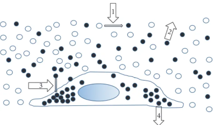
Figure 1: Targets of atherosclerotic and antiatherogenic drug ther-apy. The figure schematically represents lumen of blood vessel and a vessel wall cell highlighting possible targets for antiatherosclerotic therapy. The first target (target 1) is atherogenic modification (desialylation) of the LDL particle in blood. The prevention of LDL modification may be an approach to antiatherosclerosis therapy. The second approach may be the selective removal of modified LDL from blood (target 2). The third approach may be based on the prevention of modified LDL accumulation in arterial cells (target 3). Additionally, another approach is the removal of excess lipids from foam cells (target 4). Taken with permission from [9].
Some antiatherosclerotic properties were attributed to vitamins-antioxidants, estrogens, and cholesterol/niacin combination, though all results show no efficacy of those substances alone. Estrogen therapy was able to delay the development of atherosclerosis and also slightly turn back time in already existent plaques, but the subject sampling was limited to menopausal females. The results have also been confounded with the influence of ongoing statin treatment, so the efficacy could not have been assessed precisely [33, 34]. Naturaceuticals may yield a noticeable therapeutic result with none or mild ill-effects. Possible mechanisms of action for the potentially effective natural drugs are represented on Figure 1. Cell-based models made it possible to test pretty much all of them, using the ability of human serum to provoke atherogenic deteriorations in in vitro and ex vivo cellular models [9]. The cell-based models allow for fast and reliable screening of botanicals potentially efficient in the treatment of atherosclerosis [8]. In this review, we will sum up the studies utilizing cell-based models for seeking new nonpharmaceutical antiatherogenic drugs.
2. Cellular Models in the Search for
Antiatherosclerosis Therapy
Biological model can be extremely useful in the development of new pharmaceutical and naturaceutical products. Unlike standard laboratory tests for safety and efficacy prior to open-ing the clinical studies on human population, biological mod-els can be deployed and analyzed quickly. For instance, it is possible to rapidly acquiesce results on activity, metabolism, and direct therapeutic effect of a potential medicine. While keeping the model simple, it is important to retain the likeness to the biological system of interest. It is possible to apply
this cellular model strategy to find the naturaceuticals with antiatherosclerotic effect [35–38].
In study [9] authors have deployed two cellular mod-els: in vitro and ex vivo. The in vitro biological model was based on the primary culture of subendothelial cells extracted from thoracic aorta of men and women of 40 to 65 years of age after 1.5–3 hours of their sudden death. The ex vivo model was utilizing the same cell cultures but deployed them in a different way. The in vitro model was aimed to assess botanical and synthetic compounds for their capability of reducing the atherogenicity of human serum as well as to avert the morbid cholesterol accumulation in the intima of blood vessels [7, 9, 28, 30]. Living cells were extracted using collagenase from a variety of regions
of aorta, and then they were cultured at 37∘C for 7–10
days in an every-day rejuvenated medium [9]. The resulting heterogeneous cell population consisted mostly of pericyte-like cells and typical and modified smooth muscle cells [9]. In order to receive a “control” cellular model possessing different properties from the aforementioned cellular model that was exposed to deteriorations, authors have also taken a healthy region of aorta and cultured cells from it in a similar fashion. Abnormal cellular lipid deposition was induced by incubating the cells with atherogenic serum taken from venous blood of atherosclerotic patients [9]. Products under investigation were added to both of the models, thus testing ability to diminish cellular lipid deposits in already affected cells and the ability to prevent lipid accumulation in relatively “control” cells accordingly. The ability to diminish cellular lipid deposition was tested on the already lipid-laden cells and the antiatherosclerotic effect was regarded as positive if there was a statistically significant decrease of intracellular cholesterol level. The ability to prevent lipid accumulation was measured on the second cellular model, cultivated from normal intimal aorta and to which atherogenic serum was added. The obtained resistance to lipid accumulation was regarded as positive result. In case both stages yielded a positive result, the investigated product was passed on to the next stages of development. The third (ex vivo) model was based on macrophages dif-ferentiated from monocytes obtained from venous blood of healthy volunteers by means of culturing for 14 days
at 37∘C. Volunteers with sufficient blood serum
athero-genicity, that is, serum ability to induce the pathological accumulation of cholesterol in the cultivated cell model, were given the investigated product. There was a series of blood collections. The first blood sample was taken prior to the product administration. After the drug administration, blood sample collection was performed after 2, 4, and 6 hours to assess the short-term effects. For the assessment of long-term effects, blood sampling was done after 4, 8, 12, and 24 hours. The serum from those blood samples was added to the culture of aortic cells or monocytes-macrophages. After that, intracellular lipid content, cellular protein content, and calculated serum atherogenic potential were assessed. This mere model allowed authors to assess the antiatherogenic effect of substances taking into account the metabolic processing in the human body. These two models weeded out noneffective botanicals and left only several
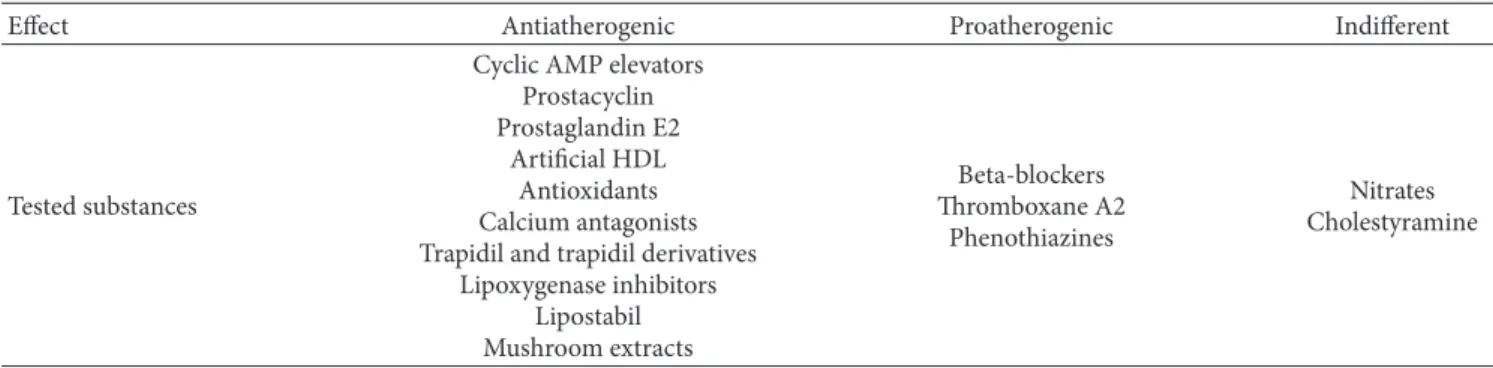
Table 1: Substances tested on cellular models.
Effect Antiatherogenic Proatherogenic Indifferent
Tested substances
Cyclic AMP elevators Prostacyclin Prostaglandin E2
Artificial HDL Antioxidants Calcium antagonists Trapidil and trapidil derivatives
Lipoxygenase inhibitors Lipostabil Mushroom extracts Beta-blockers Thromboxane A2 Phenothiazines Nitrates Cholestyramine
With permission from [9].
nonpharmaceutical compounds. Three drugs were developed and taken for further evaluation in the form of clinical studies [8].
3. Clinical Trials to Evaluate the
Antiatherosclerotic Activity of
Developed Drugs
There are three clinical trials that were aimed to test the efficacy and safety of the naturaceutical drugs, screened with cell models. Cultured aortic cell in vitro model was used to find potentially efficient natural agents fighting early-stage atherosclerosis.
3.1. Allicor. In this cell-based assay, 31 substances were assessed [39]. The effects of several substances are summa-rized in Table 1. The majority of investigated substances were pertaining to the flavonoid group. The chemicals under investigation were assessed for cytotoxicity via measurement of the absolute protein ratio and cell viability using try-pan blue. Authors have weeded out eight substances with cytotoxic effect, three of which had proatherogenic activity. Then two more substances with proatherogenic activity and four substances with no activity were eliminated, summing up to fourteen substances excluded totally. Authors based their short-list botanical substances on literature studies. Botanicals like spirulina (Spirulina platensis), onion (Allium cepa), wheat germs (Triticum vulgaris), hill-growing saltwort (Salsola collina), beetroot (Beta vulgaris), garlic (Allium sativum), licorice (Glycyrrhiza glabra), and extract of pine straw (Pinus sylvestris) have been evaluated in this study. These substances were tested in the aforementioned ex vivo model.
Authors used garlic powder in capsules for its effective-ness: six hours after the administration, the baseline rate of atherogenicity was three times lower. Based on these premises, authors have developed a natural-based product called “Allicor.” Clinical study confirmed the antiatheroscle-rotic effect of Allicor at the level of the vascular wall. The primary endpoint of the clinical trial was the rate of cIMT progression/regression [8]. The subjects in this clinical trial were all men, aged 40–74. 257 individuals were screened
and randomized to the study [8]. The inclusion criteria were the following: carotid atherosclerosis; absence of a prolonged treatment (more than 2 months per year) with sugar-lowering drugs, diuretics, and vasoactive medicines; the maximum cIMT of 1-2 mm in order to be detectable during the first B-mode ultrasound examination; systolic blood pressure below 160 mmHg; and diastolic blood pressure below 90 mmHg. 257 subjects were randomized into 2 treatment groups— Allicor treatment group and control group [8]. 196 patients in total were eligible for the assessment of the main end-point. The primary aim of the trial was to confirm or reject efficiency of Allicor in influencing the rate of cIMT progression/regression. In the treatment group, 32.3% of patients had their cIMT increased, while 47.3% of patients had a significant decrease in cIMT. In the control (placebo) group, 48.5% of subjects had an increase in cIMT, and 30.1% of the subjects had a decrease in cIMT. cIMT dynamics in the treatment group were significantly different, as compared to the control (placebo) group. Result of the trial was that the
average cIMT regression in the treatment group was 0.022±
0.007 mm per year. The divergence of total cIMT regression rate between control and treatment groups after two years of administration was still detectable and the gain was in favor of the treatment group. Decrease of patients’ blood serum ability to cause intracellular cholesterol accretion in the ex vivo cell culture test took place. In the treatment group, serum atherogenicity decreased nearly by 30% from the baseline level in as soon as 3 months after the start of administration, and the effect retained its magnitude till the end of the study, while the placebo group, on the contrary, had no sig-nificant changes. Decrease of patients’ serum atherogenicity was directly correlated with reduction of cIMT. In either of groups, level of HDL cholesterol has risen significantly during the study, while changes in triglyceride level were not significant [8]. Adverse events distribution was random, and none of the fatal serious adverse events were associ-ated with the treatment. Summing up, antiatherosclerotic effectiveness of Allicor was approved, and cIMT regression induced by Allicor was demonstrated clearly in the study [8]. Allicor successfully passed through clinical trials phases I to III and was recommended as an effective product for long-term therapy of an early stage of atherosclerosis [8].
4 BioMed Research International 3.2. Inflaminat. Inflammation is a constant companion of
vascular regions affected by atherosclerotic lesions [40]. The most important interleukins in atherosclerotic process are 6 and 1. 1 promotes local inflammation, while IL-6 is a proinflammatory factor and an important marker of inflammation in coronary atherosclerotic lesions. Inflam-mation theory of atherogenesis is extensively tested using in vivo models in order to assess the efficiency of inflam-mation inhibition in the antiatherosclerotic therapy. Before the occurrence of clinical manifestations, elevated level of inflammation markers can be registered in the blood serum. Current efforts are focused on assessing how important the role of cytokines is in chronic malicious conditions, and a direct anticytokine therapy is under development now. But, for the moment, the majority of the potential anticytokine medications are on the research stages, while those which already went through clinical trials have not shown desired efficacy and specificity. Taking that into account, there are some studies that show efficiency of compounds of natural origin in the inhibition of IL-1, IL-6, and tumor necrosis factor-alpha (TNF-𝛼).
Thus, the aim of this research was to create a naturaceu-tical product possessing anti-inflammatory properties as a supplement in patients with atherosclerosis [41]. Utilizing the same in vitro cellular model as in the screening stage for Allicor (see above), 31 selected botanicals were tested for their anticytokine activity. Of those, only 5 have shown the ability to hamper IL-1 expression, namely, violet, calendula, elder, hawthorn, and St. John’s wort.
Authors obtained a combination of three active ingredi-ents (elder, calendula, and violet) which was effective in the inhibition of expression of both IL-1 and TNF-𝛼. The product was given the name Inflaminat and it was placed into pro-duction at INAT-Pharma, Russia [41]. Inflaminat’s activities in anticytokine and antiatherogenic roles were assessed in a series of studies. The comparison with Diclofenac and Allicor was made. Authors recruited healthy volunteers, males and females with 52–69 years of age, whose blood possessed atherogenicity and proinflammatory activity. Once collecting the baseline blood sample, patients were given a dose of investigational product or comparator. After that, blood was taken after 2, 4, and 8 hours. In ex vivo model, Inflaminat was able to significantly decrease the IL-1 concentration for nearly 25% from the baseline level, while for Diclofenac the decrease was 49%. 8 hours after administration, TNF-𝛼 expression decreased by 9% for Inflaminat and by 39% for Diclofenac. 8 hours after administration, Inflaminat was able to reduce serum atherogenicity by 64% compared to the initial level: Allicor by nearly 50% and Diclofenac by 13%. This way, anticytokine and antiatherogenic properties of Inflaminat were demonstrated. The results were comparable to those for Diclofenac and Allicor. This fact shows the efficacy of the tested drug on biological models and its safety for healthy volunteers [42]. The initial double-blinded randomized placebo-controlled trial was aimed to assess efficacy and safety of Inflaminat in subjects with subclinical carotid atherosclerosis [41, 42].
Clinical trial of Inflaminat included men aged 40– 74 with asymptomatic atherosclerosis. Exclusion criteria
were the following: cardiovascular or cerebrovascular dis-ease; comorbidity that needed continuous treatment (more than 2 moths per year); signs to surgical interventions of atherosclerosis located in extracranial region of the bra-chiocephalic system; and individual intolerance to treat-ment with anti-inflammatory drugs. 78 patients entered the screening period. Systolic blood pressure decreased by 19 mmHg and diastolic arterial blood pressure decreased by 6 mmHg; total cholesterol level decreased by 49 ml/dL and LDL cholesterol decreased by 51 mg/dL in the treat-ment group. 10-Year prognostic risks for myocardial infarc-tion, sudden cardiac death, and ischemic heart disease decreased by 10%. cIMT of the right common carotid
artery decreased by 62𝜇m in average. At the same time, in
the control group, statistically significant decrease of total cholesterol was detected, probably due to diet correction recommendations received by all patients in the beginning of the trial. Nevertheless, LDL cholesterol, triglycerides, atherogenic index, and cIMT have not decreased. The results of the initial double-blinded randomized placebo-controlled clinical trial of Inflaminat approve that Inflaminat has antiatherosclerotic effect and caused a regression of atherosclerotic lesions at the dawn of the disease. Treat-ment with Inflaminat also had positive effects on different cardiovascular risk factors and caused the lowering of the prognostic risks of ischemic heart disease and myocardial infarction.
3.3. Karinat. Atherosclerosis-related pathologies are the main reason of sudden death in women (up to 73%). Nearly 55% of deaths in females are due to cardiovascular pathologies, and this proportion is higher than that in males. So, it turns out that while the major attention was paid to prevention of the atherosclerosis in men, prevention of atherosclerosis in women was left vastly underscored. The new trend in the biology is establishing new therapies for prevention and therapy of atherosclerosis in women [43, 44]. Till now, there has been no effective therapy that would have been able to improve the quality of life and avert atherosclerosis progression in menopausal women at the same time. Hormone replacement therapy is effective against osteoporosis and in alleviating the menopause symptoms but has oncogenic features and even stimulates the cardiovascular disorders.
Natural phytoestrogens bear similarity to human estro-gens and have ability to block estrogen receptors [53]. This research [54] aimed to develop a new breed of drugs for menopausal women that would reduce the menopause symp-toms and at the same time prevent the onset of atheroscle-rotic process without serious adverse effects. Authors have searched for the most effective and safe phytoestrogens in terms of positive, satisfactory, or negative antiatherosclerotic effects and pharmacodynamics on in vitro and ex vivo models. Several substances have been weeded out; the rest have been considered as promising, that is, had no detected cytotoxicity and possessed a stable effect. Grape, soybean, sage, carrot, orange, garlic, licorice, onion, hop, green tea, focus, kelp, calendula, clover, hawthorn, elder, and violet were
relatively easy to obtain and had high concentrations of the desired substances.
Antiatherosclerotic and antiatherogenic activities of the chosen plants were assessed on the in vitro and ex vivo models. A vast majority of the plants were eliminated due to the lack of activity or absence of long-term effect or low availability. The final list of promising compounds was the following: tannin from grape stone, garlic, hop, sage, and green tea leave. The final formula of active ingredients was proposed in accordance with minimal effective doses of each substance. A product called Karinat was developed. Follow-ing the successful laboratory study, the final naturaceutical product was ready for a clinical trial [43, 44, 54]. The initial double-blinded, randomized, placebo-controlled study was conducted in order to evaluate the efficacy and safety of the pills. 131 women were examined during the prescreening period. Exclusion criteria were the following: treatment with hypolipidemic drugs in a period of 6 months prior to the screening; treatment with sugar-lowering drugs for more than 2 months per year, treatment with beta-blockers and calcium antagonists; vestiges of hormone replacement therapy; vestiges of myocardial infarction and/or diagnosed acute cerebrovascular disorders and/or chronic cardiovas-cular insufficiency and/or pulmonary thromboembolism; the history of carcinoma; uncontrolled arterial hypertension more than 145/95 mmHg; chronic renal insufficiency; hep-atic cirrhosis; and individual intolerance to any of Karinat compounds. Totally, 131 females complied with the inclusion criteria. The average age of the participants was 64.8 years. Generally, subjects were having mild overweightness, high-normal arterial blood pressure, mild risk of ischemic disease and myocardial infarction, and subclinical atherosclerosis. Total cholesterol levels decreased in both control and treat-ment groups, but in the treattreat-ment group this decrease was due to LDL cholesterol, while in control group it was due to changes in HDL cholesterol level. In the treatment group, the
cIMT increase rate was only 6𝜇m/year, while in the control
group cIMT increase rate was more than 100𝜇m/year. The
placebo group confirms that there is an active atherosclerotic process in postmenopausal women going on. Considering the initial study design and short time span of therapy, none of detected changes in cIMT were considered as statisti-cally significant. The active and aggressive atherosclerosis progression in study participants was also confirmed by the increase of atherosclerotic plaques size. In the control group, the growth rate of already existing plaques topped up to 40% per year, while in the treatment group the growth rate of already existing plagues was 27% per year. The results of the initial double-blinded randomized placebo-controlled clinical trial of Karinat approve the fact that in post-menopausal women Karinat has antiatherosclerotic effect and causes regression of existing atherosclerotic lesions by 1.5 times.
4. Conclusions
Atherosclerosis is one of the most important medical and social problems, taking the blame for the development of cardiovascular pathologic conditions, which account for
more than half of all death cases. Patients on different stages of the disease have to take the same medications, while an aggressive therapy at the initial stages of the disease may result in adverse events. Currently, the most known antiatherosclerotic drugs are statins. They have been primary developed as drugs lowering serum cholesterol through inhibiting hepatic cholesterol biosynthesis. However, it soon became clear that antiatherosclerotic effects of statins could not be explained entirely by blood cholesterol reduction. Statins possess pleiotropic atheroprotective effects that are independent of cholesterol-lowering [55]. These noncholes-terol effects “alter the expression of endothelial nitric oxide synthase, the stability of atherosclerotic plaques, the pro-duction of proinflammatory cytokines and reactive oxygen species, the reactivity of platelets, and the development of cardiac hypertrophy and fibrosis” [55].
The exploitation of the concept of serum atherogenicity and the development of the effective cellular models allowed for the evaluation of the “direct antiatherosclerotic” activity of drug substances. As a result of the studies described in this overview, three natural nonpharmaceutical products have been created: Allicor, Karinat, and Inflaminat. All the drugs were tested on healthy subjects and assessed on in vitro and ex vivo models and demonstrated a significant success in decreasing serum atherogenicity. The major outcomes of the treatment were the decrease of cIMT and decrease of intracellular cholesterol accretion. Allicor was assessed in pilot and basic clinical trials, while Karinat and Inflaminat only started their life cycle with pilot studies, and the studies of the latter two are still to be continued.
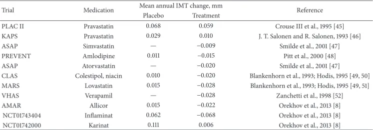
Table 2 compares the data on the antiatherosclerotic activities of Allicor, Inflaminat, and Karinat with similar data obtained in clinical trials of lipid-lowering drugs. All clinical trials had similar designs. It should be noted that Allicor, Inflaminat, and Karinat as direct antiatherosclerotic drugs realizing their effects at the arterial wall level possessed not less pronounced antiatherosclerotic activity compared to the lipid-lowering drugs that implement their pharmacological effects at the level of cholesterol circulating in blood or having pleiotropic antiatherosclerotic effects [55]. Moreover, Allicor and Inflaminat were as effective or even more effective compared to statins (Table 2). Thus, the drugs developed using our approach have been proven as very effective antiatherosclerotic drugs.
Our approaches are based on the use of in vitro and ex vivo cellular models. The effectiveness of these approaches has been confirmed by the assessment of such surrogate
marker of atherosclerosis as a cIMT. Of course, these
approaches have their limitations because they do not allow us to estimate the influence of drugs developed on clinical manifestations of atherosclerosis such as myocardial infarc-tion, stroke, and sudden death. This assessment is a challenge for further research. It is not known yet whether the mecha-nisms of Allicor, Inflaminat, and Karinat antiatherosclerotic actions are implemented via main cardiovascular risk factors or via exclusively alterations in arterial wall. Unlike statins, these drugs are not potent lipid-lowering medications. With regard to Allicor, it was shown that it possesses a mild or moderate hypotensive and cholesterol-lowering effects [40].
6 BioMed Research International
Table 2: The comparative data from clinical trials on carotid atherosclerosis regression.
Trial Medication Mean annual IMT change, mm Reference Placebo Treatment
PLAC II Pravastatin 0.068 0.059 Crouse III et al., 1995 [45] KAPS Pravastatin 0.029 0.010 J. T. Salonen and R. Salonen, 1993 [46] ASAP Simvastatin — −0.009 Smilde et al., 2001 [47] PREVENT Amlodipine 0.011 −0.015 Pitt et al., 2000 [48] ASAP Atorvastatin — −0.020 Smilde et al., 2001 [47]
CLAS Colestipol, niacin 0.010 −0.020 Blankenhorn et al., 1993; Hodis, 1995 [49, 50] MARS Lovastatin 0.015 −0.028 Blankenhorn et al., 1993; Hodis, 1995 [49, 51] VHAS Verapamil — −0.028 Zanchetti et al., 1998 [52]
AMAR Allicor 0.015 −0.022 Orekhov et al., 2013 [8] NCT01743404 Inflaminat 0.062 −0.068 Orekhov et al., 2013 [8] NCT01742000 Karinat 0.111 0.006 Orekhov et al., 2013 [8]
Allicor demonstrated antidiabetic and antiviral actions. In men with cerebral atherosclerosis, it has been demonstrated that 14-day treatment inhibited ADP-induced platelet aggre-gation by 25.4% and increased plasma fibrinolytic activity by 22.4%. One more study was performed in high-risk patients to evaluate the changes of prognostic cardiovascular risk that was calculated using algorithms derived from Framingham and Muenster Studies. Twelve-month treatment lowered 10-year prognostic risk of CHD by 13.2% in men and by 7.1% in women. Ten-year prognostic risk of acute myocardial infarction and sudden coronary death was lowered by 26.1% in men. On the other hand, all three drugs, namely, Allicor, Inflaminat, and Karinat, possess antiatherogenic potential in ex vivo model. There is no doubt that the antiatherogenic effect realizing at a level of the arterial wall makes a substantial contribution to the clinical effects of these drugs.
Competing Interests
The authors declare that there are no competing interests.
Acknowledgments
The work was supported by Ministry of Education and Sci-ences, Russia (Grant no. 14.W02.16.6995-Scientific School). The authors express their gratitude to Andrey V. Grechko from Federal Scientific Clinical Center for Resuscitation and Rehabilitation, Moscow, Russia, for editing the paper.
References
[1] A. Simmons, K. Steffen, and S. Sanders, “Medical therapy for peripheral arterial disease,” Current Opinion in Cardiology, vol. 27, no. 6, pp. 592–597, 2012.
[2] G. S. Berenson, S. R. Srinivasan, W. Bao, W. P. Newman III, R. E. Tracy, and W. A. Wattigney, “Association between multiple cardiovascular risk factors and atherosclerosis in children and young adults,” New England Journal of Medicine, vol. 338, no. 23, pp. 1650–1656, 1998.
[3] H. C. McGill Jr., E. E. Herderick, C. A. McMahan et al., “Atherosclerosis in youth,” Minerva Pediatrica, vol. 54, no. 5, pp. 437–447, 2002.
[4] E. M. Tuzcu, S. R. Kapadia, E. Tutar et al., “High prevalence of coronary atherosclerosis in asymptomatic teenagers and young adults evidence from intravascular ultrasound,” Circulation, vol. 103, no. 22, pp. 2705–2710, 2001.
[5] K. M. Anderson, P. W. F. Wilson, P. M. Odell, and W. B. Kannel, “An updated coronary risk profile. A statement for health professionals,” Circulation, vol. 83, no. 1, pp. 356–362, 1991.
[6] F. G. R. Fowkes, D. Rudan, I. Rudan et al., “Comparison of global estimates of prevalence and risk factors for peripheral artery disease in 2000 and 2010: a systematic review and analysis,” The Lancet, vol. 382, no. 9901, pp. 1329–1340, 2013.
[7] V. V. Tertov, A. N. Orekhov, V. S. Repin, and V. N. Smirnov, “Dibutyryl cyclic AMP decreases proliferative activity and the cholesteryl ester content in cultured cells of atherosclerotic human aorta,” Biochemical and Biophysical Research Communi-cations, vol. 109, no. 4, pp. 1228–1233, 1982.
[8] A. N. Orekhov, I. A. Sobenin, N. V. Korneev et al., “Anti-atherosclerotic therapy based on botanicals,” Recent Patents on Cardiovascular Drug Discovery, vol. 8, no. 1, pp. 56–66, 2013. [9] A. N. Orekhov, “Direct anti-atherosclerotic therapy;
develop-ment of natural anti-atherosclerotic drugs preventing cellular cholesterol retention,” Current Pharmaceutical Design, vol. 19, no. 33, pp. 5909–5928, 2013.
[10] A. Babelova, D. G. Sedding, and R. P. Brandes, “Anti-atherosclerotic mechanisms of statin therapy,” Current Opinion in Pharmacology, vol. 13, no. 2, pp. 260–264, 2013.
[11] X.-H. Yu, N. Jiang, P.-B. Yao, X.-L. Zheng, F. S. Cayabyab, and C.-K. Tang, “NPC1, intracellular cholesterol trafficking and atherosclerosis,” Clinica Chimica Acta, vol. 429, pp. 69–75, 2014. [12] E. Profumo, B. Buttari, L. Saso, and R. Rigan`o, “Pleiotropic effects of statins in atherosclerotic disease: focus on the antiox-idant activity of atorvastatin,” Current Topics in Medicinal Chemistry, vol. 14, no. 22, pp. 2542–2551, 2014.
[13] E. A. Stein and F. J. Raal, “New therapies for reducing low-density lipoprotein cholesterol,” Endocrinology and Metabolism Clinics of North America, vol. 43, no. 4, pp. 1007–1033, 2014. [14] I. Mendel, N. Yacov, D. Harats, and E. Breitbart,
atherosclerosis,” Current Pharmaceutical Design, vol. 21, no. 9, pp. 1185–1195, 2015.
[15] A. L. Catapano, M. Farnier, J. M. Foody et al., “Combination therapy in dyslipidemia: where are we now?” Atherosclerosis, vol. 237, no. 1, pp. 319–335, 2014.
[16] N. Artom, F. Montecucco, F. Dallegri, and A. Pende, “Carotid atherosclerotic plaque stenosis: the stabilizing role of statins,” European Journal of Clinical Investigation, vol. 44, no. 11, pp. 1122–1134, 2014.
[17] J. G. Robinson, “Starting primary prevention earlier with statins,” American Journal of Cardiology, vol. 114, no. 9, pp. 1437– 1442, 2014.
[18] Y. J. Chee, H. H. V. Chan, and N. C. Tan, “Understanding patients’ perspective of statin therapy: can we design a better approach to the management of dyslipidaemia? A literature review,” Singapore Medical Journal, vol. 55, no. 8, pp. 416–421, 2014.
[19] G. Klose, U. Laufs, W. M¨arz, and E. Windler, “Familial hyper-cholesterolemia: developments in diagnosis and treatment,” Deutsches Arzteblatt International, vol. 111, no. 31-32, pp. 523– 529, 2014.
[20] M. J. Wilkinson, L. J. Laffin, and M. H. Davidson, “Overcoming toxicity and side-effects of lipid-lowering therapies,” Best Prac-tice and Research: Clinical Endocrinology and Metabolism, vol. 28, no. 3, pp. 439–452, 2014.
[21] D. M. Diamond and U. Ravnskov, “How statistical deception created the appearance that statins are safe and effective in primary and secondary prevention of cardiovascular disease,” Expert Review of Clinical Pharmacology, vol. 8, no. 2, pp. 201– 210, 2015.
[22] G. Riccioni, “The effect of antihypertensive drugs on carotid intima media thickness: an up-to-date review,” Current Medici-nal Chemistry, vol. 16, no. 8, pp. 988–996, 2009.
[23] M. Velasco, E. Rojas, and V. B. Pirela, “Approach to a suc-cessful selection of antihypertensive drugs for the patient with atherosclerosis,” American Journal of Therapeutics, vol. 20, no. 4, pp. 442–447, 2013.
[24] R. H. Hern´andez, M. J. Armas-Hern´andez, M. Velasco, Z. H. Israili, and M. C. Armas-Padilla, “Calcium antagonists and atherosclerosis protection in hypertension,” American Journal of Therapeutics, vol. 10, no. 6, pp. 409–414, 2003.
[25] T. Kojima, K. Miyauchi, T. Yokoyama et al., “Azelnidipine and amlodipine anti-coronary atherosclerosis trial in hypertensive patients undergoing coronary intervention by serial volumet-ric intravascular ultrasound analysis in Juntendo university (ALPS-J),” Circulation Journal, vol. 75, no. 5, pp. 1071–1079, 2011. [26] S. Lim, M. J. Quon, and K. K. Koh, “Modulation of adiponectin as a potential therapeutic strategy,” Atherosclerosis, vol. 233, no. 2, pp. 721–728, 2014.
[27] A. Simon, J. Gari´epy, D. Moyse, and J. Levenson, “Differential effects of nifedipine and co-amilozide on the progression of early carotid wall changes,” Circulation, vol. 103, no. 24, pp. 2949–2954, 2001.
[28] A. N. Orekhov, V. V. Tertov, K. A. Khashimov, S. S. Kudryashov, and V. N. Smirnov, “Evidence of antiatherosclerotic action of verapamil from direct effects on arterial cells,” The American Journal of Cardiology, vol. 59, no. 5, pp. 495–496, 1987. [29] S. E. Akopov, A. N. Orekhov, V. V. Tertov, K. A. Khashimov, E.
S. Gabrielyan, and V. N. Smirnov, “Stable analogues of prosta-cyclin and thromboxane A2display contradictory influences on atherosclerotic properties of cells cultured from human aorta
The effect of calcium antagonists,” Atherosclerosis, vol. 72, no. 2-3, pp. 245–248, 1988.
[30] A. N. Orekhov, V. V. Tertov, Kh. A. Khashimov, S. A. Kudryashov, and V. N. Smirnov, “Antiatherosclerotic effects of verapamil in primary culture of human aortic intimal cells,” Journal of Hypertension, vol. 4, pp. S153–S155, 1986.
[31] H. Ikeda, J. Minamikawa, Y. Nakamura et al., “Comparison of effects of amlodipine and angiotensin receptor blockers on the intima-media thickness of carotid arterial wall (AAA study: amlodipine vs. ARB in atherosclerosis study),” Diabetes Research and Clinical Practice, vol. 83, no. 1, pp. 50–53, 2009. [32] W. F. Terpstra, J. F. May, A. J. Smit, P. A. De Graeff, B.
Meyboom-De Jong, and H. J. G. M. Crijns, “Effects of amlodipine and lisinopril on intima-media thickness in previously untreated, elderly hypertensive patients (the ELVERA trial),” Journal of Hypertension, vol. 22, no. 7, pp. 1309–1316, 2004.
[33] Z. Heidarzadeh, B. Asadi, M. Saadatnia, A. Ghorbani, and F. Fatehi, “The effect of low-dose combined oral contraceptive pills on brachial artery endothelial function and common carotid artery intima-media thickness,” Journal of Stroke and Cerebrovascular Diseases, vol. 23, no. 4, pp. 675–680, 2014. [34] E. F. Wolff, Y. He, D. M. Black et al., “Self-reported menopausal
symptoms, coronary artery calcification, and carotid intima-media thickness in recently menopausal women screened for the Kronos early estrogen prevention study (KEEPS),” Fertility and Sterility, vol. 99, no. 5, pp. 1385–1391, 2013.
[35] N. Waili, K. Salom, A. Ghamdi, M. J. Ansari, A. Al-Waili, and T. Al-Al-Waili, “Honey and cardiovascular risk factors, in normal individuals and in patients with diabetes mellitus or dyslipidemia,” Journal of Medicinal Food, vol. 16, no. 12, pp. 1063–1078, 2013.
[36] K. Ried, C. Toben, and P. Fakler, “Effect of garlic on serum lipids: an updated meta-analysis,” Nutrition Reviews, vol. 71, no. 5, pp. 282–299, 2013.
[37] A. L. Hopkins, M. G. Lamm, J. L. Funk, and C. Ritenbaugh, “Hibiscus sabdariffa L. in the treatment of hypertension and hyperlipidemia: a comprehensive review of animal and human studies,” Fitoterapia, vol. 85, no. 1, pp. 84–94, 2013.
[38] A. K. Rai, P. Debetto, and F. Dabbeni Sala, “Molecular regulation of cholesterol metabolism: HDL-based intervention through drugs and diet,” Indian Journal of Experimental Biology, vol. 51, no. 11, pp. 885–894, 2013.
[39] A. N. Orekhov, “Direct atherosclerotic and anti-atherogenic effects of garlic,” European Journal of Clinical Research A, vol. 3, pp. 1–12, 1992.
[40] V. P. Karagodin, I. A. Sobenin, and A. N. Orekhov, “Antiatherosclerotic and cardioprotective effects of time-released garlic powder pills,” Current Pharmaceutical Design, vol. 22, no. 2, pp. 196–213, 2016.
[41] T. Gorchakova, I. Suprun, I. Sobenin, and A. Orekhov, “Com-bined anti-inflammatory and anti-atherogenic activity of natu-ral drug inflaminat—a perspective for long-term atherosclerosis prevention and treatment,” Atherosclerosis Supplements, vol. 8, no. 1, article 224, 2007.
[42] T. Gorchakova, V. Myasoedova, I. Sobenin, and A. Orekhov, “Abstract: P387 atherosclerosis prevention with the anti-inflammatory dietary supplement Inflaminat,” Atherosclerosis Supplements, vol. 10, article no. 387, 2009.
[43] N. A. Nikitina, I. A. Sobenin, V. A. Myasoedova et al., “Antiatherogenic effect of grape flavonoids in an ex vivo model,” Bulletin of Experimental Biology and Medicine, vol. 141, no. 6, pp. 712–715, 2006.
8 BioMed Research International
[44] L. Sobenin, N. Nikitina, V. Myasoedova, V. Korenmaya, E. Khalilov, and A. Orekhov, “4P-1202 Antiatherogenic properties of isoflavones from phytoesrogen-rich botanicals,” Atheroscle-rosis Supplements, vol. 4, no. 2, p. 339, 2003.
[45] J. R. Crouse III, R. P. Byington, M. G. Bond et al., “Pravastatin, lipids, and atherosclerosis in the carotid arteries (PLAC-II),” The American Journal of Cardiology, vol. 75, no. 7, pp. 455–459, 1995. [46] J. T. Salonen and R. Salonen, “Ultrasound B-mode imaging in observational studies of atherosclerotic progression,” Circula-tion, vol. 87, no. 3, pp. II56–II65, 1993.
[47] T. J. Smilde, S. Van Wissen, H. Wollersheim, M. D. Trip, J. J. P. Kastelein, and A. F. H. Stalenhoef, “Effect of aggressive versus conventional lipid lowering on atherosclerosis progres-sion in familial hypercholesterolaemia (ASAP): a prospective, randomised, double-blind trial,” Lancet, vol. 357, no. 9256, pp. 577–581, 2001.
[48] B. Pitt, R. P. Byington, C. D. Furberg et al., “Effect of amlodipine on the progression of atherosclerosis and the occurrence of clinical events,” Circulation, vol. 102, no. 13, pp. 1503–1510, 2000. [49] H. N. Hodis, “Reversibility of atherosclerosis-evolving perspec-tives from two arterial imaging clinical trials: the cholesterol lowering atherosclerosis regression study and the monitored atherosclerosis regression study,” Journal of Cardiovascular Pharmacology, vol. 25, supplement 4, pp. S25–S31, 1995. [50] D. H. Blankenhorn, R. H. Selzer, D. W. Crawford et al.,
“Beneficial effects of colestipol-niacin therapy on the common carotid artery: two- and four-year reduction of intima-media thickness measured by ultrasound,” Circulation, vol. 88, no. 1, pp. 20–28, 1993.
[51] D. H. Blankenhorn, S. P. Azen, D. M. Kramsch et al., “Coronary angiographic changes with lovastatin therapy: the Monitored Atherosclerosis Regression Study (MARS),” Annals of Internal Medicine, vol. 119, no. 10, pp. 969–976, 1993.
[52] A. Zanchetti, E. Agabiti Rosei, C. Dal Pal`u, G. Leonetti, B. Magnani, and A. Pessina, “The Verapamil in Hypertension and Atherosclerosis Study (VHAS): results of long-term ran-domized treatment with either verapamil or chlorthalidone on carotid intima-media thickness,” Journal of Hypertension, vol. 16, no. 11, pp. 1667–1676, 1998.
[53] V. B. Gencel, M. M. Benjamin, S. N. Bahou, and R. A. Khalil, “Vascular effects of phytoestrogens and alternative menopausal hormone therapy in cardiovascular disease,” Mini-Reviews in Medicinal Chemistry, vol. 12, no. 2, pp. 149–174, 2012.
[54] V. Myasoedova and I. Sobenin, “Background, rationale and design of clinical study of the effect of isoflavonoid-rich botanicals on natural history of atherosclerosis in women,” Atherosclerosis Supplements, vol. 9, no. 1, p. 171, 2008.
[55] A. Oesterle, U. Laufs, and J. K. Liao, “Pleiotropic Effects of Statins on the Cardiovascular System,” Circulation Research, vol. 120, no. 1, pp. 229–243, 2017.
Submit your manuscripts at
https://www.hindawi.com
Pain
Research and TreatmentHindawi Publishing Corporation
http://www.hindawi.com Volume 2014
World Journal
Hindawi Publishing Corporation
http://www.hindawi.com Volume 2014
Hindawi Publishing Corporation
http://www.hindawi.com Volume 2014
Toxins
Journal of
Vaccines
Journal ofHindawi Publishing Corporation
http://www.hindawi.com Volume 2014
Hindawi Publishing Corporation
http://www.hindawi.com Volume 2014
Antibiotics
Toxicology
Journal ofHindawi Publishing Corporation
http://www.hindawi.com Volume 2014
Stroke
Research and Treatment Hindawi Publishing Corporationhttp://www.hindawi.com Volume 2014
Drug Delivery
Journal of Hindawi Publishing Corporationhttp://www.hindawi.com Volume 2014
Hindawi Publishing Corporation
http://www.hindawi.com Volume 2014
Advances in Pharmacological Sciences
Tropical Medicine
Hindawi Publishing Corporationhttp://www.hindawi.com Volume 2014
Medicinal ChemistryInternational Journal of Hindawi Publishing Corporation
http://www.hindawi.com Volume 2014
Addiction
Journal of Hindawi Publishing Corporationhttp://www.hindawi.com Volume 2014
Hindawi Publishing Corporation
http://www.hindawi.com Volume 2014 BioMed
Research International Emergency Medicine International
Hindawi Publishing Corporation
http://www.hindawi.com Volume 2014
Hindawi Publishing Corporation
http://www.hindawi.com Volume 2014
Diseases
Hindawi Publishing Corporation
http://www.hindawi.com Volume 2014 Anesthesiology Research and Practice
Scientifica
Hindawi Publishing Corporation
http://www.hindawi.com Volume 2014
Journal of
Hindawi Publishing Corporation
http://www.hindawi.com Volume 2014
Pharmaceutics
Hindawi Publishing Corporation
http://www.hindawi.com Volume 2014