HAL Id: tel-03173551
https://tel.archives-ouvertes.fr/tel-03173551
Submitted on 18 Mar 2021HAL is a multi-disciplinary open access archive for the deposit and dissemination of sci-entific research documents, whether they are pub-lished or not. The documents may come from teaching and research institutions in France or abroad, or from public or private research centers.
L’archive ouverte pluridisciplinaire HAL, est destinée au dépôt et à la diffusion de documents scientifiques de niveau recherche, publiés ou non, émanant des établissements d’enseignement et de recherche français ou étrangers, des laboratoires publics ou privés.
Mechanism of L,D-transpeptidase inhibition by
β-lactams and diazabicyclooctanes
Zainab Edoo
To cite this version:
Zainab Edoo. Mechanism of L,D-transpeptidase inhibition by β-lactams and diazabicyclooctanes. Microbiology and Parasitology. Sorbonne Université, 2019. English. �NNT : 2019SORUS565�. �tel-03173551�
Sorbonne Université
Ecole doctorale 515 « Complexité du Vivant »
Laboratoire de Structures Bactériennes Impliquées dans la Modulation de la Résistance aux Antibiotiques
Centre de Recherche des Cordeliers, UMRS 1138, Equipe 12
Mechanism of
L
,
D
-transpeptidase inhibition by
β-lactams and diazabicyclooctanes.
Mécanisme d’inhibition des
L
,
D
-transpeptidases par les
β-lactamines et les diazabicyclooctanes.
Par Zainab Edoo
Thèse de doctorat de Biochimie
Dirigée par Jean-Emmanuel Hugonnet
Présentée et soutenue publiquement le 22 novembre 2019
Devant un jury composé de :
Pr. Sandrine Betuing, Sorbonne Université
Présidente
Dr. Alain Baulard, Institut Pasteur de Lille
Rapporteur
Dr. Luiz Pedro Carvalho, Francis Crick Institute
Rapporteur
Dr. Célia Caillet-Saguy, Institut Pasteur
Examinatrice
Pr. Jean-Marie Frère, Université de Liège
Examinateur
Acknowledgement
I would like to start by thanking the members of my thesis jury: the rapporteurs, Alain Baulard and Luiz Pedro Carvalho, and the examinateurs, Sandrine Betuing, Célia Caillet-Saguy, and Jean-Marie Frère, for accepting to evaluate my work, and Jean-Emmanuel Hugonnet for supervising my PhD.
I am grateful to the Fondation pour la Recherche Médicale for financial support and to the doctoral school for their assistance.
Doing a PhD was harder than I thought but more rewarding than I could have imagined. This
achievement would not have been possible without the support and guidance of many individuals.
Thank you Jean-Emmanuel (Manu) for your unfailing encouragement during my thesis. You have been of great help and advice throughout the years. Thank you for believing in me!
I am deeply grateful to the head of the lab, Michel Arthur. Thank you for your invaluable guidance and for being so generous with your time. I have learned so much from you.
I would like to thank the members of my thesis committee, Houssain Benabdelhak, Thierry Touzé, Guennadi Sezonov, and Wladimir Sougakoff, for their feedback and warm encouragement.
I had the great opportunity to collaborate with these wonderful people during my thesis: Mélanie Etheve-Quelquejeu, Laura Iannazzo, and Flavie Bouchet (Université de Paris); Ines Gallay and Herman van Tilbeurgh (Université Paris-Saclay); Catherine Bougault and Jean-Pierre Simorre (Institut de Biologie Structurale); Lionel Dubost (Muséum National d’Histoire Naturelle); Waldemar Vollmer (Newcastle University); Nadine Bongaerts, Edwin Wintermute, and Ariel Lindner (Centre de Recherches Interdisciplinaires); Landys Lopez Quezada and Carl Nathan (Weill Cornell Medicine).
My sincere appreciation goes to the past and current members of Equipe 12. It has been a pleasure to work with all of you. Special thanks to Grazyna for keeping us in line and treating us with cakes, Sébastien for patiently walking me through enzyme kinetics during my Master’s internship, Matthieu for answering my chemistry-related questions, Delphine for her technical help, Antoine for the cheerfulness he brought to our office, Jean-Luc for his excellence, and David for being the best labmate. Cheers to my fellow PhD students with whom I shared the ups and downs of the last three years, and the interns, the youngest being 5-year-old Margot, who joined the lab for brief but fun periods.
My heartfelt gratitude goes to the teachers who have inspired and encouraged me over the years.
I am forever indebted to my family and friends, near and far. I have been truly blessed! You all hold a very special place in my heart.
Henri, thank you for always managing to make me laugh!
To my family – Thank you for your unwavering support and love. I owe it all to you.
ZAK forever
♥3
Foreword
Antibacterial chemotherapy is one of the most essential contributions of medicine to human health in the last century. Ever since the discovery of the first antibiotic, penicillin, peptidoglycan biosynthesis has been one of the preferred targets for the discovery of new antibiotics.
Peptidoglycan is an essential component of bacterial cells. It is composed of glycan chains, which are connected by short peptide stems. In most bacteria, the synthesis of the cross-links between the peptide stems is catalyzed by the Penicillin-Binding Proteins (PBPs). As their name implies, the PBPs are targeted by penicillin, which was the first discovered member of the β-lactam family. β-lactams have since been extensively developed for antibacterial activity against positive and Gram-negative bacteria. However, their use is threatened by numerous resistance mechanisms, the most important of which is the production of β-lactamases. The development of β-lactams that are not hydrolyzed by β-lactamases and of β-lactamase inhibitors have nonetheless contributed towards defeating emerging resistance.
Peptidoglycan cross-links are not only formed by the classical PBPs but also by an unrelated family of enzymes, the L,D-transpeptidases (LDTs). This unusual mode of cross-linking is predominant in a few pathogenic bacteria, including Mycobacterium tuberculosis. PBPs and LDTs are structurally unrelated, function with different catalytic nucleophiles (Ser vs. Cys, respectively), and use different peptidoglycan precursors for the cross-linking reaction. In contrast to PBPs, which are potentially inactivated by all β-lactam classes, LDTs are efficaciously inactivated only by the carbapenem class. The two objectives of the thesis are to investigate the mechanism of inhibition of LDTs by carbapenems and to explore whether the diazabicyclooctane scaffold, recently developed for β-lactamase inhibition, also inactivates LDTs.
The manuscript will begin with an introduction on the role and synthesis of peptidoglycan, the mode of action of β-lactams, and the catalytic activities of PBPs and LDTs. The introduction is followed by a description of the specific objectives of the thesis. The result section is based on published papers and is divided into two parts focused on the mechanism of inhibition of LDTs by β-lactams and diazabicyclooctanes. The last section consists of a discussion on the implications of the findings regarding the catalytic mechanism of LDTs for optimization of inhibitors. In parallel to the main objectives of the thesis, I contributed to two studies investigating the LDTs from Clostridium difficile and inhibition of LDTs by copper(II) ions. These two publications appear in an annex at the end of the manuscript.
4
Table of Content
ACKNOWLEDGEMENT 2 FOREWORD 3 TABLE OF CONTENT 4 LIST OF FIGURES 6 LIST OF TABLES 7 LIST OF ABBREVIATIONS 8INTRODUCTION
9 1. PEPTIDOGLYCAN BIOSYNTHESIS 10 1.1ROLE OF PEPTIDOGLYCAN 101.2PEPTIDOGLYCAN STRUCTURE AND SYNTHESIS 11
1.3ANTIBIOTICS TARGETING PEPTIDOGLYCAN SYNTHESIS 27
2. THE β-LACTAM ANTIBIOTICS 28
2.1MODE OF ACTION OF β-LACTAMS 28
2.2CLASSES OF β-LACTAMS 29 2.3RESISTANCE TO β-LACTAMS 32 3. β-LACTAMASES 34 3.1CLASSIFICATION OF β-LACTAMASES 34 3.2β-LACTAMASE INHIBITORS 37 4. PENICILLIN-BINDING PROTEINS 41 4.1CATALYTIC MECHANISM 42 4.2CLASSIFICATION OF PBPS 45 4.3INHIBITION OF PBPS BY β-LACTAMS 50 5. L,D-TRANSPEPTIDASES 53 5.1IDENTIFICATION OF LDTS 53
5.2ROLE OF LDTS IN VARIOUS BACTERIA 56
5.3STRUCTURE OF LDTS 64
5.4CATALYTIC MECHANISM OF L,D-TRANSPEPTIDATION 68
5.5INHIBITION OF LDTS BY β-LACTAMS 70
OBJECTIVES 81
RESULTS
821. MECHANISM OF INHIBITION OF L,D-TRANSPEPTIDASES BY β-LACTAMS 83
PUBLICATION 1 85
PUBLICATION 2 85
PUBLICATION 3 85
2. INHIBITION OF L,D-TRANSPEPTIDASES BY A NOVEL DIAZABICYCLOOCTANE SCAFFOLD AND MODE OF
ACTION OF THE MOLECULES IN M. TUBERCULOSIS 87
5
DISCUSSION
911.MODELS ACCOUNTING FOR ACYLATION OF L,D-TRANSPEPTIDASES BY β-LACTAMS 92
2.BASIS FOR FLUORESCENCE QUENCHING OBSERVED UPON L,D-TRANSPEPTIDASE ACYLATION BY β-LACTAMS 95 3.THE REACTIVITY OF β-LACTAMS HAS A KEY ROLE IN THE EFFICACY OF L,D-TRANSPEPTIDASE ACYLATION 95 4.MULTIPLE POTENTIAL TARGETS OF DBOS IN M. TUBERCULOSIS 96 5.OPTIMIZATION OF INHIBITORS TARGETING L,D-TRANSPEPTIDASES 100
ANNEX
102ANNEX 1 103
ANNEX 2 103
6
List of Figures
Figure 1. Structure of the bacterial cell wall. ... 10
Figure 2. The peptidoglycan subunit and its synthesis. ... 12
Figure 3. Common variations found in the peptidoglycan subunit of E. coli, E. faecium, and M. tuberculosis. ... 15
Figure 4. Structure of MurJ and the conformational change required for its activity as a flippase. .... 17
Figure 5. Peptidoglycan polymerization scheme. ... 18
Figure 6. Structure of the monofunctional glycosyltransferase MtgA of S. aureus. ... 20
Figure 7. Structure of RodA and model of peptidoglycan synthesis by the RodA-PBP2 pair. ... 23
Figure 8. Analogy between the natural substrate of PBPs and β-lactams. ... 28
Figure 9. Mechanisms of β-lactam resistance in Gram-negative bacteria. ... 33
Figure 10. Reaction scheme for the hydrolysis of β-lactams by serine β-lactamases. ... 35
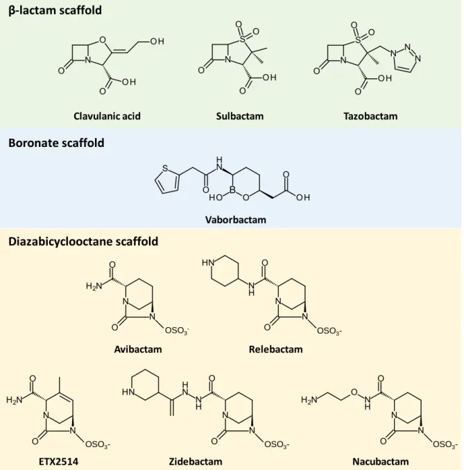
Figure 11. β-lactamase inhibitors of current interest. ... 37
Figure 12. Proposed mechanism for acylation of class A β-lactamases by clavulanate. ... 38
Figure 13. Reversible inactivation of β-lactamases by avibactam. ... 40
Figure 14. D,D-carboxypeptidation and D,D-transpeptidation catalyzed by PBPs. ... 42
Figure 15. Penicillin-binding domain and conserved motifs of B. subtilis PBP4a. ... 43
Figure 16. Structure of PBP1B of E. coli. ... 45
Figure 17. Reaction scheme for acylation of PBPs by β-lactams. ... 50
Figure 18. Model for peptidoglycan synthesis in mycobacteria. ... 60
Figure 19. Structural diversity in the domain organization of LDTs. ... 65
Figure 20. Structure of Ldtfm and its catalytic triad. ... 66
Figure 21. Binding of β-lactams and of acyl donor and acceptor substrates to Ldtfm. ... 67
Figure 22. Formation of 33 cross-links by LDTs. ... 68
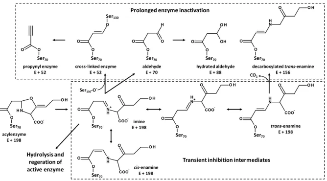
Figure 23. Variations in absorbance and in protein fluorescence during acylation of Ldtfm by imipenem and proposed reaction scheme. ... 73
Figure 24. Acylation of Ldtfm by a representative of the penams, cephems, and carbapenems. ... 76
Figure 25. Dual-active cephems for M. tuberculosis. ... 78
Figure 26. Oxyanion-forming catalytic mechanism for acylation of LdtBs by carbapenems. ... 79
Figure 27. Concerted catalytic mechanism proposed for acylation of Ldtfm by carbapenems. ... 80
Figure 28. Models proposed for acylation of LDTs by β-lactams. ... 94
7
List of Tables
Table 1. Structures of the most common β-lactam classes and of one representative of each class. . 30 Table 2. Antibiotic susceptibility and proportion of 33 cross-links in the D344S and M512 strains. 54 Table 3. An example of the variation in the proportion of 33 cross-links among selected bacterial species from four phyla. ... 57
8
List of Abbreviations
A2pm 2,6-diaminopimelic acid
Alr Alanine racemase
aPBP Class A penicillin-binding protein bPBP Class B penicillin-binding protein C55-(P)P Undecaprenyl (pyro)phosphate
cCys-Asn Cyclic Cys-Asn dipeptide
cPBP Class C penicillin-binding protein
DBO Diazabicyclooctane
Ddl D-alanine:D-alanine ligase
ESBL Extended-spectrum β-lactamase
GlcNAc N-acetylglucosamine
HMW High molecular weight
LDT L,D-transpeptidase
LMW Low molecular weight
MD Molecular dynamics
MIC Minimal inhibitory concentration
MOP Multidrug/oligosaccharidyl-lipid/polysaccharide MRSA Methicillin-resistant Staphylococcus aureus
MurNAc N-acetylmuramic acid
NCDAA Non-canonical D-amino acid
PASTA Penicillin-binding protein and serine/threonine associated domain
PBP Penicillin-binding protein
PDB Protein Data Bank
QM/MM Quantum mechanics/molecular mechanics SEDS Shape, elongation, division, and sporulation
UB2H UvrB domain 2 homologue
9
10
1. Peptidoglycan Biosynthesis
1.1 Role of Peptidoglycan
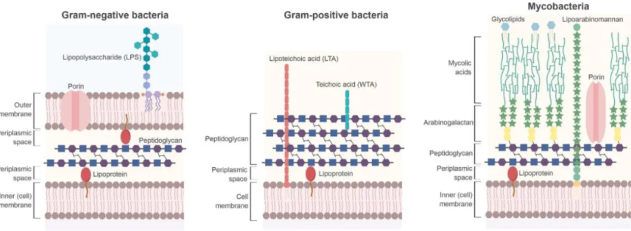
Peptidoglycan is a unique and essential component of most bacterial cells. It is a three-dimensional net-like macromolecule that surrounds the cytoplasmic membrane (Figure 1) (Holtje, 1998). Peptidoglycan is a few nm thick and is located between the inner and outer membranes in Gram-negative bacteria (Matias et al., 2003). Gram-positive bacteria lack an outer membrane and the thickness of their peptidoglycan can vary between 30 and 100 nm (Silhavy et al., 2010). The main role of peptidoglycan is to confer resistance against the turgor pressure of the cytoplasm (Whatmore & Reed, 1990). Cell lysis occurs when peptidoglycan is damaged by enzymes, such as lysozyme, or antibiotics. The biophysical properties of peptidoglycan also allow it to maintain cell shape (Harold, 1990) (Bailey et al., 2019). In addition, peptidoglycan serves as an anchor for cell wall polymers (Vollmer et al., 2008a). In Gram-negative bacteria, such as Escherichia coli, Braun’s lipoprotein is covalently linked to peptidoglycan and provides a tether for the outer membrane, thus controlling the architecture of the cell envelope (Asmar et al., 2017; Hantke & Braun, 1973). In Gram-positive bacteria, in addition to surface proteins, wall teichoic acids are covalently attached to peptidoglycan, contributing to the structure and functions of the cell envelope (Dramsi et al., 2008; Neuhaus & Baddiley, 2003). The essential roles of peptidoglycan make its biosynthesis a validated target for the development of antibiotics. Indeed, the most commonly used antibiotics today are the β-lactams, which target key enzymes in the peptidoglycan biosynthetic pathway (Kong et al., 2010).
Figure 1. Structure of the bacterial cell wall.
Peptidoglycan of Gram-negative bacteria (left) is located in the periplasm between the inner and outer membranes. It serves as a tether for lipoproteins. The outer membrane contains porins and lipopolysaccharides. Gram-positive bacteria (middle) do not contain an outer membrane. Their peptidoglycan is composed of more layers than Gram-negative bacteria and serves as an attachment for wall teichoic acids. Lipoteichoic acids are attached to the cell membrane. Mycobacteria (right) are acid-fast bacteria. They have a complex cell wall that contains glycolipids, mycolic acids, arabinogalactan polysaccharide, and a peptidoglycan network. From reference (Porfirio et al., 2019).
11
1.2 Peptidoglycan Structure and Synthesis
Peptidoglycan is composed of linear glycan strands that are cross-linked by short peptide stems (Holtje, 1998). The peptidoglycan subunit is a disaccharide composed of N-acetylglucosamine (GlcNAc) linked by a β-1→4 glycosidic bond to N-acetylmuramic acid (MurNAc), which is attached to a peptide stem containing five amino acids (Figure 2A). Alternating units of GlcNAc and MurNAc constitute a glycan chain. Adjacent chains are linked to one another by a peptide bridge formed between their respective peptide stems. The chemical composition of peptidoglycan can vary according to bacterial species and growth conditions. Secondary modifications in the glycan strands include N-deacetylation, O-acetylation, and N-glycolylation (Vollmer, 2008). There are also inter- and intra-species variations in the length of glycan strands (Vollmer et al., 2008a). In Gram-negative bacteria, the pentapeptide stem is typically composed of L-alanine-γ-D-glutamic acid-meso-2,6-diaminopimelic acid (A2pm)-D
-alanine-D-alanine (Quintela et al., 1995). The most common variation in the peptide stem occurs at position 3, where most Gram-positive bacteria contain an L-lysine, whose ε-amine can be linked to a side-chain containing one or more amino acids (Schleifer & Kandler, 1972). Amidation of D-Glu and/or A2pm is
also frequent. Variations in the glycan chain and the peptide stem are detailed below (Chapter 1.2.1b). The presence of MurNAc, of non-protein amino acids, and of γ-bonded D-Glu contributes to the uniqueness of peptidoglycan as a polymer, allowing bacteria to resist chemical and enzymatic degradation. For instance, the strong ether bond between the peptide stem and the lactic acid moiety of MurNAc ensures resistance to chemical degradation while the presence of D-amino acids confers resistance to peptidases (Cava et al., 2011b).
Peptidoglycan biosynthesis can be divided into two main stages based on their localization (Figure 2B). The first stage takes place in the cytoplasm where the disaccharide-pentapeptide unit is synthesized as a lipid-linked precursor, lipid II, before being flipped across the cell membrane (van Heijenoort, 1998). The second stage occurs on the outside surface of the cytoplasmic membrane and involves incorporation of the precursor into the existing peptidoglycan, as well as cross-linking of glycan chains (van Heijenoort, 2001a).
12 Figure 2. The peptidoglycan subunit and its synthesis.
(A) Structure of a typical disaccharide-pentapeptide found in Gram-negative bacteria. GlcNAc is linked to MurNAc, which is attached to a pentapeptide stem. The muropeptide is colored according to the color code used in B. (B) Synthesis of the peptidoglycan subunit and peptidoglycan polymerization. The sugars, GlcNAc and MurNAc, are synthesized and the pentapeptide stem assembled in the cytoplasm. The lipid II intermediate, containing the disaccharide-peptide subunit, is flipped across the cell membrane for peptidoglycan polymerization on the external surface of the membrane.
1.2.1 Synthesis of the Peptidoglycan Precursor
a) Synthesis of Lipid II
The synthesis of the disaccharide-pentapeptide building block of peptidoglycan takes place in the cytoplasm and involves nucleotide precursors and the Glm and Mur enzymes (Figure 2B) (van Heijenoort, 1998). Uridine 5’-pyrophosphate-N-acetylglucosamine (UDP-GlcNAc) is synthesized from fructose-6-phosphate in four successive steps. Fructose-6-phosphate is first converted to glucosamine-6-phosphate, which is then transformed to glucosamine-1-phosphate. The latter is acetylated and uridylated to form UDP-GlcNAc. The first two steps are catalyzed by GlmS and GlmM, respectively, while the last two steps are catalyzed by two domains of the same enzyme, GlmU (Dutka-Malen et al., 1988; Mengin-Lecreulx & van Heijenoort, 1994, 1996). UDP-MurNAc is formed from UDP-GlcNAc in two steps (van Heijenoort, 1998). The MurA transferase catalyzes the transfer of enolpyruvate from phosphoenolpyruvate to GlcNAc, yielding UDP-GlcNAc-enolpyruvate. The MurB reductase then catalyzes enolpyruvate reduction to D-lactoyl, thus forming UDP-MurNAc.
Each stepwise addition of amino acids onto the D-lactoyl moiety of MurNAc is catalyzed by a specific Mur ligase (MurC, D, E, and F) using adenosine 5’-triphosphate (ATP). The final product of these
GlcNAc L-Ala D-Glu A2pm D-Ala D-Ala MurNAc B A UDP MurA MurB
UDP UDP UDP GlmS GlmM GlmU Fructose-6-phosphate UDP UDP-GlcNAc UDP-MurNAc MurJ Lipid I Cytoplasmic membrane Cytoplasm Peptidoglycan polymerization P MraY MurC L-Ala MurD D-Glu MurE A2pm MurF D-Ala-D-Ala UDP P P MurG P P UDP Lipid II P P P P Glycan strand Peptide stem Cross-link Pi P C55-P
13 reactions is UDP-MurNAc-pentapeptide. The Mur ligases share the same reaction mechanism: formation of an acylphosphate intermediate through activation of the carboxyl group of UDP-MurNAc (or of the last amino acid added) by ATP, followed by nucleophilic attack by the amino group of the condensing amino acid, leading to elimination of phosphate and formation of an amide bond (Bouhss et al., 2002). The identity of the amino acid that is incorporated in the peptide stem depends on the substrate specificity of the Mur enzymes (Barreteau et al., 2008).
MurC catalyzes the addition of the first amino acid to the peptide stem. In most bacteria, L-alanine is added although the addition of glycine or L-serine has been observed in some rare cases (Schleifer & Kandler, 1972). MurC shows strict stereospecificity since it cannot catalyze the addition of D-Ala to UDP-MurNAc (Liger et al., 1995). MurD catalyzes the addition of the second residue to the peptide stem. In all species, this amino acid is D-glutamic acid although later modifications can convert it to D -isoglutamine (discussed in the next subchapter). MurE catalyzes the addition of the third amino acid, which is either A2pm in mycobacteria and most Gram-negative bacteria or L-Lysine in most
Gram-positive bacteria (Schleifer & Kandler, 1972). MurE is highly specific in most species. For instance, addition of L-Lysine instead of A2pm in E. coli leads to cell lysis (Mengin-Lecreulx et al., 1999). Like for
the amino acid at position 2, the one at position 3 can be modified in later stages (discussed in the next subchapter). MurF catalyzes the last assembly step, i.e. addition of a dipeptide composed of the fourth and fifth residues to the peptide stem. The most common dipeptide consists of two D-Ala. However, some vancomycin-resistant strains contain D-Ala-D-Ser or D-Ala-D-Lac.
The synthesis of the amino acids in the pentapeptide stem that are essentially found in peptidoglycan, D-Glu, A2pm, and D-Ala, is discussed here. D-Glu is produced from L-Glu by MurI, a glutamate racemase,
that catalyzes the interconversion of the D- and L-enantiomers (Doublet et al., 1993). In some bacterial species, the formation of D-Glu can also be catalyzed by a second enzyme, D-amino acid aminotransferase (D-AAT), which uses D-Ala and α-ketoglutarate as substrates to produce pyruvate in addition to D-Glu (Fotheringham et al., 1998). A2pm is the last intermediate in the biosynthetic
pathway of L-Lys from L-Asp (Scapin & Blanchard, 1998). An alanine racemase catalyzes the conversion of L-Ala to D-Ala prior to the formation of the D-Ala-D-Ala dipeptide, catalyzed by a ligase, D-Ala:D-Ala ligase (Ddl) (Neuhaus & Lynch, 1964).
After assembly of the pentapeptide stem on MurNAc, the MraY transferase catalyzes the transfer of the phospho-MurNAc-pentapeptide moiety from UDP-MurNAc-pentapeptide to a membrane acceptor, undecaprenyl phosphate (C55-P) (Bouhss et al., 2008). This yields the first lipid intermediate,
lipid I. The transferase MurG then catalyzes the transfer of GlcNAc from UDP-GlcNAc to lipid I, yielding lipid II. The carrier lipid is required for the transport of the hydrophilic peptidoglycan precursor from
14 the cytoplasm across the hydrophobic cytoplasmic membrane. The synthesis of C55-P is catalyzed by
the UppS synthase, and the BacA and PAP2 phosphatase families, which respectively catalyze the synthesis of undecaprenyl pyrophosphate (C55-PP) and its subsequent dephosphorylation.
b) Common Variations in the Pentapeptide Stem and Glycan
Chain
Additional modifications of the pentapeptide stem (briefly mentioned above) occur after formation of the lipid intermediates, often at the level of lipid II (Figure 3) (Vollmer et al., 2008a). Some of the most frequent modifications are discussed here. The α-carboxyl group of D-Glu at position 2 is amidated to form D-isoGln in most Gram-positive bacteria and mycobacteria. Amidation of the ε-carboxyl group of A2-pm at position 3 is also frequent. For instance, the pentapeptide stem of Mycobacterium
tuberculosis is amidated at both positions 2 and 3 and it was recently shown that amidation of A2pm
by the AsnB amidotransferase is essential for growth of M. tuberculosis (Ngadjeua et al., 2018). Many Gram-positive bacteria contain a peptide side-chain on the ε-amino group of the L-Lys residue at position 3 (Schleifer & Kandler, 1972). For example, in Staphylococcus aureus, L-Lys is bonded to a penta-glycine chain, the stepwise addition of glycines being catalyzed by FmhB, FemA, and FemB (Schneider et al., 2004). In Enterococcus faecium, Aslfm catalyzes addition of D-Asp, through its
β-carboxyl group, to L-Lys (Bellais et al., 2006). In mature peptidoglycan, the α-carboxyl group of D-Asp is partially amidated.
In addition to the pentapeptide stem, the sugar moieties constituting the glycan strands can also be modified (Figure 3) (Vollmer, 2008). However, compared to modifications on the peptide stem, these modifications (except N-glycolylation) are likely to occur on polymerized peptidoglycan since cytoplasmic precursors containing modifications on their sugars have not been detected and known modifying enzymes have a predicted extracytoplasmic localization (Vollmer & Tomasz, 2000). Many Gram-positive and Gram-negative bacteria contain a proportion of MurNAc residues that are O-acetylated at position 6 (Weadge et al., 2005). This modification contributes to resistance of Gram-positive bacteria to one of the most important compounds of the host defense system, lysozyme, a muramidase that cleaves the glycosidic bond between C1 of MurNAc and C4 of GlcNAc (Bera et al.,
2005). Another modification present in various bacteria, including Bacillus and Streptomyces species, that can contribute to lysozyme resistance is de-N-acetylation of GlcNAc and MurNAc, (Atrih et al., 1999) (Hugonnet et al., 2014). In the spore peptidoglycan of Bacillus species, deacetylated MurNAc is further altered to contain a δ-lactam ring, generated by the formation of an intramolecular bond between the carboxyl of the lactyl group at position 3 (which is normally attached to the peptide stem) and the amino group at position 2 (Atrih et al., 1996). This modification requires a single enzyme, the
15 CwlD amidase, which catalyzes the cleavage of the peptide stem from MurNAc (Sekiguchi et al., 1995). In mycobacteria, a fraction of the muramic acid residues is N-glycolylated rather than N-acetylated at the amino group at position 2 (Mahapatra et al., 2005). As opposed to the other modifications in the glycan strands, N-glycolylation occurs in the cytoplasm following synthesis of UDP-MurNAc-pentapeptide (Raymond et al., 2005). It is speculated that the extra hydroxyl group of glycolate increases hydrogen bonding and thus stability within the cell wall (Brennan & Nikaido, 1995). The glycan strands of most Gram-negative bacteria terminate with 1,6-anhydroMurNAc, which contains an intramolecular ether bond between C1 and C6 (Holtje, 1998). Muropeptides containing
1,6-anhydroMurNAc, whose release from peptidoglycan is catalyzed by lytic transglycosylases, can act as signaling molecules for induction of a β-lactam-resistance mechanism involving the expression of chromosomally encoded β-lactamases (discussed in Chapter 3) (Holtje et al., 1994).
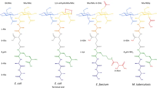
Figure 3. Common variations found in the peptidoglycan subunit of E. coli, E. faecium, and M.
tuberculosis.
The variations found in the peptidoglycan subunits, compared to that of E. coli, are shown in red. The terminal end of glycan chains in E. coli usually contains 1,6-anhydroMurNAc. The peptidoglycan subunit of E. faecium is O-acetylated on MurNAc and contains D-iGln and L-Lys substituted by D-iAsn. The subunit of M. tuberculosis is N-glycolylated on MurNAc and amidated on D-Glu and A2pm.
GlcNAc L-Ala D-Glu A2pm D-Ala D-Ala
MurNAc 1,6-anhydroMurNAc MurNAc-6-OAc
D-iGln L-Lys D-iAsn D-iGln A2pm-NH2 MurNGly E. coli E. coli
16
c) Translocation of Lipid II
After biosynthesis of lipid II on the cytoplasmic face of the cell membrane, it has to be transported or flipped across the membrane to the extracellular surface to be used for peptidoglycan polymerization (Figure 2B). Experiments carried out with fluorescently labeled peptidoglycan precursors showed that the rate of non-catalyzed flipping across the membrane was not sufficient to account for peptidoglycan synthesis (Weppner & Neuhaus, 1978). Two membrane proteins have been debated as being the flippase of E. coli, MurJ and FtsW.
MurJ, an essential protein in many bacteria, was first proposed as the lipid II flippase based on bioinformatics analyses (Inoue et al., 2008; Ruiz, 2008). It is a member of the MOP (Multidrug/Oligosaccharidyl-lipid/Polysaccharide) exporter superfamily (Hvorup et al., 2003). Depletion of MurJ in E. coli leads to defects in peptidoglycan synthesis, accumulation of peptidoglycan lipid-linked intermediates, and cell lysis (Inoue et al., 2008). A structural model of MurJ revealed the presence of a central, solvent-exposed cavity containing several charged residues, an essential structural motif for the function of MurJ as a flippase (Figure 4) (Butler et al., 2013). MurJ showed flippase activity in an in vivo assay, which did not reveal flippase activity for inactivated variants of MurJ or for FtsW (Sham et al., 2014). Lipid II titration experiments using mass spectrometry showed that MurJ had a higher affinity for lipid II than FtsW and that the vancomycin antibiotic (which binds to the terminal D-Ala4-D-Ala5 of the pentapeptide stem) forms a ternary complex with MurJ bound to
lipid II (Bolla et al., 2018). In a proposed model, lipid II binds to MurJ when it is in an inward-facing conformation, which causes MurJ to change its conformation to an outward-facing one, thus flipping lipid II to the extracytoplasmic surface (Figure 4) (Kuk et al., 2017). The membrane potential is necessary for its activity since membrane depolarization locks MurJ in the outward-facing conformation, thus blocking lipid II transport (Rubino et al., 2018). Recent studies have demonstrated that MurJ has both a lateral and a midcell localization and that its midcell localization depends on the availability of its substrate, lipid II, and on peptidoglycan polymerization at midcell (Liu et al., 2018).
FtsW is a member of the Shape, Elongation, Division, and Sporulation (SEDS) protein family. An in vitro fluorescence assay revealed that compared to MurJ, purified FtsW promoted translocation of lipid II analogues in membrane vesicles (Mohammadi et al., 2011). However, as mentioned above, results published since have shown evidence attributing this function to MurJ. An alternate role as a peptidoglycan polymerase has been identified for FtsW (Taguchi et al., 2019). Recent studies have demonstrated that FtsW (and its homolog, RodA) catalyze the synthesis of glycan strands (Meeske et al., 2016). The first structure of a SEDS protein was recently published for RodA, showing that it lacks a transmembrane channel that would be necessary for lipid II translocation (Sjodt et al., 2018). The
17 polymerase activity of FtsW and RodA is further discussed in Chapter 1.2.2b. However, the possibility that FtsW could act as both a peptidoglycan polymerase and a flippase has not been categorically ruled out.
In Gram-positive Bacillus subtilis mutants lacking MOP family members, a previously uncharacterized non-MOP protein, Amj, has been shown to also possess flippase activity (Meeske et al., 2015). Amj was able to functionally replace MurJ in E. coli.
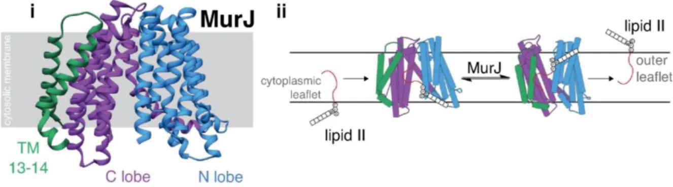
Figure 4. Structure of MurJ and the conformational change required for its activity as a flippase. (i) Structure of MurJ from Thermosipho africanus (PDB 5T77) in its inward-facing conformation. The N lobe (blue), the C lobe (purple), and transmembrane folds 13-14 (green) are organized in an N-shaped architecture. (ii) The proposed mechanism for lipid II flipping consists of a change of conformation upon substrate binding from an N-shaped inward-facing conformation to a V-N-shaped outward-facing conformation. From reference (Caveney et al., 2018).
1.2.2 Peptidoglycan Polymerization
Once lipid II is flipped to the extracytoplasmic surface of the cell membrane, the peptidoglycan disaccharide-pentapeptide subunit can be incorporated into the peptidoglycan network. Polymerization of glycan chains and cross-linking of peptide stems are catalyzed by glycosyltransferases and transpeptidases, respectively (Figure 5). Both activities are essential for peptidoglycan synthesis.
Some enzymes only possess glycosyltransferase or transpeptidase activity while others possess both activities: class A Penicillin-Binding Proteins (aPBPs) possess both a glycosyltransferase and a transpeptidase domain while monofunctional glycosyltransferases and class B PBPs (bPBPs) possess only glycosyltransferase and transpeptidase activity, respectively. Transglycosylation can proceed independently from transpeptidation but transpeptidation does not occur without prior or simultaneous transglycosylation, suggesting that peptidoglycan polymerization essentially starts with glycan chain elongation (van Heijenoort, 2001a). The newly formed glycan chains are subsequently incorporated into the preexisting peptidoglycan through the formation of cross-links between the peptide stems of the glycan strands.
18 In addition to the classical glycosyltransferases, aPBPs and monofunctional glycosyltransferases, conclusive evidence has recently emerged demonstrating that the SEDS proteins also possess glycosyltransferase activity (Emami et al., 2017; Meeske et al., 2016). Likewise, aPBPs and bPBPs are not the only enzymes capable of catalyzing the formation of cross-links between peptide stems. The L,D-transpeptidases (LDTs) constitute a structurally distinct transpeptidase family that catalyzes the synthesis of peptide cross-links (Figure 5).
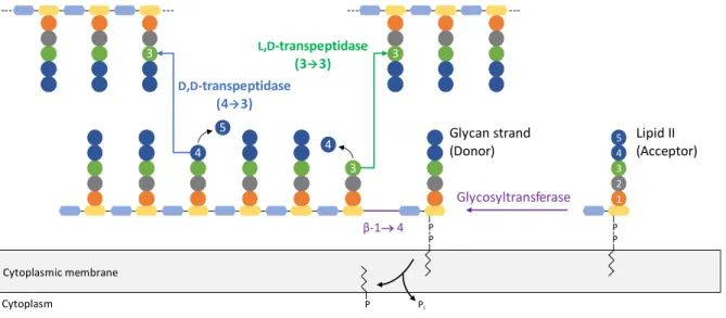
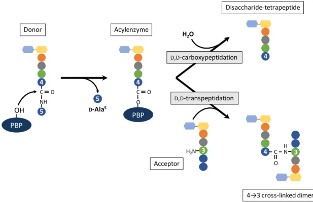
Figure 5. Peptidoglycan polymerization scheme.
Once lipid II is flipped across the cytoplasmic membrane, it is polymerized into growing glycan strands by glycosyltransferases that catalyze the formation of a β-1→4 glycosidic bond (purple) between GlcNAc of lipid II and MurNAc of the glycan chain. This reaction frees the phospholipid moiety of the glycan chain. Newly formed glycan strands are incorporated into the existing peptidoglycan by formation of peptide cross-links. D,D -transpeptidases use a pentapeptide donor substrate and catalyze the formation of 4→3 cross-links (blue) between the peptide stems. L,D-transpeptidases use a tetrapeptide donor substrate and catalyze the formation of 3→3 cross-links (green) between the peptide stems.
Peptidoglycan synthesis has to be spatially and temporally regulated for bacterial cell shape to be maintained. Rod-shaped bacteria possess two multi-protein complexes to direct peptidoglycan polymerization to specific regions of the cell (Carballido-Lopez & Formstone, 2007; Ricard & Hirota, 1973). As their names imply, the elongasome (or Rod system) and the divisome ensure peptidoglycan synthesis during bacterial elongation and division, respectively (Typas et al., 2011). Each machinery is coordinated by a cytoskeletal protein, actin-like MreB or tubulin-like FtsZ (Bi & Lutkenhaus, 1991; Jones et al., 2001). MreB assembles into short filaments that move the proteins responsible required for lateral peptidoglycan synthesis in a circumferential manner along the long axis of rod-shaped bacteria (Garner et al., 2011). FtsZ polymerizes to form the Z-ring and recruits the protein complex responsible for septal peptidoglycan synthesis prior to separation of daughter cells (den Blaauwen et al., 2017).
Growth of the peptidoglycan network not only requires the glycosyltransferase and transpeptidase activities of synthases but also hydrolytic enzymes, which have an important role to play in the maintenance and remodeling of peptidoglycan (Heidrich et al., 2001) (Singh et al., 2012). The
D,D-transpeptidase (43) Glycosyltransferase β-1 4 L,D-transpeptidase (33) Pi P P 1 2 3 4 5 P P P 3 3 4 4 5 3 Lipid II (Acceptor) Glycan strand (Donor) Cytoplasmic membrane Cytoplasm
19 peptidoglycan hydrolases consist of lytic transglycosylases, amidases, endopeptidases, and carboxypeptidases. The activity of these enzymes has to be regulated to prevent uncontrolled peptidoglycan hydrolysis, which can lead to cell lysis.
From the peptidoglycan synthases to the hydrolases, bacteria contain multiple, redundant or semi-redundant enzymes for each reaction (Typas et al., 2011). This is in sharp contrast to the cytoplasmic steps of peptidoglycan synthesis where each reaction is catalyzed by only one enzyme. Compared to the cytoplasm, the periplasm is less buffered and consequently more affected by changes in the environment. The higher redundancy for the extracytoplasmic steps could thus reflect the need for an efficient adaptation to changing environmental conditions, such as pH, osmolality, ionic concentration, and the presence of peptidoglycan-targeting antibacterial agents (Peters et al., 2016).
a) Glycosyltransferases
Glycosyltransferase modules exist either as monofunctional enzymes or as the N-terminal domain of bifunctional aPBPs (Goffin & Ghuysen, 1998; Wang et al., 2001). Class A PBPs have traditionally been considered as the main glycosyltransferases since monofunctional glycosyltransferases are not widely conserved and are not essential in most bacteria (Denome et al., 1999; Reed et al., 2011). Most glycosyltransferases belong to the GT51 family according to the carbohydrate-active enzymes (CAZy) database. They contain about 200 amino acids, which are mostly arranged in alpha helices that form a globular domain (Figure 6) (Sauvage & Terrak, 2016). The domain is divided into two sub-domains, the “head” and “jaw” regions, by a cavity containing the active site. The “jaw” region contains a hydrophobic surface for its partial burial in the cell membrane. Glycosyltransferases possess five signature motifs, of which motif 1 contains the catalytic Glu residue (Goffin & Ghuysen, 1998) (Terrak & Nguyen-Disteche, 2006).
20 Figure 6. Structure of the monofunctional glycosyltransferase MtgA of S. aureus.
Two structures of MtgA, PDB 3VMR (yellow) and PDB 3HZS (red), are superimposed (Heaslet et al., 2009; Huang et al., 2012). The glycosyltransferase inhibitor, moenomycin (green) and a lipid II analogue (cyan) are represented as sticks in the donor and acceptor sites, respectively. The mobile region between the donor and acceptor sites is encircled. The protein is anchored in the cell membrane via its N-terminal helix. From reference (Sauvage & Terrak, 2016).
Glycan strand synthesis is initiated by the transfer of the disaccharide-peptide unit from one lipid II to another. Elongation of glycan chains is sequential and proceeds through several rounds of transglycosylation since each reaction adds two sugar units (GlcNAc and MurNAc) to the growing glycan strand. Glycosyltransferases catalyze the formation of a β-1→4 glycosidic bond between C1 of
MurNAc of the growing glycan strand (i.e. its reducing end), which acts as the donor, and C4 of GlcNAc
of lipid II, which is the acceptor (Figure 5) (Ward & Perkins, 1973) (Perlstein et al., 2007). Two sub-sites have been proposed for fixation of the donor and acceptor substrates (Figure 6) (Lovering et al., 2007). Studies on the specificity of the donor and acceptor sites have demonstrated that the length of the lipid chain of the donor substrate is crucial. A lipid II mimic with a length of ten carbon atoms (or less) is not used as substrate and a length of thirty-five carbon atoms is required to produce a glycan strand of similar length as the natural substrate (Perlstein et al., 2010; Ye et al., 2001). The acceptor site is less stringent since substrate analogues containing a lipid chain of at least twenty carbon atoms are accepted. Binding of substrate analogues in the acceptor site shows allosteric activation of the donor site and increases the binding affinity of an inhibitor, moenomycin, to the donor site, suggesting a cooperative mechanism between the two sites (Bury et al., 2015). After each round of transglycosylation, the growing glycan chain, containing n+1 disaccharide units, must move from the acceptor site to the donor site to allow binding of a new lipid II in the acceptor site. Translocation of
21 the glycan chain could be facilitated by a protruding and mobile region located between the donor and acceptor sites (Figure 6) (Sauvage & Terrak, 2016). Transglycosylation frees the C55-PP moiety of lipid
II, which is flipped back to the cytoplasmic face of the cell membrane to be recycled and used in the formation of lipid I (van Heijenoort, 2001b).
b) SEDS Proteins
Monofunctional glycosyltransferases and aPBPs are not the only enzymes capable of catalyzing the polymerization of glycan strands. Deletion of all aPBPs leads to growth defects but is not fatal for B. subtilis, which does not naturally produce monofunctional glycosyltransferases, suggesting that unidentified glycosyltransferase(s) collaborating with bPBPs, which are monofunctional transpeptidases, could ensure peptidoglycan polymerization (McPherson & Popham, 2003). A similar observation was made in E. faecalis, which also lacks monofunctional glycosyltransferases. E. faecalis is viable and resistant to moenomycin (a glycosyltransferase inhibitor) in the absence of aPBPs, suggesting that moenomycin does not target the unidentified glycosyltransferase(s) (Arbeloa et al., 2004). Overproduction of PBP2, a bPBP, and the SEDS protein RodA was shown to be sufficient for peptidoglycan synthesis in membrane extracts from an E. coli strain whose aPBPs had been inhibited by antibiotics (Ishino et al., 1986). While the transpeptidase activity required for peptidoglycan synthesis was clearly shown to be due to PBP2, it was less clear for the glycosyltransferase activity. Based on their knowledge of PBPs at the time, the authors thought that RodA was unlikely to be a glycosyltransferase and postulated that it was instead a regulator of PBP2, which would be responsible for both the transpeptidase and the glycosyltransferase activities or that PBP2 and RodA formed a complex, which would then act as a bifunctional transpeptidase-glycosyltransferase. Thirty years went by before the mystery was solved.
No earlier than 2016, RodA and its homolog FtsW, ubiquitous members of the SEDS family, were shown to possess glycosyltransferase activity (Cho et al., 2016; Emami et al., 2017; Henrichfreise et al., 2016; Meeske et al., 2016; Taguchi et al., 2019). RodA and FtsW share highly conserved folds and catalytic residues. Overexpression of RodA in B. subtilis lacking aPBPs attenuates growth defects and leads to an increase in glycosyltransferase activity (Meeske et al., 2016). Purified RodA also shows glycosyltransferase activity, which is not inhibited by moenomycin. Depletion of RodA in B. subtilis leads to enlarged and spherical cells while depletion of FtsW results in elongated filamentous cells (Boyle et al., 1997; Gamba et al., 2016; Henriques et al., 1998; Kobayashi et al., 2003). SpoVE, another SEDS protein encoded by B. subtilis, is not essential for growth but is necessary for peptidoglycan synthesis of the spore cortex (Fay et al., 2010; Henriques et al., 1992).
22 Both RodA and FtsW form glycosyltransferase-transpeptidase cognate pairs with bPBPs (Figure 7). The RodA-PBP2 and FtsW-PBP3 pairs have been demonstrated to form the core synthases of the elongasome and divisome, respectively, and to be more conserved among bacterial species than aPBPs, opposing the widely held idea that the aPBPs are the main peptidoglycan synthases in bacteria (Meeske et al., 2016). Single molecule tracking experiments in E. coli revealed that RodA-PBP2 displayed circumferential motion along the long axis, similar to the elongasome protein MreB (Garner et al., 2011). In contrast, aPBPs showed distinct localization dynamics from the elongasome and were not essential for lateral peptidoglycan synthesis (Cho et al., 2016; Meeske et al., 2016).
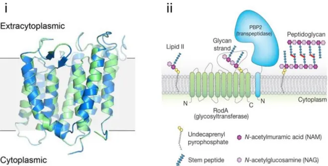
The first crystal structure of a SEDS protein was recently obtained using evolutionary covariation analysis for fold prediction since conventional methods, such as molecular replacement, which requires structural data of a homologous protein, and heavy-atom phasing methods were not applicable (Ovchinnikov et al., 2015; Sjodt et al., 2018). Evolutionary coupling analyses are based on the premise that pairs of residues interacting with each other will co-evolve to preserve their interactions. The crystal structure of RodA from Thermus thermophiles shows the presence of ten transmembrane folds, large extracellular loops and short intracellular loops (Figure 7). The transmembrane domain contains a highly conserved cavity, which was demonstrated to be essential for the polymerase activity of RodA by mutational studies in B. subtilis and E. coli (Meeske et al., 2016). The cavity is next to a hydrophobic groove, which could contain the substrate binding sites of RodA. The extracellular loops also contain catalytically essential residues. Strong evolutionary couplings of RodA and bPBP residues predicted a transmembrane binding interface for their interaction, which has been validated by mutagenesis studies for the FtsW-PBP3 pair (Fraipont et al., 2011; Leclercq et al., 2017).
RodA and FtsW use a similar transglycosylation mechanism as classical glycosyltransferases. They use reducing end addition for glycan chain elongation, where the growing glycan chain acts as donor and lipid II as acceptor (Welsh et al., 2019). Lipid length was found to be a requirement for substrate binding at the donor and acceptor sites with binding at the donor site requiring a minimum lipid length of thirty carbon atoms and the acceptor site tolerating shorter lipid chains.
23 Coordination of the transglycosylase activity of SEDS proteins and the transpeptidase activity of bPBPs is critical to prevent the toxic accumulation of uncross-linked glycan chains (Cho et al., 2014). A regulation mechanism of RodA by its cognate bPBP was unveiled in E. coli, in which the peptidoglycan polymerase activity of RodA-PBP2 complexes was shown to be stimulated by PBP2 variants (Rohs et al., 2018). The amino acid substitutions in these variants bypassed the requirement for MreC, another protein belonging to the elongasome, by mimicking the conformational changes induced by interaction of PBP2 with MreC. The PBP2 variants also promote the formation of MreB filaments, leading to an enhancement in the formation of active elongasome complexes (Hussain et al., 2018). Two modes of regulation of SEDS proteins by bPBPs have been identified in S. aureus (Reichmann et al., 2019). Although cocci lack an elongasome, S. aureus possesses both FtsW and RodA. The FtsW-PBP1 pair was shown to be required for inward septal peptidoglycan synthesis and for stabilization of the divisome while RodA-PBP3 promoted sidewall peptidoglycan synthesis at the future division site. The first mode of regulation concerns PBP1, which was shown to be essential not because of its transpeptidase activity but rather because it stimulates the essential glycosyltransferase activity of FtsW (Taguchi et al., 2019). The second mode of regulation concerns PBP3, which is not only required for localization of RodA at midcell but is also expected to modulate its activity.
Figure 7. Structure of RodA and model of peptidoglycan synthesis by the RodA-PBP2 pair.
(i) Superposition of the crystal structures of wild-type RodA (green) and RodA D255A (blue) (PDB 6BAR and 6BAS, respectively). (ii) A new model has emerged in which SEDS proteins form a complex with their cognate bPBPs for peptidoglycan polymerization. Shown here is RodA in complex with PBP2. FtsW forms a similar complex with its cognate bPBP. Not shown are the other elongasome proteins that ensure directional peptidoglycan synthesis. From reference (Sjodt et al., 2018).
Studies on Listeria monocytogenes and B. subtilis showed that expression of the SEDS proteins could be induced in response to stresses targeting the cell wall, such as treatment by β-lactams or
24 moenomycin (Emami et al., 2017; Meeske et al., 2016; Rismondo et al., 2019). L. monocytogenes encodes two FtsW and three RodA homologs. Of these, FtsW2 and RodA3 are part of the same operon, which is only minimally expressed under standard laboratory conditions (Lobel & Herskovits, 2016). Its expression is regulated by CesRK, a two-component system, which is activated by cell envelope stress, such as the presence of cell wall-targeting antibiotics and lysozyme (Kallipolitis et al., 2003). Indeed, expression of FtsW2 and RodA3 was induced in the presence of β-lactam antibiotics and moenomycin, which respectively inactivate the transpeptidase and glycosyltransferase activities of PBPs. Similarly, deletion of aPBPs in B. subtilis led to a dependence on the alternative sigma factor, SigM for survival; SigM-mediated expression of RodA is necessary and sufficient for viability and for the natural resistance of B. subtilis to moenomycin (Emami et al., 2017; Meeske et al., 2016; Salzberg et al., 2011).
The essential roles of RodA and FtsW in nearly all bacteria make them promising targets for antibiotics. A chemical screen of extracts from various actinomycete strains has led to the identification of a potential RodA inhibitor that showed antibacterial activity against B. subtilis depleted of aPBPs and acted synergistically with moenomycin against wild-type cells (Emami et al., 2017). Further work is required to elucidate the structure of the inhibitor.
c)
D
,
D
-transpeptidases
D,D-transpeptidases catalyze formation of cross-links between the peptide stems of adjacent glycan chains. In E. coli, it is estimated that around 40 to 60% of the total number of peptides is cross-linked (Glauner et al., 1988). Most bacteria contain 43 cross-links, which are formed between D-Ala at position 4 of the donor peptide and A2pm (in E. coli) at position 3 of the acceptor peptide (Figure 5)
(Schleifer & Kandler, 1972). The donor substrate has to contain a pentapeptide stem because in the first step of the reaction, cleavage of the D-Ala4-D-Ala5 bond provides the necessary energy for
formation of the cross-link (Terrak et al., 1999). The synthesis of 43 cross-links is catalyzed by the D,D-transpeptidase activity of bifunctional aPBPs or monofunctional bPBPs (detailed in Chapter 4). Depending on the growth phase, PBPs are responsible for the synthesis of 87% (stationary phase) to 94% (exponential phase) of all cross-links in E. coli (Pisabarro et al., 1985).PBPs were first identified by their ability to covalently bind penicillin and other β-lactams (Suginaka et al., 1972). The β-lactam antibiotics are discussed in Chapter 2.
d)
L
,
D
-transpeptidases
A second type of cross-links, formed between the A2pm3 residues of a donor and an acceptor peptide,
25 et al., 1988; Pisabarro et al., 1985; Quintela et al., 1995). The proportion of 33 cross-links in E. coli increases from 6% during exponential growth to 13% in the stationary phase (Pisabarro et al., 1985). Some pathogenic bacteria, such as M. tuberculosis, contain a higher proportion (60% to 80%) of 33 cross-links than 43 cross-links in their peptidoglycan in all phases of growth (Kumar et al., 2012; Lavollay et al., 2008). The enzymes responsible for the synthesis of 33 cross-links, the L,D -transpeptidases (LDTs), were first identified in the early 2000s by the host laboratory. They form a new family of peptidoglycan synthases. LDTs are structurally distinct from PBPs and their catalytic residue is a cysteine instead of a serine. Moreover, the substrate requirement for LDTs is different from PBPs since they require a tetrapeptide instead of a pentapeptide stem. LDTs are inhibited only by the carbapenem class of β-lactams. LDTs are further detailed in Chapter 5.
e) Peptidoglycan Hydrolases
Peptidoglycan hydrolases include lytic transglycosylases, amidases, carboxypeptidases, and endopeptidases. They have a non-negligible role in the formation of a robust and stable peptidoglycan network. Hydrolases are particularly important for the insertion of new material into the peptidoglycan layer while maintaining its thickness. Their activity has to be tightly regulated in order to prevent uncontrolled hydrolysis of peptidoglycan, which ultimately leads to cell lysis. One hypothesis regarding the regulation of the hydrolytic activity of these enzymes is that they exist in multi-enzyme complexes that also contain peptidoglycan synthases (Holtje, 1998). Hydrolases have also been shown to be regulated by proteins, either other hydrolases or non-catalytic proteins that act as their cognate regulators (Morlot et al., 2010; Uehara et al., 2009).
Lytic transglycosylases catalyze the cleavage of the glycosidic bond between MurNAc and GlcNAc in glycan strands, leading to the concomitant formation of disaccharide-peptides with a 1,6-anhydro bond on MurNAc (Holtje et al., 1975). Amidases catalyze the cleavage of the bond connecting peptide stems to MurNAc (Vollmer et al., 2008b). Carboxypeptidases catalyze the hydrolysis of the terminal ends of peptide stems. Endopeptidases catalyze the hydrolysis of amide bonds present inside peptide stems or of PBP- or LDT-catalyzed cross-links.
E. coli encodes around twenty hydrolases, which include multiple enzymes for each hydrolytic activity. None of these is essential for cell viability with mutants depleted of several hydrolases displaying mild phenotypes or none at all. However, some of the hydrolases have defined functions. The AmiA, AmiB, and AmiC amidases and their EnvC and NlpD regulators have redundant roles in septal peptidoglycan cleavage, which is essential for separation of daughter cells. Cells depleted of all three amidases grow as long unseparated chains, a phenotype that is exacerbated upon additional depletion of lytic
26 transglycosylases and endopeptidases (Heidrich et al., 2001). Cell elongation also necessitates hydrolases since deletion of the genes encoding the redundant endopeptidases, MepS, MepM, and MepH, which specifically cleave 43 cross-links, leads to inhibition of peptidoglycan synthesis, followed by cell lysis (Singh et al., 2015; Singh et al., 2012). Another endopeptidase, MepK, was recently shown to specifically hydrolyze LDT-catalyzed 33 cross-links (Chodisetti & Reddy, 2019). The carboxypeptidase activity of the class C PBP, PBP6b, which has no significant role under standard growth conditions, becomes important for normal morphology and growth in acidic conditions (see Chapter 4) (Peters et al., 2016). The expression, stability, and activity of the protein increase at pH 5. Similarly, a shift of temperature from 37°C to 30°C revealed an increase in activity for a lytic transglycosylase, MltA (Lommatzsch et al., 1997).
Peptidoglycan fragments released by the catalytic activities of the hydrolases can be recycled in the cytoplasm. Uptake from the periplasm requires the Opp and Mpp permeases in E. coli. In the cytoplasm, the fragments are further hydrolyzed by various enzymes for re-entry into the peptidoglycan biosynthetic pathway (Uehara et al., 2005). In addition, peptidoglycan turnover products can act as signals for the expression of chromosomally encoded β-lactamases, enzymes that catalyze the degradation of β-lactam antibiotics (see Chapter 3) (Jacobs et al., 1997). Inhibition of the transpeptidation of glycan strands by β-lactams can also induce turnover of nascent peptidoglycan by lytic transglycosylases, such as Slt (Cho et al., 2014). This induces a futile cycle of peptidoglycan synthesis and degradation, which depletes the pool of peptidoglycan precursors and contributes to the bactericidal activity of β-lactams.
In M. tuberculosis, RpfB (or Resuscitation Promoting Factor B) is a lytic transglycosylase, which is able to stimulate “resuscitation” of M. tuberculosis from dormancy (Kana & Mizrahi, 2010). RpfB interacts with RipA (or Rpf interacting protein A), an essential endopeptidase that catalyzes the hydrolysis of the amide bond between D-iGln2 and A
2pm3 of the peptide stem (Sassetti et al., 2003). Their concerted
activity leads to the formation of a 1,6-anhydro-disaccharide-dipeptide, which was shown to be the minimal structure required for resuscitation of dormant cells (Nikitushkin et al., 2015).
27
1.3 Antibiotics Targeting Peptidoglycan Synthesis
Peptidoglycan being an essential and unique component of most bacterial cells, its biosynthetic pathway is a validated target for the development of antibiotics. An overview of the most common peptidoglycan-targeting antibiotics is presented here.
Among antibiotics targeting the synthesis of peptidoglycan precursors are the natural products, fosfomycin and D-cycloserine (Barreteau et al., 2008). Fosfomycin targets the first Mur enzyme in the peptidoglycan biosynthetic pathway, MurA, which catalyzes the formation of UDP-GlcNAc-enolpyruvate, the precursor of UDP-MurNAc. D-cycloserine acts as a competitive inhibitor of two enzymes, Alr and Ddl, which catalyze the formation of D-Ala by racemization of L-Ala and the condensation of two D-Ala molecules to form a dipeptide, respectively (Prosser & de Carvalho, 2013). D-cycloserine is a second-line antibiotic used in the treatment of drug-resistant tuberculosis.
The formation of the lipid precursor is targeted by bacitracin, a mixture of natural cyclic polypeptides, which sequesters the undecaprenyl pyrophosphate (C55-PP) lipid carrier and thus prevents its
dephosphorylation to undecaprenyl phosphate (C55-P)(Stone & Strominger, 1971).
Lipid II can also be sequestered by a number of antibiotics. Glycopeptides, such as vancomycin, bind to the terminal D-Ala4-D-Ala5 end of the pentapeptide stem of lipid II (Kahne et al., 2005). Nisin, a
lantibiotic, has a dual mode of action since it binds to the pyrophosphate moiety of lipid II and it targets lipid II molecules to form pores in the cytoplasmic membrane (Hsu et al., 2004). Ramoplanin is a cyclic glycodepsipeptide, which binds to the pyrophosphate moiety and MurNAc of lipid II (Hamburger et al., 2009).
Polymerization of glycan strands can be inhibited by moenomycin, which is not clinically used due to poor pharmacokinetic properties (Ostash & Walker, 2010). This molecule does not inhibit the glycosyltransferase activity of SEDS proteins.
The transpeptidases responsible for cross-linking of glycan strands are inhibited by β-lactams. These antibiotics are discussed in the next chapter.
28
2. The β-lactam antibiotics
The β-lactams are the most successful and commonly used antibiotics against infectious diseases due to their broad antibacterial spectrum, oral availability, lack of toxicity, and good pharmacokinetic properties (Kong et al., 2010). Since the discovery of penicillin by Alexander Fleming in 1928 and its subsequent use in medicine in the 1940s, several new classes and generations of β-lactams have been developed and optimized for clinical use against infectious diseases.
2.1 Mode of Action of β-lactams
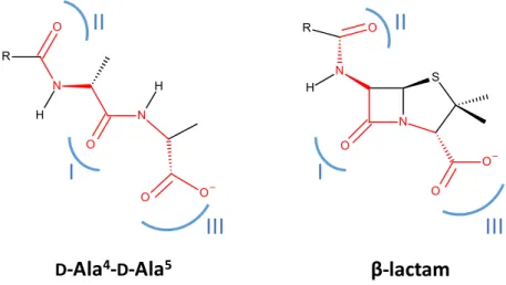
The mode of action of β-lactams was identified about forty years after the discovery of penicillin(Park & Strominger, 1957; Tipper & Strominger, 1965). β-lactams are suicide inhibitors of the Penicillin-Binding Proteins (PBPs), whose inactivation leads to cell death. PBPs are efficaciously inactivated by β-lactams through the formation of a stable acylenzyme between the catalytic serine of PBPs and the carbonyl moiety of the β-lactam ring. This reaction is thought to occur because of the analogy between the structure of the β-lactam ring and that of the D-Ala4-D-Ala5 end of the peptidoglycan precursor
(Tipper & Strominger, 1965). The similarity between β-lactams and the natural substrates of PBPs resides in the distribution of electrons in the β-lactam ring, which is proposed to mimic the distribution of three electrostatic-negative wells at the terminal D-Ala4-D-Ala5 of the natural substrate of PBPs
(Figure 8).
Figure 8. Analogy between the natural substrate of PBPs and β-lactams.
Although the terminal D-Ala4-D-Ala5 end of the pentapeptide substrate of PBPs and penams have a common
backbone (red), the analogy between the peptidoglycan precursor and β-lactams lies in a similar distribution of electrons at positions I, II, and III (blue).
I
II
III
I
II
β-lactam
D-Ala
4-
D-Ala
5III
29 In contrast to the reaction with their natural substrate, in which the enzyme is liberated in the final step, acylation of the active-site serine of PBPs by β-lactams is stable, leading to inactivation of the enzyme. The chain of events from PBP inhibition to cell lysis and bacterial death is not clearly understood and is under investigation in various bacteria (Cho et al., 2014; Chung et al., 2009; Giesbrecht et al., 1998). Loss of catalytic activity of essential PBPs has been proposed to result in an imbalance in peptidoglycan metabolism due to uncontrolled hydrolysis of the peptidoglycan network by hydrolases, which would lead to a loss of cell wall integrity and ultimately, to cell lysis (Cho et al., 2014; Park & Strominger, 1957; Tomasz & Waks, 1975).
2.2 Classes of β-lactams
The β-lactam antibiotics in clinical use are natural, semi-synthetic, or synthesized compounds (Bush & Bradford, 2016). The core structure of β-lactams is the four-membered azetidin-2-one (β-lactam) ring. β-lactams are divided into several classes based on their β-lactam-adjacent ring. The most commonly used β-lactams are the penams, cephems, carbapenems, penems, monobactams, and clavams (Table 1).
30 Table 1. Structures of the most common β-lactam classes and of one representative of each class.
β-lactam class Structure Side-chain(s) of a representative
Penam Ampicillin Cephem Ceftriaxone Carbapenem Imipenem Penem Faropenem Monobactam Aztreonam Clavam Clavulanic acid 4 3 2 1 5 6 4 3 2 1 5 6 7 4 3 2 1 5 6 4 3 2 1 5 6 4 3 2 1 5 6 R = R1 = R = R = R2 = R1 = R2 = R1 = R2 =
31
Penams
Penams, also known as penicillins, contain a thiazolidine ring, which is saturated and is composed of five atoms including sulfur at position 4. Various penams have been obtained by semi-synthesis by modifying the side-chain of the core penam structure, 6-aminopenicillanic acid (6-APA), which is composed of the β-lactam and thiazolidine rings and a primary amine R group(Dalhoff & Thomson, 2003). Some penams, known as amidinopenicillins, such as temocillin, contain a methoxy group at position 6 (instead of a hydrogen atom) in an attempt to prevent hydrolysis of the lactam ring by β-lactamases (Slocombe et al., 1981).
Cephems
Cephems, also known as cephalosporins, contain a dihydrothiazine ring, which has an unsaturation at C2-C3 and a sulfur at position 5(Dalhoff & Thomson, 2003). Cephems are the most abundant β-lactam class and the most widely used β-lactams in antibacterial therapy (Hamad, 2010). There are currently five generations of cephalosporins, which differ by their antibacterial spectrum. Most cephalosporins derive from the core 7-aminocephalosporinic acid (7-ACA) by semi-synthesis. Similarly to penams, some cephems, known as cephamycins, contain a methoxy group at position 7 instead of a hydrogen (Page, 2007). Although cephamycins, such as cefoxitin, were developed to prevent hydrolysis by β-lactamases, they can act as inducers of cephalosporinase (β-lactamases that hydrolyze cephalosporins) expression(Neu, 1986). Another downside of the methoxy group is that it reduces the antibacterial activity of cephamycins and amidinopenicillins against Gram-positive bacteria (Neu, 1974).
Carbapenems
Carbapenems contain a pyrroline ring, which is unsaturated at C2-C3 (Papp-Wallace et al., 2011). Carbapenems have certain structural characteristics that contribute to their antibacterial activity. Their side-chains are different from those found in penams and cephems. All carbapenems contain a hydroxyethyl group at position 6 of the β-lactam ring. The side-chain (R1) linked at position 3 of the pyrroline starts with a sulfur atom. The C5-C6 bond has a trans configuration in carbapenems, compared to the cis configuration of penams and cephems. Position 4 of the pyrroline is substituted by a methyl group (R2) for all carbapenems, except thienamycin and imipenem. Owing to their broad-spectrum activity and high potency, carbapenems are considered as last resort antibiotics. An inconvenience of carbapenems is that they can only be administered intravenously.
32
Penems
The β-lactam-adjacent ring of penems is similar to that of carbapenems except for a sulfur atom at position 4 (Dalhoff & Thomson, 2003). Compared to carbapenems, the faropenem penem can be administered orally (Schurek et al., 2007). (Carba)penems have shown potent antibacterial activity against M. tuberculosis both in vitro, in mice models, and in a clinical trial (Dhar et al., 2015; Diacon et al., 2016; Hugonnet et al., 2009; Kumar et al., 2012) (see Chapter 5.5.2).
Monobactams
Monobactams contain only one cycle, the β-lactam ring(Sykes et al., 1981). Aztreonam is the only clinically used monobactam (Bush & Bradford, 2016). Monobactams have a more restrained antibacterial spectrum than the other β-lactam classes. Aztreonam is one of the few β-lactams that has a highly preferred target among the PBPs. It preferentially targets the class B PBP, PBP3, which is present in the divisome of E. coli.
Clavams
Clavams are inhibitors of β-lactamases(Bush & Bradford, 2016). They are used in association with the other β-lactam classes to target β-lactamase-producing bacteria. Clavulanic acid is the only natural representative of the clavams (see Chapter 3.2.1).
2.3 Resistance to β-lactams
Bacteria have developed several resistance mechanisms to combat β-lactams (Figure 9)(Walsh, 2000). In Gram-negative bacteria, β-lactams have to cross the outer membrane in order to penetrate the periplasm to target the PBPs. β-lactams cross the outer membrane by using porins, which has led to the emergence of amino acid substitutions preventing β-lactam entry, reducing the permeability of the outer membrane(Cohen et al., 1988). Even if the β-lactam manages to reach the periplasm, it can be subjected to export by multidrug efflux pumps (Zgurskaya & Nikaido, 2000). The PBPs themselves can harbor substitutions (target modification) to alter the acylation efficacy by β-lactams (Goffin & Ghuysen, 1998). This is more common in Gram-positive bacteria, in which PBP-encoding genes are mutated or intrinsically resistant PBPs are expressed (see Chapter 4.3.2). In Gram-negative bacteria, the main resistance mechanism is the expression of β-lactamases, which rapidly hydrolyze β-lactams before they can inactivate their targets (Bush, 2018) (see Chapter 3). Bypass of the transpeptidase activity of PBPs by another enzyme family, the L,D-transpeptidases (LDTs), has been shown to confer
33 resistance to penams and cephems in laboratory-selected mutants of E. faecium and E. coli (Hugonnet et al., 2016; Mainardi et al., 2000) (see Chapter 5).
Figure 9. Mechanisms of β-lactam resistance in Gram-negative bacteria.
β-lactams penetrate Gram-negative bacteria by porins present in the outer membrane. β-lactam-susceptible bacteria (orange background) are killed due to inactivation of PBPs by β-lactams. Bacteria develop resistance (blue background) by reducing expression of porins (reduced permeability), by producing efflux pumps (antibiotic efflux), by expressing β-lactamases that efficiently hydrolyze β-lactams (antibiotic degradation), and by expressing “low-affinity” PBPs, which are not efficiently inactivated by β-lactams (target modification).
β-lactam Penicillin Binding Protein (PBP) Susceptibility
Reduced permeability
Antibiotic efflux
Antibiotic degradation Target modification
β-lactamase Efflux pump Porin Low-affinity PBP PBP Periplasm Cytoplasm