RESEARCH OUTPUTS / RÉSULTATS DE RECHERCHE
Author(s) - Auteur(s) :
Publication date - Date de publication :
Permanent link - Permalien :
Rights / License - Licence de droit d’auteur :
Bibliothèque Universitaire Moretus Plantin
Institutional Repository - Research Portal
Dépôt Institutionnel - Portail de la Recherche
researchportal.unamur.be
University of Namur
Validation of a chemiluminescent assay for specific SARS-CoV-2 antibody
Tré-Hardy, Marie; Wilmet, Alain; Beukinga, Ingrid; Dogné, Jean-Michel; Douxfils, Jonathan;
Blairon, Laurent
Published in:
Clinical Chemistry and Laboratory Medicine DOI:
10.1515/cclm-2020-0594
Publication date: 2020
Document Version Peer reviewed version
Link to publication
Citation for pulished version (HARVARD):
Tré-Hardy, M, Wilmet, A, Beukinga, I, Dogné, J-M, Douxfils, J & Blairon, L 2020, 'Validation of a
chemiluminescent assay for specific SARS-CoV-2 antibody', Clinical Chemistry and Laboratory Medicine, vol. 58, no. 8, pp. 1357-1364. https://doi.org/10.1515/cclm-2020-0594
General rights
Copyright and moral rights for the publications made accessible in the public portal are retained by the authors and/or other copyright owners and it is a condition of accessing publications that users recognise and abide by the legal requirements associated with these rights. • Users may download and print one copy of any publication from the public portal for the purpose of private study or research. • You may not further distribute the material or use it for any profit-making activity or commercial gain
• You may freely distribute the URL identifying the publication in the public portal ? Take down policy
If you believe that this document breaches copyright please contact us providing details, and we will remove access to the work immediately and investigate your claim.
Marie Tré-Hardy*, Alain Wilmet, Ingrid Beukinga, Jean-Michel Dogné, Jonathan Douxfils
and Laurent Blairon
Validation of a chemiluminescent assay for
specific SARS-CoV-2 antibody
https://doi.org/10.1515/cclm-2020-0594 Received April 26, 2020; accepted May 9, 2020
Abstract
Objectives: Faced with the COVID-19 pandemic and its
impact on the availability and quality of both therapeu-tic and diagnostherapeu-tic methods, the Belgian authorities have decided to launch a procedure for additional evaluation of the performance of serological tests offered for sale on the national territory. This has been proposed with a double aim: (1) an in-depth verification of the analytical and clini-cal performances presented by the manufacturer and (2) an economy of scale in terms of centralized validation for all the laboratories using the tests subject to evaluation.
Methods: A retrospective validation study was
con-ducted including the serum of 125 patients in order to determine the analytical and clinical performances of the LIAISON®SARS-CoV-2 from DiaSorin® detecting
anti-SARS-CoV-2 IgG and to compare its clinical performance with the enzyme-linked immunosorbent assay (ELISA) test from Euroimmun®, one of the first commercially
avail-able tests allowing the detection of anti-SARS-CoV-2 IgA and IgG.
Results: The performances of the LIAISON®
SARS-CoV-2 satisfied all the acceptance criteria and provided
“real world” analytical and clinical performances very close to the ones reported by the manufacturer in its insert kit. Comparison between the LIAISON®SARS-CoV-2 and
the ELISA method did not reveal any difference between the two techniques in terms of sensitivities and specifici-ties regarding the determination of the IgG.
Conclusions: This study reports the validation of the
LIAISON®SARS-CoV-2 allowing to detect IgG antibodies
specifically directed against SARS-CoV-2. The analytical and clinical performances are excellent, and the automa-tion of the test offers important rates, ideal for absorbing an extension of testing.
Keywords: CLIA; COVID-19; ELISA; SARS-CoV-2.
Introduction
On December 31, 2019, the World Health Organization (WHO) was alerted to the appearance of several cases of pneumonia of unknown origin in the city of Wuhan (China). Few weeks later, the pathogen causing this pneu-monia was identified: it is a new coronavirus called SARS-CoV-2, the associated disease being designated by the term COVID-19 [1]. Since then, a global health crisis has set in and the pandemic continues to grow: As of May 6, the virus has already spread to 187 countries and territories [2], the number of confirmed cases exceeds 3.6 million and the number of deaths worldwide stands at 243,401 [3]. The number of cases diagnosed, however, only reflects a frac-tion of the actual number of infecfrac-tions as a large number of countries only test severe cases.
The diagnosis of SARS-CoV-2 infection is essential for the control of the epidemic, the establishment of protective measures and the therapeutic management of patients. The WHO recommends detection of the viral genome in respiratory samples for the diagnosis COVID-19. However, even if RT-qPCR is considered the “gold standard”, many pre-analytical and analytical limitations have recently been described. First, it has been shown that the sensitivity of this method can vary depending on the quality of the sample, the stage but
*Corresponding author: Marie Tré-Hardy, Department of Laboratory
Medicine, Iris Hospitals South, rue Jean Paquot 63, 1050 Brussels, Belgium; Faculty of Medicine, Université libre de Bruxelles, Brussels, Belgium; and Department of Pharmacy, Namur Research Institute for Life Sciences, University of Namur, Namur, Belgium, Phone: +32 2 641 48 21, Fax: + 32 2 641 48 24,
E-mail: [email protected]
Alain Wilmet, Ingrid Beukinga and Laurent Blairon: Department of
Laboratory Medicine, Iris Hospitals South, Brussels, Belgium
Jean-Michel Dogné: Department of Pharmacy, Namur Research
Institute for Life Sciences, University of Namur, Namur, Belgium
Jonathan Douxfils: Department of Pharmacy, Namur Research
Institute for Life Sciences, University of Namur, Namur, Belgium; and Qualiblood sa, Namur, Belgium. https://orcid.org/0000-0002-7644-5298 (J. Douxfils)
2 Tré-Hardy et al.: Validation of a chemiluminescent assay for specific SARS-CoV-2 antibody
also the severity of the disease, leading to approximately 20% of false-negative cases [4–7].
Furthermore, access to RT-qPCR tests remains limited. Despite significant efforts in Belgium to increase the number of RT-qPCR testing, this method will not alone cover rapid and massive screening of the population.
Considering the health emergency, many cheaper and convenient serological tests have rapidly been developed and continue to reach the market. To date, more than 183 different CE marked tests have been identified, including 131 rapid tests [8]. The pandemic having impacted both the usual distribution chains and the commercial offer, the risk of fraud and the release of products of question-able or fluctuating quality have significantly increased, leading to urgent appropriate measures by the competent authorities to control the market [9]. In order to improve the quality and provide health care professionals with clearer information of the current commercial offer, the Belgian competent national authority has therefore decided to introduce an additional step allowing tests to benefit from a specific recommendation. The participa-tion of the manufacturers to this addiparticipa-tional step is on a voluntary basis.
Three categories of immunoassays exist and allow, depending on the tests, to detect the presence of IgG, IgA and IgM in response to a SARS-CoV-2 infection: automated CLIA (chemiluminescent immunoassay) tests, ELISA (enzyme-linked immunosorbent assay) tests manual or automated and finally rapid immunochromatographic tests. Many hopes are based on these serological tests which could play a complementary diagnostic role to the RT-qPCR and help in answering several questions such as the deconfinement of patients according to their degree of immunity, the discrepancy of the results between CT scan and RT-qPCR, the evaluation of individual and col-lective immunity and allow the carrying out of large-scale epidemiological analyses. However, the lack of enough documentation of the current comparative studies does not allow a proper evaluation of the analytical and clini-cal performances of these different serologiclini-cal tests and to date, a serological reference method is still lacking.
This study carries an important epidemiological objective but is also part of the deconfinement strategy led by the testing group of the task force deployed in Belgium to manage the current health crisis. The validation of the CLIA test (LIAISON®SARS-CoV-2 IgG kit, DiaSorin®,
Saluggia, Italy) described in this study responds to an official request made by this group of experts. The CLIA test (DiaSorin®), which recently obtained CE marking
(04/17/2020), has another major advantage: there is already a large implementation of LIAISON XL® analyzer
(DiaSorin®) in Belgium (n = 81), allowing the test to be
carried out on the whole territory. The choice of automa-tion is preferable to respond to the large number of testing needed in the next weeks.
The main objective of this study is to evaluate and compare the clinical performance of the LIAISON®
SARS-CoV-2 IgG kit detecting anti-SARS-SARS-CoV-2 IgG with the ELISA test (Euroimmun Medizinische Labordiagnos-tika®, Lübeck, Germany) allowing the detection of
anti-IgA and IgG SARS-CoV-2. This study is also the first national validation model described to date and the first study to report the performance of the LIAISON®
SARS-CoV-2 IgG kit.
This validation will allow the routine implementation of a serological test in all Belgian laboratories by rational-izing its use for clinical purposes and sparing heavy vali-dation steps consuming time, samples and reagents.
Materials and methods
Study design
This retrospective study was conducted from April 16 to 20, 2020 at the clinical biology laboratory of the Iris Sud Hospitals (HIS-IZZ, Brussels, Belgium). All the sera (n = 125) originate from blood sam-ples taken during previous clinical requests for diagnostic purposes and were stored in the laboratory serum biobank at ≤− 20 °C. Among these 125 samples, 81 samples were included in the specificity sis. The remaining 44 samples were included in the sensitivity analy-sis. This study has been approved by the Ethical Committee of the HIS-IZZ (ethical agreement number: CEHIS/2020-13).
Population
Blood samples positive for COVID-19 were collected from patients with mild, severe or critical infection based on the extent of anomalies observed on CT scan: moderate (10%–25%), extensive (25%–50%), severe (>50%) or critical >75% and on clinical symptoms (headache, fever, fatigue, cough and sore throat, myalgia, shortness of breath or digestive signs). Patients were considered positive according to the results of the RT-qPCR. The delay between the first onset of symp-toms and the RT-qPCR is variable but has been estimated at 4 days (±1 day) in our cohort.
Sample collection: Blood samples were collected in serum
col-lection tubes (BD Vacutainer SST II advance, BD, Plymouth, UK) according to standardized operating procedure. Samples were then centrifuged at 3500 rpm (2451 g) for 10 min. Serum was then col-lected, and samples were analyzed as soon as possible. In case the analyses were delayed, samples were aliquoted and stored between 2 and 8 °C for a maximum of 3 days. If the storage was higher than 3 days, serum sample were stored at ≤− 20 °C. If samples were stored
for a longer period of time at ≤− 20 °C, frozen samples were thawed 1 h at room temperature on the day of the analysis. Re-thawed sam-ples are vortexed and centrifuged before the analysis. Sera were not inactivated before measuring antibodies.
Analytical procedures
The quantitative analysis of the anti-SARS-CoV-2 IgG antibodies directed against the subunits (S1) and (S2) of the virus spike pro-tein was carried out using the LIAISON®SARS-CoV-2 IgG kit on
a LIAISON®XL analyzer in accordance with the manufacturer’s
instructions. On the same day, a semi-quantitative analysis of the anti-SARS-CoV-2 IgG antibodies directed against the spike protein subunit (S1) was carried out using the ELISA method (Euroimmun Medizinische Labordiagnostika®) after specific programming on the
ETI-Max 3000® controller (DiaSorin®) according to the
manufactur-er’s instructions. For each ELISA plate, a ratio between the extinc-tion of the serum samples and the calibrator was calculated. The interpretation criteria provided by the manufacturers are provided in Table 1.
Evaluation of the analytical performances of the LIAISON®SARS-CoV-2 IgG kit
Evaluation of the performance was performed in accordance with the Clinical and Laboratory Standards Institute (CLSI) EP 15-A3 docu-ment [10]. The acceptance criteria were defined according to the performance reported by the manufacturer and are summarized in Table 2.
Trueness: Trueness has been evaluated by comparing the average
value obtained on 20 replicates of two levels of quality control (QC) to the target values indicated by the manufacturer, i.e. <6 AU/mL for the low QC level and between 15.0 and 45.0 AU/mL for the high QC level.
Precision: Precision has been evaluated by analyzing the
repeata-bility (expressed as intra-run CV) and the reproducirepeata-bility (expressed as inter-run CV) of the method. The two levels of controls were run in triplicate for 5 consecutive days.
Limit of blank, detection and quantification: The diluent provided
by the manufacturer was used as a blank sample to determine the limit of blank (LoB), detection (LoD) and quantification (LoQ). The LoB has been determined by running the blank sample on three sep-arate occasions to verify that the results are well <1.0 AU/mL. The LoD and the LoQ have been determined by running 30 analyses of the blank sample using the following equations according to the SH GTA 04 document – revision 1 of the COFRAC [11].
– Limit of detection = mean of the 30 measurements + 3*standard deviation
– Limit of quantification = mean of the 30 measurements + 10*standard deviation
Linearity assessment: Linearity was evaluated according to CLSI
EP-06. The patient sample with the highest concentration observed during the clinical evaluation (i.e. 148 AU/mL) was run in triplicate and then diluted by a factor of 2 on five consecutive dilutions using the diluent provided by the manufacturer. Each dilution was then run in triplicate.
Evaluation of the carry-over: A sample with a high level of
antibod-ies was run in triplicate (A1, A2 and A3) followed by a negative sample also run in triplicate (B1, B2 and B3). The ratio is calculated using the following equation: (B1 – B3/A3 – B3) × 100. Carry-over below 1% is considered satisfactory and is not linked to significant interference.
Evaluation and comparison of the clinical performances of the LIAISON®SARS-CoV-2 IgG kit (DiaSorin®) with the
ELISA SARS-CoV-2 test system (Euroimmun Medizinische Labordiagnostika®)
Assessment of the clinical specificity of the two serological assays: Several samples (n = 81) were tested to assess the
cross-reactivity. Seventy-three sera from COVID-19-negative patients but who had other viral, bacterial, parasitic or autoimmune patholo-gies that could be considered as confounding factors were included in the study. Two sera from COVID-19-negative patients but positive to another strain of coronavirus (i.e. one serum was positive to the NL63 strain and one serum was positive to the OC43 strain) were analyzed. Sera positive for the following viral, bacterial and infec-tion from parasite origin were included to assess the possible cross-reactivity: HBsAg (n = 7), HAV IgM (n = 3), adenovirus (n = 1), HSV IgM and CMV IgM (n = 1), IgM CMV (n = 8), IgM parvovirus B19 (n = 5), HIV (n = 1), ASLO (antistreptolysin O) (n = 4), anti-Treponema pallidum antibody (n = 1), IgG Borrelia (n = 1), IgM Mycoplasma pneumoniae (n = 10), Toxoplasma gondii IgM (n = 16). The cross-reactivity of the following autoimmune pathologies was also assessed: rheumatoid factor (n = 1), anti-TPO antibody (n = 7), RAI (search for irregular agglutinins) (n = 4), direct coombs (n = 1). Finally, one serum with a high level of total IgM (9.01 g/L) (normal range: 0.40–2.30 g/L), one serum with high total IgA (4.47 g/L) (normal range: 0.70–4.00 g/L) and six sera from COVID-19-negative healthy subjects with no history
Table 1: Interpretation criteria of the CLIA LIAISON®SARS-CoV-2 IgG
kit and of the ELISA method on the ETI-Max 3000® controller.
Test Result Interpretation
CLIA method <12.0 AU/mL Negative
Between 12.0 AU/mL
and <15.0 AU/mL Doubtful
a
≥15.0 AU/mL Positive ELISA method Ratio <0.8 Negative
Ratio ≥0.8 and <1.1 Doubtful
Ratio ≥1.1 Positive
aProcedure: For the doubtful sample with the LIAISON®SARS-CoV-2
IgG kit, the sample must be retested in duplicate. If at least two of three results are doubtful, the sample will be positive. If two of the results/three are <12.0 AU/mL, the sample will be negative.
4 Tré-Hardy et al.: Validation of a chemiluminescent assay for specific SARS-CoV-2 antibody
of known autoimmune pathologies and without any acute infection of viral or bacterial origin were included in the study. In these six sera, residues from old viral infections were present: IgG parvovirus B19 (n = 1), VCA and IgG CMV (n = 2), IgG HZV and IgG Rubella (n = 2), HBV antibody (n = 1). All these samples were collected in 2019 before the start of the COVID-19 outbreak and were stored at −20 °C.
Assessment of the clinical sensitivity of the two serological assays: A total of 44 sera collected at ≥14 days since the date of the
confirmation of the diagnostic by RT-qPCR were analyzed to assess the clinical sensitivity.
Statistical analyses
Statistical analyses were carried out using MedCalc version 10.4.0.0 (MedCalc Software, Ostend, Belgium). Descriptive statistics were used to analyze the data. Sensitivity was defined as the proportion of correctly identified COVID-19-positive patients who were ini-tially positive by RT-qPCR SARS-CoV-2 determination in respiratory samples. Specificity was defined as the proportion of naive par-ticipants who were classified as positive as analyzed by one of the two methods tested in this study. The clinical performance of the LIAISON®SARS-CoV-2 IgG kit (DiaSorin®) and of the ELISA methods
(Euroimmun Medizinische Labordiagnostika®) was examined using
receiver operator characteristic (ROC) curves. The ROC area under the curve (AUC) was calculated as the fraction true positive and false positive determined according to the manufacturer’s cut-off values for positive results.
Results
Evaluation of the analytical performances of
the LIAISON
®SARS-CoV-2 IgG kit (DiaSorin
®)
Trueness
The low QC level showed a mean value of 0.06 ± 0.01 AU/ mL over the 20 samples tested. The high QC level showed a mean value of 30.15 ± 0.86 AU/mL. These results agree with the acceptance criteria and are in line with speci-fications provided by the manufacturer. However, the manufacturer does not provide a degree of uncertainty for its two QC levels but only reports a range, preventing a proper assessment of the trueness. Therefore, when avail-able, trueness should be estimated with other methods or experiments.
Precision
Table 2 summarizes the repeatability and reproducibility results. These results agree with the acceptance criteria and are in line with the CVs provided by the manufacturer.
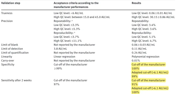
Table 2: Acceptance criteria for the evaluation of the analytical performances of the LIAISON®SARS-CoV-2 IgG kit.
Validation step Acceptance criteria according to the
manufacturer performances Results
Trueness Low QC level: <6 AU/mL Low QC level: 0.06 ± 0.01 AU/mL
High QC level: between 15.0 and 45.0 AU/mL High QC level: 30.15 ± 0.86 AU/mL
Precision Repeatability: a Repeatability:
Low QC level: ≤3.3% Low QC level: 3.4%
High QC level: ≤5.3% High QC level: 3.6%
Reproducibility: a Reproducibility:
Low QC level: ≤3.7% Low QC level: 5.1%
High QC level: ≤11.1% High QC level: 4.7%
Limit of blank Not reported by the manufacturer 0.06 ± 0.03 AU/mL
Limit of detection 3.8 AU/mL 0.11 AU/mL
Limit of quantification Not reported by the manufacturer 0.24 AU/mL
Linearity Linear regression Polynomial regression
Carry-over Not reported by the manufacturer 0.01%
Specificity Cut-off of the manufacturer
≥ 99% Cut-off of the manufacturer100%
Adapted cut-off (>6.1 AU/mL)
99% Sensitivity after 2 weeks Cut-off of the manufacturer
97% Cut-off of the manufacturer91%
Adapted cut-off (>6.1 AU/mL)
100%
aThe results refer to the groups of samples investigated and are not guaranteed specifications, as differences may exist between
The intra- and inter-run CVs were within the range reported by the manufacturer for the two levels of QC, except for the reproducibility for the low QC level (3.7% as reported by the manufacturer vs. 5.1% as reported by this validation study).
Limit of blank, detection and quantification
The LoB, LoD and LoQ are 0.06 ± 0.03 AU/mL, 0.11 AU/mL and 0.24 AU/mL, respectively. Only the LoD is reported by the manufacturer, i.e. 3.8 AU/mL, and the LoD calculated by the user according to the CLSI EP17-A2 document is far below this value which is in agreement with the accept-ance criteria [12].
Linearity
Results from the linearity study are reported in Figure 1. A regression analysis for second-order poly-nomials was performed. The regression equation was: y = 37.73 + 1.60 × − 0.006 x2. No statistically significant
dif-ference was observed between the measured and expected values (p < 0.001).
Carry-over
The following values have been obtained for the different samples and the different runs: A1 = 146 AU/mL, A2 = 140 AU/mL, A3 = 145 AU/mL, B1 = 0.275 AU/mL, B2 = 0.282 AU/ mL and B3 = 0.291 AU/mL. The carry-over is 0.01%.
Evaluation and comparison of the clinical
performances of the LIAISON
®SARS-CoV-2
IgG kit (DiaSorin
®) with the ELISA
SARS-CoV-2 test system (Euroimmun Medizinische
Labordiagnostika
®)
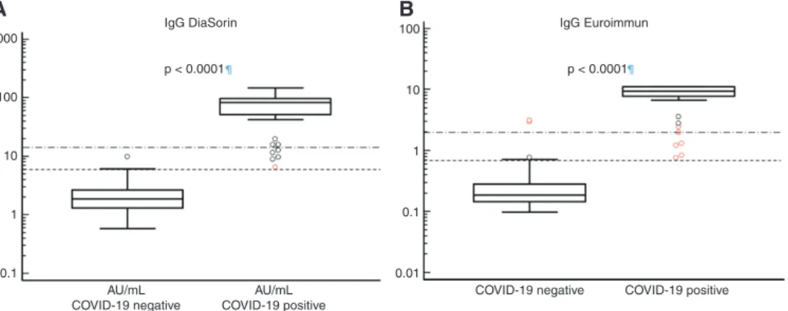
Among the 125 samples evaluated 2 weeks after the RT-qPCR positive detection, and according to manufac-turer’s cut-off, the LIAISON®SARS-CoV-2 IgG kit
identi-fied 40 true positives and 81 true negatives. Four samples were classified as false negative and none as false posi-tive (Figure 2). On the same cohort, the ELISA SARS-CoV-2 test identified 42 true positives and 79 true negatives. Two samples were false positive, and two samples were false negative. The specificity and the sensitivity were 100% (95% CI: 95%–100%) and 91% (95% CI: 79%–96%), and
0 20 40 60 80 100 120 140 160 160 140 120 100 80 60 40
Expected value, AU/mL
Measured value, AU/mL
Figure 1: Linearity assessment for the LIAISON®SARS-CoV-2 S1/S2
IgG antibody assay.
0.1 AU/mL COVID-19 negative COVID-19 negative AU/mL COVID-19 positive COVID-19 positive 1 10 100 0.01 0.1 1 10 100 1000 p < 0.0001 p < 0.0001 IgG DiaSorin IgG Euroimmun
A B
Figure 2: LIAISON®SARS-CoV-2 and ELISA SARS-CoV-2 IgG antibody performance at more than 2 weeks after a positive RT-qPCR
determination (n = 125 samples).
6 Tré-Hardy et al.: Validation of a chemiluminescent assay for specific SARS-CoV-2 antibody
98% (95% CI: 91%–99%) and 96% (95% CI: 85%–99%) for the LIAISON®CoV-2 IgG kit and the ELISA
SARS-CoV-2 test system, respectively, using the cut-off provided by the manufacturer. The kappa index was 0.93 for the two tests.
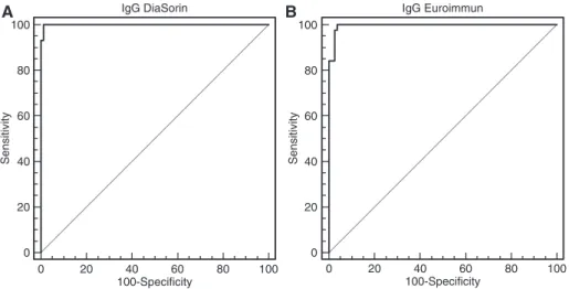
The cut-offs provided by the ROC curve analyses (i.e. >6.1 and >0.708 for the LIAISON®SARS-CoV-2 IgG kit and
the ELISA SARS-CoV-2 test system, respectively) improve the performance of the tests. Among the 125 samples tested, the use of these adapted cut-offs permits the correct reclassification of the four false-negative cases with the LIAISON®SARS-CoV-2 IgG kit to the detriment of
one false-positive case. For the ELISA SARS-CoV-2 test, the use of the adapted cut-offs permitted the correct reclas-sification of the two false-negative cases to the detriment of one false-positive case (n = 3 in total). The specificity and sensitivity were 99% (95% CI: 93%–100%) and 100% (95% CI: 92%–100%), and 96% (95% CI: 90%–96%) and 100.0% (95% CI: 92%–100%) (Figure 3) and the kappa index was 0.98 and 0.95 for the LIAISON®SARS-CoV-2 IgG
kit and the ELISA SARS-CoV-2 test system, respectively, using the adapted cut-offs. There was no statistically sig-nificant difference between the two tests in terms of clini-cal performance (p = 0.493).
Assessment of clinical specificity
From the results obtained above, interference from certain antibodies or antigens produced following viral, bacterial
or parasitic infections or following autoimmune patholo-gies reveals to be relatively low with a specificity of 99% (95% CI: 93%–100%) and 96% (95% CI: 90%–96%) for the LIAISON®SARS-CoV-2 IgG kit and the ELISA SARS-CoV-2
test system, respectively, using the adapted cut-offs. Using the cut-offs provided by the manufacturers, the specificity was 100% (95% CI: 95%–100%) and 98% (95% CI: 91%– 99%), a result not statistically and clinically different from the adapted cut-off.
Discussion
This study is the first to describe the analytical and clini-cal performances of the LIAISON®SARS-CoV-2 IgG kit from
DiaSorin® in comparison with the ELISA method from
Euroimmun Medizinische Labordiagnostika®, the first
ELISA testing that reached the market for the quantita-tive assessment of IgG and IgA directed against the spike protein subunit (S1). Only one study has previously evalu-ated the analytical performance of another CLIA test, the MAGLUMI™ 2000 Plus (New Industries Biomedical Engi-neering Co®, Shenzhen, China) but this was on a smaller
cohort of patients and samples and this study did not compare the performance of the CLIA assay with another test [13]. Also, regardless of the technique used (CLIA vs. ELISA), it is important to note that there is a difference in terms of antigenic targets. Namely, the DiaSorin® kit
pro-vides an additional antibody detection target with the S2 protein which is involved in the virus fusion machinery
IgG DiaSorin 0 20 40 60 80 100 100 A B 80 60 40 20 0 100-Specificity Sensitivity IgG Euroimmun 0 20 40 60 80 100 100 80 60 40 20 0 100-Specificity Sensitivity
Figure 3: LIAISON®SARS-CoV-2 and ELISA SARS-CoV-2 IgG antibody performance at more than 2 weeks after a positive RT-qPCR
determination (n = 125 samples).
The adapted cut-offs were the following: >6.1 for the DiaSorin assay and >0.708 for the Euroimmun assay. ROC curves reported excellent specificity and sensitivity of 99% (95% CI: 93-100%) and 100% (95% CI: 92-100%), and 96% (95% CI: 90-96%) and 100.0% (95% CI: 92-100%) for the LIAISON®SARS-CoV-2 IgG kit and the ELISA SARS-CoV-2 test system, respectively, using the adapted cut-offs.
while the kit from Euroimmun Medizinische Labordiag-nostika® only detects the S1 protein.
The analytical and clinical performances of the LIAISON®SARS-CoV-2 satisfied all the acceptance
cri-teria and provided “real world” analytical and clini-cal performances very close to the ones reported by the manufacturer in its insert kit with the exception of the reproducibility for the low QC level, i.e. 3.7% as reported by the manufacturer vs. 5.1% as reported by this valida-tion study. However, an inter-assay CV around 5% is con-sidered sufficient. Also, regarding the assessment of the trueness, the manufacturer does not provide a degree of uncertainty for its two QC levels but only reports a range, preventing adequate comparison. Therefore, when avail-able, trueness should be estimated with other methods or experiments. Evaluation of the LoQ has not been deter-mined using the 20% CV method due to the high reagent consumption such an evaluation requires. Comparison of this method of LoQ determination with the results obtained by the 10*SD method as performed in this study has to be done to confirm our results.
Comparison between the LIAISON®SARS-CoV-2 and
the ELISA method did not reveal any difference between the two techniques in terms of sensitivities and specifi-cities regarding the determination of the IgG. However, based on the cut-off provided by the manufacturers, two results were considered doubtful with the LIAISON®
SARS-COV-2 while one sample was considered doubtful with the ELISA methods. Adaptation of cut-off as determined by the ROC curve analyses highly improved the clinical performances of the tests from the second week following the positive RT-qPCR determination, with a sensitivity of 100% and a specificity of 99%. In comparison, adapta-tion of the cut-off for the ELISA SARS-CoV-2 test showed a sensitivity of 100% and a specificity of 96% on the same set of samples suggesting that, by adapting the cut-off, the LIAISON®SARS-CoV-2 shows at least, if not better,
per-formances than the ELISA testing. The results obtained during this study confirm previous observation that the production of IgG is detectable in symptomatic patients from the second week following the positive RT-qPCR determination [14]. We recommend each center to reestab-lish their own cut-off to improve the clinical performance and avoid false-negative results. Other studies have found significant differences in sensitivity when comparing the ELISA SARS-CoV-2 test from Euroimmun Medizinische Labordiagnostika® with other ELISA tests. According to
the study by Lassaunière et al. conducted on 111 patients, the Wantai®SARS-CoV-2 test (Wantai Biological Pharmacy
Enterprise®, Beijing, China), which detects total
antibod-ies, showed a specificity of 100% and a sensitivity of 90%
while the test from Euroimmun Medizinische Labordiag-nostika® revealed a specificity of 96% and a sensitivity of
65% [15].
Specificity of IgG antibody detection in
samples with known antibodies directed
against different targets
Using the adapted cut-offs, only one false-positive sample (i.e. a sample positive for AgHBs) was detected with the LIAISON®SARS-CoV-2 while three false positives were
reported with the ELISA technique. The sera that resulted in a cross-reaction showed anti-TPO antibodies (n = 1), anti-HAV IgM (n = 1) and ASLO (n = 1). Other studies have also observed false positives with the same ELISA method and reported interference with sera positive for IgM directed against influenza A, -influenza B, anti-adenovirus and anti-hCoV-HKU1 [15, 16]. According to the cross-reactivity studies described in the insert kit of the LIAISON®SARS-CoV-2, three samples out of 168
ana-lyzed also showed cross-reactivity with samples positive for anti-HBV (n = 1), anti-influenza (n = 1) and rheumatoid factor (n = 1). Although we have not tested the interfer-ence potential of anti-influenza A and B antibodies, we have observed probable new interference with anti-TPO antibodies (n = 1), anti-HAV IgM (n = 1) and ASLO (n = 1). Another limitation of our cross-reaction study is related to the very low number of antibody positive sera from other viruses of the Coronaviridae family. Given the rarity of these samples, only antibodies specifically directed against the NL63 and OC43 viruses were tested and did not show cross-reactivity.
Choice of the technique to determine the
presence of IgG antibodies and clinical
relevance
To date, an IgG protection threshold has not yet been demonstrated. However, if such a threshold should be established soon, the ELISA technique will probably be less efficient in determining a protection index as it is a semi-quantitative method, but additional data will be nec-essary to confirm this assumption. However, from a practi-cal point of view, the LIAISON®SARS-CoV-2 assay offers a
rate of random access tests of up to 170 tests/h while the ELISA technique from Euroimmun Medizinische Labor-diagnostika® adapted on the ETI-max 3000® controller
has more limited capacities with less flexible batch work and up to 160 tests/day but it integrates the possibility to combine the analysis of both IgA and IgG.
8 Tré-Hardy et al.: Validation of a chemiluminescent assay for specific SARS-CoV-2 antibody
The validation of SARS-CoV-2 serological methods is currently crucial to detect patients exposed to the virus including asymptomatic patients, to provide missing epi-demiological data in Belgium and other countries and potentially be able to detect a protective IgG threshold in the population. Serological testing carried out in the population will also be a very useful epidemiological information to compare the immunological status of a population with other countries and perhaps help in the development of a predictive visualization on the evolution of the epidemic. Routine use of this technique will also allow other serological studies to be carried out based on well-targeted population clusters in the hope of announc-ing the end of the pandemic when 50%–60% of the popu-lation have been in contact with the virus [17]. In addition to this deconfinement strategy, serological tests will also assess the potential effectiveness of vaccine trials and antibody-mediated therapies [18, 19].
Conclusions
In conclusion, this study is the first to report the valida-tion of a new CLIA test, the LIAISON®SARS-CoV-2 from
DiaSorin® allowing to detect IgG antibodies specifically
directed against SARS-CoV-2. The analytical and clinical performances are excellent, especially after adapting the cut-offs of the assays, and the automation of the test offers important rates, ideal for absorbing an extension of testing.
Acknowledgments: The sera with antibodies against NL63
and OC43 viruses were kindly provided by Professor Eli-zaveta Padalko, University Hospital Ghent. The authors also thank all the clinical laboratory staff for technical assistance.
Research funding: None declared.
Author contributions: All authors have accepted
respon-sibility for the entire content of this manuscript and approved its submission.
Competing interests: Authors state no conflict of interest. Ethical approval: The study was approved by the ethical
committee of the Iris Hospitals South.
References
1. Chan JF, Yuan S, Kok KH, To KK, Chu H, Yang J, et al. A familial cluster of pneumonia associated with the 2019 novel coronavirus indicating person-to-person transmission: a study of a family cluster. Lancet 2020;395:514–23.
2. Coronavirus Resource Center. https://coronavirus.jhu.edu/map. html. Accessed: 06 May 2020.
3. World Health Organization. Coronavirus disease 2019 (COVID-19) Situation Report – 106, 2020.
4. Yang Y, Yang M, Shen C, Wang F, Yuan J, Li J, et al. Evaluating the accuracy of different respiratory specimens in the laboratory diagnosis and monitoring the viral shedding of 2019-nCoV infec-tions. medRxiv 2020.
5. Zhang W, Du RH, Li B, Zheng XS, Yang XL, Hu B, et al. Molecular and serological investigation of 2019-nCoV infected patients: implication of multiple shedding routes. Emerg Microbes Infect 2020;9:386–9.
6. Xie X, Zhong Z, Zhao W, Zheng C, Wang F, Liu J. Chest CT for typical 2019-nCoV pneumonia: relationship to negative RT-PCR testing. Radiology 2020:200343.
7. Ai T, Yang Z, Hou H, Zhan C, Chen C, Lv W, et al. Correla-tion of chest CT and RT-PCR testing in coronavirus disease 2019 (COVID-19) in China: a report of 1014 cases. Radiology 2020:200642.
8. Sars-CoV-2 Diagnostics Pipeline. https://www.finddx.org/covid-19/pipeline/. Accessed: 23 April 2020.
9. Newton PN, Bond KC. 53 signatories from countries. COVID-19 and risks to the supply and quality of tests, drugs, and vaccines. Lancet Glob Health 2020. doi:10.1016/S2214-109X(20)30136-4.
10. CLSI. User Verification of Precision and Estimation of Bias; Approved Guideline – Third Edition. CLSI Document EP15-A3. Wayne, PA: Clinical and Laboratory Standards Institute; 2014. 11. Cofrac. Guide Technique d’Accréditation de Vérification (Portée A)/Validation (Portée B) des Méthodes en Biologie Médicale – Document SH GTA 04 (révision 01), 2015.
12. CLSI. Evaluation of Detection Capability for Clinical Laboratory Measurement Procedures; Approved Guideline – 2nd Edition. CLSI Document EP17-A2 2012; Wayne, PA: Clinical and Labora-tory Standards Institute.
13. Padoan A, Cosma C, Sciacovelli L, Faggian D, Plebani M. Analyti-cal performances of a chemiluminescence immunoassay for SARS-CoV-2 IgM/IgG and antibody kinetics. Clin Chem Lab Med 2020. doi:10.1515/cclm-2020-0443.
14. Jin Y, Wang M, Zuo Z, Fan C, Ye F, Cai Z, et al. Diagnostic value and dynamic variance of serum antibody in coronavirus disease 2019. Int J Infect Dis 2020;94:49–52.
15. Lassaunière R, Frische A, Harboe ZB, Nielsen ACY,
Fomsgaard A, Krogfelt KA, et al. Evaluation of nine commercial SARS-CoV-2 immunoassays. medRxiv 2020;2020.04.
09.20056325.
16. Okba NM, Muller MA, Li W, Wang C, GeurtsvanKessel CH, Corman VM, et al. Severe acute respiratory syndrome coronavirus 2- spe-cific antibody responses in coronavirus disease 2019 patients. Emerg Infect Dis 2020. doi:10.3201/eid2607.200841. 17. Joint European Roadmap towards lifting COVID-19
contain-ment measure. https://ec.europa.eu/info/sites/info/files/ communication_-_a_european_roadmap_to_lifting_coronavi-rus_containment_measures_0.pdf. Accesed: 24 Apr 2020. 18. Casadevall A, Pirofski LA. The convalescent sera option for
containing COVID-19. J Clin Invest 2020;130:1545–8. 19. Madore DV, Meade BD, Rubin F, Deal C, Lynn F, Meeting C.
Utilization of serologic assays to support efficacy of vaccines in nonclinical and clinical trials: meeting at the crossroads. Vac-cine 2010;28:4539–47.