Compromised SARS-CoV-2-specific placental antibody transfer
The MIT Faculty has made this article openly available. Please share
how this access benefits you. Your story matters.
Citation
Atyeo, Caroline et al. "Compromised SARS-CoV-2-specific placental
antibody transfer." Cell 184, 3 (February 2021): P628-642.e10 ©
2020 The Author(s)
As Published
https://doi.org/10.1016/j.cell.2020.12.027
Publisher
Elsevier BV
Version
Final published version
Citable link
https://hdl.handle.net/1721.1/130585
Terms of Use
Creative Commons Attribution 4.0 International license
Compromised
SARS-CoV-2-specific placental antibody transfer
Graphical abstract
Highlights
d
SARS-CoV-2-specific antibodies have a decreased placental
transfer
d
SARS-CoV-2-spike antibodies have altered glycosylation
profiles in pregnant women
d
Pregnant women with SARS-CoV-2 during the third trimester
have elevated IgG levels
d
SARS-CoV-2-specific antibody transfer is efficient after
second-trimester infection
Authors
Caroline Atyeo, Krista M. Pullen,
Evan A. Bordt, ..., Douglas Lauffenburger,
Andrea G. Edlow, Galit Alter
Correspondence
[email protected] (A.G.E.),
[email protected] (G.A.)
In Brief
Atyeo et al. reveal a deficiency in
SARS-CoV-2-antibody placental transfer among
women infected during the third
trimester. While bulk antibody transfer
remains unaltered,
SARS-CoV-2-antibodies show perturbed Fc
glycosylation profiles and elevated IgG
and FCGR3A placental expression that
suggest compensation for poor transfer.
Atyeo et al., 2021, Cell184, 628–642
February 4, 2021ª 2020 The Author(s). Published by Elsevier Inc.
Article
Compromised
SARS-CoV-2-specific placental antibody transfer
Caroline Atyeo,1,2,14Krista M. Pullen,3,14Evan A. Bordt,4Stephanie Fischinger,1,5John Burke,1Ashlin Michell,1
Matthew D. Slein,1Carolin Loos,1,3Lydia L. Shook,6Adeline A. Boatin,6Laura J. Yockey,6,7David Pepin,8
Marie-Charlotte Meinsohn,8Ngoc Minh Phuong Nguyen,8Maeva Chauvin,8Drucilla Roberts,9Ilona T. Goldfarb,6
Juan D. Matute,10Kaitlyn E. James,6Lael M. Yonker,10Lisa M. Bebell,11Anjali J. Kaimal,6Kathryn J. Gray,12
Douglas Lauffenburger,3Andrea G. Edlow,6,13,15,*and Galit Alter1,15,16,*
1Ragon Institute of MGH, MIT, and Harvard, Cambridge, MA 02139, USA
2PhD Program in Virology, Division of Medical Sciences, Harvard University, Boston, MA 02115, USA 3Department of Biological Engineering, Massachusetts Institute of Technology, Cambridge, MA 02139, USA
4Department of Pediatrics, Lurie Center for Autism, Massachusetts General Hospital, Harvard Medical School, Boston, MA 02114, USA 5PhD Program in Immunology and Virology, University of Duisburg–Essen, Essen 47057, Germany
6Department of Obstetrics, Gynecology and Reproductive Biology, Massachusetts General Hospital, Harvard Medical School, Boston,
MA 02114, USA
7Department of Medicine, Massachusetts General Hospital, Harvard Medical School, Boston, MA 02114, USA
8Pediatric Surgical Research Laboratories, Department of Surgery, Massachusetts General Hospital, Harvard Medical School, Boston,
MA 02114, USA
9Department of Pathology, Massachusetts General Hospital, Harvard Medical School, Boston, MA 02114, USA 10Department of Pediatrics, Massachusetts General Hospital, Harvard Medical School, Boston, MA 02114, USA
11Division of Infectious Diseases, Massachusetts General Hospital, MGH Global Health, and Harvard Medical School, Boston,
MA 02114, USA
12Department of Obstetrics, Gynecology and Reproductive Biology, Brigham and Women’s Hospital, Harvard Medical School, Boston,
MA 02115, USA
13Vincent Center for Reproductive Biology, Massachusetts General Hospital, Boston, MA 02114, USA 14These authors contributed equally
15These authors contributed equally 16Lead contact
*Correspondence:[email protected](A.G.E.),[email protected](G.A.) https://doi.org/10.1016/j.cell.2020.12.027
SUMMARY
SARS-CoV-2 infection causes more severe disease in pregnant women compared to age-matched
non-preg-nant women. Whether maternal infection causes changes in the transfer of immunity to infants remains
un-clear. Maternal infections have previously been associated with compromised placental antibody transfer,
but the mechanism underlying this compromised transfer is not established. Here, we used systems serology
to characterize the Fc profile of influenza-, pertussis-, and SARS-CoV-2-specific antibodies transferred
across the placenta. Influenza- and pertussis-specific antibodies were actively transferred. However,
SARS-CoV-2-specific antibody transfer was significantly reduced compared to influenza- and
pertussis-spe-cific antibodies, and cord titers and functional activity were lower than in maternal plasma. This effect was
only observed in third-trimester infection. 2-specific transfer was linked to altered
SARS-CoV-2-antibody glycosylation profiles and was partially rescued by infection-induced increases in IgG and
increased FCGR3A placental expression. These results point to unexpected compensatory mechanisms
to boost immunity in neonates, providing insights for maternal vaccine design.
INTRODUCTION
Over 44,000 pregnant women in the U.S. have been infected with SARS-CoV-2, and, with an estimated 140 million births annually worldwide, the number of pregnant women infected this year alone is likely in the millions (CDC, 2020). Although up to 16% of pregnant women test positive for SARS-CoV-2 in geographic
hotspots (Breslin et al., 2020; Sutton et al., 2020), pregnant women and neonates are excluded from vaccine and therapeu-tic trials due to enhanced safety standards required for this pop-ulation. Previous work has shown that both newborns and preg-nant women are particularly susceptible to respiratory infections, including influenza and respiratory syncytial virus (RSV) (Zaman et al., 2008;Rasmussen et al., 2012;Gerretsen and Sande, 2017;
ll
Reeves et al., 2019;Liu et al., 2020). Recent data demonstrate that a greater proportion of neonates and infants have severe or critical illness upon SARS-CoV-2 infection compared to older pediatric counterparts (Kim et al., 2020;Dong et al., 2020). Given the immature nature of the newborn’s immune system, coupled with anticipated delays in vaccine deployment to pregnant women and children, infants are highly vulnerable during the SARS-CoV-2 pandemic.
Neonates rely on the transfer of maternal immunoglobulin G (IgG) across the placenta for protection against pathogens. For most pathogens, umbilical cord titers of IgG are higher than in maternal blood (Gonc¸alves et al., 1999;Munoz et al., 2014; Mar-tinez et al., 2019), due to endosomal transport of IgG across the syncytiotrophoblast cell barrier from maternal to fetal circulation (Firan et al., 2001). These antibodies are transferred by the neonatal Fc receptor (FcRn), which is found in high concentra-tions on the placental syncytiotrophoblast (Leach et al., 1996;
Simister et al., 1996). Placental IgG transfer begins during the first trimester but increases exponentially during pregnancy, with the majority of transfer occurring during the third trimester (Fouda et al., 2018). Recent studies point to selective transfer of IgG across the maternal-fetal interface based on subclass (Palmeira et al., 2012;Wilcox et al., 2017;Langel et al., 2020) and Fc-glycan profile (Jennewein et al., 2019;Martinez et al., 2019;Langel et al., 2020). Across the IgG subclasses, IgG1 an-tibodies are transferred preferentially, followed by IgG3, IgG2, and IgG4 (Palmeira et al., 2012;Vidarsson et al., 2014). Antibody glycosylation, a post-translational modification, impacts the transfer of IgG across the placenta. Among IgG1 antibodies, gal-actosylated antibodies are transferred preferentially, potentially as a result of enhanced binding to both placental FcRn and FCGR3 (Kibe et al., 1996;Jennewein et al., 2019), enabling the selective transfer of specific antibody subpopulations to arm ne-onates most effectively in the setting of pathogen exposure. Recent reports have demonstrated infection-induced changes of Fc-glycan profiles in SARS-CoV-2-infected individuals ( Chak-raborty et al., 2020), raising the possibility that SARS-CoV-2 infection during pregnancy influences the quality of transferred immunity. However, the impact of altered glycosylation on maternal-to-neonatal antibody transfer remains unclear.
Maternal infection may alter the ability of antibodies to transfer across the placenta, in part by altering glycosylation. Prior studies have found that both maternal HIV and malaria infection result in reduced placental transfer of non-disease-specific anti-bodies (Okoko et al., 2001;Farquhar et al., 2005;Cumberland et al., 2007;Ogolla et al., 2015). This compromised transfer has been attributed to infection-associated alterations in anti-body glycosylation (Martinez et al., 2019) and to hypergamma-globulinemia in the infected mother, resulting in competition for binding to FcRn on the placenta (Englund, 2007). Less is known about placental transfer of disease-specific antibodies in the setting of maternal acute infection. Maternal:cord transfer ratios of ~1.0 have been noted in maternal Dengue (DENV) or Zika (ZIKV) acute viral infections (Perret et al., 2005;Castanha et al., 2019) in contrast to ratios of 1.5 or greater typically observed for vaccinatable pathogens, such as influenza and pertussis (Gonc¸alves et al., 1999;Heininger et al., 2009;Munoz et al., 2014;Castanha et al., 2019;Singh et al., 2019;Collier et al.,
2020). These lower-than-expected transfer ratios suggest that features of de novo antibodies generated in the setting of acute infection, such as glycosylation profile, may drive less efficient placental transfer. However, the precise mechanism of these al-terations in placental antibody transfer in the setting of recent infection is not known and has yet to be elucidated in maternal SARS-CoV-2 infection.
To address these gaps, we profiled the influenza, pertussis, and SARS-CoV-2 humoral immune response across 22 matched maternal:cord dyads from mothers who tested positive for SARS-CoV-2 infection in the third trimester of pregnancy (COVID+) and 34 contemporaneously enrolled maternal-cord
dyads that were SARS-CoV-2 negative (COVID–) using systems serology (Chung et al., 2015). Influenza hemagglutinin (HA)-and pertussis pertactin (PTN)-targeting antibodies were trans-ferred efficiently from COVID–mothers and COVID+ mothers. In contrast, we observed significantly decreased transfer of SARS-CoV-2-specific IgG and antibody functions across multi-ple SARS-CoV-2 specificities compared to HA. While similar glycan-dependent bulk (total IgG) placental transfer was observed across COVID– and COVID+ dyads, compromised
glycan-dependent transfer was noted for SARS-CoV-2-specific antibodies. In addition, while higher IgG levels in COVID–women
were associated with compromised placental transfer of HA-specific antibodies, increases in total IgG and placental co-localization of FCGR3A with FcRn played compensatory roles in augmenting SARS-CoV-2 antibody transfer across the COVID+ dyads. These data provide mechanistic insights into
the changes in antibody glycosylation and placental Fc-receptor expression that contribute to compromised third-trimester anti-body transfer to infants, pointing to opportunities to bolster neonatal immunity to SARS-CoV-2 and beyond.
RESULTS
SARS-CoV-2-specific antibodies are transferred inefficiently to the neonate
Past reports have described infection-driven alterations in placental IgG transfer to the neonate (Okoko et al., 2001; Farqu-har et al., 2005;Cumberland et al., 2007;Ogolla et al., 2015;
Martinez et al., 2019), but it is unclear whether placental transfer dynamics shift with SARS-CoV-2 infection. To address this question, we profiled humoral immune responses to three respi-ratory pathogens in COVID+and COVID–pregnant women and
their neonates’ cord blood to gain a deeper understanding of neonatal immunity following maternal SARS-CoV-2 infection. Cohort demographics are depicted inTable 1. Mean time from symptom onset to delivery was 30.4 ± 19.8 days. No neonates were infected with SARS-CoV-2; thus, COVID+infection status throughout the manuscript refers only to the mother (Edlow et al., 2020). We first compared the relative titer of antibodies in maternal plasma and cord blood against influenza hemagglu-tinin (HA) and pertussis pertactin (PTN). As expected, increased titers of HA- and PTN-specific IgG1 were observed in the cord compared to maternal blood in both COVID–and COVID+
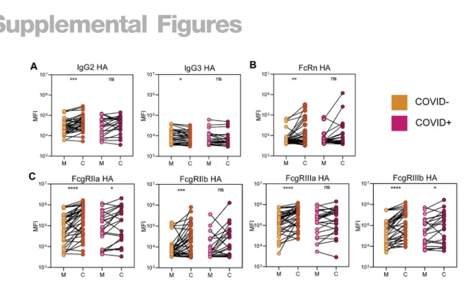
moth-er:cord dyads (Figure 1A). Similarly, all HA-specific subclasses and Fc receptor (FcR) binding were increased in cord blood compared to the maternal plasma across the dyads, with the
exception of IgG3, which has been shown to have low affinity for FcRn compared to other IgG subclasses (Vidarsson et al., 2014) (Figure S1). These data demonstrate conserved efficient transfer of HA- and PTN-specific antibodies in the setting of SARS-CoV-2 infection, albeit less significant skewing (lower p value dif-ferences) of antibodies in the cord in COVID+dyads across the placenta (Figures 1A andS1).
To determine whether the same pattern was present for CoV-2-specific antibodies, we profiled levels of SARS-CoV-2 receptor binding domain (RBD), spike (S), and nucleo-capsid (N)-specific responses across the COVID+mother:cord pairs (Figures 1B andS2). Surprisingly, rather than the expected active transfer to the cord, resulting in higher cord titers compared to maternal titers, reduced titers of RBD- and S-spe-cific antibodies were present in the cord, and N-speS-spe-cific titers were stable or slightly reduced in the cord. To further compare changes in transfer across all antigen specificities and dyads, we plotted IgG1 transfer ratios against each antigen by maternal infection status (Figure 1C). Expected transfer ratios (>1) were observed for HA- and PTN-specific antibodies in COVID– mothers. In contrast, a significant defect in transfer was visual-ized across antibody subclasses and FcR-binding to RBD, S, and N, including the S1 and S2 domain of the S protein ( Fig-ure 1C, 1D, andS2). In addition, while enhanced transfer of
an-tibodies able to bind FcRs was observed for HA and PTN, there was a reduced transfer of SARS-CoV-2-specific, FcR-binding antibodies (Figures S1andS2). Moreover, in a second indepen-dent set of mother:cord pairs, compromised SARS-CoV-2 anti-body transfer was confirmed in mothers infected during the third trimester (Figure S3A). Interestingly, this defect was exclusive to third-trimester infection, as efficient SARS-CoV-2 antibody transfer was observed for mothers infected during the second trimester (Figure S3B). In addition, in the third-trimester-infected cohort, time from infection had no significant effect on transfer rates (Figure S3C). Collectively, these data show that maternal third-trimester SARS-CoV-2 infection has a profound impact on SARS-CoV-2-specific neonatal antibody transfer compared to transfer of other pathogen-specific antibodies.
Compromised SARS-CoV-2-specific functional antibody transfer
To further dissect the impact of SARS-CoV-2 infection on anti-body transfer, we examined whether preferential transfer occurred for any humoral immune function across the dyads. Similar to titer, increased HA-specific functional antibodies were transferred to the cord across both the COVID– and COVID+pairs (Figures 2A–2D). The only exceptions were the transfer of antibodies that induce monocyte phagocytosis
Table 1. Demographics of cases and controls
COVID Negative (n = 33)a COVID Positive (n = 22)b P Valuec
Maternal age (median, IQR) 33 (30, 37) 33 (28, 39) 0.91 Gestational age (mean, SD) 39.2 (1.5) 38.0 (4.1) 0.16 Parity (median, IQR) 1 (0, 2) 1 (0, 1) 0.65
Race 0.02
Asian 2 (6%) 0 (0%)
Black/African American 0 (0%) 2 (9%)
White 26 (79%) 10 (45%)
Other 4 (12%) 4 (18%)
More than one race 0 (0%) 2 (9%) Unknown/not reported 1 (3%) 4 (18%)
Ethnicity 0.02
Hispanic or Latino 7 (21%) 13 (59%) Not Hispanic or Latino 24 (73%) 8 (36%) Unknown/Not Reported 2 (6%) 1 (5%) Severityd Asymptomatic – 6 (27%) Mild – 9 (41%) Moderate – 3 (14%) Severe – 4 (18%)
Time from symptom onset to delivery, in days (median, IQR) – 30.5 (8.5, 44) N/A Time from last influenza vaccination, in days (median, IQR) 200 (178, 209) 204 (188.5, 215.5) 0.82 Time from last pertussis vaccination, in days (median, IQR) 69 (51.75, 81.25) 72 (57, 81) 0.31
a
n = 33 COVID negative (34 dyads, one mother of di-di twins).
b
No neonates tested positive for SARS-CoV-2 by RT-PCR of nasopharyngeal swab.
c
Significant differences between groups were determined using Pearson’s chi-square test for categorical variables, and Mann-Whitney U test or Stu-dent’s t test for continuous variables (median, IQR and mean, SD, respectively).
dSeverity determinations were made based on published criteria from the Society for Maternal-Fetal Medicine and the National Institutes of Health.
ll
(ADCP), which were not preferentially transferred in COVID+
mothers (Figure 2A), and natural killer (NK) cell-degranulation-inducing antibodies (CD107a) that were transferred less effi-ciently across COVID+and COVID–dyads (Figure 2B). HA-spe-cific NK cell chemokine secretion- (macrophage inflammatory protein-1b or MIP-1b), neutrophil phagocytosis- (ADNP), and complement deposition (ADCD)-inducing antibodies were trans-ferred efficiently across both COVID+and COVID–dyads (
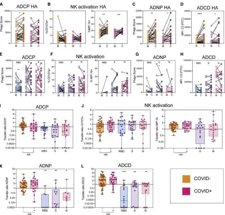
Fig-ures 2B–2D). In contrast, significantly reduced or erratic transfer was noted for RBD, S, and N-specific antibody functions in COVID+dyads (Figures 2E–2H). For SARS-CoV-2 specificities, there was a decrease, though not always significant, in functional antibodies able to drive ADCP, ADNP, and ADCD in cord blood compared to maternal plasma (Figures 2E, 2G, and 2H). In contrast, the levels of SARS-CoV-2-specific NK cell-activating antibodies were not significantly reduced between maternal plasma and cord blood (Figure 2F).
To determine whether antibodies with certain functions were selectively transferred, a transfer ratio was calculated for each specificity and function, where a transfer ratio of 1 denoted
equivalency in antibody effector function across the maternal:-cord pairs. Collectively, median transfer ratios for HA-directed functions were above 1. No significant differences were noted in the transfer ratios for HA-directed functions between COVID+ and COVID– dyads, indicating more efficient transfer of HA-directed functions compared to SARS-CoV-2-HA-directed functions (Figures 2I–2L). The median transfer ratios for all functions except NK activation were below 1 for SARS-CoV-2 specificities, indicating a decrease, although not always significant, in the transfer of antibodies able to drive ADNP, ADCD, and ADCP against SARS-CoV-2 in the cord. The higher transfer ratio of SARS-CoV-2 NK cell chemokine secretion-inducing antibodies (MIP-1b) (Figure 2J), despite lower SARS-CoV-2-specific IgG levels in the cord (Figure 1), suggests conserved preferential placental transfer of these antibodies to the neonates compared to antibodies able to drive neutrophil and complement activation (Figures 2K and 2L, respectively), as has been shown previously (Jennewein et al., 2019). Taken together, these findings point to relatively normal placental antibody transfer of non-SARS-CoV-2 antibodies in the setting of maternal SARS-CoV-2 infection but a
Figure 1. Inefficient placental transfer of SARS-CoV-2 antibodies
(A) The dot plots show the relative hemagglutinin- (HA) (left) and pertactin- (PTN) (right) specific IgG1 titers for both COVID-negative (COVID–
, orange) and COVID-positive (COVID+, pink) maternal-cord pairs. The dotted line indicates background PBS levels. Significance was determined by a Wilcoxon signed-rank test, *p < 0.05, ***p < 0.001, ****p < 0.0001.
(B) The dot plot shows the IgG1 titer against SARS-CoV-2 receptor binding domain (RBD, left), spike (S) (middle), and nucleocapsid (N) (right). The lines connect maternal:cord pairs. The dotted line shows the PBS background level. Significance was determined by Wilcoxon signed-rank test, *p < 0.05.
(C) The dot plots show the transfer ratio of HA, PTN, RBD, S, and N antibodies. The transfer ratio of COVID–
dyads are shown in orange, whereas the transfer ratios of COVID+
dyads are shown in pink. The horizontal dotted line indicates a 100% transfer efficiency (equivalent levels across both compartments). Significance was determined by using a one-way ANOVA followed by Tukey’s multiple comparisons test, where ‘‘ha’’ indicates a significant difference compared to the COVID+
HA transfer ratio and ‘‘p’’ indicates a significant difference compared to the COVID+
PTN transfer ratio. **p < 0.01, ***p < 0.001, ****p < 0.0001. (D) The heatmap shows the median PBS-background corrected transfer ratio of HA, PTN, RBD, S, and N across all antibody subclasses and FcR binding profiles. See alsoFigures S1,S2, andS3.
Figure 2. Functional HA-, but not SARS-CoV-2-, specific antibodies are transferred efficiently across the placenta
(A–D) The dot plots illustrate antibody-dependent monocyte phagocytosis (ADCP, A), natural killer cell (NK) activation (B), antibody-dependent neutrophil phagocytosis (ADNP, C), and antibody-dependent complement deposition (ADCD, D) (PBS background corrected) in maternal plasma (M) and cord-blood plasma (C) against hemagglutinin (HA). Lines connect maternal:cord pairs. COVID–
dyads are represented in orange, and COVID+
dyads are represented in pink. Significance was determined by a Wilcoxon signed-rank test, *p < 0.05, **p < 0.01, ***p < 0.001, ****p < 0.0001.
(E–H) The dot plots show the ADCP (E), NK activation (F), ADNP (G), and ADCD (H) activity (PBS background corrected) in maternal blood (M) and cord plasma (C) for the SARS-CoV-2 antigens RBD, spike (S), and nucleocapsid (N). Lines connect maternal:cord dyads. Significance was determined by a Wilcoxon signed-rank test, *p < 0.05, **p < 0.01.
(I–L) The box-and whisker plots show the transfer-ratios for ADCP (I), NK activation (J), ADNP (K), and ADCD (L) against HA, RBD, S, and N. For HA, COVID–
dyads are represented in orange and COVID+
dyads are shown in pink. For each SARS-CoV-2 antigen, significance was determined against the HA activity using the matched COVID+
dyad. A one-way ANOVA followed by Tukey’s multiple comparison test was performed to determine significance, *p < 0.05, **p < 0.01, ***p < 0.001.
ll
distinct loss of neutrophil phagocytosis- and complement-inducing SARS-CoV-2-specific functional antibody transfer, two functions that have been linked to enhanced antiviral control following natural infection in adults (Atyeo et al., 2020).
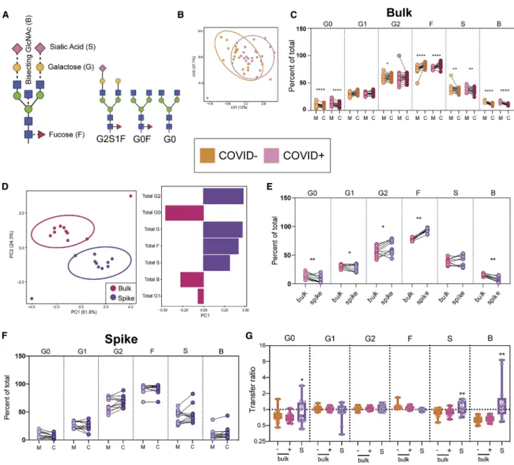
Altered SARS-CoV-2-specific glycan profiles shift placental transfer profiles
Given the emerging role of antibody Fc glycosylation in placental transfer efficiency (Jennewein et al., 2019;Martinez et al., 2019), we next probed whether changes in SARS-CoV-2 antibodies themselves could account for differences in transfer efficiencies. Fc glycosylation (Figure 3A) was captured on bulk and SARS-CoV-2-S-specific antibodies. Bulk antibody Fc-glycan profiles were indistinguishable across COVID+and COVID–mothers (
Fig-ures 3B,S4A, and S4D), with similar bulk Fc glycosylation trans-fer patterns across COVID+ and COVID– dyads (Figure 3C), consistent with previously observed selection of galactosylated Fc glycans across the placenta (Jennewein et al., 2019;Borghi et al., 2020). The conserved bulk antibody transfer profiles across the COVID+and COVID–dyads point to the preserved ability of the placenta to preferentially transfer IgG with certain glycosylation features in the setting of SARS-CoV-2 infection.
Antigen-specific antibody subpopulations may harbor distinct antibody Fc-glycan profiles compared to bulk circulating anti-bodies (Mahan et al., 2016). Thus, to determine whether there were SARS-CoV-2-antibody-specific changes to Fc-glycan pro-files, we next compared spike-specific Fc glycosylation to bulk antibodies across the COVID+mothers. In contrast to the bulk
antibody Fc profiles (Figure 3B), which did not differ across mothers, spike (S)-specific Fc-glycan profiles were significantly different within COVID+ mothers compared to bulk Fc-glycan profiles (Figure 3D, left), marked by enhanced digalactosylation (G2), fucosylation (F), and reduced agalactosylation (G0) and bisecting-n-acetyl-glucosamine (b-GlcNAc, B) on S-specific antibodies (Figures 3D, right, and 3E). As expected, these glycan-profile differences resulted in altered antibody transfer (Figure 3F). Consistent with bulk transfer glycan profiles, digalac-tosylated (G2) antibody transfer efficiency was above 1 for S-specific transfer (Figures 3F and 3G). Transfer ratios were per-turbed for agalactosylated (G0), sialyated (S), and b-GlcNAc (B) levels on S-specific antibodies compared to bulk antibody trans-fer in the COVID+ and COVID– dyads (Figure 3G). Increased transfer of G0 S-specific antibodies relative to bulk antibodies in COVID+dyads may suggest preferential transfer of
inflamma-tory afucosylated, bisected, G0 antibodies in the setting of maternal SARS-CoV-2 infection (Figure 3G). However, whether perturbed glycan transfer profiles were related to altered placental transfer of SARS-CoV-2 antibodies, or simply linked to the overall availability of different Fc-glycan profiles on maternal SARS-CoV-2-specific antibodies, remained unclear.
Signatures of FCGR3A-selective transfer in COVID+dyads
To determine whether the placental transfer differences were sim-ply related to different levels of glycosylated antibodies on the maternal side, or whether the transfer signatures were conserved across COVID+and COVID–mothers, we next examined the over-all humoral Fc profiles of HA- and PTN-specific or non-specific
antibodies in the setting of maternal SARS-CoV-2 infection or non-infection. To define distinct features between maternal plasma and cord blood across antibody populations, multivariate dimensionality reduction was coupled to multivariate visualiza-tion, using Elastic Net regularization and feature reduction with multi-level orthogonal partial least-squares discriminant analysis (m-OPLSDA). As expected, non-SARS-CoV-2-specific antibody profiles were distinct between maternal and cord blood (Figures 4A,S4B, andS4D), with a clear enrichment of digalactosyated an-tibodies and NK cell enhancing anan-tibodies in the cord (Figure 4B). COVID+and COVID–samples were interspersed and not resolv-able (Figure 4A), highlighting that SARS-CoV-2 infection does not alter the overall profile of transferred non-SARS-CoV-2-spe-cific antibodies. Given that Elastic Net selects a minimal set of fea-tures in order to prevent overfitting of the data, we next generated a correlation network to determine additional features that corre-lated with the features included in the model (Figure 4C). Elastic Net-selected glycan features G2FB and G1FB-G2 were highly correlated with other galactosylated glycan features and nega-tively correlated with agalactosylated glycans. This bias toward highly galactosylated and fewer agalactosylated bulk antibodies in the cord emphasizes the importance of conserved glycan sieving in both COVID+and COVID–dyads.
Similar to HA, PTN, and bulk antibodies, SARS-CoV-2-spe-cific antibody glycan profiles were clearly distinct in maternal versus cord blood (Figures 4D and S3C–3E). Maternal blood was enriched for fucosylated/agalactosylated S-specific anti-bodies (Spike G0F), S2-specific IgG1, and neutrophil functional antibodies, while the cord blood had elevated NK cell-activating nucleocapsid (N)-specific antibodies, S-specific afucosylated/ agalactosylated (Spike G0), and N-specific monocyte phago-cytic antibodies (Figure 4E). The enrichment of S2-domain-spe-cific antibodies, recently found to cross-react across coronavi-ruses (Ng et al., 2020), was linked to a large S2- and S-specific functional network (Figure 4F). Given that IgG1 S2 was enriched in maternal blood, this network pointed to a lack of transfer of many SARS-CoV-2-specific antibodies, consistent with univari-ate data (Figures 1 and 2). The selective transfer of afucosy-lated/agalactosylated (G0) Fcs to the cord but the retention of fu-cosylated/agalactosylated (G0F) Fcs in the maternal circulation pointed to fucosylation-based antibody selection across the placenta, likely driven by FCGR3A, which preferentially binds to afucosylated glycans (Jennewein et al., 2019). Thus, consis-tent with previous data, the placenta appears to exhibit altered galactose transfer, but similar fucose transfer in COVID+and COVID–women, continuing to selectively sieve NK functional
an-tibodies out of the SARS-CoV-2 maternal humoral response. Time from infection in third-trimester pregnancy contributed minimally to placental sieving (Figure S3E). These data suggest that reduced SARS-CoV-2-specific antibody transfer is not related to altered placental Fc-glycan sieving activity but rather to a bias against the glycoforms found dominantly on SARS-CoV-2-specific antibodies.
Elevated IgG levels augment SARS-CoV-2 antibody transfer
Despite evidence of conserved Fc-glycan selection across the placenta for SARS-CoV-2-specific antibodies, it remained
Figure 3. Spike-specific Fc-glycan profiles diverge from bulk antibody profiles
(A) The figure represents a typical Fc glycan. The solid lines between sugars represent the linkages that are always present, whereas the dotted lines represent sugars that can be added.
(B) An orthogonal partial least-squares discriminant analysis (OPLSDA) model was built using bulk glycan features from COVID–
(orange) and COVID+
(pink) maternal plasma samples. This orthogonal approach ensures that maximal variance between groups is captured in the first latent variable (LV1). The dot plot shows the distribution of each maternal sample, with ellipses encompassing the 95% confidence interval for each group.
(C) The dot plot shows the percent of each glycoform in bulk antibodies in maternal (M) and cord (C) samples. The glycoforms are denoted as follows: aga-lactosylated (G0), monogaaga-lactosylated (G1), digaaga-lactosylated (G2), fucosylated (F), sialylated (S), and bisected n-acetyl-glucosamine (GlcNAc) (B). Connected lines signify maternal:cord dyads. Significance was determined using a Wilcoxon signed-rank test, *p < 0.05, **p < 0.01, ***p < 0.001, ****p < 0.0001. (D) A multi-level principal component analysis (m-PCA) was built using bulk (pink) and spike-specific (purple) antibody glycan data from COVID+
mothers for which matched data were available. Each maternal sample is represented as a dot on the scores plot (left). The bar graph (right) shows the loadings on principal component 1 (PC1) of each glycan feature.
(E) The dot plot shows the percentage of each glycoform in maternal bulk (pink) and spike-specific (purple) antibodies. The lines represent glycan data from the same mother. Significance was determined using a Wilcoxon signed-rank test, *p < 0.05, **p < 0.01, ***p < 0.001, ****p < 0.0001.
(F) The dot plot shows the percent of each glycoform in spike-specific antibodies in maternal (M) and cord (C) samples. Significance was determined using a Wilcoxon signed-rank test
(G) The boxplots show the transfer ratio of each glycoform for bulk (orange/pink) and spike-specific (purple) antibodies. For bulk glycan data, COVID–
dyads are represented in orange and COVID+
dyads are represented in pink. Spike (S)-specific glycan data are shown in purple. Significance was determined by a Mann-Whitney test between COVID+
bulk and S-specific antibodies for each glycan feature, *p < 0.05, **p < 0.01, ***p < 0.001, ****p < 0.0001. See alsoFigure S4.
ll
unclear whether additional infection-related immune perturba-tions could influence transplacental transfer. Mounting evidence points to a critical role of infection-induced hypergammaglobuli-nemia in reducing antibody transfer (Okoko et al., 2001;Farquhar et al., 2005; Martinez et al., 2019), postulated to be due to increased competition for binding to FcRn and other Fc
recep-tors important for placental transfer. Thus, we measured total IgG titer in maternal and cord blood to determine whether alter-ations in IgG levels tracked with changes in antibody transfer (Figures 5A and 5B). While the women in our study did not exhibit clinical hypergammaglobulinemia, we noted increased IgG levels in COVID+mothers even weeks after infection. Total IgG
Figure 4. Placental sieving signatures of HA- and spike-specific antibodies
(A) Multi-level orthogonal partial least square discriminant analysis (m-OPLSDA) was performed to distinguish between profiles from mother and cord plasma using HA, PTN, and bulk glycan data. Triangular and circular data points on the scores plot represent mother and cord samples, respectively. Infection status is indicated by color (orange representing COVID–
and pink representing COVID+
individuals). The ellipses show the 95% confidence interval for the mother/cord populations.
(B) The bar graph illustrates the loadings on latent variable 1 (LV1) of the Elastic Net-selected antibody features from the m-OPLSDA shown in (A). The color indicates the group in which each feature is enriched (dark pink for cord samples, light pink for maternal plasma).
(C) A pairwise spearman correlation test was performed to determine features that correlate with the Elastic Net-selected features (bolded nodes) selected for the m-OPLSDA model shown in (A). Features with correlation of coefficients >|0.75| and p < 0.05 are shown in the networks. Positive correlations are shown in purple, and negative correlations are shown in pink. Nodes are colored by features type, with glycan data shown in purple, antibody titer shown in yellow, and FcR binding shown in blue.
(D) A m-OPLSDA model was built to classify COVID+
mother and cord plasma using SARS-CoV-2-specific antibody features and spike-specific glycan data. Only samples for which spike-specific glycan data were available were included in the model. The scores plot shows the separation between the cord samples (dark pink) and the maternal samples (light pink), where each dot represents a sample. The ellipses show the 95% confidence interval for each group.
(E) The bar graph shows the loadings on latent variable 1 (LV1) of the Elastic Net-selected features for the m-OPLSDA model built in (D). The color indicates the group in which each feature is enriched (dark pink for cord samples, light pink for maternal samples).
(F) A pairwise spearman correlation test was performed to determine features that were correlated with the Elastic Net-selected features (bold nodes) that were selected for the m-OPLSDA model shown in (D). Features with correlation of coefficients >|0.75| and p < 0.05 are shown in the networks. Positive correlations are shown in purple and negative correlations are shown in pink. Nodes are colored by feature type, with glycan data shown in purple, antibody titer shown in yellow, and FcR binding shown in blue.
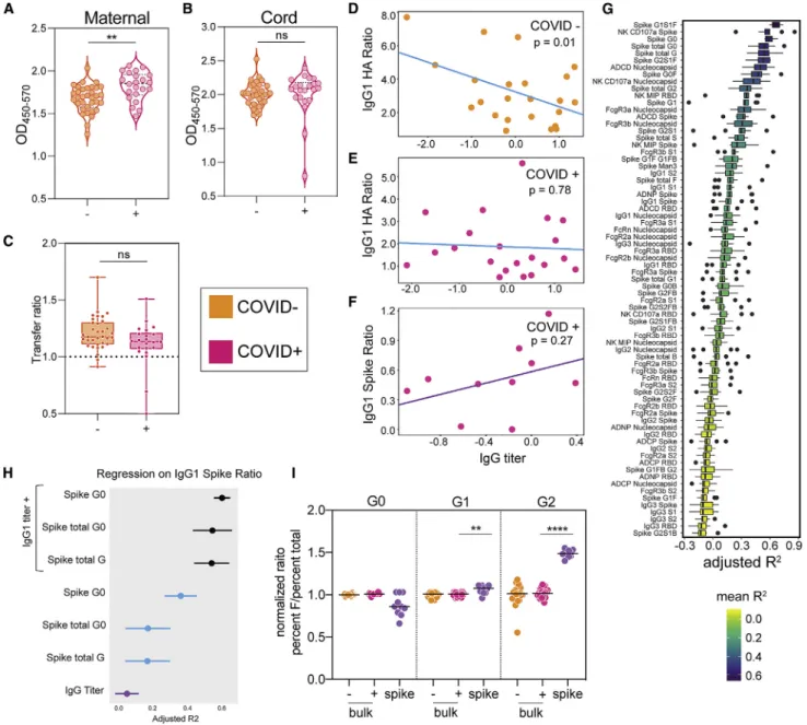
Figure 5. Elevated IgG compensates for glycosylation changes to improve SARS-CoV-2-specific antibody transfer
(A and B) A total IgG ELISA was run on COVID–
(orange) and COVID+
(pink) maternal plasma (A) and cord blood (B). Significance was determined by a Mann-Whitney test. *p < 0.05, **p < 0.01, ***p < 0.001, ****p < 0.0001.
(C) The boxplots show the transfer ratio, calculated as cord OD/maternal OD. The dotted line shows a transfer ratio of 1. Significance was determined by a Mann-Whitney test.
(D–F) The dot plots illustrate the correlation between IgG titer and IgG1 HA (D and E) or IgG1 Spike (F) transfer ratio in COVID negative (orange) and COVID positive (pink) individuals. The direction of correlation is indicated by the color of the regression line (neg, blue; pos, purple).
(G) 2-feature linear regression models were built to determine which feature, in combination with total IgG titer, was most predictive of SARS-CoV-2-specific antibody transfer ratio. The boxplots show the adjusted R2
values for the top performing models over 10 leave-one-out cross-validated trials.
(H) The dots represent the adjusted R2values of three of the top 2-feature models (G) to each of the individual features predictive performance of SARS-CoV-2-specific antibody transfer. For the 1-feature regression models, data points are colored by the direction of correlation with IgG1 spike transfer ratio (neg, blue; pos, purple). Error bars illustrate standard deviation of adjusted R2
after 10 rounds of leave-one-out cross-validation.
(I) The dot plots represent the ratio of percent fucosylated to total percent for each galactose feature. For each galactose glycoform, the data were normalized by dividing by the average ratio of the bulk COVID–
. Significance was only calculated between bulk COVID+
and spike-specific antibodies. Significance was determined by Mann-Whitney test, *p < 0.05, **p < 0.01, ***p < 0.001, ****p < 0.0001.
ll
titer was significantly higher in COVID+mothers (Figure 5A) but not their cord blood (Figure 5B). Instead, total IgG transfer ratios trended lower in COVID+mothers, highlighting the critical role of
the placenta in creating a bottleneck to restrict antibody entry into the neonate (Figure 5C). Transfer efficiency for HA-specific antibodies was inversely correlated to IgG levels in COVID– dyads (Figure 5D). Conversely, in COVID+mothers, increasing IgG levels did not affect HA-IgG transfer (Figure 5E), suggesting a compensatory mechanism may exist during SARS-CoV-2 infection to prevent IgG-level competition observed with hyper-gammaglobulinemia in other infections (Okoko et al., 2001; Far-quhar et al., 2005). In contrast to HA-specific antibody transfer in COVID– mothers, SARS-CoV-2-specific antibody transfer trended higher with increased IgG levels (Figure 5F), likely due to the accompanying increase of SARS-CoV-2 antibodies along with overall increased IgG levels.
To understand the influence of IgG levels on SARS-CoV-2-antibody transfer, the pairwise influence of elevated IgG and all individual Fc-features on transplacental transfer was examined. While previous reports have shown that galactosylation is impor-tant for transfer across the placenta, IgG levels appeared to selectively augment G1S1F, NK cell-activating (CD107a and MIP-1b), afucosylated/agalactosylated (G0), fucosylated/aga-lactosylated (G0F), and G2S1F transfer, among the top 10 IgG-co-correlates in predicting transfer (Figure 5G). Additionally, increasing IgG levels tracked with improved transfer of comple-ment- (ADCD) enhancing antibodies, and FCGR3 transfer was found among the top transfer predictors, highlighting the impor-tance of this FcR in placental sieving.
To specifically explore the impact of IgG-levels on shifting Fc-glycan transfer during SARS-CoV-2 infection, the influence of IgG level combined with galactosylation was compared to the glycan effect on transfer alone. IgG level alone was a poor pre-dictor of transfer (Figure 5H, bottom). In contrast to patterns seen in pertussis-specific antibody transfer (Jennewein et al., 2019), SARS-CoV-2-specific agalactosylated/afucosylated Fc glycans were enriched in cord blood. Interestingly, in the pres-ence of elevated IgG, all glycans transferred more efficiently, albeit agalactosylated/afucosylated glycans were the most associated with IgG1 spike transfer (Figure 5H), pointing to IgG level enhancement of all S-specific antibodies with a slight pref-erence for afucosylated antibodies. These data suggest that elevated IgG levels may compensate for poor SARS-CoV-2-spe-cific antibody transfer.
Previous studies suggest that the placenta preferentially trans-fers digalactosylated antibodies (Kibe et al., 1996;Jennewein et al., 2019;Borghi et al., 2020). However, SARS-CoV-2 antibody transfer consistently pointed to enhanced transfer of agalactosy-lated/afucosylated spike-specific antibodies (Figures 4E, 5G, and 5H). To understand this selection based on fucose content, we next dissected the fucosylation pattern on antibodies with varying degrees of galactosylation (Figure 5I). Whereas the level of fucosylation was conserved across galactosylated bulk anti-bodies in COVID+and COVID–mothers (Figures 3B and 3C), agalactosylated spike-specific antibodies showed a trend to-ward a decreased proportion of fucosylation (Figure 5I, left). Conversely, fucosylation was significantly higher on galactosy-lated antibody subpopulations (Figure 5I, middle and right).
Thus, although maternal spike-specific antibodies exhibit enhanced galactosylation (Figures 3D and 3E), these glycans also contain high levels of fucose, which may reduce the likeli-hood of transfer in the setting of collaborative FcRn and FCGR3A transplacental transfer (Pincetic et al., 2014).
Increased FCGR3A and FcRn colocalization in SARS-CoV-2 infection
Preferential transfer of agalactosylated/afucosylated SARS-CoV-2 antibodies points to a critical role for FCGR3A selection during placental transfer. To probe whether placental Fc recep-tor expression patterns were altered by maternal SARS-CoV-2 infection, we quantified the levels of FcRn, FCGR3A, and the co-localization of these receptors, which have been previously iden-tified as instrumental in placental antibody transfer (Jennewein et al., 2019). While no difference was observed in FcRn expres-sion, a slight increase in FCGR3A was observed in placentas from COVID+mothers (Figures 6A and 6B). Moreover, colocali-zation of the two receptors was significantly increased in pla-centas from COVID+mothers (Figures 6A and 6B), suggesting that enhanced FCGR3A expression and FCGR3A/FcRn colocal-ization may compensate for galactose changes and promote the specific selection of afucosylated antibodies able to drive enhanced NK cell function. Taken together, these data suggest that elevated IgG levels may limit the transfer of total antibodies but facilitate the transfer of current-infection-specific antibodies recently generated by the pregnant mother. Enhanced FCGR3A/ FcRn colocalization may be a compensatory third-trimester pregnancy-specific immune mechanism, allowing the placenta is able to continue to select functionally optimized antibodies that drive enhanced NK cell activation to deliver the best immu-nity possible to the neonate.
DISCUSSION
Specific populations are at greatest risk in the SARS-CoV-2 pandemic, including individuals with significant medical comor-bidities, the elderly, racial and ethnic minorities, patients of low socioeconomic status, and pregnant women (Ellington et al., 2020; Mehra et al., 2020; Richardson et al., 2020; Wadhera et al., 2020;Goldfarb et al., 2020). While SARS-CoV-2 infection is not known to be widespread in newborns and infants, this pe-diatric population is particularly vulnerable to severe disease upon SARS-CoV-2 infection (Dong et al., 2020), with substantial morbidity and death reported (Liguoro et al., 2020;Mithal et al., 2020;Ovalı, 2020). These reports, coupled with the potential for neonates and infants to develop the severe Kawasaki-like dis-ease called multisystem inflammatory syndrome in children (MIS-C) (Feldstein et al., 2020), and high viral loads in this age group that may act as a reservoir of viral spread (Bialek et al., 2020;Yonker et al., 2020), highlight the urgency of understand-ing factors that contribute to disease outcomes in this popula-tion. While initial pregnancy literature focused on detecting ver-tical transmission and placental infection (Alzamora et al., 2020;Hosier et al., 2020;Patane` et al., 2020;Penfield et al., 2020;Vivanti et al., 2020), it has become increasingly clear that vertical transmission and placental infection are rare events ( Fla-herman et al., 2020;Edlow et al., 2020). As our data and others
demonstrate, this does not eliminate risk for SARS-CoV-2 infec-tion for neonates and infants, who may be more vulnerable to infection due to relative lack of maternally transferred humoral immunity, especially relative to other vaccinatable respiratory in-fections. Despite the disproportionate susceptibility of pregnant women and their newborns to infections, they are among the last to receive vaccines due to required enhanced safety stringency. Understanding the gaps in the immune response in these popu-lations may provide key insights for the rational selection and design of therapeutics and vaccines able to selectively protect this vulnerable population.
Here, we report that, while bulk and non-SARS-CoV-2-spe-cific antibody transfer to the cord is intact in COVID+mothers, SARS-CoV-2-specific antibody transfer is significantly compro-mised in the third trimester of pregnancy, related to perturbed Fc glycosylation. This deficiency is not observed when infection occurs in the second trimester of pregnancy, pointing to a vulnerability arising from third-trimester infection and providing critical insights for pregnancy-specific disease pathogenesis and vaccine design. Changes in antibody glycosylation have been noted following infections, with unique glycan profiles emerging on pathogen-specific antibodies (Ackerman et al., 2013). These changes in Fc glycosylation give disease-specific antibodies the capacity to recruit innate immune effector
func-tions, aimed at controlling the pathogen more effectively ( Acker-man et al., 2013;Mahan et al., 2016). As expected, overall bulk, HA- and PTN-specific Fc-binding profiles were unaltered across the COVID+and COVID–mothers, given that these hu-moral immune responses are not leveraged during SARS-CoV-2 infection. Thus, SARS-SARS-CoV-2 infection during pregnancy does not disrupt the general antibody glycome, despite preg-nancy itself being associated with changes in antibody glyco-sylation (van de Geijn et al., 2009; Bondt et al., 2013). Conversely, SARS-CoV-2-specific antibodies did exhibit per-turbed glycosylation, potentially aimed at leveraging immune functions to protect the mother from the virus. Alterations in placental expression of Fc-receptors that facilitate the transfer of maternal antibodies to the neonate were also noted. Overall, these data point to a delicate, previously unappreciated bal-ance during third-trimester pregnancy, whereby infection re-sults in both changes in pathogen-specific antibodies posi-tioned to protect the dyad, and changes within the placenta, aimed at compensating for Fc changes that otherwise would render transfer inefficient.
While compromised transfer has been noted in prior studies of de novo infection during pregnancy (Perret et al., 2005;
Castanha et al., 2019), the specific antibody Fc-alterations and compensatory placental changes that occur, aimed at
Figure 6. Increased FcRn/FCGR3A colocalization following SARS-CoV-2 infection
(A) Placental tissue sections from COVID+
and COVID–
mothers were stained for FcRn (purple). FCGR3A (red), and placental alkaline phosphatase (PLAP, green), a trophoblast marker.
(B) The dot plots represent the intensity of FcRn, FCGR3, and FcRn/FCGR3 colocalization in placental tissues. Significance was determined by an unpaired Student’s t test with Welch’s correction, **p < 0.01.
ll
maintaining antibody transfer to infants, have not been described. The perturbations in SARS-CoV-2 antibody glycan profiles resulted in both reduced transfer and altered glycan-transfer sieving. The neonates received higher levels of agalac-tosylated, sialylated, and bisected SARS-CoV-2-specific anti-bodies, in contrast to the highly galactosylated nature of bulk antibody transfer. Among SARS-CoV-2-specific antibodies, agalactosylated/fucosylated (G0F) antibodies were retained in the mothers, but agalactosylated/afucosylated (G0) antibodies were selectively transferred to the cord. Given that fucose re-duces binding to FCGR3A (Subedi and Barb, 2016), which regulates antibody transfer in the placenta (Jennewein et al., 2019), this discrepancy in the transfer of agalactosylated antibodies highlights a selective-transfer based on fucose con-tent. Spike-specific antibodies with Fc galactosylation had increased fucosylation compared to bulk antibodies with Fc-galactosylation, potentially explaining the decreased transfer efficiency of SARS-CoV-2-specific antibodies. Consistent with the role of fucose in regulating NK cell activity and FCGR3A binding (Alter et al., 2018), the selective transfer of SARS-CoV-2-specific NK cell-activating antibodies pointed to conserved principles of enhanced functional antibody transfer. Additionally, SARS-CoV-2 infection was associated with a se-lective increase in FCGR3A and FCGR3A/FcRn colocalization, potentially driving enhanced selection and transfer of these afu-cosylated antibodies. These findings may be relevant for devel-oping COVID–vaccine strategies, with vaccine regimens able to drive high levels of afucosylated and galactosylated antibodies in order to optimize placental transfer.
While perturbed placental transfer of infection-associated anti-bodies were observed across two independent third-trimester infection cohorts, antibody transfer normalized following sec-ond-trimester infection, suggesting that inflammation-induced al-terations in SARS-CoV-2-specific-glycan profiles may resolve over time from infection. These data suggest that changes in Fc glycosylation proximal to the time of infection may be the most significant contributor to poor placental transfer. Whether anti-bodies with perturbed glycosylation profiles will also be observed following third-trimester vaccination with a de novo immunogen remains unclear. These data point to a critical nuance in anti-body-transfer dynamics, where pre-existing antibodies may be transferred exponentially during the third trimester, but de novo produced antibodies may transfer more effectively if induced earlier in pregnancy. Thus, understanding how de novo antibody transfer varies by trimester may point to critical windows in preg-nancy that may be most desirable for induction of antibodies through vaccination to optimize protection for both the mother and her infant.
Reduced maternal-to-neonatal placental antibody transfer has been noted in the setting of maternal chronic and acute infection, including HIV, dengue, and malaria infection, among others (Okoko et al., 2001;Farquhar et al., 2005;Atwell et al., 2016). While SARS-CoV-2 infection is largely transient and not typically associated with hypergammaglobulinemia, elevated IgG titers were noted in COVID+women. HA- and PTN-antibody
transfer was largely conserved across COVID+ and COVID– dyads, suggesting that the ability of the placenta to transfer an-tibodies was overall not compromised. However, IgG titer was
associated with restricted HA-specific antibody transfer in COVID–dyads and enhanced SARS-CoV-2 antibody transfer in COVID+dyads, the latter likely by simply augmenting overall
anti-body levels including SARS-CoV-2-specific antibodies. Together with increased placental colocalization of FCGR3A and FcRn on syncytiotrophoblasts, the presence of higher levels of SARS-CoV-2 antibodies may increase the probability of delivering the most functional SARS-CoV-2 antibodies to the neonate, namely, afucosylated antibodies that activate the anti-viral NK cell response.
Despite the unfavorable transfer profile of SARS-CoV-2 anti-bodies during de novo third-trimester infection, and the known increased risk of hospitalization and ICU admission when dis-ease occurs in neonates and infants, severe infection in infants and neonates still remains relatively rare compared to adults (Zeng et al., 2020;Ovalı, 2020;Yang et al., 2020;Dong et al., 2020;Kim et al., 2020). Low prevalence of neonatal infection despite suboptimal antibody protection is likely attributable to a combination of low expression of ACE2 receptors in nasal and other respiratory epithelia (Bunyavanich et al., 2020), behavioral modifications such as maternal masking and hand hygiene, and some degree of maternal-to-cord anti-SARS-CoV-2 antibody transfer in neonates. However, as infections rise, increasing numbers of newborns will be delivered to women infected across the trimesters of pregnancy, and new vulnerabil-ities as well as new principles of antibody transfer may emerge in this population that will be among the last to receive vaccination. Defining the rules of placental antibody selection and transfer in the setting of infection, marked by specific antibody Fc-alter-ations and linked Fc-receptor expression profile changes in the placenta, may provide critical insights for next generation vac-cine development to ensure that both pregnant women and their neonates are empowered with robust levels of immunity against SARS-CoV-2 and beyond.
Limitations of study
There are several limitations in this study. First, since these sam-ples were collected during the first months of the SARS-CoV-2 pandemic, the sample size of COVID+mothers was small.
How-ever, we were able to validate inefficient antibody transfer in the third trimester in a second independent cohort. We were also able to isolate the vulnerability to the third trimester of pregnancy by the comparison to a cohort of women infected in the second trimester. Whether the first trimester is also a time of vulnerability can only be defined in future studies. While these data are con-trary to pre-existing vaccine transfer efficiency data that suggest that third-trimester vaccination is associated with the highest level of placental transfer (Ben-Hur et al., 2005; Abu Raya et al., 2014), these observations may be exclusively related to boosting of pre-existing immunity for repeat vaccination. Differ-ences between the transfer of antibodies generated by de novo infection or vaccination and repeat vaccination can be ad-dressed in animal studies.
Second, given that some of the COVID+mothers were asymp-tomatic, we were unable to precisely define days since symptom onset for these individuals. Thus, the time from infection analysis was conservatively estimated based on time of testing. More ac-curate symptom-related timing analysis and repeated sampling
during pregnancy may provide enhanced resolution on how the evolving immune response contributes to antibody transfer to neonates.
Finally, antibodies may be transferred to neonates via breast milk that arm mucosal tissues against viral infection. Future studies aimed at profiling the humoral immune response across gestation and across transfer mechanisms (placental, mam-mary) may provide a full picture of the pregnancy-related vulner-abilities that must be addressed via vaccination. Overall, the data presented here point to a deficiency in antibody transfer from third-trimester infection that may extend beyond SARS-CoV-2 infection. Understanding how SARS-CoV-2-specific antibody transfer is impacted by gestational age at infection and Fc glyco-sylation profiles has profound implications for vaccine design and vaccination strategies in order to provide the greatest pro-tection for both mother and neonate.
STAR+METHODS
Detailed methods are provided in the online version of this paper and include the following:
d KEY RESOURCES TABLE d RESOURCE AVAILABILITY
B Lead contact B Materials availability
B Data and code availability statement
d EXPERIMENTAL MODEL AND SUBJECT DETAILS
B Sample Cohort B Cell Lines
B Primary Immune Cells
d METHOD DETAILS B Functional Assays B Luminex B Glycan analysis B ELISA B Immunofluorescence
d QUANTIFICATION AND STATISTICAL ANALYSIS
B Univariate Analyses B Multivariate Analyses B Correlation Networks
ACKNOWLEDGMENTS
We thank Nancy Zimmerman, Mark and Lisa Schwartz, an anonymous donor (financial support), Terry and Susan Ragon, and the SAMANA Kay MGH Research Scholars award for their support. We acknowledge support from the Ragon Institute of MGH, MIT and Harvard, the Massachusetts Consortium on Pathon Readiness (MassCPR), the NIH (3R37AI080289-11S1, R01AI146785, U19AI42790-01, U19AI135995-02, U19AI42790-01, 1U01CA260476 – 01, CIVIC5N93019C00052), the Gates Foundation Global Health Vaccine Accelerator Platform funding (OPP1146996 and INV-001650), and the Musk Foundation.
AUTHOR CONTRIBUTIONS
Conceptualization and Methodology, G.A., A.G.E., and C.A.; Investigation, C.A., S.F., J.B., A.M., M.D.S., D.P., M.-C.M., and N.M.P.N.; Formal Analysis, C.A., K.M.P., C.L., and D.L.; Validation, G.A., A.G.E., C.A., K.M.P., E.A.B., L.L.S., A.A.B., L.J.Y., J.D.M., K.E.J., L.M.Y., A.J.K., K.J.G., D.P., and D.R.; Writing – Original Draft, C.A., A.G.E., and G.A.; Writing – Review & Editing
and Supervision, C.A., K.M.P., E.A.B., S.F., J.B., A.M., M.D.S., C.L., L.L.S., A.A.B., L.J.Y., I.T.G., M.C., J.D.M., K.E.J., L.M.Y., L.M.B., A.J.K., K.J.G., D.L., A.G.E., and G.A.
DECLARATION OF INTERESTS
G.A. is the founder of Seromyx. K.J.G. has consulted for BillionToOne, Quest Diagnostics, Illumina, and Aetion. A.A.B. has consulted for Microchips Biotech and is also a Scientific Advisory Board Member for Reproductive Health Inves-tors Alliance. D.P. owns stock in Gilead Sciences, BioNano Genomics, Biogen, Bluebird Bio, ImmunoGen, Pfizer, and Bristol-Myers Squibb. Any opinion, find-ings, and conclusions or recommendations expressed in this material are those of the authors(s) and do not necessarily reflect the views of the National Science Foundation. Received: August 28, 2020 Revised: October 16, 2020 Accepted: December 15, 2020 Published: December 23, 2020 REFERENCES
Abu Raya, B., Srugo, I., Kessel, A., Peterman, M., Bader, D., Gonen, R., and Bamberger, E. (2014). The effect of timing of maternal tetanus, diph-theria, and acellular pertussis (Tdap) immunization during pregnancy on newborn pertussis antibody levels - a prospective study. Vaccine 32, 5787–5793.
Ackerman, M.E., Moldt, B., Wyatt, R.T., Dugast, A.S., McAndrew, E., Tsoukas, S., Jost, S., Berger, C.T., Sciaranghella, G., Liu, Q., et al. (2011). A robust, high-throughput assay to determine the phagocytic activity of clinical antibody samples. J. Immunol. Methods 366, 8–19.
Ackerman, M.E., Crispin, M., Yu, X., Baruah, K., Boesch, A.W., Harvey, D.J., Dugast, A.S., Heizen, E.L., Ercan, A., Choi, I., et al. (2013). Natural variation in Fc glycosylation of HIV-specific antibodies impacts antiviral activity. J. Clin. Invest. 123, 2183–2192.
Alter, G., Ottenhoff, T.H.M., and Joosten, S.A. (2018). Antibody glycosylation in inflammation, disease and vaccination. Semin. Immunol. 39, 102–110.
Alzamora, M.C., Paredes, T., Caceres, D., Webb, C.M., Valdez, L.M., and La Rosa, M. (2020). Severe COVID-19 during Pregnancy and Possible Vertical Transmission. Am. J. Perinatol. 37, 861–865.
Atwell, J.E., Thumar, B., Robinson, L.J., Tobby, R., Yambo, P., Ome-Kaius, M., Siba, P.M., Unger, H.W., Rogerson, S.J., King, C.L., and Karron, R.A. (2016). Impact of placental malaria and hypergammaglobulinemia on transplacental transfer of respiratory syncytial virus antibody in Papua New Guinea. J. Infect. Dis. 213, 423–431.
Atyeo, C., Fischinger, S., Zohar, T., Slein, M.D., Burke, J., Loos, C., McCulloch, D.J., Newman, K.L., Wolf, C., Yu, J., et al. (2020). Distinct Early Serological Sig-natures Track with SARS-CoV-2 Survival. Immunity 53, 524–532.
Ben-Hur, H., Gurevich, P., Elhayany, A., Avinoach, I., Schneider, D.F., and Zus-man, I. (2005). Transport of maternal immunoglobulins through the human placental barrier in normal pregnancy and during inflammation. Int. J. Mol. Med. 16, 401–407.
Bialek, S., Gierke, R., Hughes, M., McNamara, L.A., Pilishvili, T., and Skoff, T. (2020). Coronavirus Disease 2019 in Children — United States, February 12– April 2, 2020. MMWR Morb. Mortal. Wkly. Rep. 69, 422–426.
Bondt, A., Selman, M.H., Deelder, A.M., Hazes, J.M., Willemsen, S.P., Wuhrer, M., and Dolhain, R.J. (2013). Association between galactosylation of immuno-globulin G and improvement of rheumatoid arthritis during pregnancy is inde-pendent of sialylation. J. Proteome Res. 12, 4522–4531.
Borghi, S., Bournazos, S., Thulin, N.K., Li, C., Gajewski, A., Sherwood, R.W., Zhang, S., Harris, E., Jagannathan, P., Wang, L.-X., et al. (2020). FcRn, but not FclcRn, but not Fc"\""galactosylation of immunoglobulin G and improve-ment of. Proc. Natl. Acad. Sci. USA 117, 12943–12951.
ll
Boudreau, C.M., Yu, W.H., Suscovich, T.J., Talbot, H.K., Edwards, K.M., and Alter, G. (2020). Selective induction of antibody effector functional responses using MF59-adjuvanted vaccination. J. Clin. Invest. 130, 662–672.
Breslin, N., Baptiste, C., Gyamfi-Bannerman, C., Miller, R., Martinez, R., Bern-stein, K., Ring, L., Landau, R., Purisch, S., Friedman, A.M., et al. (2020). Coro-navirus disease 2019 infection among asymptomatic and symptomatic preg-nant women: two weeks of confirmed presentations to an affiliated pair of New York City hospitals. Am. J. Obstet. Gynecol. MFM 2, 100118.
Brown, E.P., Dowell, K.G., Boesch, A.W., Normandin, E., Mahan, A.E., Chu, T., Barouch, D.H., Bailey-Kellogg, C., Alter, G., and Ackerman, M.E. (2017). Multi-plexed Fc array for evaluation of antigen-specific antibody effector profiles. J. Immunol. Methods 443, 33–44.
Bunyavanich, S., Do, A., and Vicencio, A. (2020). Nasal Gene Expression of Angiotensin-Converting Enzyme 2 in Children and Adults. JAMA 323, 2427–2429.
Castanha, P.M.S., Souza, W.V., Braga, C., Arau´jo, T.V.B., Ximenes, R.A.A., Al-buquerque, M.F.P.M., Montarroyos, U.R., Miranda-Filho, D.B., Cordeiro, M.T., Dhalia, R., et al.; Microcephaly Epidemic Research Group (2019). Perinatal analyses of Zika- and dengue virus-specific neutralizing antibodies: A micro-cephaly case-control study in an area of high dengue endemicity in Brazil. PLoS Negl. Trop. Dis. 13, e0007246.
CDC. (2020). Data on COVID-19 during Pregnancy. Available at: https:// www.cdc.gov/coronavirus/2019-ncov/cases-updates/special-populations/ pregnancy-data-on-covid-19.html(Accessed: 13 December 2020). Chakraborty, S., Gonzalez, J., Edwards, K., Mallajosyula, V., Buzzanco, A.S., Sherwood, R., Buffone, C., Kathale, N., Providenza, S., Xie, M.M., et al. (2020). Symptomatic SARS-CoV-2 infections display specific IgG Fc structures. medRxiv.https://doi.org/10.1101/2020.05.15.20103341.
Chung, A.W., Kumar, M.P., Arnold, K.B., Yu, W.H., Schoen, M.K., Dunphy, L.J., Suscovich, T.J., Frahm, N., Linde, C., Mahan, A.E., et al. (2015). Dissect-ing Polyclonal Vaccine-Induced Humoral Immunity against HIV UsDissect-ing Systems Serology. Cell 163, 988–998.
Collier, A.Y., Borducchi, E.N., Chandrashekar, A., Moseley, E., Peter, L., Teo-doro, N.S., Nkolola, J., Abbink, P., and Barouch, D.H. (2020). Sustained maternal antibody and cellular immune responses in pregnant women infected with Zika virus and mother to infant transfer of Zika-specific antibodies. Am. J. Reprod. Immunol. 84, e13288.
Cumberland, P., Shulman, C.E., Maple, P.A., Bulmer, J.N., Dorman, E.K., Ka-wuondo, K., Marsh, K., and Cutts, F.T. (2007). Maternal HIV infection and placental malaria reduce transplacental antibody transfer and tetanus anti-body levels in newborns in Kenya. J. Infect. Dis. 196, 550–557.
Dong, Y., Mo, X., Hu, Y., Qi, X., Jiang, F., Jiang, Z., and Tong, S. (2020). Epide-miology of COVID-19 among children in China. Pediatrics 145, e20200702.
Edlow, A.G., Li, J.Z., Collier, A.Y., Atyeo, C., James, K.E., Boatin, A.A., Gray, K.J., Bordt, E.A., Shook, L.L., Yonker, L.M., et al. (2020). Assessment of maternal and neonatal SARS-CoV-2 viral load, transplacental antibody trans-fer, and placental pathology in pregnancies during the COVID-19 pandemic. JAMA Netw. Open 3, e2030455.
Ellington, S., Strid, P., Tong, V.T., Woodworth, K., Galang, R.R., Zambrano, L.D., Nahabedian, J., Anderson, K., and Gilboa, S.M. (2020). Characteristics of Women of Reproductive Age with Laboratory-Confirmed SARS-CoV-2 Infection by Pregnancy Status — United States, January 22–June 7, 2020. MMWR Morb. Mortal. Wkly. Rep. 69, 769–775.
Englund, J.A. (2007). The influence of maternal immunization on infant immune responses. J. Comp. Pathol. 137 (Suppl 1 ), S16–S19.
Farquhar, C., Nduati, R., Haigwood, N., Sutton, W., Mbori-Ngacha, D., Richardson, B., and John-Stewart, G. (2005). High maternal HIV-1 viral load during pregnancy is associated with reduced placental transfer of measles IgG antibody. J. Acquir. Immune Defic. Syndr. 40, 494–497.
Feldstein, L.R., Rose, E.B., Horwitz, S.M., Collins, J.P., Newhams, M.M., Son, M.B.F., Newburger, J.W., Kleinman, L.C., Heidemann, S.M., Martin, A.A., et al.; Overcoming COVID-19 Investigators; CDC COVID-19 Response Team
(2020). Multisystem inflammatory syndrome in U.S. Children and adolescents. N. Engl. J. Med. 383, 334–346.
Firan, M., Bawdon, R., Radu, C., Ober, R.J., Eaken, D., Antohe, F., Ghetie, V., and Ward, E.S. (2001). The MHC class I-related receptor, FcRn, plays an essential role in the maternofetal transfer of g-globulin in humans. Int. Immu-nol. 13, 993–1002.
Fischinger, S., Fallon, J.K., Michell, A.R., Broge, T., Suscovich, T.J., Streeck, H., and Alter, G. (2019). A high-throughput, bead-based, antigen-specific assay to assess the ability of antibodies to induce complement activation. J. Immunol. Methods 473, 112630.
Flaherman, V.J., Afshar, Y., Boscardin, J., Keller, R.L., Mardy, A., Prahl, M.K., Phillips, C., Asiodu, I.V., Berghella, W.V., Chambers, B.D., et al. (2020). Infant Outcomes Following Maternal Infection with SARS-CoV-2: First Report from the PRIORITY Study. Clin. Infect. Dis., ciaa1411.
Fouda, G.G., Martinez, D.R., Swamy, G.K., and Permar, S.R. (2018). The Impact of IgG transplacental transfer on early life immunity. Immunohorizons
2, 14–25.
Gerretsen, H.E., and Sande, C.J. (2017). Development of respiratory syncytial virus (RSV) vaccines for infants. J. Infect. 74 (Suppl 1 ), S143–S146.
Goldfarb, I.T., Clapp, M.A., Soffer, M.D., Shook, L.L., Rushfirth, K., Edlow, A.G., Boatin, A.A., Kaimal, A.J., Barth, W.H., Jr., and Bryant, A.S. (2020). Prevalence and severity of coronavirus disease 2019 (COVID-19) illness in symptomatic pregnant and postpartum women stratified by hispanic ethnicity. Obstet. Gynecol. 136, 300–302.
Gonc¸alves, G., Cutts, F.T., Hills, M., Rebelo-Andrade, H., Trigo, F.A., and Bar-ros, H. (1999). Transplacental transfer of measles and total IgG. Epidemiol. Infect. 122, 273–279.
Heininger, U., Riffelmann, M., Leineweber, B., and Wirsing von Koenig, C.H. (2009). Maternally derived antibodies against Bordetella pertussis antigens pertussis toxin and filamentous hemagglutinin in preterm and full term new-borns. Pediatr. Infect. Dis. J. 28, 443–445.
Hosier, H., Farhadian, S.F., Morotti, R.A., Deshmukh, U., Lu-Culligan, A., Campbell, K.H., Yasumoto, Y., Vogels, C.B., Casanovas-Massana, A., Vijaya-kumar, P., et al. (2020). SARS-CoV-2 infection of the placenta. J. Clin. Invest.
130, 4947–4953.
Jennewein, M.F., Goldfarb, I., Dolatshahi, S., Cosgrove, C., Noelette, F.J., Krykbaeva, M., Das, J., Sarkar, A., Gorman, M.J., Fischinger, S., et al. (2019). Fc Glycan-Mediated Regulation of Placental Antibody Transfer. Cell
178, 202–215.
Karsten, C.B., Mehta, N., Shin, S.A., Diefenbach, T.J., Slein, M.D., Karpinski, W., Irvine, E.B., Broge, T., Suscovich, T.J., and Alter, G. (2019). A versatile high-throughput assay to characterize antibody-mediated neutrophil phago-cytosis. J. Immunol. Methods 471, 46–56.
Kibe, T., Fujimoto, S., Ishida, C., Togari, H., Wada, Y., Okada, S., Nakagawa, H., Tsukamoto, Y., and Takahashi, N. (1996). Glycosylation and placental transport of immunoglobulin G. J. Clin. Biochem. Nutr. 21, 57–63.
Kim, L., Whitaker, M., O’Halloran, A., Kambhampati, A., Chai, S.J., Reingold, A., Armistead, I., Kawasaki, B., Meek, J., Yousey-Hindes, K., et al. (2020). Hos-pitalization Rates and Characteristics of Children Aged < 18 Years Hospital-ized with Laboratory-Confirmed COVID-19 - COVID-NET, 14 States, March 1–July 25, 2020. MMWR Morb. Mortal. Wkly. Rep. 69, 1081–1088.
Langel, S.N., Otero, C.E., Martinez, D.R., and Permar, S.R. (2020). Maternal gatekeepers: How maternal antibody Fc characteristics influence passive transfer and infant protection. PLoS Pathog. 16, e1008303.
Leach, J.L., Sedmak, D.D., Osborne, J.M., Rahill, B., Lairmore, M.D., and An-derson, C.L. (1996). Isolation from human placenta of the IgG transporter, FcRn, and localization to the syncytiotrophoblast: implications for maternal-fetal antibody transport. J. Immunol. 157, 3317–3322.
Liguoro, I., Pilotto, C., Bonanni, M., Ferrari, M.E., Pusiol, A., Nocerino, A., Vidal, E., and Cogo, P. (2020). SARS-COV-2 infection in children and newborns: a systematic review. Eur. J. Pediatr. 179, 1029–1046.