RESEARCH OUTPUTS / RÉSULTATS DE RECHERCHE
Author(s) - Auteur(s) :
Publication date - Date de publication :
Permanent link - Permalien :
Rights / License - Licence de droit d’auteur :
Dépôt Institutionnel - Portail de la Recherche
researchportal.unamur.be
University of Namur
Recruitment and activation of circulating neutrophils after sinus surgery
Watelet, J. B.; Chatelain, B.; Eloy, Ph; Dogne, J. M.; Mullier, F.
Published in: B-ENT
Publication date: 2015
Document Version
Publisher's PDF, also known as Version of record Link to publication
Citation for pulished version (HARVARD):
Watelet, JB, Chatelain, B, Eloy, P, Dogne, JM & Mullier, F 2015, 'Recruitment and activation of circulating neutrophils after sinus surgery', B-ENT, vol. 11, no. 1, pp. 1-10.
General rights
Copyright and moral rights for the publications made accessible in the public portal are retained by the authors and/or other copyright owners and it is a condition of accessing publications that users recognise and abide by the legal requirements associated with these rights. • Users may download and print one copy of any publication from the public portal for the purpose of private study or research. • You may not further distribute the material or use it for any profit-making activity or commercial gain
B-ENT, 2015, 11, 1-10
Introduction
Functional endoscopic sinus surgery (FESS), is the most widely accepted therapy for chronic rhino-sinusitis with or without nasal polyps (CRSwNP and CRSsNP, respectively) after failure of
non-invasive medical treatments.1 The procedure
re-moves inflammatory mucosa, aiming to restore both ventilation and drainage of the sinus cavities
and thus to improve patients’ quality of life.2
Post-operative wound healing is a highly organized process involving coagulation, inflammation, cell proliferation, and matrix deposition or remodeling; the process is orchestrated by a wide variety of cytokines and growth factors. The quality of healing significantly influences the functional outcome
after surgery, and inadequate repair processing can lead to the development of infection or formation of obstructive scar tissue, necessitating corrective surgery.
Neutrophils represent the largest fraction of immune cells in peripheral blood. They are the first immune cells to reach the wound site upon injury; neutrophil chemotaxis is a critical first step during inflammation and wound healing. Upon arrival at damaged tissue, neutrophils form a first line of defense, sterilizing the wound by secreting anti-microbial peptides, proteolytic enzymes, and reac-tive oxygen species, and contributing to wound re-solution. However, neutrophil infiltration can also lead to destruction of host tissues and persistent inflammation.
Recruitment and activation of circulating neutrophils after sinus surgery
J.-B. Watelet1, B. Chatelain2, Ph. Eloy3, J.-M. Dogne4 and F. Mullier2
1Department of Otorhinolaryngology, Ghent University Hospital, Belgium; 2Laboratory of Hematology-Namur
Thrombosis and Hemostasis Center, CHU Dinant-Godinne UCL Namur, Belgium; 3Department of Otorhinolaryngology,
Head and Neck Surgery, CHU Dinant-Godinne UCL Namur, Belgium; 4Department of Pharmacy. Namur Thrombosis
and Hemostasis Center, University of Namur, Belgium
Key-words. Wound healing; neutrophils; cell migration; oxidative stress; neutrophil activation
Abstract. Recruitment and activation of circulating neutrophils after sinus surgery. Objective: After failure of
pharmacological treatment, sinus surgery is the recommended alternative treatment for chronic sinusitis with or without nasal polyps. During post-operative healing, adequate local neutrophil activation plays an important role in the repair process. This pilot study aimed to systematically explore the participation of circulating neutrophils in early-phase wound repair of the nasal and paranasal mucosa after sinus surgery, with a special focus on neutrophil recruitment and activation patterns.
Methodology: We conducted a single-center outcome study of patients undergoing sinus surgery. Whole blood samples
were collected from eleven patients before surgery and at post-surgical time points of 1 hour and 1, 7, 14, and 30 days. Hematological analysis was conducted to count circulating neutrophils and evaluate their overall activation status. Using flow cytometry, neutrophil expression of membrane CD11b, CD11c, and CD15 was also measured, and oxidative burst analysis was performed.
Results: After sinus surgery, neutrophilia increased by 1 hour after surgery, reached a maximum at Day 1, and showed
a gradual return toward baseline by Day 30. The oxidative burst initially decreased during the first hours after surgery, increased at Day 14, and returned toward normal by Day 30. Lewis X factor and the expression of CD11b and CD11c exhibited a bimodal change over time, in an inverted phase compared to the oxidative reaction.
Conclusions: Circulating neutrophils are involved in the first phase of wound healing after sinus surgery as indicated by
increased abundance, early membrane changes, and the modulation of their oxidative capacities.
Source of funding: This pilot study was supported by an educational grant from Shering-Plough, Belgium.
Presented at the Annual Meeting of the Royal Belgian Society of Ear, Nose, Throat, Head and Neck Surgery, Palace of Academies, Brussels, March 20, 2010.
failure of appropriate pharmacological treatment
(following the EP3OS guidelines1). Samples from
both groups were pooled in order to observe only the common mechanisms.
The inclusion criteria were the following: adult patients (over 18 years of age), who were scheduled for primary or revision surgery after failure of medical treatment, and who signed an informed consent form (following approval by the Hospital Ethics Committee). Within the group of recruited patients, no patient had a contraindication regarding the use of topical glucocorticosteroids. To avoid possible confounding effects of pharmacological agents, the use of oral antibiotics or corticosteroids was forbidden within the month preceding the sinus surgery, and the use of topical glucocorticosteroids or vasoconstrictors two weeks before. All patients with active inflammatory, infectious, or metabolic disorders were excluded from the study because of effects on inflammatory parameter measure-ments. Before surgery, all patients exhibited normal systemic inflammatory cell counts. All 3 patients in the CRSwNP group had asthma, while none of the CRSsNP patients reported asthma. One CRSwNP patient reported aspirin sensitivity, and 1 CRSsNP patient exhibited positive skin tests for common aeroallergens.
One hour before surgery, blood samples were collected and considered to be the baseline (sample H-1). Additional blood samples were obtained after 1 hour (H + 1), 1 day (D + 1), 7 days (D + 7), 14 days (D + 14), and 30 days (D + 30). At this final time point, the surgeon endoscopically evaluated the mucosa to determine that it had healed normally and to confirm that no disease recurrence was found.
Methods
The same surgeon (PE) performed all procedures with the same technique and approximately to the same surgical extent. During the postoperative period, all patients received the same treatment: nasal lavages with isotonic physiologic solution, nasal ointment with Vaseline, and, from post-operative D + 7, nasal corticosteroids.
Blood sampling
Blood was drawn in Venosafe® terephthalate
poly-ethylene tubes (Terumo Europe, Leuven, Belgium) containing 109 mM dipotassium ethylenediamine-Recent insights into wound healing after sinus
surgery have revealed the pivotal role played by neutrophils and their massive local release of matrix
metalloproteinase 9 (MMP-9).3 MMP-9 is stored in
neutrophil granules and it likely facilitates migration inside respiratory tissues, as is seen during asthma. The severity of neutrophilic inflammation appears to influence the degree of post-operative fibrosis and may be a useful parameter with respect to
prog-nosis for the long-term healing process.4
Con-comitantly, circulating neutrophils initiate micro-biocidal functions, which can allow the acquired immune system enough time to generate sterilizing immunity and immunological memory. The vas cu-lar compartment could theoretically act as a reser-voir of neutrophils to support local healing pro-cesses and to provide defense mechanisms against any hematogenous extension of local inflam mation or any potential infection.
In this pilot study, both the recruitment and the activation of circulating neutrophils were assessed during the early post-operative phase of wound healing after sinus surgery. The neutrophil count, oxidative ability, and the membrane expression of adhesion molecules were measured in chronic rhinosinusitis (CRS) patients after surgery. We recruited patients with endoscopically normal healing and pooled tissue samples from different chronic rhinosinusitis subgroups in order to isolate the common principles leading to normal mucosa recovery from components of the initial inflam-matory process. This analysis contributes to the characterization of the reservoir and protective roles played by circulating neutrophils in response to acute surgical trauma and during wound repair after upper airway surgery.
Materials and methods
Patients
Eleven consecutive patients suffering from therapy-resistant CRS were surgically treated with FESS in the Department of Otorhinolaryngology, CHU Mont-Godinne Dinant, Belgium. At 30 days after the procedure, patients exhibited no local recurrence or secondary infection and were thus considered “normal healers.” The median age was 53 years (range: 36-88 years). Six were male and 5 were female. Three patients were treated for CRSwNP and 8 patients for CRSsNP, all of them after
Circulating neutrophils after sinus surgery 3 5 μl of CD14-PE (Clone = MΦP9, Becton Dickin-son, San Jose, CA, USA. Concentration not provi-ded by the manufacturer), and 5 μl of 100 mg/mL CD45-AmCyan (Clone = 2D1,Becton Dickinson, San Jose, CA, USA). After 5 min of incubation at 37°C, 1 ml of HBSS was added. Autofluorescence, Fluorescence Minus One (FMO), and samples from which PMA had been omitted served as three different negative controls. A BDIS FACS AriaI® flow cytometer (BD Biosciences, San Jose, CA,
USA) with a 488 nm Coherent® SapphireTM solid
state light source was used for FCM measurements. Diva-6 software (Becton-Dickinson, San Jose, CA, USA) was used for all data acquisition.
Acquisition was performed at a low rate while recording 10,000 neutrophil events.
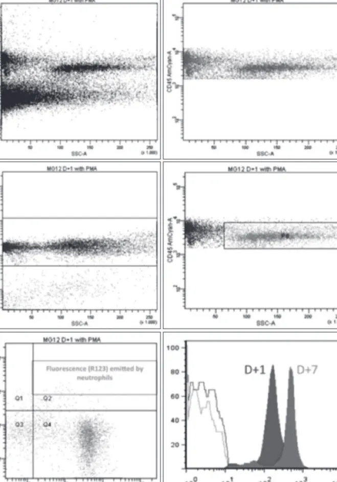
Cell fragments and unnucleated cells were excluded using DRAQ-5. Fluorescence due to rhodamine-123 was measured on neutrophils (CD45 + /CD14-) and monocytes (CD45 + /CD14 +) (Figure 1). Data acquisition was started only after one minute to ensure a constant flow for repro-ducibility and accuracy of the results.
Detection of membrane antigens
50 μl of EDTA-treated whole blood was labeled with 5 µl of a saturating amount of CD11b-FITC (clone = bear1 IOtest, Beckman Coulter, Suarlée, Belgium), CD11c-PE 31,25 µg/mL (clone = B-ly6, BD Pharmingen, San Jose, CA, USA), CD45-PerCP 6 µg/mL (clone = 2D1, Becton Dickinson), CD15-APC 5mg/0,5mL (clone = HI98, BD Phar-mingen, San Jose, CA, USA), and CD16-PB 6,25 µg/mL (clone = 3G8 BD Pharmingen, San Jose, CA, USA). After 15 min of incubation in the dark, 2 ml of Phosphate Buffered Saline was added. Autofluorescence and FMO were used as negative controls. Acquisition was performed as described for oxidative burst measurement. CS&T beads (Becton Dickinson, San Jose, CA, USA) were used for weekly adjustment of the photomultiplier vol-tages in order to maintain fluorescence targets,
according to Euroflow recommendations.7 This
calibration allows comparison of Mean Fluo-rescence Intensity measurements.
Statistical analysis
Statistical analysis was performed with Medcalc version 11.1.1.0 (Gent, Belgium). The results are expressed as median value with interquartile range tetraacetic acid (EDTA K2), heparin, or citrate. All
the measurements were performed without delay after sampling.
Measurement of hematological parameters
Routine hematology fluorescence flow cytometry of whole blood samples and processed/stimulated whole blood samples was carried out on a XE-2100 (Sysmex Corporation, Kobe, Japan), an auto mated analyzer that performs leukocyte, red blood cell (RBC), and platelet (PLT) analysis using several analytical channels: side-scatter light analysis of intracellular com plexity, forward scatter measure-ments of cellular size, and fluorescence intensity measurements of cellular nucleic acid (NA)/protein content. Finally, changes in neutrophil granularity were measured by Neutro-X in the DIFF channel, which detects neutrophilic hypo- and hyper granula-tion. The mini mal blood sample volume required for cell blood count (CBC) was 200 μl in automatic mode.
Measurement of oxidative burst5
The oxidative burst measurement was carried out by flow cytometry (FCM) after phorbol my-ristate acetate (PMA) (Sigma-Aldrich, Bornem, Belgium.Concentration: 50 ng/ml) neutrophil acti-vation. Dihydrorhodamine 123 (DHR) (Invitrogen, Life Technologies, Gent, Belgium. Concentration 5 mM) laser dye was used for studies of reactive
oxygen species (ROS) production. DHR freely
enters the cell membrane, and after ROS-mediated oxidation to rhodamine 123 it emits a bright fluorescent signal. Since rhodamine 123 binds to cellular and mitochondrial membranes, the fluo-rescent signal is predominantly localized inside the cell. DHR is specifically responsive to H2O2
accumulation.6
a) PMA activation
An aliquot of 100 µl heparinized whole blood was incubated with 400 µl Hanks’ balanced salt solution (HBSS) and 6.5 µl PMA for 25 minutes at 37°C. b) Oxidative burst measurement
After incubation, the mixture was added to 1 µl of 5 mM DRAQ-5 (Biostatus, Leicestershire, UK), which is a saturating amount, 10 μl of diluted (1/100) dihydrorhodamine 123 (Invitrogen, Mole-cular Probes, Life Technologies, Gent, Belgium),
Figure 1
Oxidative burst: flow cytometry.
The samples taken on Day 1 and Day 7 after surgery were compared. Initial isolation of cells expressing CD45, a leukocyte common antigen, isolated all leukocytes. Afterwards, cell fragments and enucleated cells were excluded using DRAQ-5. After exposure to phorbol myristate acetate (PMA), fluorescence due to rhodamine-123 was measured on neutrophils (CD45 + /CD14-).
Circulating neutrophils after sinus surgery 5
(mean: 3.71 × 10³ cells/µl) but was not significant.
At D + 1, the neutrophil count was significantly
higher than baseline values (5.08 × 10³ cells/µl,
p <0.01) and than the values at D + 30 (p <0.05)
with a mean of 3.69 × 10³ cells/µl. The mean
neutrophil count gradually diminished from D + 1 to D + 30. Considering the normative values for this test (normal range 2.10-6.30 cells/µl), only the maximal values at H + 1, D + 1, and D + 14 were considered to be elevated.
Oxidative burst
The oxidative burst, evaluated by the concentration of rhodamine 123 measured after PMA activation, (Figure 4, solid line) was significantly reduced at H + 1 (p <0.01) compared to baseline values. After reduction of the value at D + 1 and D + 7, the mean value increased significantly at D + 14 (p <0.01), compared to baseline, reaching values of up to 22.049 arbitrary units (AU). The Neutro-X values (Figure 4, dotted line) also showed a significant initial decrease (p <0.1, at D + 1) and a significant increase (p <0.2) at D + 14 after surgery, before decreasing again by D + 30. At D + 14, the relative (IQR). Comparison of serial measurements (H + 1,
D + 1, D + 7, D + 14, and D + 30) was performed using the Wilcoxon test. A p-value lower than 0.1 was noted as a trend, and a p-value lower than 0.05 was considered to be statistically significant.
Results
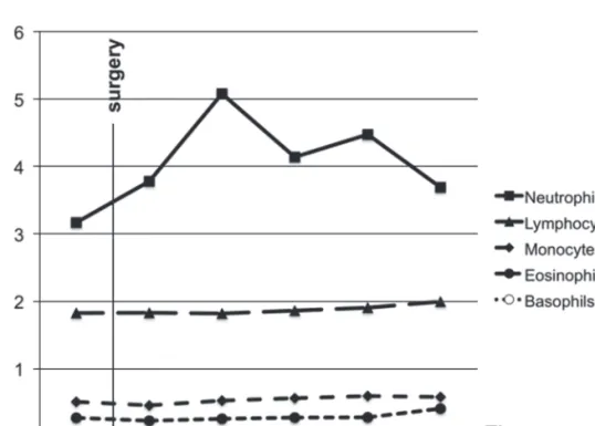
Circulating blood cell counts after sinus surgery With the exception of neutrophils, blood cell populations remained stable after sinus surgery (Figure 2). The neutrophil group showed increases at time points one hour and one day after surgery (H + 1 and D + 1), and values gradually returned to normal by D + 30 (Figure 2).
Neutrophil count after sinus surgery
At baseline, the mean neutrophil count was 3.16 ×
103 cells/µl, ranging from 1.71 to 4.36 × 10³ cells/µl
(Figure 3). After sinus surgery, the amount of circu-lating neutrophils showed a significant increase at the timepoint D + 1, and then a progressive reduction until D + 30. An early post-operative increase in the neutrophil count was noted at the H + 1 timepoint
Figure 2
Circulating blood cell counts: mean (×1000 cells/ml) after sinus surgery.
The total count of neutrophils and other cell populations are expressed as the mean (×1000 cells/ml). Time points before and after surgery are as follows: H-1, before surgery; H + 1, one hour after surgery; D + 1, one day after surgery; D + 7, seven days after surgery; D + 14, fourteen days after surgery; D + 30, thirty days after surgery.
Figure 3
Neutrophil count.
The mean, minimum, and maximum neutrophil count is shown (×1000 cells/ml) across the following time points: H-1, before surgery; H + 1, one hour after surgery; D + 1, one day after surgery; D + 7, seven days after surgery; D + 14, fourteen days after surgery; D + 30, thirty days after surgery. The normal, baseline value was determined to be 2100-6300 cells/ml.
Figure 4
Oxidative burst.
The oxidative burst was measured by dihydroxyrhodamine reaction (full line) and Neutro-X (dotted line) across the following time points: H-1, before surgery; H + 1, one hour after surgery; D + 1, one day after surgery; D + 7, seven days after surgery; D + 14, fourteen days after surgery; D + 30, thirty days after surgery.
Circulating neutrophils after sinus surgery 7
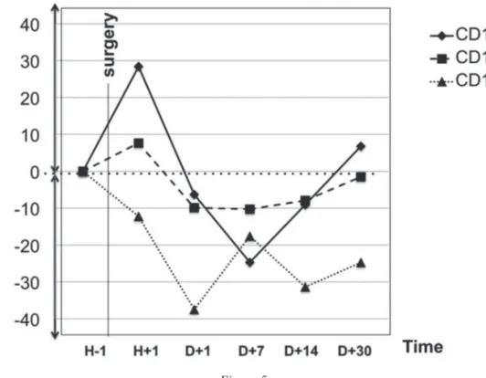
expression, which reached 37% of baseline at D + 1. After D + 1, the trend was to a slow but irregular return toward normal. These differences in CD15 expression were statistically significant between H-1 and H + 1 (p <0.05), between H + 1 and D + 1 (p <0.01), and between D + 1 and D + 7 (p <0.05).
Discussion
In this exploratory pilot study, we have shown that, in contrast to other subpopulations of inflamma-tory cells, the circulating neutrophils are actively involved in the process of wound healing after sinus
surgery. Multiple neutrophil-related changes are
evident in these patients: increased circulating neutrophils, increased oxidative burst activity, and changes in membrane expression of integrins, as determined by flow cytometry. These observations were made in patients suffering from CRS during the early defense and wound healing period fol-lowing sinus surgery, in a homogenous group of patients who healed well. This study illustrates how the circulating reserve of inflammatory cells acts during the post-surgical repair process and could offer new insights into the role of this cell population variation in Neutro-X index reached 96% of the
baseline value.
Expression of adhesion molecules
The membrane expression of CD11b and CD11c followed a similar trajectory during the early post-operative period, with a limited increase at H + 1, a slight decrease at D + 7, and a trend toward baseline at D + 30 (Figure 5). At H + 1, the CD11b changes were more marked, with a relative variation of 28% from baseline, when compared to CD11c, which showed a 7% variation from baseline. However, these differences were not statistically significant. At D + 1, a slightly negative relative variation was observed for both (-6% for CD11b and -9% for CD11c). This limited reduction persisted until the D + 30 timepoint, where a trend toward normal values was observed. The membrane expression of CD11b was significantly higher at D + 7 compared to D + 14 and D + 30 (p <0.05). The changes ob-served in membrane expression of CD11c were not significant.
With regard to CD15, the post-operative period was characterized by a reduction in membrane
Figure 5
Neutrophil CD11b, CD11c, and CD15 expression.
Neutrophil CD11b, CD11c, and CD15 expression, expressed in relative variation (%) from baseline. Measurements are presented for the following time points: H-1, before surgery; H + 1, one hour after surgery; D + 1, one day after surgery; D + 7, seven days after surgery; D + 14, fourteen days after surgery; D + 30, thirty days after surgery.
ferent phases of the healing process. In our study, both quantitative and qualitative measurements were performed to investigate the activation profile of circulating neutrophils during the early phase of upper airway repair. Many intriguing changes were observed, though not always significant in magnitude.
During cardiovascular surgery18 or in other
wound repair models,10 the total neutrophil count
increases in the first post-operative hour and returns to normal after several weeks. Our present results monitoring sinus surgery patients are consistent with results from post-surgery monitoring after cardiopulmonary bypass, in which an induced neutrophilia was observed during the first days of
the healing period.19
Additional studies have demonstrated the rapid adaptative profile of neutrophils during the early post-operative period. In vitro or in animal models of tissue repair, superoxide anion pro duction by neutrophils has been studied under different stimu-latory conditions: for example, with granulocyte-macrophage colony-stimulating factor (GM-CSF), with bacteria (Escherichia coli), or with a combina-tion of tumor necrosis factor alpha (TNFalpha) and N-formyl-methionyl-leucyl-phenylalanine
(FMLP).20 Inhibitory conditions have also been
studied, such as experimentally employing
anti-CD11b antibody to reduce myofiber damage.20 In
our study, we monitored the burst oxidative reaction during the first days after sinus surgery using flow
cytometry.5 The changes observed appeared directly
linked to structural variations in the Neutro-X labeling, and they occurred in inverted phase with the induced neutrophilia. Our results were similar
to those of Fung et al.,19 who observed a decline in
neutrophil respiratory burst activity after cardio-pulmonary bypass until day 5 post-surgery, with concomitant changes in surface molecule expres-sion, which may reduce the neutrophil activation response. However, contradictory results have also been published regarding the neutrophil oxidative burst after surgery. Increased neutrophil phago-cytosis and oxidative burst activity were detected during the first post-operative days after minor
surgery in children.21 In another study, after major
abdominal surgery and in response to endotoxin priming, the neutrophil response to environmental stimulants was found to be suppressed initially, but
then increased after three days.22 A human wounding
assay has indicated a change in responsiveness in the interactions between blood and peripheral
airway compartments in chronic airway disease. A recent study describes the general mechanism of neutrophil chemotaxis and the function of neutro-phils in inflamed tissue during chronic
inflam-matory disease or at the site of injured mucosa;8
however, the fine-grained mechanisms involved in inflammatory cell recruitment and trafficking remain only partially understood. Generation of an intravascular chemokine gradient directs neutrophil migration through the healthy tissue toward foci of
damage.9 However, the neutrophil migration seen
during wound repair seems not to be exclusively
unidirectional or monophasic.10 In an in vivo animal
model, spatiotemporal photolabeling of neutrophils demonstrated that they emerge from the hemato-poietic tissue in close proximity to injured tissue, and that they can show both forward and reverse migration between the wound and the vasculature, contributing to both local resolution at the site of the tissue injury and to systemic dissemination of
wound-sensitized neutrophils.11
Several conditions can significantly affect neu-tro phil trafficking during wound healing. Age-related defects in early neutrophil recruitment may alter the dynamics of the inflammatory phase of wound healing, having impacts on macrophage recruitment, bacterial clearance, and wound
clo-sure.12 Changes in neutrophil recruitment that are
induced by systemic infection or by diabetes can alter both the inflammatory and the proliferative
processes at remote sites of injury.13
By shaping immune responses, neutrophils contribute to tissue repair, as described above; however, neutrophils can also lead to tissue break-down. It has been observed that local activation of recruited neutrophils may cause a variable degree
of tissue destruction14 either directly or through
local activation of other inflammatory cells15 and
this can affect the repair outcome. For example, an epinephrine-induced persistent neutrophil migra-tion to the wound site occurs by an IL-6 mediated mechanism, and this process in turn impairs wound
repair.16 The involvement of neutrophils in the local
airway repair process seems even more complex because, they also mediate amplification of the defense mechanisms, by systematic release of a wide range cytokines such as TGF-β, TNF-α,
IL-1β, IL-3, IL-6, IL-8, and GM-CSF.17
It is possible that many regulatory processes modulate the neutrophil activity profile during
dif-Circulating neutrophils after sinus surgery 9 study of the biological profile exhibited by patients who healed poorly after sinus surgery and com-parisons with repair processes observed after other types of surgery also merits further exploration in the future.
Conclusions
In this pilot study, we demonstrated for the first time that systemic neutrophils are significantly involved in the early phase of upper airway defense and repair after surgical trauma. Neutrophils exhibited early-phase membrane changes facilita-ting migration and then, in a second phase, exhibited changes supporting oxidative response. These data provide new evidence supporting a reservoir-like role for the vascular compartment and may also be of diagnostic or therapeutic importance for patients who may be susceptible to poor healing from airway damage.
Acknowledgements
We acknowledge Y. CORNET and N. BAILLY from the Laboratory of Hematology-Namur Thrombosis and Hemostasis Center, CHU Dinant-Godinne UCL Namur, for standardization of techniques, calibrations and regular measurements and B. BERTRAND, M.D., from the Department of Otorhinolaryngology, Head and Neck Surgery, CHU Dinant-Godinne UCL Namur, for support of the clinical aspects of this study.
References
1. Fokkens WJ, Lund VJ, Mullol J, Bachert C, Alobid I, Baroody F, Cohen N, Cervin A, Douglas R, Gevaert P, Georgalas C, Goossens H, Harvey R, Hellings P, Hopkins C, Jones N, Joos G, Kalogjera L, Kern B, Kowalski M, Price D, Riechelmann H, Schlosser R, Senior B, Thomas M, Toskala E, Voegels R, Wang de Y, Wormald PJ. European Position Paper on Rhinosinusitis and Nasal Polyps. Rhinology. 2012;50 suppl 23:S1-298. 2. Saedi B, Sadeghi M, Akhavan-Khaleghi N, Seifmanesh H.
Impact of endoscopic sinus surgery on the quality of life of patients with nasal polyposis. B-ENT. 2014;10(1):59-65. 3. Watelet JB, Claeys C, Van Cauwenberge P, Bachert C.
Predictive and monitoring value of matrix metallo-proteinase-9 for healing quality after sinus surgery. Wound
Repair Regen. 2004;12(4):412-418.
4. Watelet JB, Demetter P, Claeys C, Cauwenberge P, Cuvelier C, Bachert C. Wound healing after paranasal sinus surgery: neutrophilic inflammation influences the outcome.
Histopathology. 2006;48(2):174-181.
5. Elbim C, Lizard G. Flow cytometric investigation of neutrophil oxydative burst and apoptosis in physiological
to chemotactic and immunoregulatory mediators observed once neutrophils have migrated to skin
lesions and have been activated.23 These conflicting
findings could be due to the fact that these various studies have been conducted with different surgical modes, different time analyses, and using different procedures, which may modulate the expression of surface receptors. Similar considerations might also apply with regard to the exploration of post-operative membrane changes.
The vascular adhesion and the transendothelial migration of neutrophils are mediated by adhe-sion molecules, particularly intercellular adheadhe-sion
molecule-124 and E-selectin, which is known to
participate in extravasation of neutrophils and lymphocytes and is also expressed during wound healing. CD11 has been the subject of many recent studies on wound healing. After major surgery or burn wounding an upregulation of baseline CD11b expression is mirrored by changes in neutrophil
adhesion,25 even in children. Expression of CD11b
shows a strong response to injury which comprises an initial increase and then a later return toward baseline, as confirmed in our study. Interestingly, the membrane expression of CD15 over time forms a perfect mirror image of the recruitment curve. We could hypothesize that, in order to counterbalance an overly massive neutrophil efflux towards the wound field, the diminished expression of mem-brane adhesion molecules can act to regulate diapedesis. Finally, the “overshooting” phase ob-served at D + 14 with respect to CD11b and CD11c expression as well as the oxidative burst could be considered to play a protective role against any hematogenous extension of inflammation or infection.
However, as both the oxidative burst reaction and the expression of CD11b can differ between peripheral and circulating neutrophils, the local physiologic wound situation cannot be safely pre-dicted based on information about the circulating neutrophil activities. Finally, it is not clear how these findings relate to neutrophil migration during lower airway inflammation.
This pilot study is limited by the small number of patients and by its monocentricity. Furthermore, by pooling data on patients exhibiting different subgroups of chronic rhinosinusitis who healed normally after surgery, the subgroup-specific principles leading to normal mucosal recovery cannot be distinguished from each other. Finally,
Alters Neutrophil Trafficking and Impairs Wound Healing by β2 Adrenergic Receptor Mediated Upregulation of IL-6. J Invest Dermatol. 2014;134(3):809-817.
17. Werner S, Grose R. Regulation of wound healing by growth factors and cytokines. Physiol Rev. 2003;83(3):835-870.
18. Kubala L, Cíz M, Vondrácek J, Cerný J, Nemec P, Studeník P, Cizová H, Lojek A. Perioperative and post-operative course of cytokines and the metabolic activity of neutro phils in human cardiac operations and heart transplantation. J Thorac Cardiovasc Surg. 2002;124(6): 1122-1129.
19. Fung YL, Silliman CC, Minchinton RM, Wood P, Fraser JF. Cardiopulmonary bypass induces enduring alterations to host neutrophil physiology: a single-center longitudinal observational study. Shock. 2008;30(6):642-648.
20. Jaeger K, Scheinichen D, Heine J, Ruschulte H, Kuse E, Winkler M, Leuwer M. GM-CSF increases in vitro the respiratory burst of human neutrophils after liver trans-plantation. Intensive Care Med. 1999;25(6):612-615. 21. Romeo C, Cruccetti A, Turiaco A, Impellizzeri P, Turiaco
N, Di Bella C, Merlino MV, Cifalà S, Basile M, Gentile C, Salpietro DC. Monocyte and neutrophil activity after minor surgical stress. J Pediatr Surg. 2002;37(5):741-744. 22. Moriwaki Y, Sugiyama M, Ozawa Y, Mochizuki Y,
Kunisaki C, Kamiya N, Yamazaki Y, Suda T. Changes in the response of neutrophils to endotoxin priming following major abdominal surgery. World J Surg. 2002;26(5):521-526.
23. Theilgaard-Mönch K, Knudsen S, Follin P, Borregaard N. The transcriptional activation program of human neutro-phils in skin lesions supports their important role in wound healing. J Immunol. 2004;172(12):7684-7693.
24. Nagaoka T, Kaburagi Y, Hamaguchi Y, Hasegawa M, Takehara K, Steeber DA, Tedder TF, Sato S. Delayed wound healing in the absence of intercellular adhesion molecule-1 or L-selectin expression. Am J Pathol. 2000; 157(1):237-247.
25. Takala AJ, Jousela IT, Takkunen OS, Jansson SE, Kyösola KT, Olkkola KT, Leirisalo-Repo M, Repo H. Time course of beta 2-integrin CD11b/CD18 (Mac-1, alpha M beta 2) upregulation on neutrophils and monocytes after coronary artery bypass grafting. CD11b upregulation after CABG surgery. Scand J Thorac Cardiovasc Surg. 1996;30(3-4): 141-148.
and pathological situations. Cytometry. 2009;75(6):475-481.
6. Walrand S, Valeix S, Rodriguez C, Ligot P, Chassagne J, Vasson MP. Flow cytometry study of polymorphonuclear neutrophil oxidative burst: A comparison of three fluorescent probes. Clin Chim Acta. 2003;331(1-2):103-110.
7. Kalina T, Flores-Montero J, van der Velden VH, Martin-Ayuso M, Böttcher S, Ritgen M, Almeida J, Lhermitte L, Asnafi V, Mendonça A, de Tute R, Cullen M, Sedek L, Vidriales MB, Pérez JJ, te Marvelde JG, Mejstrikova E, Hrusak O, Szczepański T, van Dongen JJ, Orfao A; EuroFlow Consortium (EU-FP6, LSHB-CT-2006-018708). EuroFlow standardization of flow cytometer instrument settings and immunophenotyping protocols. Leukemia. 2012;26(9):1986-2010.
8. Kolaczkowska E, Kubes P. Neutrophil recruitment and function in health and inflammation. Nat Rev Immunol. 2013;13(3):159-175.
9. McDonald B, Pittman K, Menezes GB, Hirota SA, Slaba I, Waterhouse CC, Beck PL, Muruve DA, Kubes P. Intra-vascular danger signals guide neutrophils to sites of sterile inflammation. Science. 2010;330(6002):362-366.
10. Li Z, Burns AR, Smith CW. Two waves of neutrophil emigration in response to corneal epithelial abrasion: dis-tinct adhesion molecule requirements. Invest Ophthalmol
Vis Sci. 2006;47(5):1947-1955.
11. Yoo SK, Huttenlocher A. Spatiotemporal photolabeling of neutrophil trafficking during inflammation in live zebrafish.
J Leukoc Biol. 2011;89(5):661-667.
12. Brubaker AL, Rendon JL, Ramirez L. Reduced neutrophil chemotaxis and infiltration contributes to delayed reso-lution of cutaneous wound infection with advanced age.
J Immunol. 2013;190(4):1746-1757.
13. Ochoa O, Torres FM, Shireman PK. Chemokines and diabetic wound healing. Vascular. 2007;15(6):350-355. 14. Lee SM, Rosen S, Weinstein P, van Rooijen N,
Noble-Haeusslein LJ. Prevention of both neutrophil and monocyte recruitment promotes recovery after spinal cord injury.
J Neurotrauma. 2011;28(9):1893-1907.
15. Dovi JV, He LK, DiPietro LA. Accelerated wound closure in neutrophil-depleted mice. J Leukoc Biol. 2003;73(4):448-455.
16. Kim MH, Gorouhi F, Ramirez S, Granick JL, Byrne BA, Soulika AM, Simon SI, Isseroff RR. Catecholamine Stress