Crystal Structure of the Minimal Cas9 from Campylobacter jejuni
Reveals the Molecular Diversity in the CRISPR-Cas9 Systems
The MIT Faculty has made this article openly available.
Please share
how this access benefits you. Your story matters.
Citation
Yamada, Mari et al. “Crystal Structure of the Minimal Cas9 from
Campylobacter Jejuni Reveals the Molecular Diversity in the
CRISPR-Cas9 Systems.” Molecular Cell 65, 6 (March 2017): 1109–
1121 © 2017 Elsevier Inc
As Published
https://doi.org/10.1016/j.molcel.2017.02.007
Publisher
Elsevier
Version
Author's final manuscript
Citable link
http://hdl.handle.net/1721.1/114953
Terms of Use
Creative Commons Attribution-NonCommercial-NoDerivs License
Detailed Terms
http://creativecommons.org/licenses/by-nc-nd/4.0/
Article
12
Crystal structure of the minimal Cas9 from
3Campylobacter jejuni reveals the molecular
4
diversity in the CRISPR-Cas9 systems
56
Mari Yamada
1,9, Yuto Watanabe
1,9, Jonathan S. Gootenberg
2,3,4,5,6, Hisato Hirano
1, F.
7
Ann Ran
2,7, Takanori Nakane
1, Ryuichiro Ishitani
1, Feng Zhang
2,3,4,5, Hiroshi
8
Nishimasu
1,8,*, Osamu Nureki
1,10,*9 10
1 Department of Biological Sciences, Graduate School of Science, The University of Tokyo,
2-11
11-16 Yayoi, Bunkyo-ku, Tokyo 113-0032, Japan 12
2 Broad Institute of MIT and Harvard, Cambridge, MA 02142, USA
13
3 McGovern Institute for Brain Research, Massachusetts Institute of Technology, Cambridge,
14
MA 02139, USA 15
4 Department of Brain and Cognitive Sciences, Massachusetts Institute of Technology,
16
Cambridge, MA 02139, USA 17
5 Department of Biological Engineering, Massachusetts Institute of Technology, Cambridge,
18
MA 02139, USA 19
6 Department of Systems Biology, Harvard Medical School, Boston, MA 02115, USA
20
7 Society of Fellows, Harvard University, Cambridge, MA 02138, USA
21
8 JST, PRESTO, 2-11-16 Yayoi, Bunkyo-ku, Tokyo 113-0032, Japan
22
9 These authors equally contributed to this work
23 10 Lead Contact 24 25 * Correspondence: 26
nisimasu@bs.s.u-tokyo.ac.jp (H.N.) and nureki@bs.s.u-tokyo.ac.jp (O.N.). 27
The RNA-guided endonuclease Cas9 generates a double-strand break at DNA target sites 1
complementary to the guide RNA, and has been harnessed for the development of a 2
variety of new technologies, such as genome editing. Here, we report the crystal structures 3
of Campylobacter jejuni Cas9 (CjCas9), one of the smallest Cas9 orthologs, in complex with 4
an sgRNA and its target DNA. The structures provided insights into a minimal Cas9 5
scaffold, and revealed the remarkable mechanistic diversity of the CRISPR-Cas9 systems. 6
The CjCas9 guide RNA contains an unexpected triple-helix structure, which is distinct 7
from known RNA triple helices, thereby expanding the natural repertoire of RNA triple 8
helices. Furthermore, unlike the other Cas9 orthologs, CjCas9 contacts the nucleotide 9
sequences in both the target and non-target DNA strands, and recognizes the 5′-10
NNNVRYM-3′ as the protospacer adjacent motif. Collectively, these findings improve our 11
mechanistic understanding of the CRISPR-Cas9 systems, and may facilitate Cas9 12
engineering. 13
Introduction 1
Bacteria and Archaea utilize CRISPR-Cas (clustered regularly interspaced short palindromic 2
repeats-CRISPR-associated) adaptive immune systems to defend themselves against foreign 3
genetic elements, such as plasmids and phages (Marraffini, 2015; Barrangou and Doudna, 2016; 4
Mohanraju et al., 2016; Wright et al., 2016). The CRISPR loci in the genome comprise a cas 5
operon and a CRISPR array, consisting of short repetitive sequences (direct repeats) separated 6
by non-repetitive sequences (spacers) derived from foreign genetic elements. The CRISPR array 7
is transcribed and processed into CRISPR RNAs (crRNAs), which associate with single or 8
multiple Cas proteins to form effector ribonucleoprotein complexes responsible for the 9
destruction of invading nucleic acids (Makarova et al., 2015; Nishimasu and Nureki, 2016). In 10
the type II CRISPR-Cas system, the Cas9 effector nuclease associates with dual guide RNAs 11
(crRNA and trans-activating crRNA (tracrRNA)), and cleaves double-stranded (ds) DNA 12
targets complementary to the crRNA guide(Garneau et al., 2010; Deltcheva et al., 2011;
13
Gasiunas et al., 2012; Jinek et al., 2012). In addition to the crRNA-target DNA 14
complementarity, Cas9-mediated target recognition requires a PAM (protospacer adjacent 15
motif), a short nucleotide sequence adjacent to the target site(Deveau et al., 2008; Garneau et
16
al., 2010). Importantly, a single-guide RNA (sgRNA), in which crRNA and tracrRNA are fused 17
with an artificial tetraloop, can also direct Cas9 to the target cleavage (Jinek et al., 2012). Thus, 18
the two component Cas9-sgRNA system has been harnessed for a variety of new technologies, 19
including genome editing(Cong et al., 2013; Jinek et al., 2013; Mali et al., 2013).
20 21
Structural studies of Streptococcus pyogenes Cas9 (SpCas9) have provided mechanistic details 22
of the RNA-guided DNA cleavage by the Cas9 enzyme. The crystal structure of SpCas9 in its 23
apo form revealed a bilobed architecture comprising an α-helical recognition (REC) lobe and a 24
nuclease (NUC) lobe (Jinek et al., 2014). The crystal structure of SpCas9 bound to the sgRNA 25
and its single-stranded DNA target clarified the recognition mechanism of the sgRNA and the 26
target DNA(Nishimasu et al., 2014). Subsequently, the crystal structure of SpCas9 bound to the
27
sgRNA and a PAM-containing DNA revealed the recognition mechanism of the 5′-NGG-3′ 28
PAM by SpCas9 (Anders et al., 2014). Moreover, the crystal structures of SpCas9 bound to the 29
sgRNA (Jiang et al., 2015) and SpCas9 bound to an R-loop (Jiang et al., 2016) demonstrated the 30
structural rearrangements in the Cas9 protein accompanying the guide RNA binding and R-loop 31
formation, respectively. 32
The Cas9 orthologs from different microbes have highly divergent sequences, function with 1
their cognate crRNA:tracrRNA guides, and recognize a variety of PAM sequences(Chylinski et
2
al., 2013; Fonfara et al., 2014; Hsu et al., 2014; Karvelis et al., 2015; Ran et al., 2015). SpCas9 3
(1,368 aa) recognizes 5′-NGG-3′ as the PAM(Mojica et al., 2009), whereas Staphylococcus
4
aureus Cas9 (SaCas9, 1,053 aa) and Francisella novicida Cas9 (FnCas9, 1,629 aa) recognize
5′-5
NNGRRT-3′ and 5′-NGG-3′ as the PAMs, respectively (Ran et al., 2015; Hirano et al., 2016). A 6
structural comparison of SpCas9(Anders et al., 2014; Nishimasu et al., 2014) with SaCas9
7
(Nishimasu et al., 2015) and FnCas9 (Hirano et al., 2016) revealed that, while they share the 8
conserved RuvC and HNH nuclease domains, their REC and Wedge (WED) domains are 9
structurally divergent, and recognize distinct structural features in their cognate RNA guides. In 10
addition, their PAM-interacting (PI) domains share a conserved core fold, but recognize distinct 11
PAM sequences, using a specific set of amino-acid residues. 12
13
Campylobacter jejuni Cas9 (CjCas9) has several unique features(Fonfara et al., 2014). First, 14
CjCas9 consists of 984 residues, and is one of the smallest Cas9 orthologs. Second, the 15
nucleotide sequences of the crRNA:tracrRNA guides for CjCas9 and the other Cas9 orthologs 16
differ substantially. Third, CjCas9 recognizes the 5′-NNNNACA-3′ PAM, whereas most Cas9 17
orthologs, as exemplified by SpCas9 (5′-NGG-3′) (Gasiunas et al., 2012; Jinek et al., 2012), 18
recognize G-rich PAMs. However, the functional mechanism of CjCas9 remains elusive, due to 19
the lack of structural information. 20
21
Here, we performed functional and structural characterizations of CjCas9. In vitro cleavage 22
experiments revealed that CjCas9 recognizes the 5′-NNNVRYM-3′ PAM, which is more 23
promiscuous than the previously reported PAMs. The crystal structure of the CjCas9-sgRNA-24
target DNA complex highlighted the remarkable mechanistic diversity of the CRISPR-Cas9 25
systems. Unlike the tracrRNAs for the other Cas9 orthologs, the CjCas9 tracrRNA has an 26
unanticipated triple-helix structure, which is distinct from known RNA triple helices. 27
Furthermore, CjCas9 recognizes the PAM nucleotides on both the target and non-target DNA 28
strands, whereas the other Cas9 orthologs recognize the PAM nucleotides on the non-target 29
DNA strand. 30
31
Results and Discussion 32
33
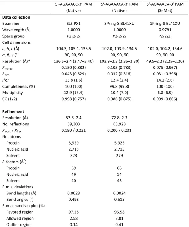
CjCas9 PAM specificity 34
Although a previous study reported that CjCas9 recognizes the 5′-NNNNACA-3′ PAM(Fonfara 1
et al., 2014), the CjCas9 PAM has not been fully characterized. To determine the CjCas9 PAM, 2
we performed the PAM discovery assay, using purified CjCas9, an sgRNA and a library of 3
plasmid DNA targets with a degenerate 7-bp PAM sequence, as described previously (Ran et 4
al., 2015; Zetsche et al., 2015). The result revealed that CjCas9 recognizes the 5′-NNNVRYM-5
3′ PAM (V is A/G/C; R is A/G; Y is T/C; M is A/C) (Figures 1A and S1), which is more 6
promiscuous than the previously reported 5′-NNNNACA-3′ PAM(Fonfara et al., 2014). Using
7
purified CjCas9 and an sgRNA, we further examined the cleavage of 13 plasmid DNA targets 8
with either 5′-AGANACC-3′, 5′-AGAANCA-3′, 5′-AGAAANA-3′ or 5′-AGAACN-3′ as the 9
PAMs. The results confirmed that CjCas9 efficiently recognizes the 5′-NNNVRYM-3′ PAM, 10
with the preference for T and C at positions 6 and 7, respectively (Figures 1B and 1C). 11
12
Crystal structure of the CjCas9-sgRNA-target DNA complex 13
To clarify the RNA-guided DNA recognition mechanism of CjCas9, we attempted to determine 14
the crystal structure of CjCas9 in complex with an sgRNA and its target DNA. However, we 15
failed to obtain diffraction-quality crystals. Previous studies revealed that the HNH nuclease 16
domain of SpCas9 is mobile and dispensable for RNA-guided DNA recognition (Nishimasu et 17
al., 2014; Jiang et al., 2015; Sternberg et al., 2015), suggesting that the flexibility of the HNH 18
domain may hamper the crystallization. We thus prepared the CjCas9-∆HNH variant lacking the 19
HNH domain (residues 481–640), in which Leu480 (RuvC-II) and Tyr641 (RuvC-III) are 20
connected by a GGGSGG linker (Figure 2A). After extensive crystallization screening, we 21
determined the crystal structure of CjCas9-∆HNH in complex with a 93-nt sgRNA, a 28-nt 22
target DNA strand, and an 8-nt non-target DNA strand containing the 5′-AGAAACC-3′ PAM, 23
at 2.4 Å resolution (Figures 2A–2C and Table 1). 24
25
The crystal structure revealed that CjCas9 adopts a bilobed architecture comprising the α-helical 26
REC lobe and a NUC lobe, as in the other Cas9 orthologs(Anders et al., 2014; Nishimasu et al.,
27
2014; Nishimasu et al., 2015) (Figure 2C), indicating that the truncation of the HNH domain 28
does not substantially affect the overall structure. The REC lobe can be divided into the REC1 29
(residues 77–234) and REC2 (residues 235–426) domains. The NUC lobe comprises the RuvC 30
(residues 1–44, 427–480 and 641–777), WED (residues 792–827), and PI domains (residues 31
828–984) (the HNH domain was truncated for crystallization). The REC and NUC lobes are 32
connected by an arginine-rich “bridge” helix (residues 45–76), while the WED and RuvC 33
domains are connected by a “phosphate lock” loop (residues 778–791), as in other Cas9 34
orthologs (Anders et al., 2014; Nishimasu et al., 2014; Nishimasu et al., 2015; Hirano et al., 1
2016). The three residues (GGS) in the GGGSGG linker between the RuvC-II and RuvC-III 2
motifs are disordered in the present structure. 3
4
The sgRNA comprises the guide segment (G1–C20), the repeat region (G21–U32), the tetraloop 5
(G33–A36), the antirepeat region (A37–U48), and the tracrRNA scaffold (A49–C93) (Figure 6
2B). The guide segment (G1–C20) and the target DNA strand (dG1–dC20) form an RNA-DNA 7
heteroduplex (Figures 2B and 2C). The repeat and antirepeat regions form the A-form-like 8
duplex (referred to as the repeat-antirepeat duplex), which consists of a wobble base pair 9
(G21•U48) and eleven Watson-Crick base pairs (U22-A47–U32-A37) (Figures 2B and 2C). The 10
RNA-DNA heteroduplex is bound within the central channel between the REC and NUC lobes, 11
while the repeat-antirepeat duplex is sandwiched between the REC1 and WED domains (Figure 12
2C). These duplexes are primarily recognized by the protein in a non-sequence-specific manner 13
(Figure S2). The target DNA strand (dC(−8)–dT(−1)) and the non-target DNA strand (dA1*– 14
dG8*) form a PAM-containing duplex, which is recognized by the WED and PI domains 15
(Figures 2B and 2C). 16
17
A recent study showed that the deletion of the HNH domain in SpCas9 impairs the non-target 18
strand cleavage by the RuvC domain, thereby suggesting that the HNH domain is required for 19
the activation of the RuvC domain (Sternberg et al., 2015). We thus examined the effect of the 20
deletion of the HNH domain on the CjCas9 activity, using in vitro cleavage assays. Our results 21
revealed that, like the D8A RuvC-inactive nickase and the H559A HNH-inactive nickase, the 22
CjCas9-ΔHNH variant functions as a nickase (Figure 2D). Importantly, the CjCas9-ΔHNH 23
variant exhibited lower cleavage activity, as compared with the H559A nickase, indicating that 24
the deletion, but not the inactivating point mutation, of the HNH domain reduces the non-target 25
strand cleavage by the RuvC domain. These observations suggested the allosteric 26
communication between the RuvC and HNH nuclease domains in CjCas9, as observed in 27
SpCas9 (Sternberg et al., 2015). 28
29
TracrRNA architecture 30
Notably, the present structure revealed that the CjCas9 tracrRNA scaffold contains a triple-helix 31
structure within a pseudoknot comprising three stem regions, which was not predicted from its 32
primary sequence (Figures 3A–3C). Stem 1 consists of four canonical base pairs (A51-U67– 33
G54-C64) and a non-canonical A50•G68 base pair, while stem 2 consists of four canonical base 34
pairs (G58-C93–G61-C90) and a non-canonical G62•A89 base pair (Figures 3B and 3C). Stem 1
3 consists of four canonical base pairs (G70-C84–G74-C81) (Figures 3B and 3C). Nucleotides 2
56/57 and 86/87 form base pairs with stem 2 and stem 1, respectively, thereby contributing to 3
the triple-helix formation. U65 in stem 1 and A89 in stem 2 base pair with A86 and U56, 4
forming an A53-U65•A86 minor-groove triple and a G62-A89•U56 major-groove triple, 5
respectively (Figures 3D and 3E). U57 base pairs with C59 and C90, forming a G92-6
C59•U57•C90-G62 base quintuple (Figure 3F). A87 and A88 base pair with G54 and U55/C64, 7
respectively, forming a U55•A88•C64-G54•A87 base quintuple (Figure 3G). 8
9
The tracrRNA scaffold is extensively recognized by CjCas9 (Figures 4A and S2). In particular, 10
A63, A76 and U80 are flipped out, and recognized by the protein in base-specific manners. The 11
nucleobase and ribose moieties of A63 form stacking interactions with the side chains of His70 12
and His67, respectively, while the N1 of A63 hydrogen bonds with the side chain of Asn74 13
(Figure 4B). A76 and U80 are accommodated within specific pockets in the PI domain (Figure 14
4A). The nucleobase of A76 is sandwiched between the side chains of Ile964 and Arg977, while 15
the N6 and N7 of A76 hydrogen bond with the main-chain carbonyl and amide groups of 16
Glu975, respectively (Figure 4C). The nucleobase of U80 is sandwiched between the side 17
chains of Phe854 and Glu980, while the N3 and O4 of U80 hydrogen bond with the main-chain 18
carbonyl group of Asp981 and the side chain of Arg832, respectively (Figure 4C). Indeed, the 19
single mutations (A63U, A76U or U80A) reduced CjCas9-mediated DNA cleavage (Figure 20
4D), confirming the functional importance of the three flipped-out nucleotides. Moreover, a 4-nt 21
substitution (nucleotides 90–93) or an 8-nt deletion (nucleotides 86–93) in the tracrRNA 3′ tail 22
abolished the CjCas9-mediated DNA cleavage (Figure 4D), indicating that the triple-helix 23
structure of the tracrRNA is critical for the activity. In addition, the SpCas9 sgRNA did not 24
support the CjCas9-mediated DNA cleavage (Figure 4D). Together, these results demonstrated 25
that CjCas9 specifically recognizes its cognate RNA guide. 26
27
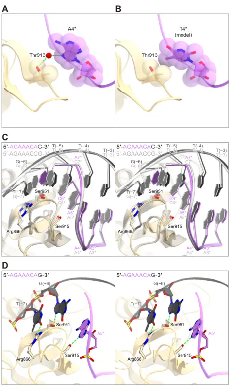
5′-NNNVRYM-3′ PAM recognition 28
In the present structure, the 5′-AGAAACC-3′ PAM-containing DNA duplex is bound to the 29
cleft between the WED and PI domains (Figure 5A). The nucleobases of dA1*–dA3* do not 30
directly contact the protein (Figures 5B and 5C), consistent with the lack of specificity for 31
positions 1–3 in the 5′-NNNVRYM-3′ PAM. The N7 of dA4* in the non-target strand forms a 32
water-mediated hydrogen bond with the side-chain hydroxyl group of Thr913 (Figures 5B and 33
5C). Modeling suggested that a steric clash could occur between the methyl group of dT4* and 34
the side chain of Thr913 (Figures S3A and S3B), consistent with the preference of CjCas9 for 1
the fourth V (A/G/C). The N7 of dA5* in the non-target strand forms a hydrogen bond with the 2
side-chain hydroxyl group of Ser915 (Figures 5B and 5C). Since N7 is common among the 3
purine nucleotides, the interaction can explain the requirement for the fifth R (A/G). Notably, 4
the nucleobase of dC6* in the non-target strand is not recognized by the protein (Figures 5B and 5
5C). Instead, the N7 of dG(−6) in the target strand forms a hydrogen bond with the side-chain 6
hydroxyl group of Ser951 (Figures 5B and 5C). These structural findings revealed that CjCas9 7
does not recognize the Y (T/C) nucleotides at position 6 in the non-target strand as the PAM, 8
but detects their complementary R (A/G) nucleotides in the target strand. Similarly, the 9
nucleobase of dC7* in the non-target strand is not recognized by the protein, whereas the O6 10
and N7 of dG(−7) in the target strand form bidentate hydrogen bonds with the side chain of 11
Arg866 (Figures 5B and 5C). In addition to the 5′-AGAAACC-3′ PAM complex, we 12
determined the crystal structure of CjCas9-∆HNH in complex with the sgRNA and the DNA 13
target containing the 5′-AGAAACA-3′ PAM (Table 1). In the 5′-AGAAACA-3′ PAM complex, 14
the dT(−7):dA7* pair in the PAM duplex undergo a slight displacement toward the PI domain, 15
as compared with the dG(−7):dC7* pair in the 5′-AGAAACC-3′ PAM complex (Figure S3C). 16
This displacement in the PAM duplex allows Arg866 to form a hydrogen bond with the O4 of 17
dT(−7) in the target strand (Figure S3D). These observations revealed that CjCas9 does not 18
recognize the M (A/C) nucleotides at position 7 in the non-target strand as the PAM, but detects 19
their complementary K (T/G) nucleotides in the target strand. The preference of CjCas9 for C 20
over A at position 7 can be explained by the bidentate hydrogen-bonding interaction between 21
dG(−7) and Arg866, in contrast to the single hydrogen-bonding interaction between dT(−7) and 22
Arg866. The single mutations of Arg866, Thr913, Ser915, and Ser951 reduced or abolished the 23
in vitro cleavage activity (Figure 5D), confirming their functional importance. Together, our
24
structural and functional data revealed that CjCas9 forms sequence-specific contacts with both 25
the target and non-target DNA strands, to achieve the recognition of the 5′-NNNVRYM-3′ 26
PAM. 27
28
Structural comparison between the Cas9 orthologs 29
A structural comparison of CjCas9 with the other Cas9 orthologs highlighted the structural 30
similarities and differences between the CRISPR-Cas9 systems, and provided insights into a 31
minimal Cas9 scaffold (Figure 6). Unlike SpCas9 (Anders et al., 2014; Nishimasu et al., 2014), 32
the smaller SaCas9 (Nishimasu et al., 2015) and CjCas9 lack the insertion subdomains within 33
the REC1 and PI domains (Figures 6A–6C). Furthermore, the WED domain of CjCas9 (36 34
amino acids) is smaller than that of SaCas9 (122 amino acids) (Figures 6A and 6C). These 1
structural differences contribute to the miniaturization of CjCas9. The REC and WED domains 2
of FnCas9, one of the largest Cas9 orthologs, adopt protein folds distinct from those of CjCas9, 3
SpCas9 and SaCas9 (Hirano et al., 2016) (Figure 6D), reinforcing the notion that FnCas9 is 4
distantly related to the other Cas9 orthologs. 5
6
Despite the structural differences in these individual domains, CjCas9 adopts the conserved 7
bilobed architecture, and accommodates the RNA-DNA heteroduplex in similar manners to 8
those of the other Cas9 orthologs (Figures 6A–6D). The sugar-phosphate backbone of the 9
sgRNA “seed” region (C13–C20) is extensively recognized by conserved arginine residues in 10
the bridge helix (Figure S4A). In addition, the backbone phosphate group between dG1 and 11
dT(−1) in the target DNA strand (referred to as the +1 phosphate(Anders et al., 2014)) interacts
12
with the main-chain amide groups of Glu790 and Thr791 and the side-chain hydroxyl group of 13
Thr791 in the phosphate lock loop, thereby facilitating target DNA unwinding (Figure S4B). 14
Indeed, the T791A mutation abolished the in vitro DNA cleavage activity (Figure S4C), 15
confirming the functional importance of the interaction between the +1 phosphate and Thr791. 16
These observations confirmed that the RNA-guided DNA targeting mechanism is highly 17
conserved among the CRISPR-Cas9 systems. 18
19
The present structure also illuminated the structural diversity of the crRNA:tracrRNA guides in 20
the CRISPR-Cas9 systems (Figures 6E–6H). The repeat-antirepeat duplexes for SpCas9, 21
SaCas9 and FnCas9 contain several unpaired nucleotides, and thus adopt distorted, distinct 22
structures (Nishimasu et al., 2014; Nishimasu et al., 2015; Hirano et al., 2016) (Figures 6F–6H). 23
In contrast, the CjCas9 repeat-antirepeat duplex adopts an A-form-like conformation (Figure 24
6E). According to these structural differences, the repeat-antirepeat duplexes are recognized by 25
the structurally divergent REC1 and WED domains in species-specific manners (Figure S5). 26
The CjCas9-REC1 adopts a conserved core fold, but has two unique loops (loops 1 and 2) that 27
interact with the repeat-antirepeat duplex. The repeat-antirepeat duplex is further recognized by 28
the WED domain, which is structurally distinct from those of the other Cas9 orthologs. 29
Furthermore, the present structure revealed the notable architectural differences in the tracrRNA 30
scaffolds. The SpCas9 and SaCas9 tracrRNA scaffolds contain three and two stem loops, 31
respectively, and the first and second stem loops are connected by a single-stranded linker 32
(although stem loop 2 of SaCas9 was truncated for crystallization) (Nishimasu et al., 2014; 33
Nishimasu et al., 2015) (Figures 6F and 6G). The FnCas9 tracrRNA scaffold contains two stem 34
loops, which are connected by a U-shaped linker (Hirano et al., 2016) (Figure 6H). In stark 1
contrast, the CjCas9 tracrRNA scaffold contains a more complicated triple-helix structure, as 2
described above (Figure 6E). 3
4
Mechanistic diversity in PAM recognition 5
Despite their limited sequence similarity, the PI domains of the Cas9 orthologs share a similar 6
core fold comprising two distorted β-sheets (β1–β3 and β4–β9) (Figures 7A–7D). In SpCas9, 7
SaCas9 and FnCas9, distinct sets of amino-acid residues in the β5–β7 region form sequence-8
specific contacts with the PAM nucleotides on the non-target DNA strand (Anders et al., 2014; 9
Nishimasu et al., 2015; Hirano et al., 2016). In SpCas9, Arg1333 and Arg1335 form bidentate 10
hydrogen bonds with the second and third Gs in the 5′-NGG-3′ PAM, respectively (Anders et 11
al., 2014) (Figure 7B). In SaCas9, Arg1015 forms a bidentate hydrogen bond with the third G in 12
the 5′-NNGRRT-3′ PAM, while Asn985, Asn986 and Arg991 form a hydrogen-bonding 13
network with the RRT nucleotides (Nishimasu et al., 2015) (Figure 7C). In FnCas9, Arg1585 14
and Arg1556 form bidentate hydrogen bonds with the second and third Gs in the 5′-NGG-3′ 15
PAM, respectively (Hirano et al., 2016) (Figure 7D). In contrast to these Cas9 orthologs, 16
CjCas9 forms sequence-specific contacts with the PAM nucleotides on the non-target strand and 17
the PAM-complementary nucleotides on the target strand (Figure 7A), illuminating the 18
mechanistic diversity of Cas9-mediated PAM recognition. Intriguingly, a recent study showed 19
that the mutations of the PAM-complementary nucleotides on the target strand abolished the 20
cleavage activity of Neisseria meningitidis Cas9(Zhang et al., 2015), suggesting that other Cas9
21
orthologs, such as N. meningitidis Cas9, also form sequence-specific interactions with both the 22
target and non-target DNA strands in the PAM duplex, as observed in CjCas9. Further studies 23
will be required to fully elucidate the mechanistic diversity in the PAM recognition by Cas9 24
orthologs. 25
26
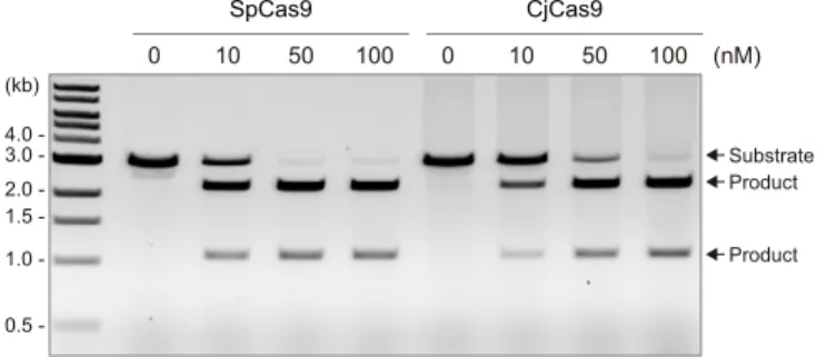
Comparison of the cleavage activities of CjCas9 and SpCas9 27
A recent study showed that the type II-C Cas9 from Corynebacterium diphtheriae (CdCas9), 28
which consists of 1084 residues and shares 21% sequence identity with CjCas9, has limited 29
unwinding and cleavage activities toward dsDNA targets, as compared with SpCas9(Ma et al.,
30
2015). This result suggested that the inefficiency of most of the type II-C Cas9 orthologs for 31
genome editing results from their limited dsDNA cleavage activities (Ma et al., 2015). To 32
examine the differences in the catalytic features of CjCas9 and SpCas9, we compared their in 33
vitro dsDNA cleavage activities. Our data revealed that, like CdCas9, CjCas9 cleaves the target
dsDNA less efficiently, as compared with SpCas9 (Figure S6). These results support the notion 1
that the type II-C Cas9 enzymes, such as CdCas9 and CjCas9, have not been harnessed for 2
genome editing at least partly because of their relatively poor activities. Thus, it is possible that 3
an engineered CjCas9 variant with improved dsDNA cleavage activity could be used for 4
eukaryotic genome editing. Although CjCas9 and CdCas9 commonly exhibit relatively weak 5
dsDNA cleavage activities, they may have distinct specificities for their cognate RNA guides. In 6
contrast to CjCas9, which is specific to its cognate sgRNA, CdCas9 promiscuously recognizes 7
the SpCas9 sgRNA as well as its cognate sgRNA (Ma et al., 2015). Further structural studies 8
will provide insights into the mechanistic diversity among the type II-C CRISPR-Cas9 systems. 9
10
Structural comparison of the CjCas9 tracrRNA and other known RNA triplexes 11
A structural comparison of the CjCas9 tracrRNA with previously characterized RNA triplexes, 12
such as the telomerase RNA subunit TER(Theimer et al., 2005), the SAM-II riboswitch
13
(Gilbert et al., 2008), and the long noncoding RNA MALAT1 (Brown et al., 2014), revealed 14
notable differences between the CjCas9 tracrRNA and the other RNA triplexes (Figure S7). 15
Notably, the CjCas9 tracrRNA lacks a canonical U•A-U triple, whereas the other three RNA 16
triplexes contain successive U•A-U triples (Figure S7). TER and SAM-II have three and seven 17
U•A-U triples in their core regions, respectively. In particular, MALAT1 forms a bipartite triple 18
helix containing stacks of five and four U•A-U triples. Moreover, in the CjCas9 tracrRNA, the 19
L1 region between the S1 and S2 regions consists of three nucleotides and is shorter than those 20
of the other three RNA triplexes (Figure S7). Thus, the CjCas9 tracrRNA adopts a distorted 21
structure, which is stabilized by the two base triples and the two base quintuples. Taken 22
together, the CjCas9-sgRNA-DNA structure revealed the previously unrecognized biological 23
role of an RNA triple helix. 24
25
Conclusions 26
The present structure of the CjCas9-sgRNA-target DNA complex unveiled the remarkable 27
diversity among the CRISPR-Cas9 systems. First, unlike the other tracrRNAs, the CjCas9 28
tracrRNA contains a triple-helix architecture, which is distinct from other known RNA 29
triplexes, thereby expanding the natural repertoire of RNA triplexes. Second, unlike other Cas9 30
orthologs, CjCas9 reads the nucleotide sequences in both the target and non-target DNA strands 31
as the PAM. Although CjCas9 has not been harnessed for genome editing applications, our 32
structural findings may provide clues for Cas9 engineering, such as miniaturization of Cas9 and 33
alteration of its PAM specificities. 34
References 1
Adams, P.D., Afonine, P.V., Bunkoczi, G., Chen, V.B., Davis, I.W., Echols, N., Headd, J.J., Hung, L.W., 2
Kapral, G.J., Grosse-Kunstleve, R.W., et al. (2010). PHENIX: a comprehensive Python-based system for 3
macromolecular structure solution. Acta Crystallogr D Biol Crystallogr 66, 213-221. 4
5
Anders, C., Niewoehner, O., Duerst, A., and Jinek, M. (2014). Structural basis of PAM-dependent target 6
DNA recognition by the Cas9 endonuclease. Nature 513, 569-573. 7
8
Barrangou, R., and Doudna, J.A. (2016). Applications of CRISPR technologies in research and beyond. 9
Nat Biotechnol 34, 933-941. 10
11
Brown, J.A., Bulkley, D., Wang, J., Valenstein, M.L., Yario, T.A., Steitz, T.A., and Steitz, J.A. (2014). 12
Structural insights into the stabilization of MALAT1 noncoding RNA by a bipartite triple helix. Nat 13
Struct Mol Biol 21, 633-640. 14
15
Chylinski, K., Le Rhun, A., and Charpentier, E. (2013). The tracrRNA and Cas9 families of type II 16
CRISPR-Cas immunity systems. RNA Biol 10, 726-737. 17
18
Cong, L., Ran, F.A., Cox, D., Lin, S., Barretto, R., Habib, N., Hsu, P.D., Wu, X., Jiang, W., Marraffini, 19
L.A., et al. (2013). Multiplex genome engineering using CRISPR/Cas systems. Science 339, 819-823. 20
21
Cowtan, K. (2006). The Buccaneer software for automated model building. 1. Tracing protein chains. 22
Acta Crystallogr D Biol Crystallogr 62, 1002-1011. 23
24
Crooks, G.E., Hon, G., Chandonia, J.M., and Brenner, S.E. (2004). WebLogo: a sequence logo generator. 25
Genome Res 14, 1188-1190. 26
27
Deltcheva, E., Chylinski, K., Sharma, C.M., Gonzales, K., Chao, Y., Pirzada, Z.A., Eckert, M.R., Vogel, 28
J., and Charpentier, E. (2011). CRISPR RNA maturation by trans-encoded small RNA and host factor 29
RNase III. Nature 471, 602-607. 30
31
Deveau, H., Barrangou, R., Garneau, J.E., Labonte, J., Fremaux, C., Boyaval, P., Romero, D.A., Horvath, 32
P., and Moineau, S. (2008). Phage response to CRISPR-encoded resistance in Streptococcus 33
thermophilus. J Bacteriol 190, 1390-1400.
34 35
Emsley, P., and Cowtan, K. (2004). Coot: model-building tools for molecular graphics. Acta Crystallogr 36
D Biol Crystallogr 60, 2126-2132. 37
38
Evans, P.R., and Murshudov, G.N. (2013). How good are my data and what is the resolution? Acta 39
Crystallogr D Biol Crystallogr 69, 1204-1214. 40
41
Fonfara, I., Le Rhun, A., Chylinski, K., Makarova, K.S., Lecrivain, A.L., Bzdrenga, J., Koonin, E.V., and 42
Charpentier, E. (2014). Phylogeny of Cas9 determines functional exchangeability of dual-RNA and Cas9 43
among orthologous type II CRISPR-Cas systems. Nucleic Acids Res 42, 2577-2590. 44
45
Garneau, J.E., Dupuis, M.E., Villion, M., Romero, D.A., Barrangou, R., Boyaval, P., Fremaux, C., 46
Horvath, P., Magadan, A.H., and Moineau, S. (2010). The CRISPR/Cas bacterial immune system cleaves 47
bacteriophage and plasmid DNA. Nature 468, 67-71. 48
49
Gasiunas, G., Barrangou, R., Horvath, P., and Siksnys, V. (2012). Cas9-crRNA ribonucleoprotein 50
complex mediates specific DNA cleavage for adaptive immunity in bacteria. Proc Natl Acad Sci U S A 51
109, E2579-2586.
52 53
Gilbert, S.D., Rambo, R.P., Van Tyne, D., and Batey, R.T. (2008). Structure of the SAM-II riboswitch 54
bound to S-adenosylmethionine. Nat Struct Mol Biol 15, 177-182. 55
Hirano, H., Gootenberg, J.S., Horii, T., Abudayyeh, O.O., Kimura, M., Hsu, P.D., Nakane, T., Ishitani, 1
R., Hatada, I., Zhang, F., et al. (2016). Structure and engineering of Francisella novicida Cas9. Cell 164, 2
950-961. 3
4
Hsu, P.D., Lander, E.S., and Zhang, F. (2014). Development and applications of CRISPR-Cas9 for 5
genome engineering. Cell 157, 1262-1278. 6
7
Jiang, F., Taylor, D.W., Chen, J.S., Kornfeld, J.E., Zhou, K., Thompson, A.J., Nogales, E., and Doudna, 8
J.A. (2016). Structures of a CRISPR-Cas9 R-loop complex primed for DNA cleavage. Science 351, 867-9
871. 10
11
Jiang, F., Zhou, K., Ma, L., Gressel, S., and Doudna, J.A. (2015). A Cas9-guide RNA complex 12
preorganized for target DNA recognition. Science 348, 1477-1481. 13
14
Jinek, M., Chylinski, K., Fonfara, I., Hauer, M., Doudna, J.A., and Charpentier, E. (2012). A 15
programmable dual-RNA-guided DNA endonuclease in adaptive bacterial immunity. Science 337, 816-16
821. 17
18
Jinek, M., East, A., Cheng, A., Lin, S., Ma, E., and Doudna, J. (2013). RNA-programmed genome editing 19
in human cells. Elife 2, e00471. 20
21
Jinek, M., Jiang, F., Taylor, D.W., Sternberg, S.H., Kaya, E., Ma, E., Anders, C., Hauer, M., Zhou, K., 22
Lin, S., et al. (2014). Structures of Cas9 endonucleases reveal RNA-mediated conformational activation. 23
Science 343, 1247997. 24
25
Karvelis, T., Gasiunas, G., Young, J., Bigelyte, G., Silanskas, A., Cigan, M., and Siksnys, V. (2015). 26
Rapid characterization of CRISPR-Cas9 protospacer adjacent motif sequence elements. Genome Biol 16, 27
253. 28
29
Leenay, R.T., Maksimchuk, K.R., Slotkowski, R.A., Agrawal, R.N., Gomaa, A.A., Briner, A.E., 30
Barrangou, R., and Beisel, C.L. (2016). Identifying and visualizing functional PAM diversity across 31
CRISPR-Cas systems. Mol Cell 62, 137-147. 32
33
Leontis, N.B., Stombaugh, J., and Westhof, E. (2002). The non-Watson-Crick base pairs and their 34
associated isostericity matrices. Nucleic Acids Res 30, 3497-3531. 35
36
Ma, E., Harrington, L.B., O'Connell, M.R., Zhou, K., and Doudna, J.A. (2015). Single-stranded DNA 37
cleavage by divergent CRISPR-Cas9 enzymes. Mol Cell 60, 398-407. 38
39
Makarova, K.S., Wolf, Y.I., Alkhnbashi, O.S., Costa, F., Shah, S.A., Saunders, S.J., Barrangou, R., 40
Brouns, S.J., Charpentier, E., Haft, D.H., et al. (2015). An updated evolutionary classification of 41
CRISPR-Cas systems. Nat Rev Microbiol 13, 722-736. 42
43
Mali, P., Yang, L., Esvelt, K.M., Aach, J., Guell, M., DiCarlo, J.E., Norville, J.E., and Church, G.M. 44
(2013). RNA-guided human genome engineering via Cas9. Science 339, 823-826. 45
46
Marraffini, L.A. (2015). CRISPR-Cas immunity in prokaryotes. Nature 526, 55-61. 47
48
Mohanraju, P., Makarova, K.S., Zetsche, B., Zhang, F., Koonin, E.V., and van der Oost, J. (2016). 49
Diverse evolutionary roots and mechanistic variations of the CRISPR-Cas systems. Science 353, 50
aad5147. 51
52
Mojica, F.J., Diez-Villasenor, C., Garcia-Martinez, J., and Almendros, C. (2009). Short motif sequences 53
determine the targets of the prokaryotic CRISPR defence system. Microbiology 155, 733-740. 54
55
Nishimasu, H., Cong, L., Yan, W.X., Ran, F.A., Zetsche, B., Li, Y., Kurabayashi, A., Ishitani, R., Zhang, 56
1
Nishimasu, H., and Nureki, O. (2016). Structures and mechanisms of CRISPR RNA-guided effector 2
nucleases. Curr Opin Struct Biol 43, 68-78. 3
4
Nishimasu, H., Ran, F.A., Hsu, P.D., Konermann, S., Shehata, S.I., Dohmae, N., Ishitani, R., Zhang, F., 5
and Nureki, O. (2014). Crystal structure of Cas9 in complex with guide RNA and target DNA. Cell 156, 6
935-949. 7
8
Ondov, B.D., Bergman, N.H., and Phillippy, A.M. (2011). Interactive metagenomic visualization in a 9
Web browser. BMC Bioinformatics 12, 385. 10
11
Ran, F.A., Cong, L., Yan, W.X., Scott, D.A., Gootenberg, J.S., Kriz, A.J., Zetsche, B., Shalem, O., Wu, 12
X., Makarova, K.S., et al. (2015). In vivo genome editing using Staphylococcus aureus Cas9. Nature 520, 13
186-191. 14
15
Sternberg, S.H., LaFrance, B., Kaplan, M., and Doudna, J.A. (2015). Conformational control of DNA 16
target cleavage by CRISPR-Cas9. Nature 527, 110-113. 17
18
Theimer, C.A., Blois, C.A., and Feigon, J. (2005). Structure of the human telomerase RNA pseudoknot 19
reveals conserved tertiary interactions essential for function. Mol Cell 17, 671-682. 20
21
Waterman, D.G., Winter, G., Parkhurst, J.M., Fuentes-Montero, L., Hattne, J., Brewster, A., Sauter, N.K., 22
Evans, G., and Rosenstrom, P. (2013). The DIALS framework for integration software. CCP4 Newsletter 23
49, 16-19.
24 25
Wright, A.V., Nunez, J.K., and Doudna, J.A. (2016). Biology and applications of CRISPR systems: 26
harnessing nature's toolbox for genome engineering. Cell 164, 29-44. 27
28
Zetsche, B., Gootenberg, J.S., Abudayyeh, O.O., Slaymaker, I.M., Makarova, K.S., Essletzbichler, P., 29
Volz, S.E., Joung, J., van der Oost, J., Regev, A., et al. (2015). Cpf1 is a single RNA-guided 30
endonuclease of a class 2 CRISPR-Cas system. Cell 163, 759-771. 31
32
Zhang, Y., Rajan, R., Seifert, H.S., Mondragon, A., and Sontheimer, E.J. (2015). DNase H activity of 33
Neisseria meningitidis Cas9. Mol Cell 60, 242-255.
34 35 36 37
Author Contributions 1
M.Y. performed in vitro cleavage experiments and crystallized the complexes, with assistance 2
from H.H. and H.N.; Y.W. initially obtained diffraction-quality crystals; J.S.G. and F.A.R. 3
performed PAM screens; M.Y., T.N., R.I. and H.N. determined the crystal structures; H.N. 4
conceived the crystallization strategy; M.Y., H.H., H.N. and O.N. wrote the manuscript with 5
help from all authors. F.Z., H.N. and O.N. supervised all of the research. 6
7
Acknowledgements 8
We thank the beamline scientists at PXI at the Swiss Light Source and BL32XU and BL41XU 9
at SPring-8 for assistance with data collection. J.S.G. is supported by a D.O.E. Computational 10
Science Graduate Fellowship. F.A.R. is a Junior Fellow at the Harvard Society of Fellows. F.Z. 11
is supported by the National Institutes of Health through NIMH (5DP1-MH100706 and 1R01-12
MH110049) and NIDDK (5R01DK097768-03), the New York Stem Cell, Simons, Paul G. 13
Allen Family, and Vallee Foundations; and B. Metcalfe. F.Z. is a New York Stem Cell 14
Foundation Robertson Investigator. F.Z. is a founder of Editas Medicine and a scientific advisor 15
for Editas Medicine and Horizon Discovery. H.N. is supported by JST, PRESTO, JSPS 16
KAKENHI Grant Numbers 26291010 and 15H01463. O.N. is supported by the Basic Science 17
and Platform Technology Program for Innovative Biological Medicine from the Japan Agency 18
for Medical Research and Development, AMED, and the Council for Science, Technology and 19
Innovation (CSTI), Cross-ministerial Strategic Innovation Promotion Program (SIP), 20
“Technologies for creating next-generation agriculture, forestry and fisheries” (funding agency: 21
Bio-oriented Technology Research Advancement Institution, NARO), and the Platform for 22
Drug Discovery, Informatics, and Structural Life Science from the Ministry of Education, 23
Culture, Sports, Science and Technology. The content is solely the responsibility of the authors 24
and does not necessarily represent the official views of the National Institute of General Medical 25
Sciences or the National Institutes of Health. 26
27
Accession Numbers 28
The accession numbers for the atomic coordinates of the CjCas9-sgRNA-DNA complexes 29
reported in this paper are Protein Data Bank: 5X2G AGAAACC-3′ PAM) and 5X2H (5′-30
AGAAACA-3′ PAM). 31
STAR METHODS 1
2
CONTACT FOR REAGENT AND RESOURCE SHARING 3
Requests for further information and reagents should be directed to the Lead Contact, Osamu 4
Nureki (nureki@bs.s.u-tokyo.ac.jp). 5
6
EXPERIMENTAL MODEL AND SUBJECT DETAILS 7
The plasmid DNAs were amplified in Escherichia coli Mach (Thermo Fisher Scientific),
8
cultured in LB medium (Nacalai Tesque) at 37°C overnight. The recombinant proteins were 9
overexpressed in E. coli Rosetta 2 (DE3) (Novagen). The E. coli cells were cultured at 37°C in 10
LB medium (containing 20 mg/l kanamycin) until the OD600 reached 0.8, and then protein
11
expression was induced by the addition of 0.5 mM isopropyl-b-D-thiogalactopyranoside 12
(Nacalai Tesque) and an incubation at 20°C for 20 h. 13
14
Sample preparation 15
The gene encoding full-length CjCas9 (residues 1–984) was codon optimized, synthesized 16
(Genscript), and cloned between the NdeI and XhoI sites of the modified pE-SUMO vector 17
(LifeSensors). For crystallization, we prepared the CjCas9-ΔHNH variant lacking the HNH 18
domain (residues 481–640), in which Leu480 (RuvC-II) and Tyr641 (RuvC-III) are connected 19
by a GGGSGG linker. The CjCas9-ΔHNH variant was created by a PCR-based method, using 20
the vector encoding the full-length CjCas9 as the template. The CjCas9-ΔHNH protein was 21
expressed at 20°C in E. coli Rosetta 2 (DE3), and was purified by chromatography on Ni-NTA 22
Superflow resin (QIAGEN). The eluted protein was purified by chromatography on a HiTrap 23
Heparin HP column (GE Healthcare), and was then dialyzed overnight at 20°C with TEV 24
protease, to remove the N-terminal His6-SUMO-tag. The CjCas9-ΔHNH protein was further
25
purified by chromatography on NiNTA and HiLoad 16/600 Superdex 200 (GE Healthcare) 26
columns. The selenomethionine (SeMet)-substituted CjCas9-ΔHNH was expressed in E. coli 27
B834 (DE3) (Novagen), and was purified using a similar protocol to that for the native protein. 28
The sgRNA was transcribed in vitro with T7 RNA polymerase, using a PCR-amplified dsDNA 29
template. The transcribed RNA was purified by 8% denaturing (7 M urea) polyacrylamide gel 30
electrophoresis. The target and non-target DNA strands were purchased from Sigma-Aldrich. 31
The purified CjCas9-ΔHNH protein was mixed with the sgRNA, the target DNA strand, and the 32
non-target DNA strand (containing either the 5′-AGAAACA-3′ PAM or the 5′-AGAAACC-3′ 33
PAM) (molar ratio, 1:1.5:2.3:3.4), and then the CjCas9-sgRNA-DNA complex was purified by 34
gel filtration chromatography on a Superdex 200 Increase column (GE Healthcare). For in vitro 1
cleavage assays, the wild type and mutants of full-length CjCas9 were expressed and purified, 2
using a protocol similar to that for CjCas9-ΔHNH. 3
4
Crystallography 5
The purified CjCas9-sgRNA-DNA complex (containing either the 5′-AGAAACA-3′ PAM or 6
the 5′-AGAAACC-3′ PAM) was grown at 20°C, using the hanging-drop vapor diffusion 7
method. Crystals were obtained by mixing 1 µl of complex solution (A260 nm = 15) and 1 µl of
8
reservoir solution (12.0–14.5% PEG 2,000, 0.4 M ammonium acetate). The SeMet-labeled 9
complex (containing the 5′-AGAAACA-3′ PAM) was crystallized under similar conditions. X-10
ray diffraction data were collected at 100 K on beamlines BL41XU at SPring-8 and PXI at the 11
Swiss Light Source. The crystals were cryoprotected in reservoir solution supplemented with 12
25% ethylene glycol. X-ray diffraction data were processed using DIALS (Waterman et al., 13
2013) and AIMLESS (Evans and Murshudov, 2013). The structure was determined by the Se-14
SAD method, using PHENIX AutoSol(Adams et al., 2010). The model was automatically built
15
using Buccaneer (Cowtan, 2006), followed by manual model building using COOT (Emsley 16
and Cowtan, 2004) and structural refinement using PHENIX (Adams et al., 2010). The final 17
models of the 5′-AGAAACA-3′ PAM complex (2.3 Å resolution) and the 5′-AGAAACC-3′ 18
PAM complex (2.4 Å resolution) were refined using native data sets. Data collection statistics 19
are summarized in Table 1. Structural figures were prepared using CueMol 20
(http://www.cuemol.org). 21
22
PAM discovery assay 23
To generate plasmid libraries containing randomized PAM sequences, synthesized ssDNA 24
oligonucleotides (Integrated DNA Technologies), consisting of seven randomized nucleotides 3′ 25
of a 20-nt target sequence, were used to generate dsDNAs through annealing to a short primer 26
and extension by the large Klenow fragment (New England Biolabs). The dsDNAs were 27
subsequently cloned into linearized pUC19 with Gibson cloning (New England Biolabs). To 28
propagate and purify the cloned plasmids, the products were used to transform >107 competent
29
Stbl3 E. coli cells (Invitrogen), which were pooled and harvested with a Maxi-prep kit 30
(QIAGEN) after overnight growth. The randomized PAM plasmid library was cleaved in vitro, 31
using purified CjCas9 with sgRNAs targeting the PAM library, and the cleavage products were 32
separated on 2% agarose E-gels (Life Technologies). The band corresponding to the un-cleaved 33
target plasmid was isolated with a Zymoclean Gel DNA Recovery Kit (Zymo Research), and 34
MiSeq sequencer (Illumina) with 150 single-end cycles. To analyze the resulting sequence data, 1
the seven nucleotide PAM region was extracted, the individual PAMs were counted, and the 2
PAM counts were normalized to the total reads for each sample. For a given PAM sequence, the 3
enrichment was measured as the log2 ratio as compared to a no-protein control, with a 0.01
4
pseudocount adjustment. PAMs above an enrichment threshold set to 3.5 were compiled and 5
used to generate sequence logos (Crooks et al., 2004). For the PAM wheel generation, 6
abundances were used to generate wheels with Krona (Ondov et al., 2011), as described in the 7
previous report (Leenay et al., 2016). The ratios of PAM abundances as compared to a no-8
protein control, with a 0.01 pseudocount adjustment, were used directly as input for Krona. 9
10
In vitro cleavage assay
11
In vitro plasmid DNA cleavage experiments were performed essentially as described previously
12
(Nishimasu et al., 2015). The EcoRI-linearized pUC119 plasmid (100 ng, 4.7 nM), containing 13
the 20-nt target sequence and the PAMs, was incubated at 37°C for 5 min with the CjCas9-14
sgRNA complex (100 nM, molar ratio, 1:1.5), in 10 µl of reaction buffer containing 20 mM 15
HEPES, pH 7.5, 100 mM KCl, 2 mM MgCl2, 1 mM DTT and 5% glycerol. Reaction products
16
were resolved on a 1% agarose gel, stained with ethidium bromide, and then visualized using a 17
Typhoon FLA 9500 imager (GE Healthcare). 18
19
METHOD DETAILS QUANTIFICATION AND STATISTICAL ANALYSES 20
In vitro cleavage experiments were performed at least three times, and representative results
21
were shown. 22
23
DATA AND SOFTWARE AVAILABILITY 24
The atomic coordinates of the CjCas9-sgRNA-DNA complexes have been deposited in the 25
Protein Data Bank, with the accession numbers PDB: 5X2G (5′-AGAAACC-3′ PAM) and 26
5X2H (5′-AGAAACA-3′ PAM). Data of in vitro cleavage experiments have been deposited in 27
the Mendeley Data repository (http://dx.doi.org/doi:10.17632/6v2dwwcgs3.1). The CueMol 28
program is available at http://www.cuemol.org. 29
Figure Legends 1
2
Figure 1 CjCas9 PAM specificity 3
(A) Motif obtained from the in vitro PAM discovery assay. 4
(B and C) In vitro cleavage assays for DNA targets with different PAMs. The linearized 5
plasmid targets with either the 5′-AGAANCA-3′, 5′-AGAAANA-3′ or 5′-AGAACN-3′ PAM 6
(B), or the 5′-AGANACC-3′ PAM (C) were incubated with CjCas9-sgRNA, and then analyzed 7
by agarose gel electrophoresis. 8
See also Figure S1. 9
10
Figure 2 Overall structure 11
(A) Domain structure of CjCas9. The HNH nuclease domain was truncated for crystallization. 12
BH, bridge helix; PLL, phosphate lock loop. 13
(B) Schematics of the sgRNA and the target DNA. TS, target strand; NTS, non-target strand. 14
(C) Overall structure of CjCas9-∆HNH in complex with an sgRNA and its target DNA. The 15
predicted location of the HNH domain is indicated by the pink circle. 16
(D) In vitro cleavage activity of CjCas9-∆HNH. The circular and linearized plasmid targets with 17
the 5′-AGAAACC-3′ PAM were incubated with wild-type CjCas9 or the three CjCas9 variants 18
(D8A, H559A and ∆HNH), and then were analyzed by agarose gel electrophoresis. The D8A 19
and H559A variants of CjCas9 correspond to the D10A and H840A nickases of SpCas9, 20
respectively. 21
See also Figure S2 and Table 1. 22
23
Figure 3 TracrRNA scaffold 24
(A) 2mFO – DFC electron density map for the tracrRNA scaffold (contoured at 2σ).
25
(B) Schematics of the tracrRNA scaffold. Non-Watson-Crick base pairs are indicated with 26
Leontis-Westhof notations (Leontis et al., 2002). The base triples and quintuples are highlighted 27
in gray and orange backgrounds, respectively. 28
(C) Structure of the CjCas9 tracrRNA scaffold (stereo view). The 3′ nucleotides involved in the 29
triple-helix formation are colored blue. Nucleotides involved in the formation of the base triple 30
and quintuple are highlighted in gray and orange backgrounds, respectively. 31
(D–G) Base triples (D and E) and base quintuples (F and G) in the triple helix. Hydrogen bonds 32
between canonical and non-canonical base pairs are depicted with green and gray dashed lines, 33
respectively. 34
1
Figure 4 TracrRNA scaffold recognition 2
(A) Binding of the tracrRNA scaffold to CjCas9 (stereo view). 3
(B and C) Specific recognition of A63 (B) and A76/U80 (C). Hydrogen bonds are depicted by 4
dashed lines. 5
(D) Functional importance of the tracrRNA scaffold. The linearized plasmid target with the 5′-6
AGAAACC-3′ PAM was incubated with CjCas9, together with either the full-length sgRNA 7
(nucleotides 1–93), the sgRNA variants, or the SpCas9 sgRNA. 1–93, the full-length sgRNA; 8
GGCG, the sgRNA variant, in whichnucleotides 90–93 (CCGC) were replaced with GGCG; 1–
9
85, the sgRNA variant, in which nucleotides 86–93 were truncated; Sp, the SpCas9 sgRNA. 10
11
Figure 5 PAM recognition 12
(A) Binding of the PAM duplex to CjCas9. 13
(B) Schematics of the PAM recognition by CjCas9. Hydrogen bonds are depicted by dashed 14
lines. Water molecules are shown as red spheres. Water-mediated hydrogen bonds between the 15
protein and the sugar-phosphate backbone are omitted for clarity. 16
(C) Recognition of the 5′-AGAAACC-3′PAM (stereo view). Water molecules are shown as red
17
spheres. Hydrogen bonds are depicted by dashed lines. 18
(D) Functional importance of the PAM-interacting residues. The linearized plasmid target with 19
the 5′-AGAAACC-3′ PAM was incubated with either the wild type or the mutants of CjCas9, 20
together with the sgRNA. 21
See also Figure S3. 22
23
Figure 6 Structural comparison of the Cas9 orthologs 24
(A–D) Structures of CjCas9 (A), SpCas9 (PDB: 4UN3) (B), SaCas9 (PDB: 5CZZ) (C), and 25
FnCas9 (PDB: 5B2O) (D) in complexes with their cognate sgRNAs and target DNAs. SpCas9 26
and FnCas9 have structurally distinct subdomains inserted within their REC1 domains 27
(previously referred to as the REC2 domains (Nishimasu et al., 2014; Hirano et al., 2016)). 28
(E–H) Structures of the sgRNAs for CjCas9 (E), SpCas9 (PDB: 4OO8) (F), SaCas9 (PDB: 29
5CZZ) (G), and FnCas9 (PDB: 5B2O) (H). The guide segments are omitted for clarity. 30
See also Figures S4–S7. 31
32
Figure 7 PAM recognition by the Cas9 orthologs 33
(A–D) PAM recognition by CjCas9 (A), SpCas9 (PDB: 4UN3) (B), SaCas9 (PDB: 5CZZ) (C), 1
and FnCas9 (PDB: 5B2O) (D). In (B), the subdomain inserted between β6 and β7 is omitted for 2
clarity. The PAMs are highlighted in purple. The conserved core β-strands are numbered. Close-3
up views for the PAM recognition are shown in right panels. 4
Table 1. Data collection and refinement statistics 5ʹ-AGAAACC-3ʹ PAM (Native) 5ʹ-AGAAACA-3ʹ PAM (Native) 5ʹ-AGAAACA-3ʹ PAM (SeMet) Data collection
Beamline SLS PX1 SPring-8 BL41XU SPring-8 BL41XU
Wavelength (Å) 1.0000 1.0000 0.9791 Space group P212121 P212121 P212121 Cell dimensions a, b, c (Å) 104.3, 105.1, 136.5 102.0, 103.9, 134.5 102.0, 104.2, 134.6 α, β, γ (°) 90, 90, 90 90, 90, 90 90, 90, 90 Resolution (Å)* 136.5–2.4 (2.47–2.40) 103.9–2.3 (2.36–2.30) 49.5–2.2 (2.25–2.20) Rmerge 0.150 (0.882) 0.105 (0.783) 0.075 (0.967) Rpim 0.043 (0.529) 0.032 (0.316) 0.031 (0.396) I/σI 13.8 (1.6) 12.4 (2.4) 14.2 (2.6) Completeness (%) 100 (100) 99.8 (99.8) 100 (100) Multiplicity 12.9 (13.4) 10.4 (7.0) 6.8 (6.9) CC (1/2) 0.998 (0.757) 0.986 (0.875) 0.999 (0.866) Refinement Resolution (Å) 52.6–2.4 72.8–2.3 No. reflections 59,303 63,923 Rwork / Rfree 0.190 / 0.221 0.200 / 0.231 No. atoms Protein 5,929 5,925 Nucleic acid 2,715 2,715 Solvent 323 279 B-factors (Å2) Protein 59 65 Nucleic acid 49 54 Solvent 40 45 R.m.s. deviations Bond lengths (Å) 0.0023 0.0024 Bond angles (°) 0.498 0.515 Ramachandran plot (%) Favored region 97.28 96.58 Allowed region 2.58 3.01 Outlier region 0.14 0.41 *Values in parentheses are for the highest resolution shell. 1
Position C GA
G
A
A TC
C
A
0.0 1.0 Bits 2 3 4 5 6 7 1A
C
B
Substrate Product Product 4.0 3.0 2.0 1.5 1.0 -(kb) 0.5 -– A G C T 5′-AGANACC-3′– ACA GCA CCA TCA AAA AGA ATA ACG ACC ACT
Substrate Product Product 5′-AGAANNN-3′ 4.0 3.0 2.0 1.5 1.0 -(kb) 0.5
-CCTTTAATCCACGCGAACCG |||||||||||||||||||| GGAAAUUAGGUGCGCUUGGCGUUUUAGUCCCUG |||||||||||| UAAAAUCAGGGAA A Guide (spacer) Repeat Antirepeat Tetraloop −1 Stem 1 Stem 2 Stem 3 56 8887 57 58 63 62 64 68 80 86 84 81 70 73 76 Triple helix 21 32 37 49 48 53 50 1 20 5′ 93 3′ 20 1 1* 8* −8 TS NTS 5′ 3′ 5′ 3′ A A U PAM AGAAACCG |||||||| TCTTTGGC U U AA | || CGCCA UG AGAAA ||||| | |||| GCGGG C UCUGCGGGG UUA | |||| | C A UCCCC AA
D
1 45 77 235 427 481 641 480 641 480 641 778 792 828 984 NUC lobe CjCas9RuvC-I BH REC1 REC2 RuvC-II HNH RuvC-III WED PI
REC lobe PLL
CjCas9-∆HNH
RuvC-I BH REC1 REC2 RuvC-II RuvC-III WED PI
Triple helix REC lobe NUC lobe (GGGSGG) WED WED RuvC RuvC
REC2 REC1 REC1 REC2
PI PI 180º TS NTS 5′ 5′ 3′ 5′ 3′ 3′ 5′ 3′ PAM Repeat Repeat Antirepeat Antirepeat 4.0 3.0 2.0 1.5 1.0 -(kb) 0.5 -WT D8A H559A Circular – WT D8A H559A Linearized – Nicked Linear Supercoiled Products ∆HNH ∆HNH
B
A
C
Stem 1 Stem 2 Stem 3 G C G G G C U C U G C G G G G U U A A U C C C C A A A U U A A U C G C C A U G A G A A A -C 49 53 55 93 57 56 60 91 87 88 63 62 58 64 70 76 84 86 80 5′ 3′ Triple helix 49 66 67 68 69 80 63 60 58 53 62 86 89 56 65 87 88 55 54 64 57 90 59 61 85 84 84 83 82 81 70 70 71 73 72 74 75 76 77 77 78 79 93 92 91 52 51 50 A63 U48 G70 A76 C84 U85 A86 G58 C93 5′ 5′ 3′ 49 66 67 68 69 80 63 60 58 53 62 86 89 56 65 87 88 55 54 64 57 90 59 61 85 83 82 81 71 73 72 74 75 76 78 79 93 92 91 52 51 50 5′ 3′ U65 A53 A86 G62 U56 A89 C59 U57 G61 G92 C90 C64 G54 U55 A88 A87 3′
C
B
D
E
G
F
A
Asn74 His67 His70 Asn66 A63 A76 U80 Ile964 Glu975 Arg977 Arg832 Glu980 Phe854 Phe982 Asp981 3′ C93 C93 A86 A86 A76 U80 U85 A63 WED RuvC REC2 REC1 PI PI BH C93 C93 A86 A86 A76 U80 U85 A63 WED RuvC REC2 REC1 PI
B
C
D
A
Substrate Product Product 4.0 3.0 2.0 1.5 1.0 -(kb) 0.5 -Sp1–93 A63U A76U U80A GGCG 1–85 –
3′ 5′ 3′ 5′ 5′ 5′ WED PI NTS TS NTS TS PAM PAM PAM 4* 5* A4* A5* 3* 2* 1* 6* 7* 8* Thr913 Ser915 Arg866 Ser951 G(−6) G(−7) A4* A5* G(−6) G(−7) 3′ 5′ 5′ Thr913 Ser915 Arg866 Ser951 NTS TS 3′ 5′ 5′ 3′ Asn827 Ser801
Tyr850 Tyr802 Thr913 Ser915 (Ser915)
Lys890
Arg780
(Met830)
Ser951 Thr916 Lys940 Arg866
Lys950 Arg793 A1* T( − 1 ) G2* C ( − 2 ) A3* T( − 3 ) A4* T( − 4 ) A5* T( − 5 ) C6* G ( − 6 ) C7* G ( − 7 ) G8* C ( − 8 )
A
C
D
B
Substrate Product Product 4.0 3.0 2.0 1.5 1.0 -(kb) 0.5Triple helix WED WED RuvC REC2 REC1 REC2 REC1 REC2 REC2 REC1 REC2 REC1 PI PI RuvC TS NTS TS NTS TS NTS 5′ 3′ 3′ 3′ 3′ Repeat Antirepeat SL1 SL2 SL1 SL1 SL2 SL3 Linker Linker Linker WED WED RuvC REC2 REC1-Ins REC1-Ins REC1-Ins REC1 PI PI-Ins PI RuvC TS NTS 5′ Repeat Antirepeat WED WED RuvC REC2 REC1 REC1 REC1 PI PI RuvC 5′ Repeat Antirepeat WED WED RuvC REC2 PI PI RuvC 5′ Repeat Antirepeat
A
B
C
D
E
F
G
H
CjCas9 R:AR duplex R:AR duplex R:AR duplex R:AR duplex CjCas9 sgRNA SpCas9 sgRNA SaCas9 sgRNA FnCas9 sgRNA SpCas9 SaCas9 FnCas9 180ºCjCas9 5′-NNNVRYM-3′ SpCas9 5′-NGG-3′ SaCas9 5′-NNGRRT-3′ FnCas9 5′-NGG-3′ 3′ 3′ 5′ 5′ 5 6 7 8 9 4 3 1 2 3′ 3′ 5′ 5′ 5 6 7 8 9 4 3 12 3′ 3′ 5′ 5′ 5 6 7 8 9 4 3 1 2 3′ 3′ 5′ 5′ 5 6 7 4 3 1 2 5′ 3′ 5′ Ser915 Arg866 Ser951 A4* A5* G(−6) G(−7) 5 6 7 5′ 5′ Asn985 Asn986 Arg1015 Arg991 G3* A4* A5* T6* 5 6 7 3′ 5′ 5′ Arg1335 Arg1333 G2* G3* 5 6 7 5′ 5′ Arg1585 Arg1556 G2* G3* 5 6 7
A
B
C
D
A A C C 6% A 4% T A 6% C 2% A C 2% G C 0.6% G C A 8 % C 0.9% T A 3 % T G C 1% C A T C 5% A 2 % G 1% C C 4% A 4 % A 5% C G 1% C G C 4% A 3% C T 1% C C C 1% A G A C 9% A 6% C T 6% A A 4% C G 1% C G C 3% C 0.6% T 0.7% T 5′ NNNNNNN3′ 4 5 6 7
Figure S1. CjCas9 PAM specificity
The results of the in vitro PAM discovery assay are represented as the PAM wheel (Leenay et al., 2016). The area of the sector indicates the relative enrichment in the library.
Figure S2. Schematic of the nucleic-acid recognition
Residues that interact with nucleic acids via their main chain are shown in parentheses. Water-mediated hydrogen bonds are omitted for clarity. Related to Figure 2. (Asp981)Arg832 (Arg234) (Lys188) (Phe792) (Glu88) (Leu90)Ser89 (Leu98) Gln172 (Phe181) Glu180 Lys794 Ser97 (Gly130) (Asn186) Asn186 Arg185 Leu787 (Leu787) (Gly785) Ser784 Lys782 Arg64 Arg57 Arg129 Arg234 Thr448 Arg49 Arg52 Arg48 Lys56 Asn821 His70 Asn74 Asn66 His67 His788 (Lys237) Arg60 Arg460 Glu779 Ile964 Lys781 Arg977 (Glu975) (Lys781) Glu980 Phe854 Arg30 Lys461 Lys457 (Ala45) Asn649 Lys257 Asn280 Asn277 Arg254 (Gly244) Lys363 Tyr131 Tyr191 Lys823 Lys62 Arg61 His124 Tyr102 Arg128 Lys69 Lys127 His393 (Asp392) (Leu146)(Ile145) (Glu790) Thr791 (Thr791) (Val184) Lys187 (Arg185) Lys413 Asn395 (Asn395) Tyr414 Ser247 G1 G2 A3 A4 A5 U6 U7 A8 G9 G10 U11 G12 C13 G14 C15 U16 U17 G18 G19 C20 C19 T18 T17 T16 A15 A14 T13 C12 C11 A10 C20 C9 G8 C7 G6 A5 A4 C3 C2 G1 U48 A47 A46 A45 A44 U43 C42 A41 G40 G39 G38 U23 U24 U22 A26 G21 U25 G62 G61 G60 C59 C90 C91 U57 A89 U56 G92 G58 C93 G68 U67 C66 U65 A86 A51 G52 A50 A49 C69 G70 G71 G72 G73 C82 C83 C84 U85 C81 U74 U75 A79 U80 A78 A76 C77 A53 C64 G54 A87 A88 U55 A63 G27 U28 C29 C30 C31 A37 U32 Bridge helix REC lobe PI domain WED domain RuvC domain Phosphate lock loop
Watson-Crick base pair Non Watson-Crick base pair Hydrogen bond/salt bridge Hydrophobic interaction Repeat-antirepeat duplex Triple helix TS RNA-DNA heteroduplex sgRNA Guide (spacer) Repeat Antirepeat Stem 2 Stem 1 Stem 3 Tetraloop
Figure S3. PAM recognition
(A and B) Interactions between Thr913 and dA4* (A) and modeled dT4* (B).
(C) Superimposition of the 5′-AGAAACC-3′ PAM complex (semitransparent gray) and the 5′-AGAAACA-3′ PAM complex (colored) (stereo view). (D) Recognition of the 5′-AGAAACA-3′ PAM (stereo view). Hydrogen bonds are depicted by dashed lines.
Related to Figure 5. A7* C7* C6* C6* A5* A5* A4*
A4* A3*A3*
T(−7) G(−7) G(−6) G(−6) T(−5) T(−5) T(−5)T(−5) T(−4)T(−4) T(−3) T(−3) T(−4) T(−4) T(−3) T(−3) Thr913 A4* Thr913 T4* (model) 5′-AGAAACAG-3′
5′-AGAAACCG-3′ 5′-5′-AGAAACCG-3′AGAAACAG-3′
A5* T(−7) G(−6) Arg866 Ser951 Ser915 A7* C7* C6* C6* A5* A5* A4*
A4* A3*A3*
T(−7) G(−7) G(−6) G(−6) Arg866 Ser951 Ser915 Arg866 Ser951 Ser915 A5* T(−7) G(−6) Arg866 Ser951 Ser915 5′-AGAAACAG-3′ 5′-AGAAACAG-3′
C
D
A
B
Figure S4. RNA-guided DNA targeting
(A) Recognition of the sgRNA seed region by CjCas9 (stereo view). The target DNA strand is omitted for clarity. (B) Recognition of the +1 phosphate by CjCas9 (stereo view).
(C) Functional importance of the phosphate lock loop. The linearized plasmid target with the 5′-AGAAACC-3′ PAM was incubated with either the wild type or the T791A mutant of CjCas9, together with the sgRNA.
Related to Figure 6. −3 Ala45 Thr448 Arg49 Arg52 Arg60 Arg57 Lys237 Arg234 Arg64 Arg129 Gly130 Arg185 Asn186 Lys188 15 16 14 13 12 17 18 19 20 21 −1 3* 2* 1* −2 −3 +1P 1 20 21 22 23 47 24 19 Thr791 Glu790 2 3 PAM PAM −1 3* 2* 1* −2 +1P 1 20 21 22 23 47 24 19 Thr791 Glu790 2 3 BH Ala45 Thr448 Arg49 Arg52 Arg60 Arg57 Lys237 Arg234 Arg64 Arg129 Gly130 Arg185 Asn186 Lys188 15 16 14 13 12 17 18 19 20 21 BH PLL PLL Substrate Product Product 4.0 3.0 2.0 1.5 1.0 -(kb) 0.5 -– WT T791A
C
B
A
Figure S5. Recognition of the repeat-antirepeat duplex by the Cas9 orthologs
(A–D) Species-specific recognition of the repeat-antirepeat duplex by the REC1/WED domains of CjCas9 (A), SpCas9 (PDB: 4UN3) (B), SaCas9 (PDB: 5CZZ) (C), and FnCas9 (PDB: 5B2O) (D).
BH WED REC1 Loop 1 Loop 2 Loop 2 Loop 1 Repeat Antirepeat BH WED REC1 Repeat Antirepeat BH WED REC1 Repeat Antirepeat BH WED REC1 Repeat Antirepeat BH WED REC1 Repeat Antirepeat BH WED REC1 Repeat Antirepeat BH WED REC1 Repeat Antirepeat BH WED REC1 Repeat Antirepeat CjCas9
A
SpCas9B
SaCas9C
FnCas9 CjCas9 SpCas9 SaCas9 FnCas9D
Figure S6. Comparison of the DNA cleavage activities of CjCas9 and SpCas9
The linearized plasmid targets with the 5′-TGG-3′ PAM and the 5′-AGAAACC-3′ PAM were incubated at 37°C for 5 min with SpCas9 (10–100 nM) and CjCas9 (10–100 nM) in the presence of their cognate sgRNAs, respectively. Related to Figure 6. Substrate Product Product 4.0 3.0 2.0 1.5 1.0 -(kb) 0.5 -10 0 50 100 SpCas9 10 0 50 100 (nM) CjCas9
Figure S7. Structures of the RNA triple helices
(A–D) Structures of the CjCas9 tracrRNA (A), the telomerase RNA subunit TER (PDB: 1YMO) (B), the SAM-II riboswitch (PDB: 2QWY) (C), and the long noncoding RNA MALAT1 (PDB: 4PLX) (D) (stereo view). The U•A-U triples are depicted as stick models. S, stem; L, loop. Related to Figure 6. 5′ 3′ S1 S2 L1 L3 S3 L2 5′ 3′ S1 S2 L1 L3 L2 5′ 3′ S1 S2 L1 L3 S3 L2 5′ 3′ S1 S2 L1 U•A-U U•A-U U•A-U L3 5′ 3′ S1 S2 L1 U•A-U U•A-U U•A-U U•A-U U•A-U U•A-U U•A-U U•A-U U•A-U U•A-U U•A-U U•A-U U•A-U U•A-U U•A-U U•A-U U•A-U 5′ 3′ S1 S2 L1 L3 L2 U•A-U U•A-U U•A-U U•A-U L3 5′ 3′ S1 S2 L1 U•A-UU•A-U L3 5′ 3′ S1 S2 L1 U•A-UU•A-U L3 CjCas9 tracrRNA
A
SAM-IIC
hTRB
MALAT1D
Table S1 Oligonucleotides, related to the STAR Methods
Oligonucleotides used to generate the CjCas9 expression vector (pE‐SUMO‐CjCas9, with the TEV recognition site between His6‐SUMO and CjCas9)
PCR template Forward primer Reverse primer
pE‐SUMO (modified) ACTCGAGCACCACCACCACC CATATGGGATCCTTGGAAGTACAAG
pColdGST‐CjCas9 CAAGGATCCCATATGGCCCGCATCCTCGCTTTC TGGTGGTGCTCGAGTTTATTTCTTAAAATCCTCGCGCTG Oligonucleotides used to introduce the CjCas9 mutations (pE‐SUMO‐CjCas9)
Mutation Forward primer Reverse primer
ΔHNH TACATTGCGCGACTGGTTC CCCCCCGGACCCCCCCCCCAATTCGATATTTATTTTGTGCAC D8A GCGATCGGAATCTCTAGTATCGGATG GAAAGCGAGGATGCGGGC
H559A GCGATCTATCCGTATAGCAGGTCATTTGAC GTCGATCTCCAGCATTTTCTCGTC T791A GAAGAGGCATTCCGCAAGGAAGAGG GCGGAATGCCTCTTCGTGCAGTGC R866A GTGGCTGCGTCGAAGAAAGGAGAGATC TCTTCGACGCAGCCACCGCCTTATTTGG T913A AGCAGTACAGTGTCCCTGATTGTGAGCAAGC CGCGAAGGCGTTATAATAAACAAATTCTGGTTCC S915A ACAGTGTCCCTGATTGTGAGCAAGC CGCGCTCGTGAAGGCGTTATAATAAACAAATTCTG S951A GCAAAGGCAATTGGCATCCAAAACCTG GCCAATTGCCTTTGCGATGACCTCCTT
DNA oligonucleotides used for crystallization
PAM sequence Target DNA strand Non‐target DNA strand AGAAACA CTGTTTCTGCCAAGCGCACCTAATTTCC AGAAACAG AGAAACC CGGTTTCTGCCAAGCGCACCTAATTTCC AGAAACCG sgRNA Cj sgRNA 1‐93 GGAAATTAGGTGCGCTTGGCGTTTTAGTCCCTGAAAAGGGACTAAAATAAAGAGTTTGCGGGACTCTGCGGGGTTACAATCCCCTAAAACCGC Cj sgRNA A63U GGAAATTAGGTGCGCTTGGCGTTTTAGTCCCTGAAAAGGGACTAAAATAAAGAGTTTGCGGGTCTCTGCGGGGTTACAATCCCCTAAAACCGC Cj sgRNA A76U GGAAATTAGGTGCGCTTGGCGTTTTAGTCCCTGAAAAGGGACTAAAATAAAGAGTTTGCGGGACTCTGCGGGGTTTCAATCCCCTAAAACCGC Cj sgRNA U80A GGAAATTAGGTGCGCTTGGCGTTTTAGTCCCTGAAAAGGGACTAAAATAAAGAGTTTGCGGGACTCTGCGGGGTTACAAACCCCTAAAACCGC Cj sgRNA GGCG GGAAATTAGGTGCGCTTGGCGTTTTAGTCCCTGAAAAGGGACTAAAATAAAGAGTTTGCGGGACTCTGCGGGGTTACAATCCCCTAAAAGGCG Cj sgRNA 1‐85 GGAAATTAGGTGCGCTTGGCGTTTTAGTCCCTGAAAAGGGACTAAAATAAAGAGTTTGCGGGACTCTGCGGGGTTACAATCCCCT Sp sgRNA GGAAAUUAGGUGCGCUUGGCGUUUUAGAGCUAGAAAUAGCAAGUUAAAAUAAGGCUAGUCCGUUAUCAACUUGAAAAAGUGGCACCGAGUCGGUGCUU Oligonucleotides used to introduce the sgRNA mutations (pUC119‐sgRNA)
Mutation Forward primer Reverse primer
A63U TCTCTGCGGGGTTACAATCCCCTAAAACCGC CCCGCAAACTCTTTATTTTAGTCCCTTTTC A76U TCAATCCCCTAAAACCGCTTGTACCGAGCTCG AACCCCGCAGAGTCCCGCAAACTCTTTATTTTAG U80A ACCCCTAAAACCGCTTGTACCGAGCTCGAATTCAC TTGTAACCCCGCAGAGTCCCGCAAACTC GGCG GGCGTTGTACCGAGCTCGAATTCACTGGCCGTCG TTTTAGGGGATTGTAACCCCGCAGAGTCCCGC Oligonucleotides used to generate the dsDNA templates for in vitro transcription
sgRNA Forward primer Reverse primer
1‐93, A63U, A76U AGCGCCCAATACGCAAAC GCGGTTTTAGGGGATTG
U80A AGCGCCCAATACGCAAAC GCGGTTTTAGGGGTTTG