HAL Id: tel-01124075
https://tel.archives-ouvertes.fr/tel-01124075
Submitted on 6 Mar 2015HAL is a multi-disciplinary open access
archive for the deposit and dissemination of sci-entific research documents, whether they are pub-lished or not. The documents may come from teaching and research institutions in France or abroad, or from public or private research centers.
L’archive ouverte pluridisciplinaire HAL, est destinée au dépôt et à la diffusion de documents scientifiques de niveau recherche, publiés ou non, émanant des établissements d’enseignement et de recherche français ou étrangers, des laboratoires publics ou privés.
Genome-wide characterization of RNA polymerase II
behavior during transcription termination and upon
UV-B stress
Akos Gyenis
To cite this version:
Akos Gyenis. Genome-wide characterization of RNA polymerase II behavior during transcription termination and upon UV-B stress. Genomics [q-bio.GN]. Université de Strasbourg, 2012. English. �NNT : 2012STRAJ056�. �tel-01124075�
UNIVERSITÉ DE
STRASBOURG
École Doctorale des Sciences de la Vie et de la Santé
IGBMC - CNRS UMR 7104 - Inserm U 964
THÈSE
présentée par:
Akos Gyenis
Soutenue le: 19 décembre 2012
Pour obtenir le grade de :
Docteur de l’Université de Strasbourg
Discipline/ Spécialité: Aspects moléculaires et cellulaires de la biologie
Etudes génomiques de la dynamique de l’ARN
polymérase II pendant l’étape de terminaison de la
transcription et après un stress causé par les UV-B
(Genome-wide characterization of RNA polymerase II
behavior during transcription termination and upon
UV-B stress)
THÈSE dirigée par:
Dr. Laszlo Tora Institut de génétique et de biologie moléculaire et cellulaire, Strasbourg
RAPPORTEURS:
Dr. Olivier Bensaude Institut de biologie de l’école normale supérieure, Paris Dr. Matthieu Gerard Institut de biologie et de technologies de Saclay
Dr. Evi Soutoglou Institut de génétique et de biologie moléculaire et cellulaire, Strasbourg
Contents
Contents ... 2
Acknowledgement... 5
List of abbreviations ... 6
Résumé de thèse ... 9
INTRODUCTION ... 20
Chapter one: RNA polymerase II regulation and pause during transcription . 21
I. The expression of genetic information... 211. DNA dependent RNA polymerases ... 21
A. Structure of Pol II ... 23
B. Carboxyl-terminal domain of Pol II ... 24
C. Posttranslational modifications of CTD ... 25
II. RNA polymerase II transcription cycle... 31
2. Transcription initiation ... 31
D. Core promoter elements ... 31
E. General transcription factors ... 34
F. Sequential assembly of the PIC complex ... 36
G. Activation dependent transcription and coactivators ... 37
a) Mediator complex... 37
b) Upstream stimulatory activity (USA) ... 38
c) Chromatin remodeling and modifications ... 38
3. Promoter escape ... 40
4. Early elongation... 41
5. Pol II pause ... 42
H. Factors contributing in Pol II pause establishment ... 44
I. Possible roles of Pol II pause ... 47
J. Promoter associated RNAs... 49
6. Transcription elongation ... 50
K. Factors to diminish Pol II pause during elongation ... 50
d) P-TEFb and DSIF ... 50
e) TFIIS ... 52
g) Elongin complex ... 53
h) The ELL family ... 54
i) CSB ... 55
L. Elongation factors with chromatin remodeling ability ... 55
j) SWI/SNF ... 55
k) Chd1 ... 56
l) FACT and Spt6 ... 56
M. Elongation factors which maintain histone modifications ... 57
7. Transcription termination and 3’ end processing ... 57
N. 3’ end processing of pre-mRNA ... 58
O. Different 3’ end processing and termination pathway on short, none-polyadenylated transcripts ... 59
P. Transcription termination I: Torpedo model ... 60
Q. Transcription termination II: Allosteric/anti-terminator model... 61
R. Chromatin remodeling factors at the 3’ end of genes... 62
8. Pol II pause downstream from 3’-end of genes ... 63
RESULTS I. ... 65
III. Genome-wide characterization of Pol II pause downstream from 3’ end of genes... 65
IV. Publication I... 66
V. Publication II. ... 67
VI. DISCUSSION I. ... 68
Chapter two: The fate of RNA polymerase II transcription upon UVB
irradiation ... 73
VII. Transcription is blocked by alterations in the DNA structure ... 73
9. Ultraviolet light... 74
S. UV-induced DNA damage ... 75
T. DNA double strand breaks ... 77
10. Persistent blocks initiate transcription coupled repair processes... 78
U. Nucleotide excision repair (NER) ... 78
m) CSB is a key element of TCR / damage recognition ... 79
n) TCR factor recruitment / damage verification... 80
o) Role of TFIIH complex in TCR ... 81
p) Late steps of TCR / DNA synthesis ... 81
A. Single strand break repair ... 84
B. Repair of double strand breaks ... 84
11. DNA damage signaling ... 85
12. Fate of arrested Pol II transcription ... 86
C. Transcriptional pause ... 86
D. Fate of Pol II during TCR ... 87
E. Ubiquitination of Pol II ... 88
RESULTS II. ... 90
VIII. Genome-wide characterization of RNA polymerase II behavior upon UVB stress (Manuscript under preparation) ... 90
IX. UNPUBLISHED RESULTS ... 92
13. Low dose of UVB is sufficient to induce DNA damage response, but is not lethal for cells 92 F. Testing cell lethality ... 92
G. Detection of DNA lesions ... 93
H. Activated DNA damage signaling... 94
I. Detection of p21 activation (I.)... 94
J. Detection of p21 activation (II.)... 95
14. Genome-wide analysis of Pol II behavior upon UVB irradiation ... 96
K. Genome-wide average Pol II occupancy profile ... 98
L. Average Pol II occupancy profile of 4500 expressed genes ... 100
X. DISCUSSION II. ... 109
XI. Materials and methods ... 114
Supplementary results ... 120
Acknowledgement
First of all I would like to thank Dr Laszlo Tora for accepting me in his lab, and for all the support, advices and guidance for my projects in the past years. The time I spent in his lab was very instructive for me.
I thank the jury members Dr Olivier Bensaude, Dr Matthieu Gerard and Dr Evi Soutoglou for spending time to read my thesis and to evaluate my work.
I am also grateful to Didier Devys for the constant scientific, non-scientific and climbing discussions, critics and help.
I specially thank every present and former lab members: Elisabeth Scheer, Jacques Bonnet, Marjorie Fournier, Krishanpal Karmodya, Eda Suer, David Umlauf, Sarina Ravens, Anne Riss, Nikolaos Vosnakis, Matthieu Stierle, Chen-Yi Wang, Guillaume Lang, Arnaud Krebs, Monika Ballarino, Anne Helmrich, Anamika Krishanpal, and Meritxell Orpinell for their help in my experiments and life in France. I thank Dr Frédéric Coin for providing me equipments for my experiments. I thank Dr Evi Soutoglou and Dr Dirk Eick for antibodies. I specially thank Tao Ye, Stéphanie Le Gras and the sequencing platform for helping me with the ChIP-seq
analyses. I thank Betty Heller and the cell culture facility for providing me cells and medium for my experiments. I also thank Gabor Papai, Judit Osz Papai, Orban Komonyi, Zita Nagy, Attila Oravecz, Emese Gazdag, Tibor Pankotai, Gabriella Pankotai-Bodo and Maria Takacs for the help to arrange my life in France. I thank for my family and friends for the constant support.
The work presented herein was carried out in Dr. Laszlo Tora’s laboratory in IGBMC Strasbourg. My studies were supported by the Fondation Pour La Recherche Médicale (FRM).
6
List of abbreviations
6 6-4PPs pyrimidine 6-4 pyrimidone photoproducts A ATAC ATM Ataxia-telangiectasia-mutated BBRE TFIIB-recognition element
C
CAK Cyclin-Activating Kinase complex
CARM1 coactivator-associated arginine methyl-transferase 1
CDC Cell Division Cycle
CDK Cyclin-Dependent Kinase
ChIP Chromatin Immunoprecipitation ChIP-seq ChIP coupled High Throughput Sequencing
CPDs Cyclobutane pyrimidine dimers CPSF cleavage and polyadenylation specificity factor
CS Cockayne syndrome
CstF Cleavage stimulation factors CTD Carboxyl-Terminal Domain
D
DCE Downstream core element
DDB DNA Damage binding protein DP DNA polymerase
DPE Downstream promoter element DSB Double strand break
DSIF DRB Sensitivity-Inducing Factor
E
EAG End of annotated gene
G
GGR Global genome repair GRO-seq Global Run-On assay coupled sequencing
GTF General Transcription Factor GW Genome-wide H H Histone HR Homologous recombination I Inr Initiator K kb Kilobase L
LEC Little elongation complex
M
7 MAZ MYC-associated zinc-finger protein
mb Megabase MD Mega Dalton MED Mediator
MLL mixed lineage leukemia mRNA messenger RNA
MTE Motif ten element
N
NC2 Negative cofactor
NELF Negative elongation factor NER Nucleotide excision repair NHEJ Non-homologous end-joining nm Nanometer
NMR Nuclear Magnetic Resonance nt Nucleotide
O
O3 Ozone
O-GlcNAc O-linked N-acetylglycosamine
ORF Open reading frame
P
P Phosphorylation PA Polyadenylated
PAR Promoter associated RNA PARP1 Poly ADP ribose polymerase 1 PIC Pre Initiation Complex
PIN1 peptidyl–prolyl cis/trans-isomerase Pol I, II, III RNA polymerase I, II, III Pol IIa, hypophosphorylated Pol II Pol IIo hyperphosphorylated Pol II P-TEFb Positive Transcription Elongation factor complex
R
RNAi RNA interference
ROS Reactive oxygen Species RPA Replication protein A
RPAP2 RNA Polymerase II-associated protein
rRNA Ribosomal RNA
S
SAGA Spt-Ada-Gcn5 acetyltransferase
SEC Super elongation complex snoRNAs small nucleolar RNA snRNP Small nuclear ribonucleoprotein
T
TCR Transcription coupled repair TF Transcription factor
tiRNA Transcription initiation related RNA TRF TBP-related factors
tRNA Transfer RNA
TSS Transcription start site
TTS Transcription termination site
8 UAS Upstream activating sequence
UB Ubiquitin
USA Upstream stimulatory activity UV Ultraviolet
X
XCPE1 X core promoter element1 XP Xeroderma pigmentosum
9
Résumé de thèse
Analyse sur l’ensemble du génome de la dynamique de l’ARN polymérase II lors de la terminaison de la transcription et en réponse à un stress aux UV-B.
Introduction:
Les cellules vivantes sont en permanence soumises à des signaux internes (processus métaboliques de base) et externes (stress environnementaux). En réponse à ces stimuli les cellules disposent de mécanismes très précisément contrôlés afin d’accéder et de transcrire les informations génétiques contenues dans l’ADN permettant ainsi de maintenir l’état physiologique des cellules. Ceci est réalisé chez les eucaryotes par la machinerie de transcription associée à l’ARN polymérase II qui permet l’expression des g ènes codant pour les protéines et pour certains ARNs régulateurs non-codants.
La transcription par l’ARN polymérase II (Pol II) est un mécanisme régulé de facon extrêmement fine nécessitant l’action séquentielle d’un grand nombre de complexes protéiques. Chaque cycle de transcription peut être divisé en trois phases principales : initiation, élongation et terminaison. Chacune de ces phases peut également être divisée en plusieurs étapes correspondant chacune à des possibilités de régulation pour l’expression des gènes et la synthèse d’ARNm.
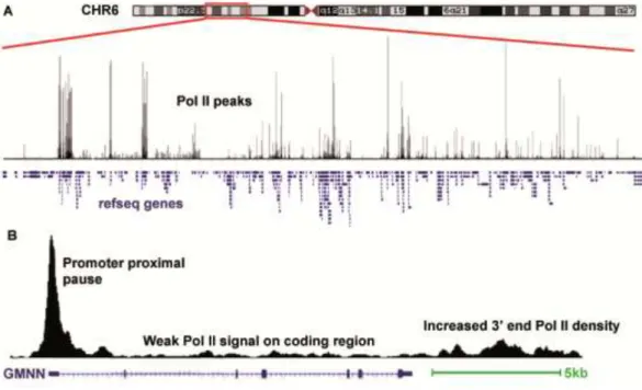
La transition entre les phases d’initiation et d’élongation de la transcription est une étape limitante au cours de laquelle la Pol II s’arrête (pause) en aval du site d’initiation après la synthèse des 25-50 premiers nucléotides. Cette pause de la polymérase a d’abord été décrite comme une étape limitante entre initiation et élongation précoce lors de la transcription de certains gènes de choc thermique chez la drosophile. Plus récemment des études sur l’ensemble du génome ont montré que ce phénomène de pause de la Pol II à proximité du promoteur constitue un mécanisme de régulation qui est conservé et observé sur un très grand nombre de promoteurs chez les eucaryotes supérieurs. Une pause ou un ralentissement de l’ARN Pol II a également été découvert en aval de la fin des gènes annotés (end of annotated genes : EAGs) mais cette observation n’a jamais été analysée de façon
10 globale par des études sur l’ensemble du génome qui se sont concentrées sur la caractérisation des pauses de la Pol II à proximité du promoteur. Ainsi, une caractérisation détaillée des gènes potentiellement régulés par une pause de la polymérase en aval des EAGs n’a pas été réalisée sur l’ensemble du génome. Il reste donc à démontrer si ce phénomène est mécanisme général observé sur l’ensemble des gènes chez les mammifères.
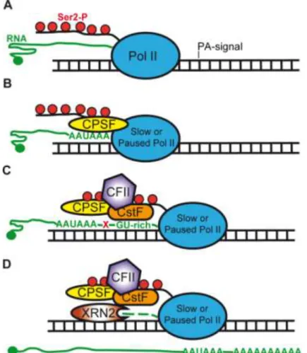
La terminaison de la transcription correspond à l’arrêt de la synthèse de l’ARN par la Pol II et au relargage de la molécule d’ARNm et de la Pol II de la matrice d’ADN. La terminaison de la transcription est cruciale pour la physiologie de la cellule. En effet, elle empêche une interférence entre la Pol II et des éléments fonctionnels de séquence d’ADN comme les promoteurs évitant ainsi que la Pol II n’initie la transcription d’un gène v oisin. La terminaison de la transcription est également liée au clivage et à la polyadénylation du transcrit natif d’ARN. Il a été montré que le ralentissement de la Pol II au site de terminaison permet au domaine C-terminal (C-terminal domain : CTD) de la Pol II de recruter et d’augmenter la concentration locale des facteurs de maturation de l’extrémité 3’ et de servir de plateforme pour l’assemblage des complexes de clivage de l’ARNm.
La terminaison de la transcription n’a pas lieu sur des sites conservés ou à une distance constante de l’extrémité 3’ des ARNm matures. Chez les mammifères, la terminaison peut se situer à une distance très variable de l’extrémité 3’ des ARNm, de quelques paires de bases à plusieurs kb. En effet des analyses par ChIP ont montré sur plusieurs gènes modèles, des densités de Pol II plus élevées en aval des extrémités 3’ des gènes (EAG) que sur l’ensemble de la séquence transcrite. Cependant il reste à déterminer ci cet enrichissement correspond à une pause, un arrêt ou un ralentissement de la polymérase. En effet, il a été montré que le signal de polyadénylation mais également des éléments de séquence en aval peuvent ralentir la progression de la Pol II en aval.
Durant ma thèse j’ai réalisé deux projets utilisant des techniques d’immunoprécipitation de la chromatine associées à des méthodes de séquençage à haut débit (ChIP-seq) afin d’analyser la distribution de l’ARN polymérase II dans deux conditions :
D’une part j’ai caractérisé sur l’ensemble du génome les pauses de la Pol II en aval du site EAG des unités de transcription dans les cellules humaines.
11 D’autre part, j’ai étudié les effets de stress génotoxiques sur la machinerie de transcription associée à la Pol II en analysant les modifications de profils de distribution de la Pol II.
Résultats 1.: Caractérisation des pauses de la Pol II en aval de la fin des unités de transcription
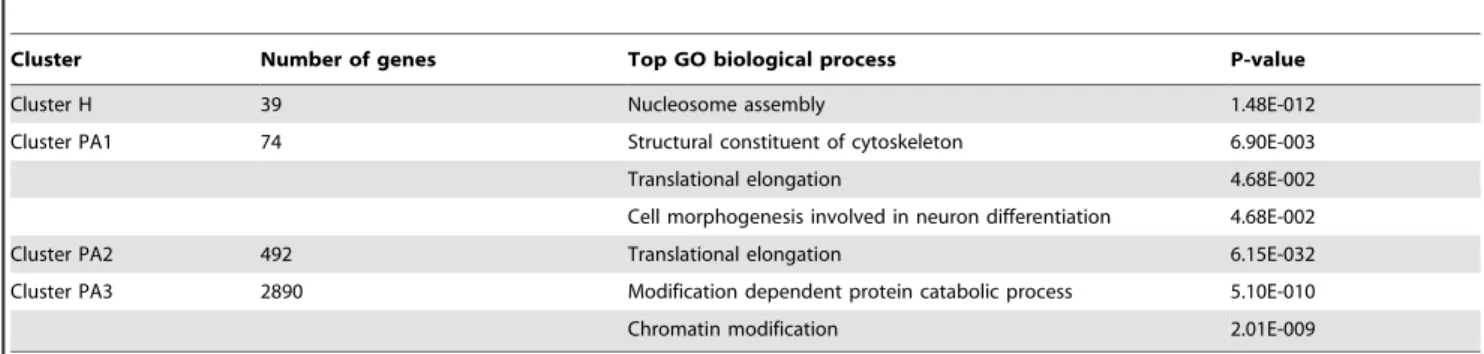
Afin de caractériser les profils de distribution de l’ARN Pol II en aval des EAGs, j’ai réalisé des expériences de ChIP-seq en utilisant un anticorps reconnaissant toutes les formes d’ARN Pol II humaine. J’ai analysé les profils de Pol II en aval de 13787 gènes qui n’ont pas de gène flanquant à +/- 4kb en amont ou en aval. Nos résultats ont été analysés en comparaison avec des données disponibles de séquençage à haut débit d’ARN naissants (Global Run On assay coupled sequencing : GRO-seq). Nos résultats montrent qu’un enrichissement de la Pol II en aval de l’extrémité des unités de transcription est une caractéristique partagée par tous les gènes exprimés et reflète la présence d’ARN Pol II active. Des analyses bioinformatiques (K-means clustering) m’ont permis de distinguer quatre groupes de gènes : le premier groupe (H) est caractérisé par un profil de pause étroit alors que les trois autres groupes (PA1-PA3) montrent un profil large ou très large, pouvant aller jusqu’à 6kb en aval des EAGs. Des analyses d’annotations (Gene Ontology) révèlent que le groupe H contient pratiquement exclusivement des gènes d’histones qui ne contiennent pas d’intron et dont les transcrits ne sont pas polyadénylés. A l’inverse, les groupes PA1-PA3 contiennent des gènes codant pour des transcrits polyadénylés. J’ai confirmé par des expériences de ChIP couplées à une analyse par qPCR les différents types de profils de distribution de Pol II décrits par analyse bioinformatique. Nos résultats sont en accord avec d’autres publications et suggèrent un lien entre le profil de distribution de la Pol II à l’extrémité 3’ des gènes histones et les mécanismes particuliers de maturation de l’extrémité 3’ de ces transcrits. Cette idée est renforcée par nos analyses fonctionnelles montrant que l’inhibition des mécanismes de polyadénylation augment la présence de l’ARN Pol II en 3’ des EAGs pour les gènes codant pour des transcrits polyadénylés. A l’inverse, cette inhibition ne change pas la distribution de la Pol II sur les gènes d’histones dont les transcrits ne sont pas polyadénylés. L’ensemble de ces résultats suggère un mécanisme qui augmenterait le
12 temps de résidence de la Pol II en aval de l’extrémité 3’ des gènes en cas de défaut de polyadénylation.
L’analyse des profils de distribution de l’ARN Pol II en aval des unités de transcription sur l’ensemble du génome, nous ont permis de montrer que le phénomène de pause de la Pol II est un mécanisme général retrouvé pour tous les gènes exprimés et qui parait conservé chez les vertébrés. Sur les gènes histones qui ne contiennent pas d’introns et dont les transcrits ne sont pas polyadénylés, le profil d’enrichissement de la Pol II en aval de l’extrémité 3’ est étroit et pourrait correspondre à des mécanismes très particuliers de maturation de l’extrémité 3’ de ces ARNm.
Résultats 2.: Analyse de la dynamique de l’ARN polymérase II au cours du stress par les UV-B.
Afin d’étudier les effets de stress génotoxiques sur la dynamique de l’ARN Pol II, j’ai utilisé une approche ChIP-seq en utilisant un anticorps reconnaissant toutes les formes de Pol II sur des chromatines préparées à partir de lignées de cellules humaines à différents temps après traitement par les UV-B.
L’analyse bioinformatique des résultats de distribution de la Pol II sur l’ensemble du génome m’a permis de montrer que des doses sub-létales d’UV-B induisent temporairement un arrêt global de la transcription. J’ai pu monter qu’un traitement par les UV-B entraine une perte progressive et majeure du signal Pol II des promoteurs des gènes exprimés. Cette perte de signal s’étend ensuite sur l’ensemble de l’unité de transcription jusqu’à quatre heures après irradiation. Cependant, la densité du signal Pol II en aval des EAGs n’est que très faiblement diminuée après irradiation. Des analyses par western blot utilisant différents anticorps reconnaissant spécifiquement les différentes formes de phosphorylation du CTD de l’ARN Pol II m’ont permis de montrer que les niveaux de ces différentes formes de Pol II ne sont pas affectées excluant un mécanisme actif de dégradation. Nous avons également identifié un groupe de gènes caractérisés par une augmentation du signal Pol II après irradiation par les UV-B. Ces gènes correspondent à des gènes de réponses aux dommages de l’ADN.
13 L’ensemble de nos résultats sont en accord avec des observations montrant qu’après irradiation par les UV-B l’arrêt temporaire de la transcription est nécessaire pour la mise en place de la réparation couplée à la transcription (transcription-coupled repair: TCR). Nous avons également observé que, six heures après traitement par les UV-B, les gènes initialement réprimés montrent ensuite une augmentation du recrutement de l’ARN Pol II par rapport au contrôle. Cette observation suggère que la machinerie transcriptionnelle de l’ARN Pol II pourrait compenser la réduction initiale de l’expression de ces gènes lors des étapes de réparation TCR.
Conclusions:
La transcription par l’ARN polymérase II a été étudiée en détail au cours des dernières décennies mais de nombreux mécanismes sont encore mal compris, empêchant ainsi de décrire un modèle décrivant tous les mécanismes de régulation de l’expression des gènes. Les projets que j’ai abordés pendant ma thèse nous ont permis d’élargir nos connaissances à propos de la transcription par la Pol II. Nous avons tout d’abord caractérisé sur l’ensemble du génome l’accumulation de la Pol II atour de l’extrémité 3’ des gènes. Nous avons observé qu’une augmentation de la présence de la Pol II en aval des gènes exprimés est une caractéristique générale conservée chez les mammifères. Nous avons également montré que les profils de distribution de la Pol II sur les gènes des histones canoniques sont différents ce qui pourrait correspondre à des mécanismes spécifiques de maturation des extrémités 3’ des ARNm correspondants. Nos études fonctionnelles renforcent l’hypothèse d’un lien entre augmentation de la densité de Pol II en aval des unités de transcription et terminaison de la transcription ainsi qu’avec la maturation de l’extrémité 3’ des ARNm.
L’étude des effets de stress génotoxiques sur la distribution de l’ARN Pol II sur l’ensemble du génome, nous a permis de montrer que des doses sub-létales d’UV-B altèrent de façon globale la machinerie de transcription. Nos résultats confirment d’autres observations suggérant que les complexes Pol II sont enlevés à la fois des sites de lésion de l’ADN mais également des promoteurs des gènes afin d’inhiber la transcription pendant les étapes de TCR. De plus, des modèles récents suggèrent qu’en présence de lésions persistantes de l’ADN qui seront lentement réparées, des sous-unités de l’ARN Pol II seront poly-ubiquitinées et dégradées pour permettre l’accès des facteurs de réparation sur ces
14 lésions et afin d’inhiber de nouveaux cycles de transcription. A l’inverse, nos résultats montrent une absence de dégradation de la Pol II après traitement avec des doses sub-létales d’UV-B ce qui pourrait s’expliquer par un mécanisme permettant une dissociation temporaire de la Pol II des promoteurs sans dégradation. Cette observation suggère que différents mécanismes pourraient être mis en jeu pour enlever la Pol II des unités de transcription pendant la réparation des lésions. L’activation de l’un ou l’autre de ces mécanismes d’éviction de la Pol II dépendrait du type de lésion impliquée et de leur persistance dans le temps.
15
ENGLISH VERSION
Genome-wide characterization of RNA polymerase II behavior during transcription termination and upon UVB stress
Summary of Thesis
Introduction:
Living cells are continuously exposed to stimuli from internal (basic metabolic processes) and external (environmental or chemical stress) sources. Therefore a very accurate and tightly regulated cellular process is needed to access and express the DNA-encoded information in order to maintain the normal physiological state of a cell. In eukaryotes this is carried out by the RNA polymerase II (Pol II) machinery, which is responsible for the expression of thousands of genes coding for proteins and non-coding regulatory RNAs.
RNA Polymerase II based transcription is one of the most highly regulated cellular process, which requires a well orchestrated, sequential action of multiple different protein complexes. Transcription cycle can be divided into three main phases: initiation, elongation and termination, however these phases are also built up from multiple sub-steps, which all represent a checkpoint and a regulatory possibility for proper mRNA synthesis and gene expression.
The transition from transcription initiation into elongation is an inefficient process, where Pol II shows tendency to stop (pause) after transcribing the first 25-50 nucleotides. This polymerase pause was first described as a rate limiting regulatory step between initiation and early elongation at the promoters of certain Drosophila heat shock genes. Recently, genome-wide studies demonstrated that promoter proximal Pol II pausing is a conserved regulatory step that is present at almost every promoters in higher eukaryotes. Surprisingly, paused or slowed down polymerases were also discovered downstream from end of genes (EAGs) however, in mammalian systems, the main focus of genome-wide study with respect to Pol II pausing was only promoter proximal pausing. Therefore the full spectrum of genes regulated by pausing downstream of EAGs has not yet been investigated
16 at a genome-wide level. Thus, it was not clear whether this phenomenon is commonly occurring among mammalian genes.
Transcription termination occurs when Pol II ceases RNA synthesis and both Pol II and the RNA molecule are released from the DNA template. Termination serves many vital functions in the cell. It prevents Pol II from interfering with downstream DNA elements, such as promoters and thus, Pol II will not enter into an unnecessary transcription of a neighbour gene. In addition, termination is functionally linked to the cleavage and polyadenylation of the nascent RNA transcript. It has been shown that the terminating or slowed down Pol II C-terminal domain (CTD) raises the local concentration of 3’ end processing factors near the nascent transcript and also serves as a platform for the assembly of the cleavage complexes.
Termination does not occur at a conserved site or constant distance from the 3’ end of the mature mRNAs. In mammals, termination can occur anywhere from a few base pairs to several kb downstream from the 3’ end of mRNA. Indeed, ChIP assays showed higher Pol II densities downstream from 3’ end of genes (EAG) than throughout the gene body on several model genes, however, it is uncertain, whether it indicates pausing, arrested or slowed down polymerases. In turn, it is known that the polyadenylation signal and certain downstream sequence elements can further negatively influence Pol II progression downstream of genes.
During my Ph.D. I carried out projects using chromatin immunoprecipitation assay coupled to high-throughput sequencing techniques (ChIP-seq) to analyse genome-wide Pol II behaviour in two aspects:
First, we characterized Pol II pausing downstream of the EAG of the transcription units in human cells genome-wide, by using high-resolution occupancy profiling.
Second, we investigated the effect of genotoxic stress on the Pol II transcription machinery by following the global alteration of Pol II occupancy profiles.
17
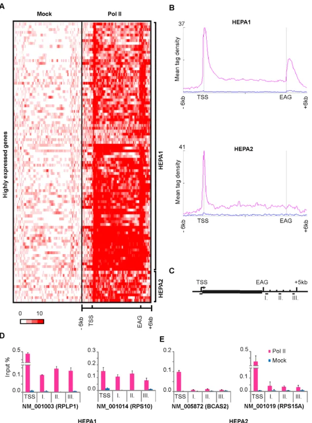
Results 1.: Characterization Pol II pausing downstream of the end of transcription units
In order to characterize Pol II occupancy profiles downstream of EAGs, we carried out ChIP-seq technique with an antibody, which recognizes all forms of human Pol II. We analyzed Pol II occupancy profiles downstream of 13787 genes which have no neighboring genes +/- 4kb up and downstream. Our results together with a Global Run on assay coupled sequencing (GRO-seq) data show that Pol II occupancy downstream of 3’ end of transcription units is a common feature of expressed genes and reflects transcriptionally active Pol IIs. We subdivided the expressed genes by K-means clustering and distinguished four main clusters, where cluster one (H) showed narrow pause profile, the rest (PA1-PA3) showed broad and very broad sometimes up to 6 kb long Pol II profiles downstream of their EAG. Gene ontology analyses showed that genes in cluster H are almost exclusively core histone genes, which are intronless and code for non-polyadenylated transcripts. The PA1-PA3 clusters represent genes coding for polyadenylated transcripts. We validated the bioinformatically isolated Pol II pause patterns with ChIP followed by qPCR detection. Our results are in good agreement with previous studies and may suggest a link between the Pol II occupancy profiles 3′ from histone genes and the different 3’ end processing mechanisms of the corresponding transcripts. This idea was corroborated by the functional studies, which showed that inhibition of polyadenylation increased Pol II occupancy 3′ of the EAGs of genes with polyadenylated transcripts, while it had no significant effect on Pol II drop at the 3′ end of the core histone genes. In addition these results suggest a mechanism, which increases the residency time of Pol II downstream of genes upon defective polyadenylation.
By characterizing Pol II occupancy profiles downstream from transcription units, we found that Pol II pausing is a genome-wide feature of expressed genes and it is conserved in mammals. On histone genes, which are coding for intronless and non polyadenylated transcripts, the Pol II occupancy profile is narrow, which may reflect the different mRNA 3’ end processing mechanism.
Results 2.: Investigation of RNA polymerase II behavior upon UVB stress.
To study the effect of genotoxic stress on the Pol II transcription machinery, we carried out ChIP-seq technique with an antibody which recognizes all form of Pol II on human cells in different time points after UVB treatment.
18 By generating heat maps from the global Pol II occupancy datasets, we found that a sub-lethal dose of UVB can temporary interrupt transcription genome-wide. Following UVB treatment we observed a progressive and massive Pol II signal loss from the promoters of expressed genes, which will then extend through the entire transcription unit, up to four hours after irradiation. Surprisingly, Pol II tag densities downstream from EAGs showed only a slightly decrease upon irradiation. To determine whether Pol II is degraded upon UVB treatment, we carried out western blot analyses with antibodies which recognize different phosphorylated forms of Pol II. Surprisingly we did not detect any decrease in the global level of Pol II. In addition, we found a subset of genes with increased Pol II signal upon irradiation, which were identified as DNA damage response genes. Our results are in good agreement with the observation that after UV irradiation transcription is arrested during the period of transcription-coupled repair (TCR). Interestingly, six hours after UVB treatment, genes that were negatively influenced, showed increased Pol II signal compared to the control state, suggesting that the Pol II transcription apparatus may compensate for the reduced gene expression associated with TCR.
Conclusions:
RNA polymerase II transcription has been extensively studied in the past decades, but lots of details are still missing to establish a model, which covers every regulatory mechanism during gene expression. With my Ph.D. projects, we managed to expand the current knowledge about Pol II transcription machinery. First we characterized the Pol II accumulation around the 3’ end of genes that was not yet investigated at a genome wide level. We found that increased Pol II occupancy downstream from expressed genes is a general feature that is conserved in mammalians. We also reported that Pol II profiles at core histone genes are different, which can be due to their unique mRNA 3’ end processing mechanism. With our functional study, we further support the link between the increased Pol II presence downstream from transcription units, transcription termination and 3’ end processing of mRNA. We also uncovered a possible regulatory mechanism which negatively influences transcription initiation upon defective polyadenylation.
By studying the effect of genotoxic stress on Pol II mediated transcription genome-wide, we show that a sublethal dose of UVB can disturb the global transcription machinery.
19 Our results are in good agreement with studies suggesting that Pol II complexes are removed not only from the DNA lesion site, but also from promoters to prevent further transcription initiation during TCR. Moreover, recent models suggest that in the presence of persistent DNA lesions, which are repaired slowly, Pol IIs can be polyubiquitinated and degraded to provide access for the repair factors to the lesion and to prevent new transcription cycles. In contrast, we did not detect Pol II degradation upon sublethal dose of UVB, which might reflect the activity of a mechanism that may facilitate the temporary dissociation of Pol II complexes from the promoters of expressed genes without degradation. This idea further suggests that different mechanisms may exist to remove Pol II from the transcriptional units during the DNA repair processes and their activation depends on the persistency and types of DNA lesions.
20
INTRODUCTION
21
Chapter one: RNA polymerase II
regulation and pause during
transcription
I. The expression of genetic information
The genetic information carried by DNA macromolecules, resides in the linear order (sequence) of nucleotides along a strand and divided into functional units (genes). Every cellular function and events require the access and activation of only a small subset of genes out of the whole genome. Eukaryotic genomes are complex (up to 25000 genetic loci in human) and organized within compact nucleoprotein (chromatin) structures. Therefore, transcription has a tightly regulated spatiotemporal control, which requires a well orchestrated recruitment of multiple protein complexes at a precise location in the nucleus in order to express the encoded information in the genome.
Living cells are constantly exposed to different intrinsic and extrinsic stimuli coming from the basic metabolic and cellular processes or from external environmental sources. In order to maintain the normal physiological state, cells have to access their DNA-encoded instructions and transcribe RNA molecules from particular genes. Most of these RNAs are processed and are translated during protein synthesis or they can act as regulators during different biochemical processes, which are required for normal cell functions such as development, differentiation and homeostasis in eukaryotic cells.
1.
DNA dependent RNA polymerases
In prokaryotes, one DNA-dependent RNA polymerase is sufficient to transcribe genes into RNA molecules required by the cell. In eukaryotes, expression of genes is shared by three distinct multi-subunit enzymes, each of which is dedicated to transcribe specific gene types. In eukaryotes RNA polymerase (Pol) I transcribes genes encoding 18S, 28S and 5.8S ribosomal RNAs (rRNAs). RNA Polymerase III is responsible for the transcription of genes coding for tRNAs 5S RNA and other small RNAs found in the nucleus and cytosol. RNA
22 Polymerase II is responsible for the transcription of all the protein-coding genes which constitute the largest group of distinct individual genes in the eukaryotic genome. In addition, it also transcribes genes coding for small non-coding RNAs, like micro RNAs (miRNAs), spliceosomal RNAs, small nucleolar RNA (snoRNAs) and a yet still unidentified fraction of intergenic or non-coding transcripts. These polymerases are the products of 31 genes: Pol I contains 14, Pol III 17 and Pol II has 12 subunits.
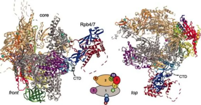
Figure 1: RNA polymerase II structure. Ribbon diagram shows two standard views front (left) and top
(right). The 12 subunits Rpb1-Rpb12 are colored according to the key (middle) between the views. Dashed lines
represent disordered loops. Eight zinc ions and the active site magnesium ion are depicted as cyan spheres and a pink sphere, respectively. The full CTD tail is not presented here. Figure adopted from (Armache et al, 2005).
Yeast and human Pol II both contain 12 subunits, (RPB1 to RPB12, with a total mass of > 0.5 MD) by decreasing order of their molecular mass (Figure 1) (Young, 1991). The 12 subunits of Pol II are highly conserved in sequence, architecture, and function thus, seven subunits of human Pol II can either partially (RPB4, RPB7, and RPB9) or completely (RPB6, RPB8, RPB10, and RPB12) substitute for the their yeast homologues. Five subunits (RPB5, RPB6, RPB8, RPB10, and RPB12) are shared between the three polymerases. The largest catalytic subunits of these polymerases (RPB1, RPB2, RPB3, and RPB6) share homology with each other and with the largest subunit of bacterial RNA polymerase subunits β-, β, α and ω, respectively. The similar sequences between RPB1 and β- as well as between RPB2 and β also refer to functional similarity: RPB1 and β-are involved in DNA binding, while RPB2 and β bind nucleotide substrates (Hampsey, 1998; Lee & Young, 2000). RPB1 and RPB2 are responsible for most of the catalytic activity of polymerase and are essential for
23 phosphodiester bond formation. Only RPB4, RPB7, RPB9 and the Carboxyl-Terminal Domain (CTD) of RPB1 are unique to Pol II (Minakhin et al, 2001; Mitsuzawa & Ishihama, 2004; Wild & Cramer, 2012).
A. Structure of Pol II
In the past twenty years, the structure of eukaryotic Pol II has been studied intensively and a wealth of structural information is provided by photocrosslinking, X-ray crystallography, NMR, and cryo-electron microscopy. These structure studies showed that the yeast Pol II easily dissociates into a 10-subunit catalytic core and a heterodimer of RPB4 and RPB7 subunits. The RPB4/RPB7 heterodimer is not essential for RNA chain elongation, although it is required for Pre-Initiation Complex (PIC) formation and mRNA maturation (Pankotai et al, 2010). [Also reviewed by (Cramer, 2004a; Cramer, 2004b; Hampsey, 1998; Lee & Young, 2000)] The two large polymerase subunits in Pol II (Rpb1 and Rpb2) form the opposite sides of the active centre. The small core subunits RPB5, RPB6 and RPB8 can bind to RPB1, RPB9 binds to RPB2 and the remaining subunits (RPB3, RPB10, RPB11 and RPB12) form a distinct subassembly that bridges between RPB1 and RPB2. Therefore, the polymerase core may be divided into three interacting subassemblies, which we refer to as RPB1 (composed of RPB1, RPB5, RPB6 and RPB8), RPB2 (consisting of RPB2 and RPB9) and RPB3 (RPB3, RPB10, RPB11 and RPB12) subassemblies, respectively (Figure 1). The central mass formed by RPB1 and RPB2 can be subdivided into four mobile elements, termed “core”, “clamp”, “shelf” and “jaw” lobe which bunk up around a positively charged “cleft” (Figure 2). The active center is buried at the base of the “cleft”. A “pore” beneath the active centre widens towards the outside, creating an inverted “funnel” through RPB1. DNA is suggested to enter at the “cleft” down the middle of the enzyme, passing between the mobile “jaws”. Beyond the active site, the DNA path is blocked by a protein “wall”. DNA-RNA hybrid formed in the active site would have to pass up the wall, at nearly right angles to the incoming DNA in the cleft. Both DNA and RNA are held in place by a massive “clamp” swinging over the cleft and active-center region. A hole in the floor of the cleft below the active site (“pore”) would allow entry of substrate nucleoside triphosphates and would also allow exit of RNA during retrograde movement of the polymerase on the DNA.
24 The location of RPB4/RPB7 was found close to the RNA exit channel and an additional role in transcriptional initiation was suggested (Armache et al, 2003; Boeger et al, 2005; Bushnell & Kornberg, 2003).
Figure 2: Structure of Rpb1. (A) Domains and domain like regions of Rpb1. The amino acid residue numbers at
the domain boundaries are indicated. (B) Ribbon diagrams, showing the location of Rpb1 within Pol II (“front” and ”top” views of the enzyme) and Rpb1 alone. Locations of NH2- and COOH-termini are indicated. Color-coding as in (A). Figure adopted from (Cramer et al, 2001).
The CTD is an unstructured extension from the catalytic core of Pol II. It is flexibly linked via an 80-residue linker to the close proximity of the RNA exit channel and provides an exposed surface to interact with proteins involved in 5’ capping, mRNA splicing, termination and 3’-end processing (Meinhart et al, 2005).
B. Carboxyl-terminal domain of Pol II
Compared to the other DNA-dependent RNA polymerases, the eukaryotic Pol II has an unusual structure what is uniquely found at the C terminus of the largest subunit (RPB1). This carboxyl-terminal domain is evolutionarily conserved in eukaryotes even as distant as yeast and humans. CTD comprises multiple tandem repeat of heptapeptides with the consensus sequence Tyr-Ser-Pro-Thr-Ser-Pro-Ser (Y1S2P3T4S5P6S7), with up to 52 repeats in
the mammalian proteins (Figure 3). Deletion of the CTD in mouse, Drosophila or yeast is lethal, demonstrating that this structure is essential for living cells.
25
Figure 3: The carboxyl-terminal domain (CTD) of Pol II. The sequence of human CTD (amino acids 1593–
1970) varies mainly at position 7 in the heptad motif in the distal (C-terminal) section of CTD. Amino acid residues that vary from the consensus motif are depicted in red. The number of repeats as well as the amino acid sequence is 100% conserved in mammals. Figure adopted from (Heidemann et al, 2012).
Interestingly, transcription, in some conditions can proceed without the CTD (Corden, 1990; McCracken et al, 1997), pointing to an ancillary, rather than fundamental role for this structure. It is also important that the heptads have to be in tandem: insertion of an alanine residue between heptads is lethal in yeast, whereas insertion of an alanine between “heptad pairs” can be tolerated (Stiller & Cook, 2004). While the CTD is essential for life, it is frequently not required for General Transcription Factor (GTF)-mediated initiation and RNA synthesis in vitro (Buratowski & Sharp, 1990; Zehring et al, 1988). This suggested that CTD tail of the RPB1 does not form part of the catalytic essence of Pol II, thus it may have important regulatory roles. Later, it became clear, that CTD serves as a scaffold for the interaction of a wide range of nuclear factors and plays a major role in transcription and co-transcriptional RNA processing of protein-coding genes, mammalian snRNA genes and yeast snoRNA genes (Gudipati et al, 2008). The CTD extends from the Pol II core enzyme close to the RNA exit channel, and this localization makes it capable for direct or indirect interaction with components of the RNA processing machinery.
C. Posttranslational modifications of CTD
Phosphorylation of the heptapeptide residues is the best-studied CTD modification. RPB1 can exist in two forms that can be distinguished on SDS PAGE: Pol IIa, when the CTD is hypophosphorylated and Pol IIo with hyperphosphorylated CTD. Pol IIa was shown to associate with the PIC at the promoter, and any phosphorylation of the CTD before this point
26 will prevent incorporation in the PIC and initiation. Transcription elongation and 3’ processing require sequential phosphorylation of Pol II. The CTD is the site of phosphorylations, with up to five potential phosphorylation sites on the tandem heptapeptide (Y1, S2, T4, S5, S7) (Baskaran et al, 1993) (Figure 3). In vivo phosphorylation
occurs mainly on serine residues (Zhang & Corden, 1991).
Proline-directed serine kinases are creating the characteristic pattern of Ser2 and Ser5 phosphorylation, which correlates with the phase of transcription on protein-coding genes in yeast and mammals (Figure 4 and 5) (Phatnani & Greenleaf, 2006). Phosphorylation of Ser5 is carried out by Cyclin-Dependent Kinase (CDK7) subunit of the TFIIH complex (see section E). Ser5 phosphorylation is needed for the recruitment of enzymes (Ceg1 and Abd1 in yeast) that will add the methyl guanosine cap to the nascent RNAs (Komarnitsky et al, 2000; Schroeder et al, 2000). Phosphorylation level of Ser5 is enriched at the promoter and decrease successively towards the 3′ end of genes. Ser5-P is dephosphorylated by Ssu72 phosphatase around 3’ end of genes which is essential for 3’ end processing of mRNAs (Steinmetz & Brow, 2003).
Phosphorylation of Ser2 is mediated by CDK9, which is the catalytic subunit of the Positive Transcription Elongation factor b (P-TEFb) complex (see section K/d). Ser2 phosphorylation increases on the elongating Pol II toward the 3’ end of genes (Figure 4 and
5) (Peterlin & Price, 2006). Ser2 phosphorylation plays an important role in the transition
from early elongation block into a productive elongating form. In addition, in vitro it was shown that splicing and polyadenylation is activated by Ser2 and Ser5 phosphorylated CTD (Hirose & Manley, 1998; Hirose et al, 1999). In higher eukaryotes, inhibition of Ser2 phosphorylation will end up in biased elongation, splicing and polyadenylation (Bird et al, 2004). However, Ser2 phosphorylation seems to play little part in the elongation of transcription of mammalian snRNA and replication-activated histone genes, (encoding relatively short RNAs) which are neither spliced nor polyadenylated (Medlin et al, 2005). In addition, the mammalian p21 protein-coding gene does not require P-TEFb activity for either elongation or RNA processing (Gomes et al, 2006), indicating that the requirement for Ser2 phosphorylation can be bypassed. Histone mRNA 3’ end formation is directed by the specialized, replication-activated histone mRNA processing signal and seems to be phospho-Ser2 (phospho-Ser2-P) independent (Discussed in section 7). In contrast, 3’ processing of snRNA genes require Ser2-P. Ser2 and Ser5 can also be phosphorylated by CDK8 and Cell Division Cycle 2
27 protein (CDC2). These kinases create the highly phosphorylated Pol IIo and Pol IIm forms that are thought to be transcriptionally inactive (Palancade & Bensaude, 2003). Dynamic dephosphorylation of Ser2 and Ser5 by CTD-specific phosphatases during the transcription cycle is essential for recycling Pol II. Two evolutionary conserved proteins (FCP1 and SSU72) dephosphorylate phospho-Ser2 and Ser5 respectively (Meinhart et al, 2005). RPAP2 (RNA Polymerase II-associated protein) also can dephosphorylate CTD-Ser5 and creates heptapeptides with phospho-Ser7, which facilitate the recruitment of P-TEFb and the subsequent phosphorylation of Ser2 (Czudnochowski et al, 2012; St Amour et al, 2012).
Ser7 is also a target of dynamic phosphorylation during the Pol II transcription cycle in eukaryotic cells (Akhtar et al, 2009; Chapman et al, 2007). Phosphorylated Ser7 is detected at the Transcription Start Site (TSS) and its levels remain high close to the 3′ end of Pol II genes (Brookes et al, 2012; Kim et al, 2010; Mayer et al, 2010) (Figure 5). Substitution of Ser7 to alanine is not lethal in yeast, but impairs viability of human cells by affecting the processing of snRNAs (Egloff et al, 2007; Schwer & Shuman, 2011). For the recruitment of the functional Integrator complex [a multi-subunit complex responsible for snRNA 3′ processing (Baillat et al, 2005)] two adjacent Ser7-P and Ser2-P marks are needed (Egloff et al, 2010). Ser7-P is further known to be a gene-specific “CTD-code” for its role in the expression and processing of snRNAs, although this mark is also present in protein-coding genes, where the Ser7-P is placed early in transcription, similar to Ser5-P, but its levels remain stable until the transcription termination site. Recent studies demonstrated that CDK7 is also the primary kinase for Ser7 phosphorylation in human and yeast cells (Boeing et al, 2010; Glover-Cutter et al, 2009; Kim et al, 2009). In the later phases of the transcription, the role of CDK7 is taken over by BUR1 a kinase which travels with the elongating Pol II placing phospho-Ser7 marks on its CTD. Although high levels of CTD Ser7-P correlate with high transcription rates (Bataille et al, 2012; Tietjen et al, 2010), the exact function of this CTD mark in transcription of protein coding genes is still unclear. Nevertheless, there might be a strong connection between Ser5 and Ser7 phosphorylation, which is suggested by data showing that CDK7 and Ssu72 act as a common kinase/phosphatase for both residues. Ssu72 removes Ser7-P immediately after cleavage and polyadenylation and thereby contributes to the reconstitution of the hypophosphorylated state of Pol II (Bataille et al, 2012; Zhang et al, 2012a).
28
Figure 4: Pol II CTD phosphorylations during transcription. (A)The CTD of Pol II which is recruited by
the PIC at the promoter is unphosphorylated. (B) Phosphorylation of Ser5 and Ser7 by the CDK7 subunit of TFIIH just after initiation helps to recruit and activate enzymes that add a methylguanosine cap (green filled circle) to the 5’ end of the nascent RNA. (C) Subsequent phosphorylation of Ser2 by CDK9 subunit of P-TEFb activates elongation and RNA processing. Phosphorylation of Tyr1 prevents the binding of termination factors to the CTD. Ser5 is dephosphorylated toward the 3’ end of the transcription unit by SSU72. (D) As Pol II reaches the termination site, the Tyr1-P signal weakens, but Thr4 is phosphorylated by PLK3 in humans and facilitates recruitment of termination factors. (E) After cleavage and polyadenylation of the 3’ end of the pre-mRNA, dephosphorylation of the CTD may help Pol II to disengage and to be recruited for another round of transcription.
Recent studies revealed that CTD threonine 4 is phosphorylated in yeast and higher eukaryotes. In humans Thr4 is phosphorylated by PLK3, but so far in yeast, no specific kinase was identified (Hsin et al, 2011) (Hintermair et al, 2012; Mayer et al, 2012). The replacement of Thr4 to alanine is not lethal in yeast (Stiller et al, 2000) however, the presence of Thr4 is essential for viability in human cells (Hintermair et al, 2012). Chromatin immunoprecipitation assay coupled sequencing (Chip-seq) datasets in yeast revealed that Thr4-P can be found mainly on the body region of genes and shows a low profile at the polyadenylation site (Mayer et al, 2012), while in human cells, phospho-Thr4 is enriched in the 3′ region of genes (Figure 5). Experiments demonstrated that Thr4 phosphorylation may have a role in the recruitment of termination factors like PCF11 (Meinhart & Cramer, 2004). When Thr4 was
29 changed into valine in chicken cells, biased processing of core histone mRNA was observed, while the basal transcription was not affected (Hsin et al, 2011).
Phosphorylation of CTD-Tyr1 was reported on human Pol II almost two decades ago (Baskaran et al, 1993), but its functional role was unknown. Genome-wide occupancy profiling revealed that this modification is associated with active genes (Mayer et al, 2012). The distribution profile correlates with the genome-wide phospho-Ser2 Pol II profile: Tyr-1 occupancy is low at promoters, then increases on the gene bodies and drops before reaching the polyadenylation site (Figure 5). This is in good agreement with experiments which demonstrated that Tyr1-P prevents CTD binding to the conserved CTD-interacting domain of termination factors. In contrast, Tyr1-P does not impair CTD binding to the distinct CTD-binding domain of elongation factor SPT6, consistent with SPT6 occupancy within the Tyr1-phosphorylated region of genes (Mayer et al, 2012). Therefore studies about phospho-Tyr1 mark explain how termination factor activities are restricted to the late phases of the Pol II transcription.
Each CTD repeat contains two prolines, embedded between phosphorylation sites. The two prolines at positions 3 and 6 of the CTD heptads can undergo conformational changes mediated by peptidyl–prolyl cis/trans-isomerases (PIN1) (Egloff & Murphy, 2008). Proline isomerization is also part of the “CTD code” which creates various binding scaffolds for other CTD associating factors. For example the cleavage and polyadenylation factor PCF11 is recruited to Ser2-P modified CTD repeats (Licatalosi et al, 2002) in combination with the relevant prolines in the energetically favored trans conformation (Meinhart & Cramer, 2004; Noble et al, 2005). Thus, the isomerization status of prolines can greatly influence the CTD phosphorylation pattern.
Serine and threonine residues can carry O-linked N-acetylglycosamine (O-GlcNAc), which is mutually exclusive with phosphorylation, meaning that this mark may be important to prevent CTD phosphorylation (Comer & Hart, 2001). Current studies propose that glycosylation of CTD Ser5 and Ser7 is carried out by GlcNAc transferase (OGT) and O-GlcNAc aminidase (OGA) during assembly of PIC (Ranuncolo et al, 2012).
The distal part of the Pol II tail comprises largely non-canonical CTD heptads with aberrations mainly occurring at position 7 (Chapman et al, 2008). Experiments showed that lysines at position 7 of the heptads and arginine 1810 of CTD repeat 31 can be specifically methylated by coactivator-associated arginine methyl-transferase 1 (CARM1) (Sims et al,
30 2011). This CTD mark can be found in hyperphosphorylated CTD in vivo. The enzymatic activity of CARM1 toward Arg1810 is repressed by phospho-Ser5 and Ser2 residues in vitro, meaning that the methylation is placed before early initiation but is still present during transcription (Sims et al, 2011).
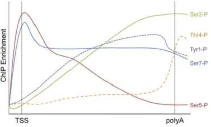
Figure 5: Average profile of CTD phosphorylation marks in genes.
Schematic representation of genome-wide occupancy profiles for all CTD phosphorylation marks. Tyr1-P, Ser2-P, Thr4-P, Ser5-P and Ser7-P show similar distribution of the modified Pol II along certain gene regions. While Ser5-P and Ser7-P peak at the transcription start site (TSS) of genes, Tyr1-P, Ser2-P and Thr4-P signals increase toward the 3′ end and polyadenylation site. Adopted from (Heidemann et al, 2012)
CTD residue Type of modification Modification added by Removed by Role Localization Serine /
Threonine
O-linked
N-acetylglycosamine O-GlcNAc transferase
O-GlcNAc aminidase
Prevent CTD phosphorylation
during PIC formation Promoters
Serine 2 Phosphorylation CDK9 /CDK8 / CDC2 FCP1 Promotes elongation / 3' end processing
Gene body / End of genes
Serine 5 Phosphorylation CDK7 / CDK8 / CDC2 SCP1 / PRAP2 / SSU72
Recruits RNA-capping enzymes /
Promoter clearance Promoters
Serine 7 Phosphorylation CDK7 / BUR1 SCP1 snRNA processing Promoter / Gene body
Tyrosine 1 Phosphorylation c-Abl proto-oncogene Prevents termination factor binding to CTD
Gene body / End of genes
Threonine 4 Phosphorylation PLK3 / CDK9? Recruits 3'-end processing /
termination factors End of genes
Proline 3/6 Isomerization PIN1 Recruits 3'-end processing /
termination factors End of genes
31
II. RNA polymerase II transcription cycle
Transcription of genes is the most highly regulated process in eukaryotic gene expression. Transcription cycle of Pol II can be divided into three phases: initiation, when the polymerase is recruited at a promoter with the help of a set of DNA binding proteins and begins to synthesize RNA, elongation, when Pol II extends the RNA transcript and termination when the polymerase and the RNA product disengage from the template.
2.
Transcription initiation
Transcription initiation starts with the recognition of a promoter region, which will serve as a platform for the assembly of the transcription factors and indicates the starting site of the transcription.
D. Core promoter elements
The Pol II core promoters contain special, evolutionally conserved sequence motifs, which are extended approximately 70 nucleotides around the Transcription Start Site (TSS) and direct the initiation of the transcription. A core promoter could be as simple as a single motif that serves as a universal transcription start site, or as complex as having a unique combination of a set of sequence instructions for each promoter (Figure 6).
We can distinguish focused promoters, where there is either a single transcription start site or a distinct cluster of start sites in a relatively short region and dispersed promoters, in which there are a number of transcription start sites distributed over a broad region with a range from up to 100 nucleotides. Some other genes might have alternate promoter sites, which are distinct and sometimes involve different type of regulation. These promoters are typically located hundreds or thousands of nucleotides apart.
In vertebrates, only about one-third of core promoters are focused core promoters, whereas the vast majority of genes contain dispersed core promoters (Juven-Gershon et al, 2008).
32 The presence of core promoters is necessary for accurate transcription initiation. These regions and their combination of short sequence elements represent an ancient and the most basic level of transcription regulation.
A core promoter can be built up from the following elements:
TATA box was the first eukaryotic promoter element to be identified. It is the most ancient and the most widely-used core promoter motif in nature. It has an A/T-rich consensus sequence [TATA(A/T)A(A/T)(A/G)], which is located approximately 20-30 nucleotides (nt) upstream of TSS in humans. The upstream T nucleotide is commonly located at -31 or -30 relative the Inr (Initiator). TATA box is present in less than 10% of human promoters. Surprisingly a wide range of A/T-rich sequences are capable of functioning as TATA boxes, consistent with the fact that TATA-Binding Protein (TBP) recognizes DNA through sequence-independent minor groove contacts. Many promoters do not contain consensus TATA boxes, or even non-consensus TATA boxes. Some TATA-less promoters retain the ability to direct transcription initiation from a specific nucleotide, whereas others can direct transcription initiation from multiple start sites (Smale, 1997; Smale & Kadonaga, 2003).
The initiator (Inr) is generally a pyrimidine-rich sequence around the TSS, PyPyA+1N(T/A)PyPy, (from nucleotide -6 to +11). In mammalians, computational analysis of thousands of transcription start sites suggests that the mammalian Inr consensus is YR, where R corresponds to the +1 start site (Carninci et al, 2006). The Inr is necessary in vitro and in vivo for a low level of accurate transcription by RNA polymerase II. Inr is capable to direct accurate transcription initiation either alone or with additional core promoter elements. These characteristics make Inr and TATA box functionally similar in two respects: both direct accurate transcription initiation by Pol II in the absence of other control elements, and both direct a high level of accurately-initiated transcription when stimulated by an upstream activator (Concino et al, 1984). The Inr is probably the most commonly occurring sequence motif in focused core promoters and it is a recognition site for a number of proteins.
33 The downstream promoter element (DPE) can be mainly found at TATA-less promoters, precisely +28 to +32 nt from TSS. It has a consensus sequence of (A/G)G(A/T)CGTG what was shown to be bound by Transcription Factor II D (TFIID). DPE is functionally dependent on Inr, therefore either promoter features a DPE sequence, it also has an Inr. TFIID binds cooperatively to DPE and Inr, therefore any mutation in either motif or alteration of the spacer region will lead to diminished level of basal transcription (Kutach & Kadonaga, 2000). Photocrosslinking studies revealed that the DPE is in close proximity to the TFIID subunits TAF6 and TAF9 (TAF stands for: TBP Associated Factor). Both contain histone fold motifs, thus it is possible that these subunits of TFIID interact with the DPE in a manner that is similar to binding of histones H3-H4 to DNA in nucleosomes (Shao et al, 2005).
Motif Ten Element (MTE) with a consensus sequence C(G/C)A(A/G)C(G/C)(G/C)AACG(G/C) can be found between +18 to +29 from TSS. It can also functionally substitute the loss of the TATA box and in addition, with the TATA box and DPE in proximity, it enhances the promoter activity in an Inr-dependent manner.
Downstream Core Element (DCE) consists of three sub-elements: SI, SII, and SIII, with a core sequence of CTTC, CTGT, and AGC, respectively. DCE occurs frequently with the TATA box, and appears to be distinct from the DPE. In a photo-crosslinking experiment TAF1 was shown to bind specifically the DCE.
XCPE1 (X core promoter element 1) motif is located from −8 to +2 relative to the +1 start site. It is present in about 1% of human core promoters, most of which are TATA-less. XCPE1 exhibits little activity by itself. [Reviewed in (Juven-Gershon et al, 2008)]
TFIIB-recognition element (BRE) with its (G/C)(G/C)(G/A)CGCC consensus sequence, can be found immediately upstream of the TATA box of 12% eukaryotic TATA-promoters. As it is in the name, TFIIB recognizes and binds BRE and its presence can
34 both negatively and positively influence basal promoter strength (Deng & Roberts, 2007).
Figure 6: Location of core promoter motifs. Each of these elements is found in only a fraction of all core
promoters. The Inr is probably the most commonly occurring core promoter motif. TATA box, MTE, DPE, DCE are recognition sites for TFIID. BREu and BREd interact with TFIIB. Figure adopted from (Juven-Gershon et al, 2008).
Large majority of the mammalian promoters, 60% of the human protein coding genes have multiple alternative start sites spread across a region. The advantage of this alternative promoter usage is that in one cell, different proteins can be generated from the same locus for different requirements.
The presence and combination of these sequence elements 5’ of transcription units determines not only the start site of transcription, but influences the promoter strength, direction of transcription, and may indicate functional differences between genes with different promoter architecture.
E. General transcription factors
The RNA polymerase enzymes do share a common property in transcribing DNA sequences, although they lack sequence-specific recognition ability to correctly specify the transcription start site unique to each gene. For site-specific initiation, additional proteins are necessary to form an initiation competent RNA polymerase complex. These essential accessory factors were collectively defined as general transcription factors (GTFs). Namely TFIIA, TFIIB, TFIID, TFIIE, TFIIF, and TFIIH (TF means transcription factor, II indicates Pol II driven transcription).
35 TFIID was first identified as a multiprotein complex which contains TBP and another 13 TBP-associated factors (TAFs) which support the promoter recognitions by binding to different promoter elements around the TSS (Fire et al, 1984; Young, 1991). Later, biochemical experiments demonstrated that the TFIID subunit composition can be different in cells at certain stages of development to provide different gene expression patterns (Muller et al, 2010). It is known that TAFs bind to the Inr, DPE, and DCE element, which allow TFIID to recognize TATA-less promoters. TBP exhibit nonspecific DNA-binding activity, which can lead to the formation of nonproductive transcription complexes. Thus, to prevent false initialization, TBP is affected by multiple negative regulations such as: TBP homodimer formation, inhibition by TAF1, BTAF1, Negative Cofactor2 (NC2) and nucleosome barrier (Goppelt et al, 1996; Kim & Burley, 1994; Kokubo et al, 1993; Nikolov & Burley, 1994; Timmers et al, 1992). The promoter recognition and TBP-TATA complex formation is positively regulated by TFIIA and TFIIB complex.
TFIIA is a complex of three proteins (α, β, γ subunit), which are encoded by two genes in higher eukaryotes. TFIIA increases the precise promoter recognition of TBP via promotion of the dissociation of TBP dimers. Furthermore, TFIIA stabilizes the forming PIC by competing with the TAF1/BTAF1 inhibition (Auble & Hahn, 1993; Coleman et al, 1999).
TFIIB is a single, 316 amino acid long polypeptide, which stabilizes the promoter bound TBP by competing with NC2 and it has also an important role in the recruitment of Pol II/TFIIF (Goppelt et al, 1996). TFIIB was further shown to recognize BREu element in a TBP independent manner via a non-conserved helix-turn-helix motif (Evans et al, 2001).
TFIIF is a heterodimer comprising two subunits, each of RAP30 and RAP74 (RNA Pol II Associated Proteins). It has multiple essential roles during the PIC formation process. Once it is bound to Pol II, it will facilitate its recruitment to the promoters and will further increase the TBP-TATA-TFIIB complex stability. As for the next step in the PIC formation it will facilitate the recruitment of TFIIE/TFIIH complexes. Later, it is
36 required for TSS selection, Pol II promoter escape and in transcription elongation (Flores et al, 1990; Yan et al, 1999).
TFIIE contains two subunits, α and β, which form a heterotetramer. TFIIE interacts with TFIIF, TFIIB, Pol II and promoter DNA. TFIIE’s α subunit is needed for the interaction with Pol II and binds close to the active center. TFIIE helps to recruit TFIIH, while it stimulates the helicase activities and the CDK7-mediated phosphorylation of the Pol II CTD (Ohkuma & Roeder, 1994; Watanabe et al, 2003).
TFIIH complex is composed of ten subunits: p89/XPB, p80/XPD, p62, p52, p44, p40/CDK7, p38/Cyclin H, p34, p8 and p32/MAT1 (Compe & Egly, 2012; Conaway & Conaway, 1989). The whole complex can be divided into two functional subcomplexes: a Cyclin-Activating Kinase complex (CAK) and a core complex. TFIIH has multiple roles during transcription: First, the core complex (XPB which is a helicase, p62, p52, p44, p34 and p8) is responsible for a DNA dependent ATPase activity. This activity is required for stable promoter opening and for the first phosphodiester bond formation which will further catalyze promoter clearance of Pol II(Kumar et al, 1998). Second, CAK subcomplex (CDK7, Cyclin H, MAT1) can phosphorylate Pol II CTD through CDK7 (Lu et al, 1992). This phosphorylation step mediates the transition from initiation to elongation phase. Third, the XPB and XPD subunits have ATP-dependent helicase activity, which is essential for promoter clearance and has an important role in DNA-damage repair too (Schaeffer et al, 1994). Fourth, E3 ubiquitin ligase activity of p44 subunit was also detected during DNA damage response (Takagi et al, 2005).
F. Sequential assembly of the PIC complex
The stepwise assembly of these GTFs at the recognized promoter will lead to a formation of a transcriptionally productive PIC. According to the sequential assembly model (Buratowski et al, 1989), first, the TATA-element is recognized and bound by TBP and the TFIID complex. Then the entry of TFIIA and TFIIB further stabilize the TBP–TFIID complex at


![Figure 7: Model for activated transcription. Transcriptional activators bind to their target sites [upstream activating sequence (UAS)] and recruit the intact form of Mediator](https://thumb-eu.123doks.com/thumbv2/123doknet/14465475.521254/39.893.120.773.521.735/activated-transcription-transcriptional-activators-upstream-activating-sequence-mediator.webp)