HAL Id: inserm-02503960
https://www.hal.inserm.fr/inserm-02503960
Submitted on 10 Mar 2020
HAL is a multi-disciplinary open access
archive for the deposit and dissemination of
sci-entific research documents, whether they are
pub-lished or not. The documents may come from
teaching and research institutions in France or
abroad, or from public or private research centers.
L’archive ouverte pluridisciplinaire HAL, est
destinée au dépôt et à la diffusion de documents
scientifiques de niveau recherche, publiés ou non,
émanant des établissements d’enseignement et de
recherche français ou étrangers, des laboratoires
publics ou privés.
pore opening
Benjamin Wacquier, Laurent Combettes, Geneviève Dupont
To cite this version:
Benjamin Wacquier, Laurent Combettes, Geneviève Dupont. Dual dynamics of mitochondrial
perme-ability transition pore opening. Scientific Reports, Nature Publishing Group, 2020, 10 (1), pp.3924.
�10.1038/s41598-020-60177-1�. �inserm-02503960�
www.nature.com/scientificreports
Dual dynamics of mitochondrial
permeability transition pore
opening
Benjamin Wacquier
1, Laurent combettes
2& Geneviève Dupont
1*Mitochondria play an essential role in bioenergetics and cellular ca2+ handling. the mitochondrial permeability transition pore (mPTP) is a non-specific channel located in the inner mitochondrial membrane. Long-lasting openings of the pore allow the rapid passage of ions and large molecules, which can result in cell death. the mptp also exhibits transient, low conductance openings that contribute to ca2+ homeostasis. Although many regulators of the pore have been identified, none of them uniquely governs the passage between the two operating modes, which thus probably relies on a still unidentified network of interactions. By developing a core computational model for mPTP opening under the control of mitochondrial voltage and ca2+, we uncovered the existence of a positive feedback loop leading to bistability. the characteristics of the two stable steady-states correspond to those of the two opening states. When inserted in a full model of ca2+ handling by mitochondria, our description of the pore reproduces observations in mitochondrial suspensions. Moreover, the model predicted the occurrence of hysteresis in the switching between the two modes, upon addition and removal of free ca2+ in the extra-mitochondrial medium. Stochastic simulations then confirmed that the pore can undergo transient openings resembling those observed in intact cells.
In most cell types, cytosolic Ca2+ is a key ion that controls major intracellular processes in health and disease1.
The signalling specificity of this ion largely relies on the spatio-temporal organisation of its stimulus-induced increases2. For example, oscillations and waves can occur due to the auto-catalytic Ca2+ release from the
endo-plasmic reticulum (ER), which acts as the main Ca2+ store. However, Ca2+ exchanges with other organelles
fur-ther extend the spatio-temporal diversity of Ca2+ signals. Among these, exchanges between the cytosol and
mitochondria play an important role. Entry of Ca2+ into mitochondria is mediated by the mitochondrial Ca2+ uniporter (MCU), while exit occurs via the Na+-Ca2+ and the H+-Ca2+ exchangers (NCLX and HCX), in
non-excitable cells3. Ca2+ uptake by mitochondria not only participates in the regulation of the cytosolic Ca2+
concentration ([Ca2+]) but also stimulates mitochondrial respiration and ATP production4.
The mitochondrial permeability transition pore (mPTP) can also transport Ca2+. Indeed, in response to a
metabolic stress or to an excessive accumulation of mitochondrial Ca2+, an increase in the permeability of the inner mitochondrial membrane (IMM) can be observed. First described in the 1970’s5–7, this phenomenon, called
permeability transition, could rapidly be ascribed to a non-selective pore. Mitochondrial Ca2+ overload and/or
oxidative stress lead to a massive and unselective opening of the pore, which allows for the transit of molecules up to 1500 Da. This includes Ca2+, metabolic substrates and ATP. Consequently, opening of the mPTP in this mode
induces the dissipation of the IMM voltage (∆Ψ) and, finally, cellular death. However, the pore can also exhibit moderate and transient openings8. Indeed, the existence of smaller conductance sub-states of the mPTP has been
demonstrated by experiments on mitochondrial suspensions9 and by electrophysiology10,11. This mode of reduced
activity contributes to Ca2+ homeostasis and thereby helps maintaining normal cellular functions8,12.
Well before its plausible molecular identification13,14, it was known that many factors, including reactive
oxy-gen species (ROS), pH, inorganic phosphates or cyclophilin D regulate the permeability transition12. Importantly,
the main drivers of mPTP opening are ∆Ψ and mitochondrial Ca2+ concentration ([Ca2+]m). Opening requires
a low voltage and a high Ca2+ load. Once open, the mPTP allows for the passage of ions, including Ca2+ itself,
which leads to mitochondrial membrane depolarisation (Fig. 1). There is no evidence of any specific regulator
1Unit of Theoretical Chronobiology, Faculté des Sciences, Université Libre de Bruxelles (ULB) CP231, B1050, Brussels,
Belgium. 2UMR-S 1174, Université Paris-Sud, INSERM, 91405, Orsay, France. *email: [email protected]
that would drive the mPTP from a low- to a large opening state, and it can thus be anticipated that this passage results from a network of feedback regulations.
In this study, we hypothesised that the two operating states of the mPTP rely on bistability. In such a scenario, two stable steady-states can coexist in a range of [Ca2+]m, which confers robustness to both states of channel opening. This assumption is based on the observation of a positive feedback loop that relies on the cross-inhibition between mPTP opening and mitochondrial voltage (∆Ψ): low voltage indeed opens the mPTP, which leads to further mitochondrial depolarisation (i.e. decrease in ∆Ψ). As mitochondrial Ca2+, known as the main mPTP
regulator, directly controls ∆Ψ, we anticipated that this positive feedback loop constitutes the core regulatory mechanism of mPTP opening. We first built a computational model formalising the mechanism just described. As assumed, the model can display bistability. We validated the model and parameter values using experiments in mitochondrial suspensions, which allow for controlled Ca2+ exchanges between mitochondria and the
extra-mitochondrial medium (em). The model then led us to rightly predict the conditions in which a hysteretic behaviour, which is a hallmark of bistability, can be observed. The robustness of bistability was assessed by a sen-sitivity analysis of the computational model. Finally, we showed that the model recapitulates the reversible, tran-sient openings of the mPTP observed in intact cells. In conclusion, this work proposes a simple dynamical mechanism by which mitochondria can safely use the mPTP in two operating modes, which differ drastically by their conductances and by their physiological implications. Put in a cellular context, the proposed bistable behav-iour not only provides robustness to the Ca2+ transport properties of the mPTP, but also ensures that, once
initi-ated, the mPTP-induced mitochondrial depolarisation is physiologically irreversible.
Model
The regulation of the mPTP by [Ca2+]
m and ∆Ψ is schematised in Fig. 2A. Based on this scheme, a single
differ-ential equation is used to describe the evolution of the fraction of open mPTP in the mitochondrial pool, noted
PTP. A small value of PTP thus corresponds to the transient, low conductance mode while a high value of this
variable can be associated to the large, long-lasting opening mode. In the following, we refer to these states as the low and high conductance modes. It should be noted that these terms do not refer to the single channel activities measured by electrophysiology. As in previous models15,16, the evolution equation includes a highly non-linear
term of opening of the mPTP, that is triggered when ∆Ψ falls below a threshold17,18. In agreement with
experi-mental data19,20, the value of the threshold is controlled by [Ca2+]
m (Cm in the model). The rate of mPTP closure
is described by a linear function. Thus, the evolution of the fraction of open mPTP is given by :
= − + − ⋅ ∆Ψ− ⋅ dPTP dt V PTP e k PTP (1 ) 1 1 , (1) op qop Cm q cl 11 Jin Ca2+ Low Ca2+ High Ca2+ Krebs cycle JPDH JO NAD+ NADH ETC JH,leak JO JH,leak JO ΔΨ ≈ 280mV ΔΨ ≈ 0mV + + + + ++ + + -- -- -- -H+ Na+ Solutes JPTP JPTP Ca H JPTP JPTP Ca H
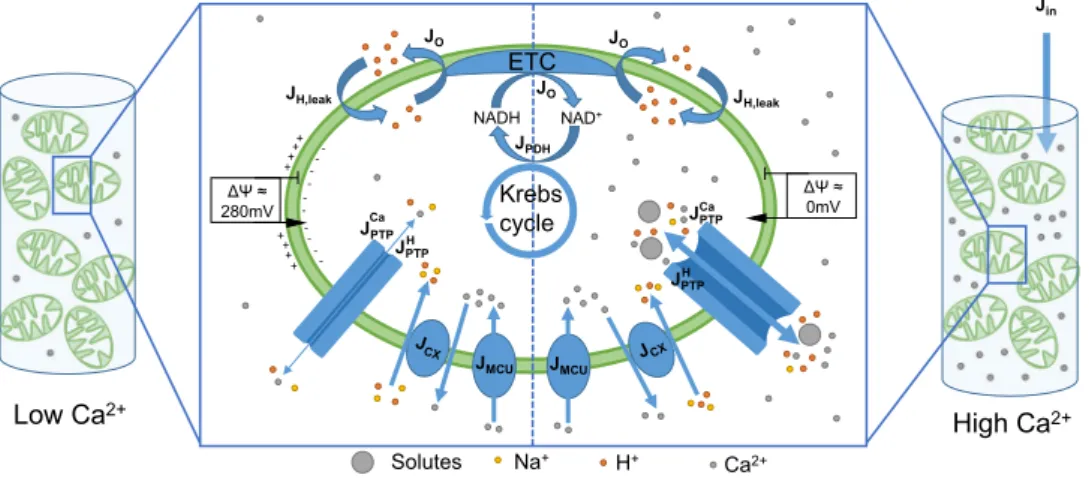
Figure 1. Schematic representation of the model describing Ca2+ dynamics and mPTP opening in
mitochondrial suspensions. The model includes Ca2+ exchanges between mitochondria and the
extra-mitochondrial medium through the Mitochondrial Ca2+ Uniporter (J
MCU), the Ca2+ exchangers (JCX), and the
mPTP (JPTPCa). The mPTP is potentially permeable to ions (H+, Mg2+, K+, ...) and small solutes. These fluxes are
gathered in JPTPH . Mitochondrial Ca2+ triggers the reduction of NAD+ in NADH (J
PDH) that can be oxidised by
the electron transport chain (JO) to generate a proton gradient. A leakage of protons into the mitochondrial space is considered (JH leak, ). ATP synthesis is not included because the medium does not contain adenine
nucleotides. The model reproduces a resting state (on the left) and a Ca2+-overloaded state (on the right). At
rest, the Ca2+ concentration in the suspension is low. Thus, the mPTP does not open fully, and the fluxes
through this pore (JPTPH , J
PTPCa) are low. In these conditions, a ∆Ψ is maintained thanks to the extrusion of
protons. Upon the addition of a massive amount of Ca2+ in the medium (J
in), the mPTP opens and becomes
highly permeable to ions and solutes. This leads to the dissipation of the ∆Ψ. See text, SI Appendix and Wacquier et al.22 for more details on the model.
www.nature.com/scientificreports
www.nature.com/scientificreports/
where Vop and kcl are rate constants of mPTP opening and closing, respectively. qop and q11 set the Ca + 2 and the
voltage dependencies of mPTP opening. When the mPTP is open, Ca2+ and protons leak through the pore. Each
ion flux depends on the electrochemical gradient and on the opening state of the mPTP. We describe these fluxes by mathematical expressions based on a simplified version of the Goldman-Hodgkin-Katz formalism21. Fluxes of
Ca2+ and H+ are described by Eqs. 2 and 3, respectively.
J V PTP C C e ( ) 1 , (2) PTPCa PTPCa m q em q 13 12 = ⋅ ⋅ − + −∆Ψ = ⋅ ⋅ + −∆Ψ J V PTP e 1 1 , (3) PTPH PTPH q q 13 12 where VPTPCa and V
PTPH are rate constants of Ca2+ and H+ fluxes through the mPTP, and q13 and q12 are coefficients
characterising the voltage dependencies of these ions fluxes through the mPTP. The concentration of H+ does not
appear in Eq. 3 as it is not a variable of the model. It is therefore implicitly included in VPTPH , and the flux JPTPH only
appears in the evolution equation for voltage (Eq. 4). Moreover, JPTPH also includes other compounds leaking
through the pore (ions and small solutes).
Experimentally, the dynamics of mPTP opening are commonly investigated in suspensions of mitochondria. To model this experimental system, Eqs. 1–3 describing mPTP opening and closing, and the associated ion fluxes, are included in a previously-published model describing Ca2+ dynamics in such a system (Fig. 1)22. This model
describes 1) Ca2+ exchanges between mitochondria and their medium via the MCU (J
MCU) and the NCLX/HCX
(JCX) 2) The Ca2+-dependent synthesis of NADH by the Krebs cycle (JPDH), and its consumption by the electron
0 5 10 15 Cm( M) 50 100 150 200 (mV) 0 5 10 15 Cm( M) 0 0.2 0.4 0.6 0.8 1 PT P
PTP
ΔΨ
C
m 0 0.25 0.5 0.75 1PTP
0 50 100 150 200(mV)
Cm = 8 µM dPTP/dt = 0 d /dt = 0ΔΨA
B
C
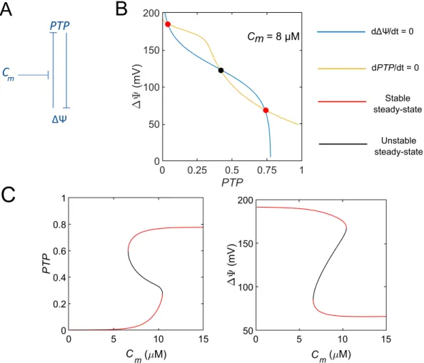
Stable steady-state Unstable steady-stateFigure 2. A two-variable model describing mPTP opening. (A) Scheme of the model. The positive feedback loop between the fraction of open mPTP (PTP) and mitochondrial voltage (∆Ψ) underlies bistability. This loop results from cross-inhibition. Cm modulates the threshold of PTP inhibition by ∆Ψ. (B) Phase space for Cm=8
µM. The yellow and blue curves represent ∆Ψ and PTP null-clines, respectively. Cem was fixed at 0.5 Mµ and
NADH at 200 µM. In red: stable steady-state. In black : unstable steady-state. (C) Bifurcation diagrams of PTP
transport chain (ETC, JO) 3) The variations of voltage induced by Ca2+ transporters, the ETC and proton leaks
(JH leak, ). Eq. 1 is thus coupled with this model.
The mitochondrial voltage varies in time according to : ∆Ψ
= . − − − . + . −
d
dt (a J1 O JH leak, JCX 2 JMCU 2 JPTPCa JPTPH )/Cp (4) Positive voltage corresponds to an excess of positive charges in the extra-mitochondrial medium. Cp scales
molec-ular fluxes into voltage changes. It includes the membrane capacitance and the Faraday constant. a1 scales NADH
consumption into voltage variations.
For the time evolution of extra-mitochondrial Ca2+ concentration (C
em), we write
dC
dtem =f Jem in( +δJCX−δJMCU+δJPTPCa), (5) where fem is the Ca2+ buffering capacity of the medium considering the rapid buffering approximation23. It
cor-responds to the ratio between free and total Ca2+ concentrations in the extra-mitochondrial medium (em). δ is
the volumic ratio between mitochondria and their medium (V Vm em/ ). Jin allows to simulate the addition of Ca2+
in the medium.
In a similar way, the time evolution of mitochondrial Ca2+ concentration (C
m) is expressed by
= − −
dC
dtm f Jm MCU( JCX JPTPCa), (6) where fm is the Ca2+ buffering capacity of mitochondria.
Finally, NADH is produced by the Krebs cycle and consumed in the ETC, which is described by
= − d NADH dt J J [ ] (7) PDH O
The detailed kinetic expressions of the fluxes are exposed in the Supplementary Material. Parameters values are listed in Table S1 and, except for the mPTP opening description, are taken from our previous models that were validated against experimental data22,24.
Results
the mptp acts as a bistable switch.
Bistability often occurs in a system including a positive feedback loop25,26. As schematised in Fig. 2A, such a loop exists between the mPTP and ∆Ψ, as a high ∆Ψ prevents theopening of the pore, while ∆Ψ is dissipated by an open mPTP due to ion leakage8. Because [Ca2+]
m controls the
value at which ∆Ψ starts inhibiting mPTP opening, Ca2+ could play a key role in the switch. To study the possible
existence of a Ca2+-controlled bistability in the mathematical description of the mPTP presented in the previous
section, we first considered a minimal model that only considers the evolution of ∆Ψ and PTP (Eqs. 1 and 4) while Cm, Cem and NADH are kept constant. We analysed the behaviour of this two-variable model on the basis of bifurcation diagrams where the steady-states of ∆Ψ and PTP are shown as a function of Cm (Fig. 2C). For low Cm (<7 µM), the system tends towards a resting state, i.e. a large ∆Ψ and an almost closed mPTP (low value of vari-able PTP). For high Cm (>11 µM), the system will always evolve towards a dissipated potential and a fully open
mPTP. For intermediate concentrations, one can observe the coexistence between these two stable steady-states, separated by an unstable one. This scenario is also visible in the phase plane, where the null-clines intersect once at low or high value of PTP, for low or high Cm respectively (not shown), and three times for intermediate Ca2+
concentrations (Fig. 2B). The two steady-states display the characteristics of the two operating modes of the mPTP described above. The first one occurs at low Ca2+, when a high ∆Ψ is established across the IMM8. The
other state is reminiscent of the behaviour of the mPTP reported at high mitochondrial Ca2+ load: the mPTP
largely opens, leading to the dissipation of ∆Ψ. Interestingly, in suspensions, it is possible to come back from the high to the low conductance mode by adding a Ca2+ chelator27. The model is possibly in agreement with this
reversible bistability, as the ordinate axis does not intersect the bistable area26.
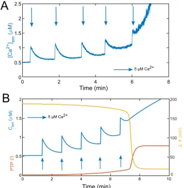
To validate the model, its behaviour must be compared to experiments. Successive additions of large amounts of Ca2+ in a suspension of mitochondria isolated from hepatocytes can induce mPTP opening. In Fig. 3A, each
arrow represents the addition of 5 µM exogenous Ca2+. Changes in Ca2+ concentration in the medium
([Ca2+]
em) are monitored by spectrophotometry22, allowing to follow Ca2+ exchanges between mitochondria and
the medium. Up to the first four additions of Ca2+ in the medium, most of the Ca2+ is sequestered by
mitochon-dria via the MCU, and the mPTP has a limited effect22. However, after the fifth addition, one observes a brutal
increase in the [Ca2+]
em (Fig. 3A). This important rise is attributed to the opening of the mPTP in its high
con-ductance mode. This opening is accompanied by the release of Ca2+ from mitochondria, which explains the fast
and sudden elevation in the medium. To compare the behaviour of the model with these experimental observa-tions, we took variations of Ca2+ and NADH concentrations into account and simulated the whole dynamical
system, defined by Eqs. 1 and 4–7 with the same values of parameters as in our previous study22. The model
faithfully reproduces the experimentally observed behaviour (Fig. 3B): after a few pulses, Cem increases all of a sudden (blue curve). This increase is associated with the opening of the mPTP (red curve), which triggers a Ca2+
www.nature.com/scientificreports
www.nature.com/scientificreports/
flux from mitochondria to the medium (via JPTPCa) while ∆Ψ is collapsing due to the important ion fluxes through
the mPTP (yellow curve).
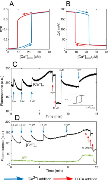
The switch-like behaviour shown in Fig. 3 is compatible with the existence of bistability, but might also rely on the existence of a sharp threshold. To investigate if the switch corresponds to a change of steady-state, we cannot draw bifurcation diagrams as a function of Cm as in the two-variable model (Fig. 2C), since Cm is a variable in the full model. We thus established pseudo bifurcation diagrams showing the values of PTP and ∆Ψ as a function of the amount of Ca2+ added during the pulses (Figs. 4A,B and S1). The blue curve is drawn by performing a series
of simulations where the steady-states of the model are sequentially computed after successive additions of 0.1 Mµ Ca2+. After a critical amount of added Ca2+, the system is driven towards a state with a large opening of the mPTP. This opening is, as expected, associated with a drop in ∆Ψ and Ca2+ efflux from mitochondria. From
this state, we then simulated the removal of Ca2+ from the medium (red curve corresponding to successive
decreases of Ca2+ of 0.1 µM). Biologically, it would correspond to the addition of a Ca2+ chelator. It is clear that
the blue and red trajectories do not coincide and that bistability also occurs in the full model. The amount of Ca2+
that has to be removed to bring the mPTP back in its low conductance mode exceeds the amount of Ca2+ that was
necessary to trigger the passage from the low to the high conductance mode. This hysteretic behaviour is typical of a bistable system26.
Hysteresis in mptp opening.
We next wondered if experiments can validate the hysteresis predicted by the model. We first checked in a control experiment that in a medium devoid of mitochondria, the addition of a given amount of Ca2+ is rapidly counterbalanced by the addition of the same amount of the high affinity Ca2+ buffer EGTA (Fig. S2). This observation is in agreement with the 1 to 1 stoichiometry of Ca2+ binding to EGTAand with the values of kon and koff (6 µM−1 s−1 and 1 s−1, respectively28). Then, on the basis of the model prediction
shown in Fig. 4, we submitted mitochondria in suspensions to successive additions of Ca2+, up to the opening of
the mPTP. As soon as the mPTP opens, we rapidly added EGTA. We then compared the amount of EGTA neces-sary to close the mPTP to the amount of Ca2+ that was necessary to open it. Following the addition of three Ca2+ pulses of 5 µM in a mitochondrial suspension, the mPTP opens and [Ca2+]
em starts to rise (Fig. 4C). Thus,
between 10 and 15 µM Ca2+ were necessary to trigger opening. Accordingly, in the absence of hysteresis, the
removal of 5 µM Ca2+ should close the mPTP. Nevertheless, the addition of 10 µM EGTA is not sufficient to close
the mPTP: the [Ca2+]
em is still high, indicating that the mPTP is still open. By contrast, the Ca2+ level decreases
drastically after a second addition of 10 µM EGTA. At this stage, the mPTP is closed or in a low conducting mode. Figure 3. Successive additions of Ca2+ leading to mPTP opening in experiments and in the model. (A)
Experiment performed in a suspension of mitochondria extracted from rat hepatocytes. The [Ca2+] em is
monitored by fluorescence using 5 Mµ Fluo-4. Following the addition of five Ca2+ pulses (blue arrows), the
mPTP opens, as attested by the sudden rise in Ca2+ concentration in the medium. (B) Simulations of the
five-variable model (Eqs. 1 and 4–7). The model successfully reproduces the [Ca2+]em behaviour (in blue), in
response to the Ca2+ pulses (implemented with J
in). The final increase in the Cem is associated with a sudden
increase in mPTP opening (variable PTP, standing for the fraction of open PTP, in red) and a fall in ∆Ψ (in yellow). See Table S1 for parameter values.
As a control, upon Ca2+ re-addition, we see a characteristic spike due to Ca2+ entry in mitochondria. This would
not be visible with an open mPTP, as mitochondria would not be able to sequester Ca2+. Finally, a further Ca2+
addition allows to open the mPTP again.
The existence of hysteresis is also supported by the simultaneous measurement of ∆Ψ and [Ca2+]
em during a
similar experiment (Fig. 4D). After 32.5 µM of added Ca2+, the drop in potential associated to mPTP opening is
observed through an increase in the fluorescence of the probe. Concomitantly, the opening of the pore is seen via the increase in the [Ca2+]
em. 40 µM EGTA are then required to bring the system back to its basal state, in
agree-ment with the presence of a hysteresis loop.
Robustness of bistability.
To be biologically relevant, bistability in mPTP dynamics must be robust with respect to cell-to-cell variations. It is thus important to check that the bistable behaviour predicted by the mathe-matical model occurs for a large range of values of the kinetic parameters. We first analysed the extent of the domain of bistability when changing the values of the parameters of the model (Fig. 5, in blue). It is visible that bistability occurs in a quite extended range of values of all parameters. The existence of bistability is sensitive to the kinetic parameters that control the opening of the pore (Vop and kcl). If the pore opens too slowly, or closes toofast, bistability is lost. Bistability is thus favoured by a given ratio between opening and closure rates (Fig. S3B).
0 10 20 30 40 ( M) 0 0.2 0.4 0.6 0.8 PT P 0 10 20 30 40 ( M) 0 50 100 150 200 (mV)
A
0 4 8 12 40 80 120 160 200 0 5 10 15 100 150 200 250 Time (min) Time (min) Fluorescence (a.u.) Fluorescence (a.u.) [Ca2+] em [Ca2+] em ΔΨ 5 µM 7.5 µM 2.5 µM 7.5 µM 7.5 µM 7.5 µM 5 µM 5 µM 5 µM 5 µM 10 µM 10 µM [Ca2+]added [Ca2+]added
[Ca2+]added
[Ca2+] addition EGTA addition
PTP
10 µM (x4)
B
C
D
Figure 4. mPTP opening corresponds to a bistable switch. Numerical bifurcation diagrams of the fraction of open mPTP, PTP (A) and ∆Ψ (B) as a function of added Ca2+. Blue and red curves stand for trajectories
following Ca2+ additions (from left to right) or Ca2+ removal (from right to left), respectively. The equivalent
bifurcation diagrams of Cm and Cem as a function of added Ca2+ are shown in Fig. S1. (C,D) Experimental
evidence for bistability of the mPTP in a hepatocyte mitochondrial suspension. Ca2+ is monitored in the
medium with 5 µM Fluo-4 (C,D) and ∆Ψ with 5 µM TMRM (SI Appendix) (D). In both cases, the
concentration of EGTA necessary to close the mPTP is larger than the concentration of Ca2+ that allowed its
opening. This highlights the existence of hysteresis as schematised in the inset in Panel (C). Panels (C,D) are representative of 12 similar experiments.
www.nature.com/scientificreports
www.nature.com/scientificreports/
The most sensitive parameter is qop, that regulates the threshold of inhibition of the pore by ∆Ψ (Fig. S3C). This parameter also alters the threshold of mPTP opening by Ca2+. Consequently, increasing this parameter shifts the
whole bifurcation curve (Fig. 4A for example) to the left, until the bistable domain disappears from positive Ca2+
concentrations. On the opposite, decreasing the parameter shifts the bistable area to the right. For high qop values, the system is thus bistable, but we consider this situation as biologically not relevant as it requires the addition of an unrealistically large amount of Ca2+. Interestingly, the existence of bistability is extremely robust with respect to changes in the parameters related to Ca2+ fluxes (V
MCU, VCX and VPTPCa). Changing these parameters mainly
impacts on the rates at which Ca2+ is exchanged between mitochondria and the medium. This relative
insensitiv-ity confirms that the relationship between ∆Ψ and PTP is the core mechanism leading to bistabilinsensitiv-ity, whereas Ca2+
controls the transition between the two states. Finally, if the ion flux through the mPTP (VPTPH ) is weak, bistability
is also lost (Fig. S3D). Indeed, the loss of ∆Ψ induced by mPTP opening is then much reduced, which weakens the positive feedback loop that generates bistability.
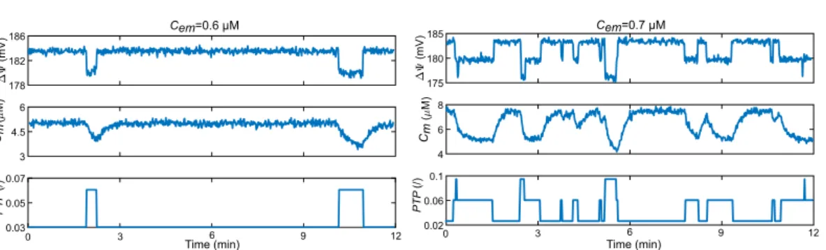
Simulation of transient openings of the mptp.
The level of opening of the mPTP in its low conduct-ance mode is in average very small. The associated Ca2+ fluxes are thus expected to be minimal. In intact cells,openings in this mode indeed take the form of short-lived, stochastic events as reported for permeabilised heart cells29 and astrocytes30. These random openings are associated with small-amplitude drops in ∆Ψ and
mitochon-drial Ca2+ and are localised to small areas of the cell.
We developed a stochastic version of the model to simulate transient mPTP openings (SI Appendix). As these open-ings remain highly localised, we simulated a small volume containing a single mitochondrion, and considered that the concentration of cytosolic Ca2+ around this mitochondrion (C
em) remains constant. An increase in Cem simulates an
efflux from the ER, triggered by an external stimulus. At low Cem, simulations predict a noisy but stationary state for ∆Ψ
and Cm, associated with a mPTP that is nearly always closed. If we increase Cem, small and rapid drops in ∆Ψ,
accompa-nied by some Ca2+ efflux from the mitochondrion, can be observed (Fig. 6). These isolated events correspond to
sud-den, random and brief openings of the pore in its low conductance mode. For higher Ca2+ concentrations, the number
of events increases, as well as the mPTP open probability (Fig. S4). If the number of events becomes too large, changes in ∆Ψ and Cm are quite irregular, as observed in heart mitochondria in suspension31.
Vop kcl qop V PTP Ca V PTP H VMCUVCX 10-1 100 101 ret e mar ap tl uaf ed eht fo % Oscillations Monostability Bistability
Figure 5. Range of bistability in the mPTP opening model. In blue: the system is bistable; in orange-coloured: the system has one stable steady-state; in yellow: the system oscillates at high PTP values. Sensitivity analysis was performed by individually varying seven different parameters from 10 to 1000 % of their default values. See Fig. S3 for bistability conditions and notes on oscillatory behaviour.
3 4.5 6 ( ) M 178 182 186 ) V m( 0 0.03 0.05 0.07 PT P (/) 4 6 8 ( M) 175 180 185 (mV ) 0.02 0.06 0.1 PT P (/) Cem=0.6 µM Cem=0.7 µM Cm Cm 3 6 9 12 0 3 6 9 12 Time (min) Time (min)
Figure 6. Stochastic opening of the mPTP. Time series of the evolution of ∆Ψ, Cm and PTP for different Cem,
obtained by simulations of a stochastic version of the model. The simulated system corresponds to the volume of a single mitochondrion exposed to a constant Cem.
Discussion
We have provided a minimal description of the dynamics of the mPTP, in which we only considered the regula-tion of the pore by its main regulators, Ca2+ and ∆Ψ. It is clear, however, that mPTP opening is inhibited by a
variety of compounds, which include Mg2+, adenine nucleotides, cyclosporin A, mitochondrial H+, metabolic
fluxes and environment, and stimulated by others, such as reactive oxygen species, organic phosphate or cyclo-philin D12,15,20,32. Although the influence of most of these factors can partly be accounted for by their influence on
∆Ψ or Ca2+, a more accurate description of the mPTP regulation would be useful to account for some
experi-mental observations. Inhibition of mPTP opening by protons is sometimes proposed as a primary mechanism of mPTP regulation33. In the present model, protons influence mPTP opening through membrane voltage, but not
directly (see Eq. 4). It is known that the mPTP is open at pH ≥7.3-7.5 and closed at pH <734,35. Because at rest,
mitochondrial pH lies between 7.2 and 7.633,36, and because an increase in [Ca2+]
m leads to an increase in
mito-chondrial pH, direct H+ regulation of the mPTP cannot on its own account for the abrupt openings of the pore
observed upon additions of Ca2+ (Fig. 3).
Focussing on the core regulatory mechanism of mPTP opening by Ca2+ and ∆Ψ allowed us to uncover the
occurrence of bistability in an intermediate range of [Ca2+]
m. This bistability underlies the well-established
exist-ence of two operating modes displaying widely different average conductances. The proposed mechanism more-over agrees with the recent observation of a common molecular nature of the two modes of the pore, which is formed from the FoF1 ATP synthase, at the interface between monomers within dimers13.
The bistable scenario proposed in this study contrasts with the description of the passage between the low- and the high-conductance mode of the pore as a switch controlled by a single threshold governed, for example, by mitochondrial pH16,33,37,38, [Ca2+]
em39, [Ca2+]m40 or mitochondrial volume41. In Bazil et al.15, the switch is
imposed by ∆Ψ, and the value of the threshold is controlled by [Ca2+]m, as in the present study. This ~200-variable
model is very detailed, as it includes regulation of the mPTP by a variety of factors as well as mitochondrial bio-energetics. It nicely reproduces experimental observations but is hardly usable for a bifurcation analysis. In a broader context, many processes governed by binary choices in cell biology have been described by bistability. This includes cell differentiation42, enzymatic reactions43, the cell cycle26,44, or bacterial communication45. One of
the main advantages of bistability-controlled transitions is their robustness. In the case of the mPTP, this property plays a key role in relation to the very nature of the pore. Indeed, when the pore enters in a high conductance mode, it provokes a decrease in ∆Ψ and [Ca2+]
m. If a threshold-associated switch was at play, this decrease would
bring the channel back in a low conductance mode, provoking in turn an increase in ∆Ψ and [Ca2+]
m, hence the
opening of the channel again. These unrealistic back and forths between a fully- and a partially-open pore do not occur in a bistable scenario, which ensures that, once initiated, the full opening of the channel is maintained up to a point where largely depolarised mitochondria lead to cell death.
Much remains to be done to assess the physiological relevance of the low conductance mode of the pore in intact cells. We have touched this question by performing stochastic simulations of the model in conditions cor-responding to a single mitochondrion facing different levels of Ca2+ in its surrounding cytosol. A stochastic
approach is necessary because openings are transient and weak. The low level of mPTP opening computed in the deterministic bifurcation diagram (Fig. 4A) corresponds to an average over extended periods of time, during which the mPTP undergoes random, discrete openings (Fig. 6). In cells, such transient openings are thought to play an important role in releasing matrix Ca2+ to maintain mitochondrial homeostasis46. In vivo, mitochondria
are morphologically and functionally heterogenous47, which can explain the large variety of changes in [Ca2+] m
that have been reported experimentally22. Moroever, mitochondria are permanently rebuilt through fusion and
fission that are promoted by mitochondrial movements48. The last two factors are expected to further increase the
variability in mPTP openings, in addition to their inherent stochastic character, which again calls for a relatively noise-insensitive mode of switching between the two conductance modes.
The mPTP is involved in the pathophysiology of many diseases, ranging form ischemia/reperfusion injury to neurodegenerative disorders49. It would be useful to integrate the present description of the mPTP as a bistable
molecular switch in comprehensive models of mitochondrial Ca2+ and metabolism15,50,51 to provide a detailed
computational support to the experimental investigation of such pathologies52.
Received: 8 July 2019; Accepted: 6 February 2020; Published: xx xx xxxx
References
1. Berridge, M. J., Bootman, M. D. & Lipp, P. Calcium - a Life and Death signal. Nature 395, 645–648, https://doi.org/10.1038/27094
(1998).
2. Dupont, G., Falcke, M., Kirk, V. & Sneyd, J. Models of Calcium Signalling, vol. 43 of Interdisciplinary Applied Mathematics (Springer International Publishing, 2016).
3. Jouaville, L. S., Ichas, F. & Mazat, J.-P. Modulation of Cell Calcium Signals by Mitochondria. Bioenergetics of the Cell: Quantitative
Aspects 371–376, https://doi.org/10.1007/978-1-4615-5653-4_24 (1998).
4. Denton, R. M. Regulation of Mitochondrial Dehydrogenases by Calcium Ions. Biochimica et Biophysica Acta (BBA) - Bioenergetics 1787, 1309–1316, https://doi.org/10.1016/j.bbabio.2009.01.005 (2009).
5. Hunter, D. R. & Haworth, R. A. The Ca2+-Induced Membrane Transition in Mitochondria. III. Transitional Ca2+ Release. Archives
of Biochemistry and Biophysics 195, 468–477 (1979).
6. Haworth, R. A. & Hunter, D. R. The Ca2+-Induced Membrane Transition in Mitochondria. II. Nature of the Ca2+ Trigger Site.
Archives of Biochemistry and Biophysics 195, 460–467 (1979).
7. Hunter, D. R. & Haworth, R. A. The Ca2+-Induced Membrane Transition in Mitochondria. I. The Protective Mechanisms. Archives
www.nature.com/scientificreports
www.nature.com/scientificreports/
8. Brenner, C. & Moulin, M. Physiological Roles of the Permeability Transition Pore. Circulation Research 111, 1237–1247, https://doi.
org/10.1161/CIRCRESAHA.112.265942 (2012).
9. Ichas, F., Jouaville, L. & Mazat, J.-P. Mitochondria Are Excitable Organelles Capable of Generating and Conveying Electrical and Calcium Signals. Cell 89, 1145–1153 (1997).
10. Szabò, I. & Zoratti, M. The Giant Channel of the Inner Mitochondrial Membrane Is Inhibited by Cyclosporin A. The Journal of Biological Chemistry 266, 3376–3379 (1991).
11. Kinnally, K. W., Campo, M. L. & Tedeschi, H. Mitochondrial Channel Activity Studied by Patch-clamping Mitoplasts. Journal of
Bioenergetics and Biomembranes 21, 497–506, https://doi.org/10.1007/BF00762521 (1989).
12. Hurst, S., Hoek, J. & Sheu, S.-S. Mitochondrial Ca2+ and Regulation of the Permeability Transition Pore. Journal of Bioenergetics and
Biomembranes 49, 27–47, https://doi.org/10.1007/s10863-016-9672-x (2017).
13. Giorgio, V. et al. Dimers of Mitochondrial ATP Synthase Form the Permeability Transition Pore. Proceedings of the National
Academy of Sciences 110, 5887–5892, https://doi.org/10.1073/pnas.1217823110 (2013).
14. Carraro, M., Checchetto, V., Szabò, I. & Bernardi, P. F-ATP Synthase and the Permeability Transition Pore: Fewer Doubts, More
Certainties. FEBS Letters, https://doi.org/10.1002/1873-3468.13485 (2019).
15. Bazil, J. N., Buzzard, G. T. & Rundell, A. E. A Bioenergetic Model of the Mitochondrial Population Undergoing Permeability
Transition. Journal of Theoretical Biology 265, 672–690, https://doi.org/10.1016/j.jtbi.2010.06.001 (2010).
16. Pokhilko, A. V., Ataullakhanov, F. I. & Holmuhamedov, E. L. Mathematical Model of Mitochondrial Ionic Homeostasis: Three
Modes of Ca2+ Transport. Journal of Theoretical Biology 243, 152–169, https://doi.org/10.1016/j.jtbi.2006.05.025 (2006).
17. Petronilli, V., Cola, C., Massari, S., Colonna, R. & Bernardi, P. Physiological Effectors Modify Voltage Sensing by the Cyclosporin A-sensitive Permeability Transition Pore of Mitochondria. Journal of Biological Chemistry 268, 21939–21945 (1993).
18. Bernardi, P., Veronese, P. & Petronilli, V. Modulation of the Mitochondrial Cyclosporin A-sensitive Permeability Transition Pore I.
Evidence for Two Separate Me2+ Binding Sites with Opposing Effects on the Pore Open Probability. The Journal of Biological
Chemistry 268, 1005–1010 (1993).
19. Giorgio, V. et al. Ca2+ Binding to F-ATP Synthase Beta Subunit Triggers the Mitochondrial Permeability Transition. EMBO reports
18, 1065–1076, https://doi.org/10.15252/embr.201643354 (2017).
20. Giorgio, V., Guo, L., Bassot, C., Petronilli, V. & Bernardi, P. Calcium and Regulation of the Mitochondrial Permeability Transition.
Cell Calcium 70, 56–63, https://doi.org/10.1016/j.ceca.2017.05.004 (2018).
21. Bertram, R., GramPedersen, M., Luciani, D. S. & Sherman, A. A Simplified Model for Mitochondrial ATP Production. Journal of
Theoretical Biology 243, 575–586, https://doi.org/10.1016/j.jtbi.2006.07.019 (2006).
22. Wacquier, B., RomeroCampos, H. E., González-Vélez, V., Combettes, L. & Dupont, G. Mitochondrial Ca2+ Dynamics in Cells and
Suspensions. The FEBS Journal 284, 4128–4142, https://doi.org/10.1111/febs.14296 (2017).
23. Smith, G. D., Wagner, J. & Keizer, J. Validity of the Rapid Buffering Approximation Near a Point Source of Calcium Ions. Biophysical Journal 70, 2527–2539 (1996).
24. Wacquier, B., Combettes, L., Tran Van Nhieu, G. & Dupont, G. Interplay Between Intracellular Ca2+ Oscillations and Ca2+
-stimulated Mitochondrial Metabolism. Scientific Reports 6, https://doi.org/10.1038/srep19316 (2016).
25. Thomas, R. & Kaufman, M. Multistationarity, the Basis of Cell Differentiation and Memory. I. Structural Conditions of
Multistationarity and Other Nontrivial Behavior. Chaos: An Interdisciplinary Journal of Nonlinear Science 11, 170, https://doi.
org/10.1063/1.1350439 (2001).
26. Ferrell, J. E. Self-perpetuating States in Signal Transduction: Positive Feedback, Double-negative Feedback and Bistability. Current
Opinion in Cell Biology 14, 140–148, https://doi.org/10.1016/S0955-0674(02)00314-9 (2002).
27. Broekemeier, K. M., Klocek, C. K. & Pfeiffer, D. R. Proton Selective Substate of the Mitochondrial Permeability Transition Pore: Regulation by the Redox State of the Electron Transport Chain. Biochemistry 37, 13059–13065 (1998).
28. Dargan, S. L. & Parker, I. Buffer Kinetics Shape the Spatiotemporal Patterns of IP3-Evoked Ca2+ Signals. The Journal of Phisiology
553, 775–788 (2003).
29. Lu, X., Kwong, J. Q., Molkentin, J. D. & Bers, D. M. Individual Cardiac Mitochondria Undergo Rare Transient Permeability Transition Pore Openings. Novelty and Significance. Circulation research 118, 834–841 (2016).
30. Agarwal, A. et al. Transient Opening of the Mitochondrial Permeability Transition Pore Induces Microdomain Calcium Transients
in Astrocyte Processes. Neuron 93, 587–605.e7, https://doi.org/10.1016/j.neuron.2016.12.034 (2017).
31. Hüser, J., Rechenmacher, C. E. & Blatter, L. A. Imaging the Permeability Pore Transition in Single Mitochondria. Biophysical Journal 74, 2129–2137 (1998).
32. Briston, T. et al. Mitochondrial Permeability Transition Pore: Sensitivity to Opening and Mechanistic Dependence on Substrate
Availability. Scientific Reports 7, https://doi.org/10.1038/s41598-017-10673-8 (2017).
33. Oster, A. M., Thomas, B., Terman, D. & Fall, C. P. The Low Conductance Mitochondrial Permeability Transition Pore Confers
Excitability and CICR Wave Propagation in a Computational Model. Journal of Theoretical Biology 273, 216–231, https://doi.
org/10.1016/j.jtbi.2010.12.023 (2011).
34. Zoratti, M. & Szabò, I. The Mitochondrial Permeability Transition. Biochimica et Biophysica Acta 1241, 139–176 (1995).
35. Antoniel, M. et al. The Unique Histidine in OSCP Subunit of F-ATP Synthase Mediates Inhibition of the Permeability Transition
Pore by Acidic pH. EMBO reports 19, 257–268, https://doi.org/10.15252/embr.201744705 (2018).
36. Azarias, G. & Chatton, J.-Y. Selective Ion Changes during Spontaneous Mitochondrial Transients in Intact Astrocytes. Plos One 6,
e28505, https://doi.org/10.1371/journal.pone.0028505 (2011).
37. Selivanov, V. et al. A Model of mitochondrial Ca2+ -induced Ca2+ Release Simulating the Ca2+ Oscillations and Spikes Generated
by Mitochondria. Biophysical chemistry 72, 111–121 (1998).
38. Makarov, V., Khmelinskii, I. & Javadov, S. Computational Modeling of In Vitro Swelling of Mitochondria: A Biophysical Approach.
Molecules 23, 783, https://doi.org/10.3390/molecules23040783 (2018).
39. Chapa-Dubocq, X., Makarov, V. & Javadov, S. Simple Kinetic Model of Mitochondrial Swelling in Cardiac Cells. Journal of Cellular
Physiology 233, 5310–5321, https://doi.org/10.1002/jcp.26335 (2018).
40. Baranov, S. V., Stavrovskaya, I. G., Brown, A. M., Tyryshkin, A. M. & Kristal, B. S. Kinetic Model for Ca2+ -induced Permeability
Transition in Energized Liver Mitochondria Discriminates between Inhibitor Mechanisms. Journal of Biological Chemistry 283,
665–676, https://doi.org/10.1074/jbc.M703484200 (2008).
41. Eisenhofer, S. et al. A Mathematical Model of Mitochondrial Swelling. BMC Research Notes 3,
https://doi.org/10.1186/1756-0500-3-67 (2010).
42. Mojtahedi, M. et al. Cell Fate Decision as High-Dimensional Critical State Transition. Plos Biology 14, e2000640, https://doi.
org/10.1371/journal.pbio.2000640 (2016).
43. Olsen, L. F., Hauser, M. J. B. & Kummer, U. Mechanism of Protection of Peroxidase Activity by Oscillatory Dynamics. European
Journal of Biochemistry 270, 2796–2804, https://doi.org/10.1046/j.1432-1033.2003.03655.x (2003).
44. Tyson, J. J. & Novak, B. Control of Cell Growth, Division and Death: Information Processing in Living Cells. Interface Focus 4,
20130070–20130070, https://doi.org/10.1098/rsfs.2013.0070 (2014).
45. Martinez-Corral, R., Liu, J., Süel, G. M. & Garcia-Ojalvo, J. Bistable Emergence of Oscillations in Growing Bacillus subtilis Biofilms.
46. Elrod, J. W. et al. Cyclophilin D Controls Mitochondrial Pore-dependent Ca2+ Exchange, Metabolic Flexibility, and Propensity for
Heart Failure in Mice. Journal of Clinical Investigation 120, 3680–3687, https://doi.org/10.1172/JCI43171 (2010).
47. Collins, T. J. Mitochondria Are Morphologically and Functionally Heterogeneous within Cells. The EMBO Journal 21, 1616–1627,
https://doi.org/10.1093/emboj/21.7.1616 (2002).
48. Liu, X., Weaver, D., Shirihai, O. & Hajnóczky, G. Mitochondrial ‘Kiss-and-run’: Interplay Between Mitochondrial Motility and
Fusion-fission Dynamics. The EMBO Journal 28, 3074–3089, https://doi.org/10.1038/emboj.2009.255 (2009).
49. Bhosale, G. & Duchen, M. R. Investigating the Mitochondrial Permeability Transition Pore in Disease Phenotypes and Drug
Screening. Current Protocols in Pharmacology e59, https://doi.org/10.1002/cpph.59 (2019).
50. Magnus, G. & Keizer, J. Minimal Model of beta-cell Mitochondrial Ca2+ Handling. The American Physiology Society 273, C717–C733
(1997).
51. Cortassa, S., Aon, M., Marbán, E., Winslow, R. L. & O’Rourke, B. An Integrated Model of Cardiac Mitochondrial Energy Metabolism
and Calcium Dynamics. Biophysical Journal 84, 2734–2755, https://doi.org/10.1016/S0006-3495(03)75079-6 (2003).
52. Maldonado, E. M., Taha, F., Rahman, J. & Rahman, S. Systems Biology Approaches Toward Understanding Primary Mitochondrial
Diseases. Frontiers in Genetics 10, https://doi.org/10.3389/fgene.2019.00019 (2019).
Acknowledgements
G.D. is Research Director at the Belgian FRS-FNRS. G.D. and B.W. benefit from a financial support from ULB. L.C., G.D. and B.W. benefit from a WBI-France exchange program (WBI, FRS, Ministère Français de l’Enseignement supérieur et de la Recherche dans le cadre des Partenariats Hubert Curien).
Author contributions
B.W., L.C. and G.D. conceived and designed the simulations and the experiments. L.C. and B.W. performed the experiments. B.W. performed the simulations. B.W., L.C. and G.D. analysed the data. B.W., L.C. and G.D. wrote the manuscript.
competing interests
The authors declare no competing interests.
Additional information
Supplementary information is available for this paper at https://doi.org/10.1038/s41598-020-60177-1. Correspondence and requests for materials should be addressed to G.D.
Reprints and permissions information is available at www.nature.com/reprints.
Publisher’s note Springer Nature remains neutral with regard to jurisdictional claims in published maps and institutional affiliations.
Open Access This article is licensed under a Creative Commons Attribution 4.0 International License, which permits use, sharing, adaptation, distribution and reproduction in any medium or format, as long as you give appropriate credit to the original author(s) and the source, provide a link to the Cre-ative Commons license, and indicate if changes were made. The images or other third party material in this article are included in the article’s Creative Commons license, unless indicated otherwise in a credit line to the material. If material is not included in the article’s Creative Commons license and your intended use is not per-mitted by statutory regulation or exceeds the perper-mitted use, you will need to obtain permission directly from the copyright holder. To view a copy of this license, visit http://creativecommons.org/licenses/by/4.0/.